0095-1137/10/$12.00 doi:10.1128/JCM.01948-09
Copyright © 2010, American Society for Microbiology. All Rights Reserved.
Sequence-Based Identification of Filamentous Basidiomycetous Fungi
from Clinical Specimens: a Cautionary Note
䌤
Anna M. Romanelli,
1Deanna A. Sutton,
2Elizabeth H. Thompson,
2Michael G. Rinaldi,
2and Brian L. Wickes
1*
Department of Microbiology and Immunology, The University of Texas Health Science Center at San Antonio, San Antonio, Texas,1
and Department of Pathology, The University of Texas Health Science Center at San Antonio, San Antonio, Texas2
Received 2 October 2009/Returned for modification 10 November 2009/Accepted 18 December 2009
The species-level identification of sterile and/or arthroconidium-forming filamentous fungi presumed to be basidiomycetes based upon morphological or physiological features alone is usually not possible due to the limited amount of hyphal differentiation. Therefore, a reliable molecular approach capable of the unambiguous identification of clinical isolates is needed. One hundred sixty-eight presumptive basidiomycetes were screened by sequence analysis of the internal transcribed spacer (ITS) and D1/D2 ribosomal DNA regions in an effort to obtain a species identification. Through the use of this approach, identification of a basidiomycetous fungus to the species level was obtained for 167/168 of the isolates. However, comparison of the BLAST results for each isolate for both regions revealed that only 28.6% (48/168) of the isolates had the same species identification by use of both the ITS and the D1/D2 regions, regardless of the percent identity. At the less stringent genus-only level, the identities for only 48.8% (82/168) of the isolates agreed for both regions. Investigation of the causes for this low level of agreement revealed that 14% of the species lacked an ITS region deposit and 16% lacked a D1/D2 region deposit. Few GenBank deposits were found to be complete for either region, with only 8% of the isolates having a complete ITS region and 10% having a complete D1/D2 region. This study demonstrates that while sequence-based identification is a powerful tool for many fungi, sequence data derived from filamentous basidiomycetes should be interpreted carefully, particularly in the context of missing or incomplete GenBank data, and, whenever possible, should be evaluated in light of compatible morphological features.
The emergence of rare but clinically significant fungi has placed a growing diagnostic burden on clinical microbiologists. Nevertheless, the accurate identification of these etiologic agents remains critically important, despite the low frequency of some species that are encountered in clinical specimens (10, 21). For filamentous fungi, identification by the use of colonial and microscopic morphologies, the major identification method, largely depends on the production of reproductive structures. Although filamentous basidiomycetes rarely cause disease, they are increasingly recognized from clinical specimens (27). How-ever, definitive identification can be problematic, with many iso-lates remaining sterile in culture (15, 23, 28). The inability to ascertain a genus or species due to the lack of observable repro-ductive structures can potentially increase the time to the report-ing of an inconclusive result and, consequently, adversely affect treatment strategies (13, 26, 29). Therefore, there is a clear need for alternative methods for the identification of fungi that do not produce morphologically distinguishing features.
Sequencing of the ribosomal genes has emerged as a useful diagnostic tool for the rapid detection and identification of fungi, regardless of whether morphologically distinct structures are produced (6, 16, 32). One of the most common ribosomal targets for sequence identification is the internal transcribed spacer (ITS) region. This region contains two informative
re-gions, ITS1 and ITS2, which are located between the 18S and 28S ribosomal subunits and which are separated by the 5.8S ribosomal subunit (8, 9). The ITS region can be amplified from a broad spectrum of fungi with primers ITS-1 and ITS-4 and can generally be recovered in a single PCR, since the amplicon is usually ⬃400 to 700 bp in length (9, 11, 17). A second variable site within the ribosomal DNA (rDNA) cluster, called the D1/D2 region, can also be amplified from a broad spectrum of fungi with primers NL-1 and NL-4, although it is usually less variable than the ITS region (19). The D1/D2 region is located toward the 5⬘end of the large ribosomal subunit (26S or 28S) and overlaps the ITS region at the ITS-4/NL-1 primer site. The combination of conserved and variable regions offers great flexibility for PCR sensitivity and specificity. The conserved sequences at the flanking ends of the D1/D2 and ITS regions provide universal PCR priming sites, while the variable inter-nal regions provide species-specific sequences in many cases (4, 7, 19).
Although the ITS region displays enough sequence variabil-ity to allow the identification of many fungi to the species level, for some fungi the sequence of the ITS region alone is not sufficient for accurate identification to the species level (1, 24). In these cases, a second locus, such as that for-tubulin or calmodulin, can be sequenced (2, 3). Unfortunately, universal priming sites, which are required to obtain an amplicon from an unknown fungus, are sacrificed for the more variable nature of these nonribosomal genes, which in turn requires enough knowledge of the strain identity to allow the selection of prim-ers that will yield a PCR product. Since sterile molds could potentially be found in any phylum, it would not be possible to
* Corresponding author. Mailing address: Department of Microbi-ology and ImmunMicrobi-ology, The University of Texas Health Science Cen-ter at San Antonio, Mail Code 7758, Room 5.027V, 7703 Floyd Curl Dr., San Antonio, TX 78229-3900. Phone: (210) 567-3938. Fax: (210) 567-6612. E-mail: [email protected].
䌤Published ahead of print on 30 December 2009.
741
on May 16, 2020 by guest
http://jcm.asm.org/
TABLE 1. Strains used in this study
Strain no. Accession no. Yra Phenotypic featuresb Source H, A, or O
sourcec
B-1 07-56 2007 Sterile, chlamydoconidia, crystal-encrusted hyphae BALd H
B-2 07-31 2007 Sterile, crystals BAL H
B-3 06-4454 2006 Arthroconidia Eye H
B-4 06-4450 2006 Arthroconidia Scalp H
B-5 06-4444 2006 Arthroconidia BAL H
B-6 06-4410 2006 Sterile, chlamydoconidia BAL H
B-7 06-4341 2006 Sterile, chlamydoconidia Sputum H
B-8 06-4285 2006 Sterile, chlamydoconidia BAL H
B-9 06-4161 2006 Arthroconidia Neck mass H
B-10 06-4137 2006 Sterile BAL H
B-11 06-4124 2006 Sterile BAL H
B-12 06-4103 2006 Arthroconidia, crystals Bronchial wash H
B-13 06-4057 2006 Sterile, mushroom smell Sputum H
B-14 06-3994 2006 Sterile, crystals, spathulate hyphae Nasal H
B-15 06-3970 2006 Arthroconidia, brown diffusing pigment BAL H
B-16 06-3924 2006 Arthroconidia BAL H
B-17 06-3906 2006 Sterile, crystal-encrusted hyphae Sputum H
B-18 06-3888 2006 Arthroconidia BAL H
B-19 06-3869 2006 Sterile Aspirate H
B-20 06-3821 2006 Arthroconidia, chlamydoconidia Bronchial wash H
B-21 06-3806 2006 Sterile Sputum H
B-22 06-3795 2006 Arthroconidia Bronchial wash H
B-23 07-312 2007 Arthroconidia, chlamydoconidia Axillary and lymph nodes A
B-24 06-3621 2006 Arthroconidia BAL H
B-25 06-3536 2006 Curved conidia, gold BAL H
B-26 06-3497 2006 Sterile BAL H
B-27 06-3349 2006 Arthroconidia Lung H
B-28 06-3341 2006 Arthroconidia BAL H
B-29 06-3321 2006 Curved conidia, gold BAL H
B-30 07-315 2007 Sterile Draining tract A
B-31 06-3788 2006 Sterile, chlamydoconidia BAL H
B-32 06-3787 2006 Sterile, chlamydoconidia Lung H
B-33 06-3769 2006 Arthroconidia BAL H
B-34 06-3768 2006 Sterile Lung H
B-35 06-3499 2006 Sterile BAL H
B-36 06-3466 2006 Sterile, chlamydoconidia BAL H
B-37 06-3460 2006 Sterile, chlamydoconidia BAL H
B-38 06-3335 2006 Sterile BAL H
B-39 06-3320 2006 Sterile, chlamydoconidia BAL H
B-40 05-1243 2005 Arthroconidia BAL H
B-41 05-1416 2005 Arthroconidia, crystal-encrusted hyphae Sputum H
B-42 05-2219 2005 Curved conidia BAL H
B-43 05-2239 2005 Arthroconidia, chlamydoconidia BAL H
B-44 05-2353 2005 Spicules Sinus H
B-45 05-2587 2005 Arthroconidia, chlamydoconidia BAL H
B-46 05-2369 2005 Sterile BAL H
B-47 07-551 2007 Sterile BAL H
B-48 07-495 2007 Sterile, chlamydoconidia, gold-brown BAL H
B-49 05-567 2005 Sterile, setal hyphae Carapace A
B-50 05-459 2005 Curved conidia Aortic graft tissue H
B-51 05-597 2005 Clamp connections, crystals BAL H
B-52 05-679 2005 Sterile, chlamydoconidia BAL H
B-53 05-1037 2005 Sterile, chlamydoconidia BAL H
B-54 05-1063 2005 Sterile, chlamydoconidia, crystals BAL H
B-55 05-1422 2005 Sterile, crystals Tissue A
B-56 05-1553 2005 Curved conidia BAL H
B-57 05-1560 2005 Sterile, crystals BAL H
B-58 05-1575 2005 Sterile, chlamydoconidia, crystals Right lower lung tissue H
B-59 05-1822 2005 Sterile BAL H
B-60 05-1853 2005 Arthroconidia, chlamydoconidia, crystals BAL H
B-61 05-1932 2005 Sterile Lung tissue H
B-62 05-2034 2005 Sterile Respiratory A
B-63 05-2061 2005 Arthroconidia, chlamydoconidia BAL H
B-64 05-2112 2005 Arthroconidia Sputum H
B-65 05-2164 2005 Sterile, crystals Sputum H
B-66 05-2269 2005 Arthroconidia, clamp connections BAL H
Continued on following page
on May 16, 2020 by guest
http://jcm.asm.org/
TABLE 1—Continued
Strain no. Accession no. Yra Phenotypic featuresb Source H, A, or O
sourcec
B-67 05-2308 2005 Sterile Sputum H
B-68 05-2341 2005 Sterile BAL H
B-69 05-2354 2005 Sterile BAL H
B-70 05-2474 2005 Sterile, crystals BAL H
B-71 05-2504 2005 Arthroconidia Cranium H
B-72 05-2586 2005 Sterile, crystals, yellow refractile hyphae BAL H
B-73 05-2588 2005 Sterile, crystals BAL H
B-74 05-2641 2005 Sterile, chlamydoconidia BAL H
B-75 06-3310 2006 Sterile BAL H
B-76 06-3308 2006 Sterile BAL H
B-77 06-3298 2006 Sterile, frequently septate hyphae BAL H
B-78 06-3297 2006 Arthroconidia Sputum H
B-79 06-3281 2006 Arthroconidia Sinus H
B-80 06-3259 2006 Arthroconidia BAL H
B-81 06-3223 2006 Arthroconidia BAL H
B-82 06-3212 2006 Curved conidia BAL H
B-83 06-3194 2006 Sterile CSF H
B-84 06-3190 2006 Sterile, skelatoid hyphae BAL H
B-85 06-3183 2006 Sterile BAL H
B-86 06-3182 2006 Arthroconidia Bronchial biopsy H
B-87 06-3176 2006 Sterile BAL H
B-88 06-3159 2006 Sterile BAL H
B-89 06-3094 2006 Sterile Sputum H
B-90 06-3093 2006 Sterile BAL H
B-91 06-3082 2006 Sterile, crystals, yellow-orange BAL H
B-92 06-3080 2006 Sterile BAL H
B-93 05-2738 2005 Sterile, chlamydoconidia, crystals, brown Sputum H
B-94 05-2742 2005 Basidiospores, phototropic Pleural fluid H
B-95 05-2777 2005 Arthroconidia, chlamydoconidia, crystals Sputum H
B-96 05-2954 2005 Sterile, crystals BAL H
B-97 05-3058 2005 Sterile BAL H
B-98 05-3255 2005 Sterile, chlamydoconidia BAL H
B-99 05-3313 2005 Arthroconidia BAL H
B-100 05-3368 2005 Sterile BAL H
B-101 05-738 2005 Sterile, refractile brown hyphae, crystals Right wrist H
B-102 05-2582 2005 Sterile, setal hyphae BAL H
B-103 05-2585 2005 Arthroconidia BAL H
B-104 07-729 2007 Sterile, chlamydoconidia Urine H
B-105 07-793 2007 Arthroconidia BAL H
B-106 07-797 2007 Arthroconidia Sputum H
B-107 05-2661 2005 Sterile, skelatoid hyphae, crystals BAL H
B-108 05-2677 2005 Sterile, crystals, clamp connections Sputum H
B-109 05-3095 2005 Arthroconidia, golden brown BAL H
B-110 05-3281 2005 Arthroconidia BAL H
B-111 07-864 2007 Arthroconidia Lung wash H
B-112 07-865 2007 Arthroconidia BAL H
B-113 07-866 2007 Arthroconidia BAL H
B-114 07-1061 2007 Sterile, crystal-encrusted hyphae BAL H
B-115 07-1076 2007 Arthroconidia BAL H
B-116 07-1092 2007 Arthroconidia, chlamydoconidia Sputum H
B-117 07-1095 2007 Arthroconidia BAL H
B-118 06-2442 2006 Arthroconidia BAL H
B-119 06-2441 2006 Produces arthroconidia BAL H
B-120 06-2439 2006 Arthroconidia, chlamydoconidia BAL H
B-121 06-2433 2006 Sterile Sputum H
B-122 06-2432 2006 Sterile, achanthohyphidia BAL H
B-123 06-2422 2006 Sterile, chlamydoconidia, crystals BAL H
B-124 06-2420 2006 Sterile, chlamydoconidia BAL H
B-125 06-2401 2006 Sterile Sputum H
B-126 06-2362 2006 Sterile, chlamydoconidia Sputum H
B-127 06-2358 2006 Sterile BAL H
B-128 06-2354 2006 Sterile BAL H
B-129 06-2341 2006 Sterile BAL H
B-130 06-2304 2006 Sterile BAL H
B-131 06-2581 2006 Arthroconidia, crystal-encrusted hyphae Right foot H
B-132 06-2571 2006 Arthroconidia Sputum H
B-133 06-2563 2006 Sterile BAL H
Continued on following page
on May 16, 2020 by guest
http://jcm.asm.org/
know for sure which gene-specific primer pair to select for use in a PCR assay, since the priming sites could be genus specific. Additionally, it is possible that sequencing of a second site could be even less informative than rDNA sequencing due to the fewer GenBank deposits for the target locus. Therefore, the goal of this study was to determine if combined sequencing of the ITS and D1/D2 regions of a large collection of mostly sterile filamentous molds, presumed to be basidiomycetes, could confirm this pre-liminary placement in the phylumBasidiomycotaas well as pro-vide an accurate species-level identification.
MATERIALS AND METHODS
Strains and media.The isolates that were used in this study were from a large collection archived in the Fungus Testing Laboratory (http://strl.uthscsa.edu /fungus/) in the Department of Pathology at the University of Texas Health Science Center at San Antonio (UTHSCSA) (Table 1). The isolates were main-tained on potato dextrose agar (PDA; Difco, Detroit, MI) slants and had pre-viously been identified as probable basidiomycetes on the basis of their macro-scopic morphology on potato flakes agar (25), their micromacro-scopic features (noted in Table 1), and their physiological features. All isolates demonstrated rapid, woolly growth that was white to cream or golden, had the ability to grow on agar containing 10g/ml benomyl (30), and failed to grow on medium containing 0.5 g/ml cycloheximide (Mycobiotic agar; Remel, Inc., Lenexa, KS). These
candi-date isolates were plated onto PDA, grown at 25°C for 4 to 7 days, and then submitted for molecular characterization.
DNA preparation.The isolates were again subcultured onto PDA and were grown for 24 h at 30°C. DNA was isolated from the hyphae by use of the Prepman Ultra reagent (Applied Biosystems, Foster City, CA), in which a small amount of material (enough to fill a loop) from each isolate was suspended in 50 l of Prepman Ultra reagent in a 0.5-ml microcentrifuge tube. The suspension was initially vortexed for 45 s to 60 s to disperse the hyphal material and was then heated for 15 min at 100°C. The suspension was vortexed briefly and was then pelleted for 5 min at a maximum speed of 16,000⫻gin a microcentrifuge. The supernatant was transferred to a new tube and stored on ice until the PCRs could be set up (within 1 h).
[image:4.585.49.538.79.427.2]PCR.PCR was performed with a 50-l volume, which contained the following: 3l of template DNA, 5l 10⫻PCR buffer, 5l of a 10M stock solution of each primer (ITS-1 forward primer [32] and NL-4 reverse primer [17, 19]), 1.5l of 10 mM deoxynucleoside triphosphates (Invitrogen, Carlsbad, CA), and 5.0 U of TriplemasterTaqDNA polymerase (Eppendorf, Westbury, NY). The PCRs were performed in an Eppendorf master thermocycler and were run with a temperature profile of 2 min at 94°C, followed by 35 cycles of 20 s at 94°C, 20 s at 60°C, and 1 min at 72°C. The 35 cycles were followed by 5 min at 72°C. A 5-l aliquot of each PCR product and a negative no-DNA control were run on a 0.7% agarose gel, stained with ethidium bromide, and documented with a DC 290 imaging system (Eastman Kodak Co., Rochester, NY) to confirm that amplifi-cation took place. The PCR products were purified with a QIAquick PCR purification kit (Qiagen, Valencia, CA), and both strands were sequenced through the original ITS-1 and NL-4 PCR primer sites. The sequences were TABLE 1—Continued
Strain no. Accession no. Yra Phenotypic featuresb Source H, A, or O
sourcec
B-134 06-2544 2006 Sterile BAL H
B-135 06-2552 2006 Sterile BAL H
B-136 06-2544 2006 Sterile Unknown H
B-137 06-2536 2006 Arthroconidia, chlamydoconidia, conidia Lung H
B-138 06-2486 2006 Sterile BAL H
B-139 06-2736 2006 Curved conidia BAL H
B-140 06-2734 2006 Sterile BAL H
B-141 06-2729 2006 Curved conidia BAL H
B-142 06-2725 2006 Arthroconidia BAL H
B-143 06-2723 2006 Sterile BAL H
B-144 06-2721 2006 Sterile BAL H
B-145 06-2687 2006 Sterile BAL H
B-146 06-2685 2006 Sterile, clamp connections BAL H
B-147 06-2683 2006 Arthroconidia BAL H
B-148 06-2670 2006 Sterile, chlamydoconidia BAL H
B-149 06-2650 2006 Arthroconidia Cornea H
B-150 06-2644 2006 Curved conidia BAL H
B-151 06-2641 2006 Sterile, clamp connections BAL H
B-152 06-2629 2006 Sterile Cornea H
B-153 06-2624 2006 Sterile BAL H
B-154 06-3057 2006 Arthroconidia BAL H
B-155 06-3035 2006 Sterile BAL H
B-156 06-3002 2006 Sterile BAL H
B-157 06-3001 2006 Sterile, chlamydoconidia BAL H
B-158 06-2997 2006 Arthroconidia BAL H
B-159 06-2951 2006 Arthroconidia BAL H
B-160 06-2949 2006 Sterile, chlamydoconidia, orange BAL H
B-161 06-2947 2006 Arthroconidia BAL H
B-162 06-2939 2006 Sterile, chlamydoconidia BAL H
B-163 06-2860 2006 Sterile, chlamydoconidia BAL H
B-164 06-2839 2006 Arthroconidia BAL H
B-165 06-2833 2006 Sterile, chlamydoconidia Sinus fluid H
B-166 06-2807 2006 Arthroconidia BAL H
B-167 07-1060 2007 Sterile Sputum H
B-168 07-1074 2007 Sterile, spicules Left sinus H
a
Year accessioned into the Fungus Testing Laboratory culture collection. b
Determined by growth on potato flakes agar at 25°C. c
H, human source; A, animal source; O, other source (e.g., the environment). d
BAL, bronchoalveolar lavage.
on May 16, 2020 by guest
http://jcm.asm.org/
obtained as overlapping runs of the two flanking primers (primers ITS-1 and NL-4), as well as runs of two internal primers (primers ITS-4 and NL-1) (9, 17, 32). Sequencing was performed at the UTHSCSA Advanced Nucleic Acids Core Facility, and data were obtained with Sequencing Analysis Software (version 5.3.1; Applied Biosystems).
Sequence analysis.The sequence data were assembled and analyzed by the use of MacVector software (MacVector, Inc., Cary, NC) and were then searched by using the ITS-1 and ITS-4 primer sequences to delineate the ITS region, as well as the NL-1 and NL-4 sequences to delineate the D1/D2 region. Each sequence was parsed into both the ITS and the D1/D2 regions and was then separately used to perform individual nucleotide-nucleotide searches with the BLASTn algorithm at the NCBI website (http://www.ncbi.nlm.nih.gov/BLAST/). The out-puts from the BLAST searches were sorted on the basis of the maximum identity and were recorded as they appeared without modification of genus or species names that may have been synonyms or teleomorphs of other genus or species names in other GenBank records. Sequence-based identities with a cutoff of 97% or greater were considered significant in this study, and the best hit was defined as the sequence with the highest maximum identity to the query sequence.
RESULTS
Morphological basidiomycete identification. The isolates
used in the study were identified as probable or presumptive basidiomycetes on the basis of their macroscopic, microscopic, and physiological features. Although a limited number of fea-tures of filamentous basidiomycetes are not diagnostic, they are suggestive for placement of the isolates in the phylum
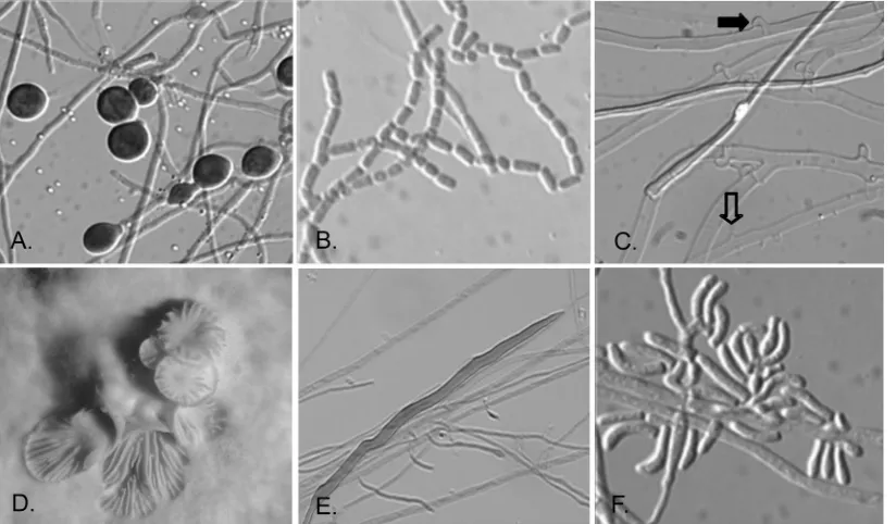
Basidiomycota. Growth is typically rapid, often up the side of the tube or plate; and colony colors are usually white, but they are sometimes cream to golden, orange, or slightly brownish on PDA. Microscopically, sterile basidiomycetes may display hy-phae only or hyhy-phae with chlamydoconidia (Fig. 1A). Some basidiomycetes do, however, produce conidia in culture. Most are arthroconidia, as seen in Fig. 1B, or compact clusters of arthroconidia, as seen for someHormographiellaspecies
(ana-morphs of someCoprinopsis[Coprinus] species). One of the more useful microscopic features for the identification of ster-ile isolates as basidiomycetes is the production of clamp con-nections, the defining characteristic for this phylum (Fig. 1C). Another important diagnostic feature of some sterile basidio-mycetes is the production of spicules along the sides of hyphae (Fig. 1C, open arrow), with or without clamp connections (Fig. 1C, closed arrow), as in the case of Schizophyllum commune
(Fig. 1C). Occasional dikaryons of this species may also pro-duce basidiocarps (Fig. 1D). The recently reported species
Inonotus(Phellinus)tropicalis(12, 15, 31), which is otherwise sterile in culture, may produce somewhat unusual hyphal ele-ments known as setal hyphae (Fig. 1E); however, these types of hyphae may occur in other genera as well. Curved conidia, which are typical ofHormographiellaspecies, may also be ob-served (Fig. 1F). The microscopic features of isolates included in this study are noted in Table 1. Finally, the ability of most basidiomycetes to grow on medium containing benomyl and their lack of growth on media containing cycloheximide further supported their probable identification.
A total of 168 filamentous isolates that had been identified as probable basidiomycetes by using the criteria cited above made up the study set that was sequenced.
Comparison of ITS and D1/D2 region BLAST results.
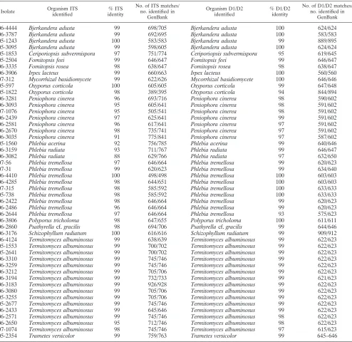
[image:5.585.89.501.70.311.2]Com-parison of the top hits from the GenBank database for the ITS and D1/D2 regions showed a number of isolates that returned the same species name for both the ITS region and the D1/D2 region (Table 2). However, when the number of disagreements was considered for the two regions, comparative ITS-D1/D2 sequencing for this set of isolates showed an overall striking lack of agreement. Although the BLASTn results for each
FIG. 1. Typical morphological features of basidiomycetes in culture. The morphological features that basidiomycetes may display in culture are shown. Microscopic features include chlamydoconidia (A), arthroconidia (B), spicules (open arrow) and clamp connections (solid arrow) of Schizophyllum commune(C), macroscopic basidiocarp of a dikaryoticSchizophyllum communeisolate (D), setal hyphae ofInonotus(Phellinus) tropicalis(E), and conidia of theHormographiellaanamorph of aCoprinussp. (F). (A, B, C, E, F) Magnifications,⫻880; (D) magnification,⫻5.
on May 16, 2020 by guest
http://jcm.asm.org/
isolate yielded a basidiomycete identification to the species level for 99.4% (167/168) of the isolates, the inconsistency of the outputs for the two regions made it impossible to assign a conclusive identification for 70.8% (119/168) of the isolates (Table 3). At the least-stringent level, in which agreement between the two sequences needed to consist only of the same genus name, regardless of the percent identity (i.e., the ITS sequence identifiedPhlebia tremellosawith 95% identity; the D1/D2 sequence identifiedPhlebia radiatawith 96% identity), only 48.8% (82/168) of the results were in agreement (Table 4). For genus and species agreement, regardless of the percent
identity (i.e., the ITS sequence identified Phlebia tremellosa
with 97% identity; the D1/D2 sequence identifiedPhlebia tre-mellosawith 93% identity), the results for only 28.6% (48/168) of the specimens agreed. For genus and species agreement with a cutoff of ⱖ97% identity, the results for only 21.4% (36/168) of the specimens agreed. Further analysis showed that of the 168 sequences, the sequence of only a single isolate (0.6%) displayed matching ITS and D1/D2 region-based genus and species names with 100% identity.
Comparison of ITS and D1/D2 GenBank deposits.In order
[image:6.585.46.540.80.560.2]to investigate possible causes for the low frequency of agreement
TABLE 2. Comparison of GenBank top hits for the ITS and D1/D2 regions which agreea
Isolate Organism ITS identified
% ITS identity
No. of ITS matches/ no. identified in
GenBank
Organism D1/D2 identified
% D1/D2 identity
No. of D1/D2 matches/ no. identified in
GenBank
06-4444 Bjerkandera adusta 99 698/705 Bjerkandera adusta 100 624/624
06-3787 Bjerkandera adusta 99 692/695 Bjerkandera adusta 100 583/583
05-1243 Bjerkandera adusta 100 583/583 Bjerkandera adusta 99 889/895
05-3095 Bjerkandera adusta 99 598/605 Bjerkandera adusta 100 624/624
05-1853 Ceriporiopsis subvermispora 97 751/774 Ceriporiopsis subvermispora 95 619/645
05-2504 Fomitopsis feei 99 646/647 Fomitopsis feei 99 646/647
06-3335 Fomitopsis rosea 98 638/647 Fomitopsis rosea 98 638/647
06-3906 Irpex lacteus 99 660/663 Irpex lacteus 100 560/560
07-312 Mycorrhizal basidiomycete 99 622/626 Mycorrhizal basidiomycete 100 646/646
05-597 Oxyporus corticola 100 605/605 Oxyporus corticola 99 647/648
05-1822 Oxyporus corticola 98 389/395 Oxyporus corticola 94 844/894
06-3281 Peniophora cinerea 96 693/716 Peniophora cinerea 98 590/602
06-3093 Peniophora cinerea 95 605/641 Peniophora cinerea 98 591/602
07-1076 Peniophora cinerea 95 505/541 Peniophora cinerea 98 591/602
06-2439 Peniophora cinerea 97 625/641 Peniophora cinerea 99 591/602
06-2581 Peniophora cinerea 96 617/641 Peniophora cinerea 97 591/602
06-2670 Peniophora cinerea 98 735/741 Peniophora cinerea 97 591/602
06-3035 Peniophora cinerea 91 775/841 Peniophora cinerea 97 587/602
05-1560 Phlebia acerina 92 756/785 Phlebia acerina 99 640/646
06-3159 Phlebia radiata 93 711/767 Phlebia radiata 99 646/647
06-3082 Phlebia radiata 88 629/766 Phlebia radiata 97 632/650
07-56 Phlebia tremellosa 97 646/664 Phlebia tremellosa 99 620/623
07-31 Phlebia tremellosa 99 620/623 Phlebia tremellosa 99 634/640
06-4410 Phlebia tremellosa 100 498/498 Phlebia tremellosa 100 603/603
06-4285 Phlebia tremellosa 98 644/651 Phlebia tremellosa 100 603/603
07-315 Phlebia tremellosa 98 585/592 Phlebia tremellosa 100 633/633
05-738 Phlebia tremellosa 98 585/592 Phlebia tremellosa 100 633/633
06-2422 Phlebia tremellosa 98 646/664 Phlebia tremellosa 99 620/623
06-2486 Phlebia tremellosa 96 646/664 Phlebia tremellosa 99 620/623
06-2644 Phlebia tremellosa 97 646/664 Phlebia tremellosa 93 575/623
06-3806 Polyporus tricholoma 98 647/655 Polyporus tricholoma 100 611/611
06-2860 Psathyrellacf.gracilis 98 694/706 Psathyrellacf.gracilis 99 644/646
06-3176 Schizophyllum radiatum 100 616/616 Schizophyllum radiatum 99 909/912
06-4124 Termitomyces albuminosus 99 638/639 Termitomyces albuminosus 99 622/623 05-1553 Termitomyces albuminosus 99 700/702 Termitomyces albuminosus 99 622/623 05-2641 Termitomyces albuminosus 99 700/702 Termitomyces albuminosus 99 622/623 06-3310 Termitomyces albuminosus 99 745/746 Termitomyces albuminosus 99 622/623 06-3259 Termitomyces albuminosus 99 745/746 Termitomyces albuminosus 99 622/623 06-3212 Termitomyces albuminosus 99 705/706 Termitomyces albuminosus 99 622/623 06-3194 Termitomyces albuminosus 99 732/733 Termitomyces albuminosus 99 621/623 06-3183 Termitomyces albuminosus 99 926/928 Termitomyces albuminosus 99 622/623 06-3080 Termitomyces albuminosus 99 705/706 Termitomyces albuminosus 99 622/623 05-3255 Termitomyces albuminosus 99 705/706 Termitomyces albuminosus 99 622/623 05-2677 Termitomyces albuminosus 99 745/746 Termitomyces albuminosus 99 622/623 06-2433 Termitomyces albuminosus 99 645/646 Termitomyces albuminosus 99 622/623 06-2571 Termitomyces albuminosus 99 745/746 Termitomyces albuminosus 98 622/623 06-2650 Termitomyces albuminosus 95 712/746 Termitomyces albuminosus 98 622/623 07-1074 Termitomyces albuminosus 98 745/746 Termitomyces albuminosus 97 615/623
05-2354 Trametes versicolor 99 759/763 Trametes versicolor 99 645–646
a
Differences in sequence matches between multiple isolates of the same species and what was returned by BLAST reflect the different percent identities of multiple GenBank records for the same species, one of which had the closest identity to our sequence but which could differ with each search. The table was sorted alphabetically.
on May 16, 2020 by guest
http://jcm.asm.org/
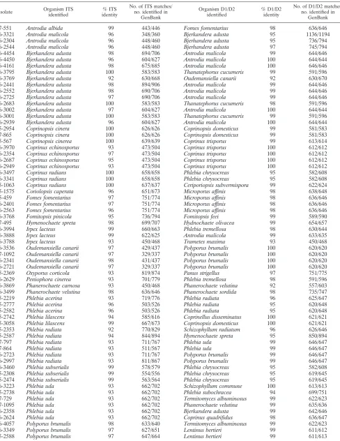
TABLE 3. Comparison of GenBank top hits for the ITS and D1/D2 regions which disagreea
Isolate Organism ITS identified
% ITS identity
No. of ITS matches/ no. identified in
GenBank
Organism D1/D2 identified
% D1/D2 identity
No. of D1/D2 matches/ no. identified in
GenBank
07-551 Antrodia albida 99 443/446 Fomes fomentarius 98 636/646
06-3321 Antrodia malicola 96 348/360 Bjerkandera adusta 95 1136/1194
06-2304 Antrodia malicola 96 448/460 Bjerhandera adusta 95 736/794
06-2544 Antrodia malicola 96 448/460 Bjerhandera adusta 97 745/794
06-4454 Bjerkandera adusta 98 694/706 Antrodia malicola 99 644/646
06-4450 Bjerkandera adusta 96 604/627 Antrodia malicola 100 644/644
06-4161 Bjerkandera adusta 98 675/685 Antrodia malicola 100 646/646
06-3795 Bjerkandera adusta 100 583/583 Thanatephorus cucumeris 99 591/596
06-3769 Bjerkandera adusta 92 630/668 Oudemansiella canarii 92 630/670
06-2441 Bjerkandera adusta 98 894/906 Antrodia malicola 99 644/646
06-2552 Bjerkandera adusta 98 690/706 Antrodia malicola 99 644/646
06-2725 Bjerkandera adusta 97 690/706 Antrodia malicola 99 644/646
06-2683 Bjerkandera adusta 100 583/583 Thanatephorus cucumeris 98 591/596
06-3002 Bjerkandera adusta 97 604/627 Antrodia malicola 100 644/644
06-3001 Bjerkandera adusta 100 583/583 Thanatephorus cucumeris 99 591/596
06-2939 Bjerkandera adusta 96 604/627 Antrodia malicola 100 644/644
05-2954 Coprinopsis cinera 100 626/626 Coprinopsis domesticus 99 581/583
07-865 Coprinopsis cinera 100 626/626 Coprinopsis domesticus 99 581/583
05-567 Coprinopsis cinerea 100 639/639 Coprinus trisporus 99 613/614
06-3970 Coprinus echinosporus 93 473/504 Coprinus trisporus 100 612/612
06-2354 Coprinus echinosporus 97 473/504 Coprinus trisporus 100 612/612
06-2687 Coprinus echinosporus 95 473/504 Coprinus trisporus 100 612/612
06-2949 Coprinus echinosporus 93 473/504 Coprinus trisporus 100 612/612
06-3497 Coprinus radians 100 658/658 Phlebia chrysocreas 95 582/608
06-3341 Coprinus radians 100 658/658 Phlebia chrysocreas 95 582/608
05-1063 Coprinus radians 100 637/637 Ceriporiopsis subvermispora 99 622/624
05-1575 Coriolopsis caperata 96 651/673 Microporus affinis 98 638/648
05-459 Fomes fomentarius 97 751/774 Microporus affinis 98 636/646
06-2401 Fomes fomentarius 97 751/774 Microporus affinis 98 636/646
06-2563 Fomes fomentarius 96 751/774 Microporus affinis 98 636/646
06-3768 Fomitopsis pinicola 95 736/794 Fomitopsis feei 99 589/590
07-495 Hymenochaete spreta 98 699/707 Hydnochaete olivacea 99 654/657
06-3994 Irpex lacteus 99 660/663 Phlebia tremellosa 98 630/644
06-3888 Irpex lacteus 99 622/625 Antrodia malicola 99 633/635
06-3788 Irpex lacteus 93 450/468 Trametes maxima 93 450/468
06-3536 Oudemansiella canarii 97 429/437 Polyporus brumalis 100 620/620
07-1092 Oudemansiella canarii 97 329/337 Polyporus brumalis 100 620/620
06-2341 Oudemansiella canarii 98 431/437 Polyporus brumalis 100 620/620
06-2721 Oudemansiella canarii 97 329/337 Polyporus brumalis 100 620/620
05-2369 Oxyporus corticola 93 819/874 Panus strigellus 97 751/775
06-2629 Peniophora cinerea 93 701/779 Phlebia tremellosa 98 591/596
06-3869 Phanerochaete carnosa 93 450/468 Phanerochaete velutina 92 557/603
06-3499 Phanerochaete velutina 98 636/646 Phanerochaete sordida 98 735/747
05-2219 Phlebia acerina 93 719/776 Phlebia radiata 96 625/647
05-2777 Phlebia acerina 96 503/526 Phlebia radiata 95 620/648
05-2582 Phlebia acerina 96 503/526 Phlebia radiata 95 620/648
05-2742 Phlebia lilascens 94 585/616 Coprinellus disseminatus 100 621/621
05-3058 Phlebia lilascens 99 667/673 Coprinopsis domesticus 100 621/621
05-2353 Phlebia radiata 92 770/829 Schizophyllum radiatum 96 626/646
05-2587 Phlebia radiata 94 844/894 Hymenochaete spreta 95 850/894
07-797 Phlebia radiata 93 711/767 Phlebia uda 99 646/647
07-864 Phlebia radiata 93 511/567 Phlebia uda 99 646/647
06-2723 Phlebia radiata 93 711/767 Polyporus brumalis 99 646/647
06-2997 Phlebia radiata 93 811/867 Polyporus brumalis 99 646/647
06-3460 Phlebia subserialis 99 578/579 Phlebia chrysocreas 95 582/608
05-2308 Phlebia subserialis 99 554/556 Phlebia chrysocreas 95 619/645
05-2474 Phlebia subserialis 99 563/564 Phlebia chrysocreas 95 619/645
06-3223 Phlebia uda 93 662/702 Schizophyllum commune 100 613/613
05-2738 Phlebia uda 93 662/702 Phlebia subochracea 94 699/751
07-729 Phlebia uda 93 662/702 Termitomyces albuminosus 99 622/623
07-1095 Phlebia uda 93 662/702 Phanerochaete velutina 99 635/636
06-2358 Phlebia uda 93 662/702 Bjerkandera adusta 99 642/646
06-2624 Phlebia uda 93 662/702 Coprinus quadrifidus 98 636/647
06-4057 Polyporus brumalis 98 633/640 Termitomyces albuminosus 99 622/623
06-3349 Polyporus brumalis 97 627/651 Lentinus bertieri 99 611/612
05-2588 Polyporus brumalis 97 647/664 Lentinus bertieri 99 611/613
Continued on following page
on May 16, 2020 by guest
http://jcm.asm.org/
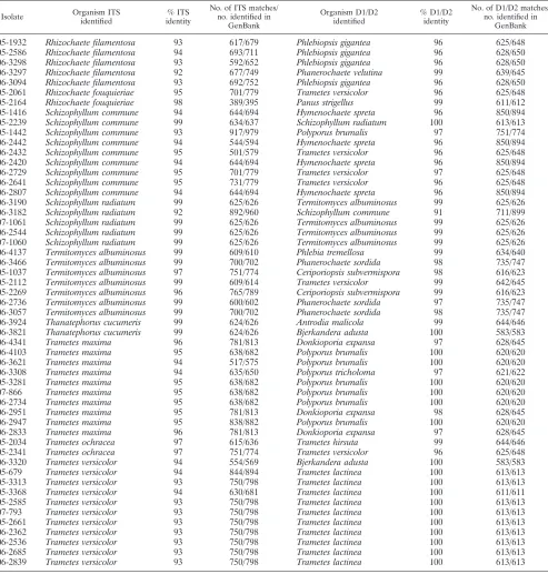
for the ITS and D1/D2 regions, the GenBank database was searched for the presence of sequence deposits that corresponded to these two sequences for each species. This analysis revealed that there were no entries in GenBank for 14% of the top ITS hits (7/50) and 16% (8/50) of the top D1/D2 hits for the isolates on our list (Table 5). Therefore, 30% of the species that were
iden-tified in this study had either an ITS or a D1/D2 sequence that matched a deposit in GenBank, but not both.
Analysis of ITS and D1/D2 GenBank deposit sequence
lengths.The top hits for each BLAST query for both the ITS
and the D1/D2 regions with an identity of 97% or greater were evaluated for their completeness, which was defined as a
se-TABLE 3—Continued
Isolate Organism ITS identified
% ITS identity
No. of ITS matches/ no. identified in
GenBank
Organism D1/D2 identified
% D1/D2 identity
No. of D1/D2 matches/ no. identified in
GenBank
05-1932 Rhizochaete filamentosa 93 617/679 Phlebiopsis gigantea 96 625/648
05-2586 Rhizochaete filamentosa 94 693/711 Phlebiopsis gigantea 96 628/650
06-3298 Rhizochaete filamentosa 93 592/652 Phlebiopsis gigantea 96 628/650
06-3297 Rhizochaete filamentosa 92 677/749 Phanerochaete velutina 99 639/645
06-3094 Rhizochaete filamentosa 93 692/752 Phlebiopsis gigantea 96 628/650
05-2061 Rhizochaete fouquieriae 95 701/779 Trametes versicolor 96 625/648
05-2164 Rhizochaete fouquieriae 98 389/395 Panus strigellus 99 611/612
05-1416 Schizophyllum commune 94 644/694 Hymenochaete spreta 96 850/894
05-2239 Schizophyllum commune 99 634/637 Schizophyllum radiatum 100 613/613
05-1442 Schizophyllum commune 93 917/979 Polyporus brumalis 97 751/774
06-2442 Schizophyllum commune 94 544/594 Hymenochaete spreta 96 850/894
06-2432 Schizophyllum commune 95 501/579 Trametes versicolor 96 625/648
06-2420 Schizophyllum commune 94 644/694 Hymenochaete spreta 96 850/894
06-2729 Schizophyllum commune 95 701/779 Trametes versicolor 97 625/648
06-2641 Schizophyllum commune 95 731/779 Trametes versicolor 96 625/648
06-2807 Schizophyllum commune 94 644/694 Hymenochaete spreta 96 850/894
06-3190 Schizophyllum radiatum 99 625/626 Termitomyces albuminosus 99 625/626
06-3182 Schizophyllum radiatum 92 892/960 Schizophyllum commune 91 711/899
07-1061 Schizophyllum radiatum 99 625/626 Termitomyces albuminosus 99 625/626
06-2544 Schizophyllum radiatum 99 625/626 Termitomyces albuminosus 99 625/626
07-1060 Schizophyllum radiatum 99 625/626 Termitomyces albuminosus 99 625/626
06-4137 Termitomyces albuminosus 99 609/610 Phlebia tremellosa 99 634/640
06-3466 Termitomyces albuminosus 99 700/702 Phanerochaete sordida 98 735/747
05-1037 Termitomyces albuminosus 97 751/774 Ceriporiopsis subvermispora 98 616/623
05-2112 Termitomyces albuminosus 99 609/614 Trametes versicolor 99 642/645
05-2269 Termitomyces albuminosus 96 765/789 Ceriporiopsis subvermispora 99 616/623
06-2736 Termitomyces albuminosus 99 600/602 Phanerochaete sordida 97 735/747
06-3057 Termitomyces albuminosus 99 700/702 Phanerochaete sordida 98 735/747
06-3924 Thanatephorus cucumeris 99 624/626 Antrodia malicola 99 644/646
06-3821 Thanatephorus cucumeris 99 624/626 Bjerkandera adusta 100 583/583
06-4341 Trametes maxima 96 781/813 Donkioporia expansa 97 628/645
06-4103 Trametes maxima 95 638/682 Polyporus brumalis 100 620/620
06-3621 Trametes maxima 94 517/575 Polyporus brumalis 100 620/620
06-3308 Trametes maxima 94 635/650 Polyporus tricholoma 97 621/622
05-3281 Trametes maxima 95 638/682 Polyporus brumalis 100 620/620
07-866 Trametes maxima 95 638/682 Polyporus brumalis 100 620/620
06-2734 Trametes maxima 95 638/682 Polyporus brumalis 100 620/620
06-2951 Trametes maxima 95 781/813 Donkioporia expansa 98 628/645
06-2947 Trametes maxima 95 838/882 Polyporus brumalis 100 620/620
06-2833 Trametes maxima 96 781/813 Donkioporia expansa 97 628/645
05-2034 Trametes ochracea 97 615/636 Trametes hirsuta 99 644/646
05-2341 Trametes ochracea 97 751/774 Trametes versicolor 96 625/648
06-3320 Trametes versicolor 94 554/569 Bjerkandera adusta 100 583/583
05-679 Trametes versicolor 94 844/894 Trametes lactinea 100 613/613
05-3313 Trametes versicolor 93 750/798 Trametes lactinea 100 613/613
05-3368 Trametes versicolor 94 630/681 Trametes lactinea 100 611/611
05-2585 Trametes versicolor 93 750/798 Trametes lactinea 100 613/613
07-793 Trametes versicolor 93 750/798 Trametes lactinea 100 613/613
05-2661 Trametes versicolor 93 750/798 Trametes lactinea 100 613/613
06-2362 Trametes versicolor 93 750/798 Trametes lactinea 100 613/613
06-2536 Trametes versicolor 93 750/798 Trametes lactinea 100 613/613
06-2685 Trametes versicolor 93 750/798 Trametes lactinea 100 613/613
06-2839 Trametes versicolor 93 750/798 Trametes lactinea 100 613/613
a
Differences in sequence matches between multiple isolates of the same species and what was returned by BLAST reflect the different percent identities of multiple GenBank records for the same species, one of which had the closest identity to our sequence but which could differ with each search. The table was sorted alphabetically on the basis of the ITS name.
on May 16, 2020 by guest
http://jcm.asm.org/
[image:8.585.46.539.81.598.2]quence whose length matched the length of our query se-quence, excluding internal deletions or insertions. Comparison of GenBank deposit sequence lengths of both the ITS and the D1/D2 regions for each isolate showed that the deposit entries for each region were largely truncated compared to the lengths of the regions that we sequenced. Of the 50 species repre-sented, only 8% of the ITS regions and 10% of the D1/D2 regions had complete sequence data in GenBank (Table 5).
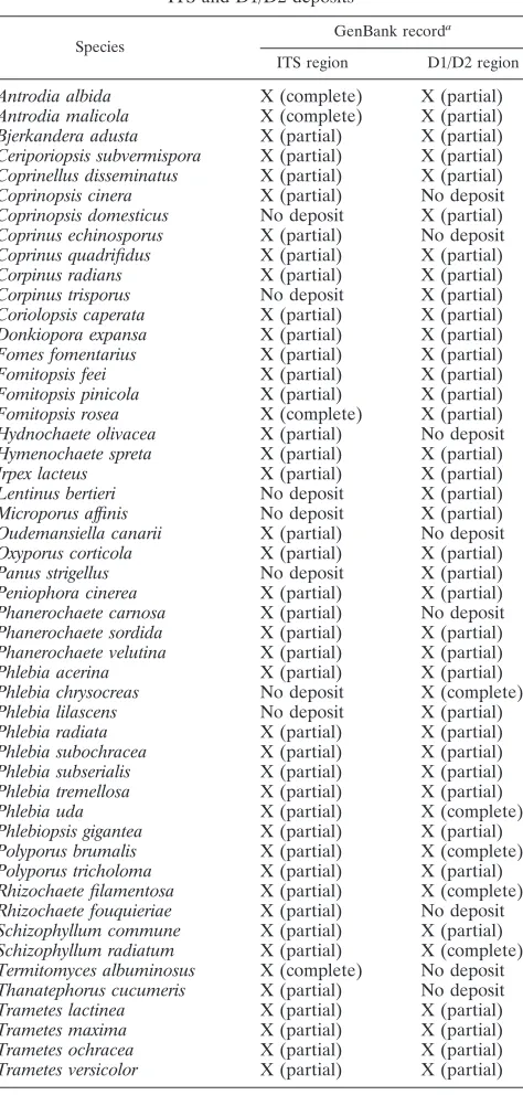
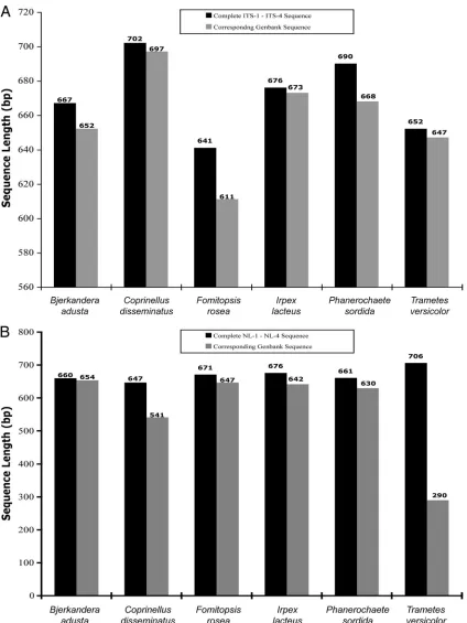
Since most of the species that we identified are rare and in many cases had only a single GenBank deposit, we selected six species that were the most redundant from the BLAST output list (Table 2) and obtained the sequence lengths from each record with the highest percent identity. Sequences were recovered forBjerkandera adusta(GenBank accession num-ber EU918694), Coprinellus disseminatus (GenBank accession number FN386275),Fomitopsis rosea(GenBank accession num-ber DQ491412), Irpex lacteus (GenBank accession number FJ462768.1),Phanerochaete sordida(GenBank accession num-ber EU118653.1), andTrametes versicolor(GenBank accession number FJ810146). The ITS sequence analysis (Fig. 2A) showed that regardless of the species, there were no complete sequences compared to our ITS sequences. The most complete sequence ofCoprinellus disseminatusfound in GenBank was 697 bp (GenBank accession number FN386275), whereas the ITS sequence that we obtained was 702 bp. The other GenBank ITS sequences varied in length and were found to be incomplete as well. The ITS sequence lengths obtained from GenBank ranged from 95% to 99% compared to the complete ITS sequences that we derived by sequencing with primers ITS-1 and ITS-4. The D1/D2 sequence length data (Fig. 2B) also proved to be largely incomplete. The most complete se-quence ofBjerkandera adustafound in the GenBank database was 654 bp (GenBank accession number AB096738), whereas the D1/D2 sequence that we obtained was 660 bp. The D1/D2 sequence lengths obtained from GenBank ranged from 41% to 99% complete compared to the complete D1/D2 sequences that we obtained by sequencing with primers NL-1 and NL-4. These data indicate that many of the current GenBank se-quences for basidiomycetes have incomplete sequence data for the regions that we used for identification. Importantly, these results were obtained for the most redundant species recov-ered from our BLAST searches and therefore would be
ex-pected to have a higher likelihood for a complete deposit due to the presence of multiple records.
DISCUSSION
The frequency of human mycoses due to filamentous fungi is steadily increasing, and mycoses mostly affect defined risk groups, such as immunocompromised or severely ill
pa-TABLE 4. ITS-D1/D2 BLAST output comparison
% Identity cutoff No.a % Agreement
Genus only agreementb 82 48.8
Genus⫹species agreement,cany % identity 48 28.6
Genus⫹species agreement, oneⱖ97% identity 48 28.6 Genus⫹species agreement, bothⱖ97% identity 36 21.4 Genus⫹species agreement, oneⱖ98% identity 43 25.6 Genus⫹species agreement, bothⱖ98% identity 32 19 Genus⫹species agreement, oneⱖ99% identity 36 21.4 Genus⫹species agreement, bothⱖ99% identity 24 14.3 Genus⫹species agreement, one 100% identity 12 7.1 Genus⫹species agreement, both 100% identity 1 0.6
aNumber of isolates of 168 isolates tested with given identity. bAny percent identity.
[image:9.585.42.284.80.193.2]cGenus plus species agreement represents BLAST outputs in which the ITS genus and species name matched the D1/D2 genus and species name.
TABLE 5. Presence of species-specific GenBank ITS and D1/D2 deposits
Species GenBank record
a
ITS region D1/D2 region Antrodia albida X (complete) X (partial) Antrodia malicola X (complete) X (partial) Bjerkandera adusta X (partial) X (partial) Ceriporiopsis subvermispora X (partial) X (partial) Coprinellus disseminatus X (partial) X (partial) Coprinopsis cinera X (partial) No deposit Coprinopsis domesticus No deposit X (partial) Coprinus echinosporus X (partial) No deposit Coprinus quadrifidus X (partial) X (partial) Corpinus radians X (partial) X (partial) Corpinus trisporus No deposit X (partial) Coriolopsis caperata X (partial) X (partial) Donkiopora expansa X (partial) X (partial) Fomes fomentarius X (partial) X (partial) Fomitopsis feei X (partial) X (partial) Fomitopsis pinicola X (partial) X (partial) Fomitopsis rosea X (complete) X (partial) Hydnochaete olivacea X (partial) No deposit Hymenochaete spreta X (partial) X (partial) Irpex lacteus X (partial) X (partial) Lentinus bertieri No deposit X (partial) Microporus affinis No deposit X (partial) Oudemansiella canarii X (partial) No deposit Oxyporus corticola X (partial) X (partial) Panus strigellus No deposit X (partial) Peniophora cinerea X (partial) X (partial) Phanerochaete carnosa X (partial) No deposit Phanerochaete sordida X (partial) X (partial) Phanerochaete velutina X (partial) X (partial) Phlebia acerina X (partial) X (partial) Phlebia chrysocreas No deposit X (complete) Phlebia lilascens No deposit X (partial) Phlebia radiata X (partial) X (partial) Phlebia subochracea X (partial) X (partial) Phlebia subserialis X (partial) X (partial) Phlebia tremellosa X (partial) X (partial) Phlebia uda X (partial) X (complete) Phlebiopsis gigantea X (partial) X (partial) Polyporus brumalis X (partial) X (complete) Polyporus tricholoma X (partial) X (partial) Rhizochaete filamentosa X (partial) X (complete) Rhizochaete fouquieriae X (partial) No deposit Schizophyllum commune X (partial) X (partial) Schizophyllum radiatum X (partial) X (complete) Termitomyces albuminosus X (complete) No deposit Thanatephorus cucumeris X (partial) No deposit Trametes lactinea X (partial) X (partial) Trametes maxima X (partial) X (partial) Trametes ochracea X (partial) X (partial) Trametes versicolor X (partial) X (partial)
aX, a sequence deposit was made for this species. No deposit, no GenBank record could be found for the corresponding sequence; partial and complete, the sequence length between the ITS-1 and ITS-4 primers (ITS region) or the NL-1 and NL-4 primers (D1/D2 region).
on May 16, 2020 by guest
http://jcm.asm.org/
[image:9.585.303.540.83.579.2]tients. In addition to the well-known opportunistic basidio-mycetous pathogenCryptococcus neoformans, other basidio-mycetous yeasts such as Malassezia spp. and Rhodotorula
spp. are now considered emergent opportunistic pathogens
[image:10.585.81.505.66.631.2]and are recovered at increasing frequencies (6, 14, 20, 22). Basidiomycetous molds, with few exceptions, are rarely re-covered as human pathogens because of the difficulty iden-tifying these fungi or the difficulty distinguishing colonizers
FIG. 2. Comparison of ITS and D1/D2 sequence lengths. (A) Comparison of ITS lengths to ITS lengths in GenBank. The sequence lengths of the ITS regions of our isolates were compared to those found in GenBank. The six species represented here were chosen on the basis of being the most redundant among the results from the BLASTn search. (B) Comparison of D1/D2 lengths to D1/D2 lengths in GenBank. The sequence lengths of the D1/D2 regions of our isolates were compared to those found in GenBank.
on May 16, 2020 by guest
http://jcm.asm.org/
from invasive isolates in patient specimens. Sterile and/or arthroconidium-forming basidiomycetes are a subset of this class and cannot be conclusively identified by standard phe-notypic methods because they do not produce distinguishing structures. Although these mostly sterile isolates may be mor-phologically identified as basidiomycetes when clamp connec-tions are present, many genera of basidiomycetes do not pro-duce clamp connections in culture. Consequently, they may simply be described as nonsporulating molds with unknown clinical significance.
In a study by Pounder et al., which also used a sequencing strategy for identification, 31 of the 48 (65%) isolates were classified as basidiomycetes (23) by use of a sequence derived from the ITS region. Under the cutoff criteria of a sequence length of at least 400 bp,ⱖ99% identity for a species-level identification, andⱖ93% identity for a genus-level identifica-tion, 92% of the isolates were identified to the genus level and 79% were identified to the species level. Because of the rela-tively high identification rate, we decided to use a similar strategy to identify our isolates. A large number of the initial ITS sequences that we obtained did not meet the 97% cutoff criterion that we established for identity. Therefore, we de-cided to add the D1/D2 region as a second locus, under the assumption that the results from D1/D2 searches would yield more identities higher than 97%, thereby allowing an identifi-cation. However, we were surprised to find that while in many cases we obtained a D1/D2 identity ofⱖ97%, we observed a striking amount of disagreement between the best hit (the highest level of identity) for the ITS search and the best hit for the D1/D2 search. The agreement between the two sequences for the same isolate was only 28.6% at any level of identity, whereas with a more stringent cutoff ofⱖ97% identity, agree-ment occurred only 21.4% of the time. We suspect that this low level of agreement would likely be the same for any mold that is rarely studied at the molecular level, whether it is sterile or not, due to the absence of searchable data in GenBank.
The low level of ITS-D1/D2 agreement led us to investigate why the results were so disparate. Of the 50 species that we identified, almost a third did not have a GenBank deposit for the ITS region or the D1/D2 region. When all significant hits (ⱖ97% identity) were considered for each search output, two-thirds (66%) of the records had either an ITS deposit or a D1/D2 deposit, but not both. As a result of this discrepancy, error can be introduced during the BLAST search output when the next-highest identity, which will be a different species, becomes the top hit in the search. We also found that the sequences in GenBank were largely incomplete compared to our query sequences. It is not clear how much sequence would need to be truncated from either or both ends before a signif-icant impact on identity occurs; however, sequence alignments demonstrate that sequence variation can occur very close (within a few bases) to the primer sites that we used (data not shown). These variable regions may not be present in the sequence if the sequence is truncated due to single-stranded sequencing, if the sequence is derived from a different primer combination or a partially overlapping region, or if the se-quencing run terminates and does not proceed through the primers. These observations, combined with known GenBank issues such as nomenclature errors (5) and poor-quality depos-its (18, 23), can complicate sequence-based identification. In
fact, fungal GenBank deposits may be more adversely affected by issues involving nomenclature than GenBank deposits for other microbial organisms. Few investigators working with fungi outside of classical mycology are well versed in the rules governing how and when the anamorph and teleomorph no-menclature is properly used. Similarly, isolates may be identi-fied by their obsolete or synonymous names, and selection of the currently accepted name is difficult even for classical my-cologists, since names are often changed on the basis of basic research, including some of the molecular techniques used in this study, and may not be widely reported or even accepted. The sequences of basidiomycetes in the GenBank database, with the possible exception of those ofC. neoformans, may be more ad-versely affected by these issues, since taxonomic studies of basidio-mycete species pathogenic for humans may be lagging similar studies of other fungi, such as the aspergilli or the fusaria, for which detailed analysis has resulted in revised classifications (2, 3). These issues were not addressed in the data analysis, since the study focused on the actual GenBank outputs; consequently, it is possible that the levels of agreement would improve slightly due to a correct agreement being masked by the erroneous or incon-sistent naming of the deposit.
Our results and the results of Pounder et al. (23) suggest that sterile molds recovered from human clinical specimens may comprise a substantial number of basidiomycetes. In fact, our study utilized a subset of sterile and/or arthroconidium-pro-ducing isolates from human clinical specimens phenotypically identified as probable basidiomycetes (on the basis of the mor-phological criteria that we used for our study) that had been sent to the Fungus Testing Laboratory. Both studies had six species in common, including Polyporus tricholoma, Irpex lacteus,Schizophyllum commune,Phlebia subserialis, Trame-tes versicolor, and Thanatephorus cucumeris. While it is highly likely that most filamentous basidiomycetes identified from clinical specimens are clinically insignificant because they are noncolonizers abundant in ambient air, a number of our specimens were from sites other than the respiratory tract that are normally sterile (i.e., cerebrospinal fluid). The host status, the route of infection, and the shear number and variety of fungal elements that a patient is exposed to likely determine whether a basidiomycosis can occur.
While this study has highlighted issues that need to be care-fully considered when sequence-based identification is em-ployed, sequence-based identification has some major diagnos-tic strengths and continues to be extremely useful to our group for fungal identification. It clearly has great diagnostic value for common fungi and/or fungi that have numerous GenBank deposits. The sequence data in GenBank are also useful if they are combined with additional nonsequence data, even if the sequencing results are somewhat ambiguous. In fact, our se-quencing results for the 168 isolates were in complete agree-ment with the preliminary morphological results, in that all BLAST results were consistent with the organism being a ba-sidiomycete. However, as a general rule, on the basis of the results of this study, we now utilize both the ITS and D1/D2 regions when we make sequence-based identifications for any fungus and double check if there is disagreement to make sure there is a GenBank deposit for both sequences. While this strategy does not guarantee that the sequence results will be 100% accurate, it can rapidly reveal whether there are enough
on May 16, 2020 by guest
http://jcm.asm.org/
data in GenBank for sequencing to even be used in the iden-tification process. In our specific study, unfortunately, there does not seem to be enough data in GenBank to identify any unknown sterile basidiomycete with a high degree of confi-dence by ITS and/or D1/D2 sequencing.
As sequencing moves toward broader acceptance in the clin-ical laboratory, an important challenge to be overcome will be the development of a process that can provide a platform that certification bodies (the Clinical Laboratory Improvement Amendments [CLIA], the College of American Pathologists [CAP]) can standardize. In fact, guidelines are now being es-tablished to facilitate standardization (11). Evaluation of the data source should clearly be included in this platform, a major part of which should be a determination of how databases, whether they are public, such as GenBank, or private, could fit into the process. Unfortunately, the choice of database is not going to be a trivial issue. Despite the known errors with GenBank records, the depth of the sequences with regard to the number of potential species included in the database can-not be matched. Even imprecise GenBank records can be informative in some cases, since some taxonomic information may be identifiable, despite incorrect genus or species names. Conversely, private or closed databases may be more accurate due to the confirmation of each entry and the deposit of high-quality sequences. However, these databases will likely sacri-fice species diversity and redundancy due to the smaller num-ber of entries. Despite this shortcoming, a closed database may be more amenable to standardization, particularly if sequences are generated specifically for the database (versus download-ing from another source), since primers, completeness, and identities can be standardized and confirmed.
In summary, this study has shown that in addition to the well-known concerns with the use of the sequences in a public database for sequence-based identification, missing data can also contribute to erroneous conclusions during searches. These errors may be caught for fungi for which substantial phenotypic data are available for comparison to the sequenc-ing results; however, when there are few phenotypic data, such as for sterile basidiomycetes or other molds, erroneous con-clusions could be quite common.
ACKNOWLEDGMENTS
A.M.R. was supported by NIDCR grant DE14318 (CO STAR). B.L.W. was supported by grant PR054228 from the U.S. Army Medical Research and Materiel Command, Office of Congressionally Directed Medical Research Programs.
REFERENCES
1.Atkins, S. D., and I. M. Clark.2004. Fungal molecular diagnostics: a mini review. J. Appl. Genet.1:3–15.
2.Balajee, S. A., A. M. Borman, M. E. Brandt, J. Cano, M. Cuenca-Estrella, E. Dannaoui, J. Guarro, G. Haase, C. C. Kibbler, W. Meyer, K. O’Donnell, A. Petti, J. L. Rodriguez, D. Sutton, A. Velegraki, and B. L. Wickes.2009. Sequence based identification ofAspergillus,Fusarium, andMucorlaes spe-cies in the clinical mycology laboratory: where should we go from here? J. Clin. Microbiol.47:877–884.
3.Balajee, S. A.2007.Aspergillusspecies identification in the clinical setting. Stud. Mycol.1:39–46.
4.Balajee, S. A., L. Sigler, and M. E. Brandt.2007. DNA and the classical way: identification of medically important molds in the 21st century. J. Med. Mycol.45:475–490.
5.Bidartondo, M. I.2008. Preserving accuracy in GenBank. Science319:1616. 6.Borman, A. M., C. J. Linton, S. J. Miles, and E. M. Johnson.2008. Molecular
identification of pathogenic fungi. J. Antimicrob. Chemother.61:7–12. 7.Bruns, T. D., T. J. White, and J. W. Taylor.1991. Fungal molecular
system-atics. Annu. Rev. Ecol. Syst.22:525–564.
8.Chen, Y. C., J. D. Eisner, M. M. Kattar, S. L. Rassoulian-Barrett, K. Lafe, U. Bui, A. P. Limaye, and B. T. Cookson.2000. Identification of medically important yeasts using PCR-based detection of DNA sequence polymor-phisms in the internal transcribed spacer 2 region of the rRNA genes. J. Clin. Microbiol.38:2302–2310.
9.Chen, Y. C., J. D. Eisner, M. M. Kattar, S. L. Rassoulian-Barrett, K. Lafe, U. Bui, A. P. Limaye, and B. T. Cookson.2001. Polymorphic internal tran-scribed spacer region 1 DNA sequences identify medically important yeasts. J. Clin. Microbiol.39:4042–4051.
10.Cirado, D. E., G. Schar, M. Altwegg, E. C. Bottger, and P. P. Bosshard.2007. Identification of moulds in the diagnostic laboratory—an algorithm imple-menting molecular and phenotypic methods. Diagn. Microbiol. Infect. Dis. 59:49–60.
11.Clinical and Laboratory Standards Institute.2008. Interpretive criteria for identification of bacteria and fungi by DNA sequencing; approved guideline. CLSI MM18-A. Clinical and Laboratory Standards Institute, Wayne, PA. 12.Davis, C. M., L. M. Noroski, M. K. Dishop, D. A. Sutton, R. M. Braverman,
M. E. Paul, and H. M. Rosenblatt.2007. Basidiomycetous fungalInonotus tropicalissacral osteomyelitis in X-linked chronic granulomatous disease. Pediatr. Infect. Dis. J.26:655–656.
13.Erjavec, Z., and P. E. Verweij.2002. Recent progress in the diagnosis of fungal infections in the immunocompromised host. Drug Resist. Updat.5:3–10. 14.Gonzalez, A., R. Sierra, R. E. Cardenas, A. Grajales, S. Restrepo, M. C. Cepero
de Garcia, and A. Celis.2009. Physiological and molecular characterization of atypical isolates ofMalassezia furfur. J. Clin. Microbiol.47:48–53.
15.Gonzalez, G. M., D. A. Sutton, E. Thompson, E. Tijerina, and M. G. Rinaldi. 2001. In vitro activities of approved and investigational antifungal agents against 44 clinical isolates of basidiomycetous fungi. Antimicrob. Agents Chemother.45:633–635.
16.Hoorfar, J., N. Cook, B. Malorny, M. Wagner, D. De Medici, A. Abdulmaw-jood, and P. Fach. 2004. Diagnostic PCR: making internal amplification control mandatory. Lett. Appl. Microbiol.38:79–80.
17.Iwen, P. C., S. H. Hinrichs, and M. E. Rupp.2002. Utilization of the internal transcribed spacer regions as molecular targets to detect and identify human fungal pathogens. J. Med. Mycol.40:87–109.
18.Janda, J. M., and S. L. Abbott.2007. 16S rRNA gene sequencing for bac-terial identification in the diagnostic laboratory: pluses, perils, and pitfalls. J. Clin. Microbiol.45:2761–2764.
19.Kurtzman, C. P., and C. J. Robnett.1997. Identification of clinically impor-tant ascomycetous yeasts based on nucleotide divergence in the 5⬘end of the large-subunit (26S) ribosomal DNA gene. J. Clin. Microbiol.35:1216–1223. 20.Mirza, S. A., M. Phelan, D. Rimland, E. Graviss, R. Hamill, M. E. Brandt, T. Gardner, M. Sattah, G. Ponce de Leon, W. Baughman, and R. A. Hajjeh. 2003. The changing epidemiology of cryptococcosis: an update from popu-lation-based active surveillance in 2 large metropolitan areas, 1992–2000. Clin. Infect. Dis.36:789–794.
21.Pfaller, M., and R. Wenzel.1992. Impact of the changing epidemiology of fungal infections in the 1990s. Eur. J. Clin. Microbiol. Infect. Dis.11:287–291. 22.Pfaller, M. A., and D. J. Diekema.2004. Rare and emerging opportunistic
fungal pathogens: concern for resistance beyondCandida albicansand As-pergillus fumigatus. J. Clin. Microbiol.42:4419–4431.
23.Pounder, J. I., K. E. Simmon, C. A. Barton, S. L. Hohmann, M. E. Brandt, and C. A. Petti.2007. Discovering potential pathogens among fungi identi-fied as nonsporulating molds. J. Clin. Microbiol.45:568–571.
24.Rakeman, J. L., U. Bui, K. LaFe, Y. Chen, R. J. Honeycutt, and B. T. Cookson. 2005. Multilocus DNA sequence comparisons rapidly identify pathogenic molds. J. Clin. Microbiol.43:3324–3333.
25.Rinaldi, M. G.1982. Use of potato flakes agar in clinical mycology. J. Clin. Microbiol.15:1159–1160.
26.Shorr, A. F., R. Lazarus, J. H. Sherner, W. L. Jackson, M. Morrel, V. J. Frasier, and M. H. Kollef.2007. Do clinical features allow for accurate prediction of fungal pathogenesis in bloodstream infections? Potential im-plications of the increasing prevalence of non-albicans candidemia. J. Soc. Crit. Care Med.35:1077–1083.
27.Sigler, L., and S. L. Abbott.1997. Characterizing and conserving diversity of filamentous basidiomycetes from human sources. Microb. Cult. Col-lect.13:21–27.
28.Steinbach, W. J., T. G. Mitchell, W. A. Schell, A. Espinel-Ingroff, R. F. Coico, T. J. Walsh, and J. R. Perfect.2003. Status of medical mycology education J. Med. Mycol.41:457–467.
29.Stevens, D. A.2002. Diagnosis of fungal infections: current status J. Anti-microb. Chemother.49:11–19.
30.Summerbell, R. C.1993. The benomyl test as a fundamental diagnostic method for medical mycology. J. Clin. Microbiol.31:572–577.
31.Sutton, D. A., E. H. Thompson, M. G. Rinaldi, P. C. Iwen, K. K. Nakasone, H. S. Jung, H. M. Rosenblatt, and M. E. Paul.2005. Identification and first report ofInonotus(Phellinus)tropicalisas an etiologic agent in a patient with chronic granulomatous disease. J. Clin. Microbiol.43:982–987.
32.White, T. J., T. D. Bruns, S. B. Lee, and J. W. Taylor.1990. Amplification and sequencing of fungal ribosomal RNA genes for phylogenetics, p. 315– 322.InPCR protocols and applications: a laboratory manual. Academic Press, New York, NY.