http://dx.doi.org/10.4236/cm.2015.62016
How to cite this paper: Gu, J.F., et al. (2015) Specificity Screening of Potential Active Components from Moutan Cortex for Rat Mesangial Cells HBZY-1 by Cell Membrane Immobilized Chromatography. Chinese Medicine, 6, 147-157.
http://dx.doi.org/10.4236/cm.2015.62016
Specificity Screening of Potential Active
Components from Moutan Cortex for Rat
Mesangial Cells HBZY-1 by Cell Membrane
Immobilized Chromatography
Junfei Gu1,2,3, Minghua Zhang1, Jiarui Yuan1,3, Bingjie Zhao1,3, Liang Feng1*, Xiaobin Jia1,
Li Zhang1, Yuesheng Wang4, Luqi Huang2*
1Key Laboratory of New Drug Delivery System of Chinese Materia Medica, Jiangsu Provincial Academy of
Chinese Medicine, Jiangsu Branch of China Academy of Chinese Medical Sciences, Nanjing, China
2State Key Laboratory Breeding Base of Dao-Di Herbs, China Academy of Chinese Medical Sciences, Beijing,
China
3College of Pharmacy, Nanjing University of Chinese Medicine, Nanjing, China
4Institute of Chinese Medicine, China Academy of Chinese Medical Sciences, Beijing, China
Email: *[email protected], *[email protected]
Received 5 May 2015; accepted 20 June 2015; published 23 June 2015
Copyright © 2015 by authors and Scientific Research Publishing Inc.
This work is licensed under the Creative Commons Attribution International License (CC BY).
http://creativecommons.org/licenses/by/4.0/
Abstract
Moutan Cortex (MC) has been demonstrated to have an inhibitive effect on inflammation and oxida-tive stress responses in mesangial cells in our previous study. However, little is known about the components of MC contributing to this benefit. In the present study, cell membrane immobilized chromatography (CMC), a fast and useful method, was presented for screening potential active components of MC. HBZY-1 cells were incubated with MC (200 μg/mL) at the optimal incubation time (90 min). HPLC-DAD analysis and LC/ESI/MS/MS were performed to distinguish the active components and identify its structural ion fragments. The results showed that eight components binding to HBZY-1 cells were mudanoside B, paeoniflorin sulfonate, paeoniflorin, tetragalloyl glu-cose (isomeride), hexagalloyl gluglu-cose, mudanopiside A, and paeonol. In conclusion, our estab-lished CMC might be a useful method for screening potential active components in complicated traditional Chinese medicines. These components might be associated with the efficacy of MC on prevention and treatment of diabetic nephropathy.
Keywords
Components, Diabetic Nephropathy
1. Background
Diabetic nephropathy (DN), one of microvascular complications in diabetic patients as the end-stage renal dis-ease, is the leading cause of high morbidity and mortality of diabetes mellitus (DM) throughout the word [1]. The optimal treatment for DN continues to evolve as newer therapies for decreasing DN progression through a multifaceted target [2]. Recent studies show that the beneficial effects of traditional Chinese medicines (TCMs) on the treatment and prevention of DN have attracted the increasing interest of researchers [3].
As a well-known traditional herbal medicine, Moutan Cortex (MC), the root bark of Paeonia suffruticosa Andr., has been used for DN treatment for a long time in China and other Asian countries [4]. It is worth noting that MC has an inhibitive effect on inflammation and oxidative stress responses in DN [5] [6]. Phytochemical study has shown that MC contains lots of chemical compounds such as paeoniflorin, oxypaeoniflorin, benzoy-loxypaeoniflorin, apiopaeonoside, benzoylpaeoniflorin, paeonol, sugars, etc. [7]. Actually, as we know, not all of components in TCMs are responsible for the disease treatment [8]. Of all the components in MC, paeonol has been investigated to have the anti-diabetic effect [9]-[11]. In addition, paeoniflorin, oxypaeoniflorin, benzoylox-ypaeoniflorin and paeonol could inhibit the aggregation of platelet which might exacerbate the response of DN [12]. It has also been investigated that paeoniflorin and oxypaeoniflorin have the antioxidant and anti-inflamma- tion activities on AGEs-induced mesangial cell damage in our present study [13]. However, little is known about the mutil-components of MC contributing to the prevention and treatment of DN.
Recently, more and more methods have been developed for the screening of active components in TCMs, such as high throughput screening, and DNA sequencing [14]-[16]. Pharmacology researches have demonstrated that the action of drugs may be derived from the interaction of bioactive component molecules with some re-ceptors or channels on cell membrane. Cell membrane immobilized chromatography (CMC), one of cell mem-brane affinity chromatography, is composed of the biologic memmem-brane immobilized part and the chromatographic analysis part. Specifically, the cell membrane of organism is used as a solid phase for targeting effective com-ponents in TCMs. Then, the potential active comcom-ponents can be analyzed by chromatographic techniques timely. The effective components in TCMs can be screened quickly and efficiently [17]. Hence, the use of CMC by bio-membrane of organism extraction together with chromatography analysis may be a powerful tool for screening potential active components in TCMs.
Therefore, the present study was conducted to establish an appropriate CMC method for screening the poten-tial active components in MC which interacted with mesangial cells. Furthermore, these active components, which were related to the protective effect of MC on DN, were revealed for the first time in our study.
2. Materials and Methods
2.1. Materials and Reagents
MC, the dried roots of Paeonia suffruticosa Andr. (Batch No. 20120415), was from Anhui Huqiao Chinese Medicine Technology Co., Ltd. (Tongling, Anhui Province). The pharmaceutical botany of the medicinal ma-terial was identified by Prof. Dekang Wu from Nanjing University of Chinese Medicine. Standard substances of paeoniflorin and paeonol, each content was more than 98% were offered by National Instisutes for Food and Drug Control. Mudanoside B, tetragalloyl glucose, hexagalloyl glucose, mudanopiside A, which the content were exceeded 98%, were supplied by BOC Sciences (NY, USA). D-glucose was purchased from Sigma (St. Louis, MO, USA). Basal DMEM medium and fetal bovine serum (FBS) was provided by Gibco (BRL, USA). All other reagents used in this study were from commercial sources.
2.2. Sample Preparation
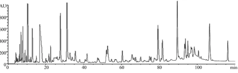
stored at 4˚C for further use. The chromatograms of MC extract were analyzed by HPLC-DAD at 254 nm (Figure 1).
2.3. Preparation of AGEs
Advanced glycation end products (AGEs) were prepared according to previous method [18] [19]. In brief, 5 g bovine serum albumin (BSA) and 9 g D-glucose were dissolved adequately in 100 mL phosphate buffer saline (PBS, 0.2 M, pH = 7.4). After being passed through 0.22 µm microporous membrane filter, the solution was then incubated in 5% CO2/95% air at 37˚C under sterile conditions. The reaction mixture was dialyzed in 0.2 M
PBS overnight to remove unincorporated glucose molecules after the incubation of 3 months. The content of AGEs was measured by human AGEs ELISA kit, the final concentration was 858 mg/mL, and then stored at 4˚C until use.
2.4. Cell Culture
Rat mesangial cells line HBZY-1 was obtained from American Type Culture Collection (ATCC; Manassas, VA, USA) and cells were maintained in low-glucose Dulbecco’s modified Eagle’s medium (DMEM) supplemented with 10% fetal bovine serum and 80 units/mL of penicillin/streptomycin. Cells were incubated in 25 cm2 plastic
cell culture dishes in 5% CO2/95% O2 at 37˚C. All medium was renewed every 2 days to provide sufficient
nu-trition. After 80% - 90% confluent layer, cells were used for the further experiments.
2.5. Cell Membrane Extraction of Sample
HBZY-1 cells were incubated in a plastic dish at a generating of 80% - 90% confluent layer. AGEs of 1 mL was added into 9 mL PBS (pH = 7.4) to induce mesangial cell dysfunction. The MC extraction (50, 100, 200 μg/mL)
was then incubated with live cells at 37˚C according to the previous description [7]. These cells were washed with 2 mL PBS to remove the unbound components for six times. The sixth washing eluate was retained for HPLC or LC/ESI/MS/MS analysis. Sequentially, 5 mL dissociation solution containing citric acid (pH 4.0) was added to dissociate the binding molecules at 37˚C for 60 min. The liquid supernatants of 5 mL were evaporated to dryness by high-purity nitrogen for 12 h at room temperature. Chromatographic methanol of 0.8 mL was added to each of them to dissolve the samples dryness. After vortex-mixing for 2 min, the supernatants of all the samples were taken and evaporated to dryness again for 5 h at room temperature. The residue was redissolved in 0.2 mL methanol followed by vortex-mixing for 2 min and centrifugation at 11,000 g for 10 min. Finally, the supernatants of all the samples were taken for HPLC and LC/ESI/MS/MS analysis.
2.6. HPLC Analysis
HPLC analysis system of Agilent 1200 series (Agilent Technologies, Santa Clara, CA, USA) equipped with a diode array detector (DAD), a quaternary pump, a column heater-cooler and an autosampler was performed to separate and analyze these components of MC and eluates. The samples were separated on an Agilent TC-C18
[image:3.595.102.499.585.707.2]column (4.6 mm × 250 mm, 5 µm). The mobile phase gradient conditions consisted of acetonitrile (A) and 0.1% formic acid (B) was 0 - 20 min, 5% - 10% A; 20 - 30 min, 10% A; 30 - 80 min, 10% - 18% A; 80 - 120 min, 18% - 50% A. The flow rate was 0.8 mL/minand the column temperature was maintained at 25˚C with a 10 µL
injection volume at 254 nm.
2.7. LC/ESI/MS/MS Analysis for Identification of Compounds
In this study, LC/ESI/MS/MS analysis was used to identify the compounds which were dissociated from cell membrane of HBZY-1 cells. The analysis was performed under negative-ion mode by ThermoQuest mass spec-trometer (San Jose, CA, USA). The optimal operating parameters were as follows: ion spray voltage, −4 kV; the nebulizer gas (N2): 0.45 L/min; curtain gas (N2): 0.2 L/min; declustering potential 1 (DP1): −20 V; focusing
po-tential (FP): −80 V; declustering potential 2 (DP2): −20 V. The mass spectrometer was detected over a range of m/z 80 to 1000 in the full scan mode.
2.8. Statistical Analysis
All data were expressed as means ± standard deviation (SD). Statistical analysis was performed by GraphPad Prism TM 5.0 statistical package with analysis of variance. Comparison between groups was using t-test. The value of p less than 0.05 was considered to be a statistically significant difference.
3. Results and Discussion
3.1. The Optimization of Conditions in Cell Membrane Immobilization
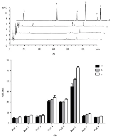
The incubation concentration and incubation time are two important factors for CMC. Taken cells viability into
consideration, different concentration extracts of MC including 50, 100, 200 μg/mL were examined to screen the
optimal incubation concentration. As shown in Figure 2(A) and Figure 2(B), after being incubated with three
concentrations of MC, the most components of MC had a high peak area at the concentration of 200 μg/mL. It indicated that MC of 200 μg/mL had a better interaction with HBZY-1 cells. To make sure a better incubation
for screening of components, 200 μg/mL MC was introduced for the cell membrane immobilization in our exp e-riments. In addition, the selected concentration is consistent with that used in the evaluation of efficacy in our previous study [20].
Another important factor in CMC is the incubation time for the binding of components of MC to HBZY-1 cells. Different incubation time might affect the binding level of molecules to targets on cell membrane. Gener-ally, the incubation time was 30 min as described by previous study in platelets or macrophages [17] [21]. In our present study, different time points including 15, 30, 60, 90, 120 min were examined for the most appropriate incubation time in HBZY-1 cells immobilized system. As shown in Figure 3(A) and Figure 3(B), only 6 peaks were found in 15 min, whereas, 8 peaks were shown after 30 min. The peak areas of these 8 peaks were in-creased from 30 min to 90 min in a time-dependent manner. However, there were no more peaks and significant increase of peak area of 8 peaks in the chromatogram at 120 min. These data suggested that 90 min was the op-timal time for the incubation.
Eventually, HBZY-1 cells were incubated with MC at the concentration of 200 μg/mL for 90 min. The optim-al incubation time and concentration were used for the immobilization.
3.2. Cell Membrane Immobilization of Components in MC
Studies show that cell membrane immobilized chromatography (CMC) is a powerful tool for screening of active compounds in TCM extract relying on the living cells incubated in the culture flask associated with the patho-genesis through the relevant passageway. Hence, there are two important factors in potential active components screening from MC in the biologic membrane immobilized part: the style of cell membrane and the pathogenic passageway participating in DN.
It is well known that the targets related to pathological changes on the cell membrane are not identical in dif-ferent types of cells. There is specific for the binding of bioactive components by the immobilization of difdif-ferent cell membranes. Reports have shown that mesangial cells, one of the main cell types of glomerulus, play impor-tant roles in the development of chronic kidney diseases including DN [22]. Hence, HBZY-1 cells were used for immobilizing potential active components of MC.
Figure 2. HPLC-DAD chromatograms was detected at 254 nm of MC after incubation with HBZY-1 cells in different con-centrations (A). The peak area of 8 components of MC in different concon-centrations (B). (a) MC of 50, (b) MC of 100, (c) MC
of 200 μg/mL at 37˚C, (d) the mixed standard of (1) mudanoside B; (3) paeoniflorin; (5) tetragalloyl glucose; (6) hexagalloyl
glucose; (7) mudanopiside A; (8) paeonol.
teraction between AGEs and their specific receptors RAGE (AGEs-RAGE) on HBZY-1 cells membrane could stimulate the over-expressions of receptors and trigger signaling events contributing to the cells damage [24]. More importantly, the active components of MC play a protective role in the pathogenesis of mesangial cells under pathological conditions. Consequently, in the present study, AGEs were introduced to induce mesangial cells dysfunction for immobilizing these potential active components of MC which were binding to cell mem-brane.
Figure 3. HPLC-DAD chromatograms detected at 254 nm of MC after incubation with HBZY-1 cells (A) and the peak area of 8 components of MC (B) at (a) 15 min, (b) 30 min, (c) 60 min, (d) 90 min, (e) 120 min, (f) the mixed standard of (1) mu-danoside B; (3) paeoniflorin; (5) tetragalloyl glucose; (6) hexagalloyl glucose; (7) mudanopiside A; (8) paeonol. *p < 0.05, (d) vs.
(a); **p < 0.01, (d) vs. (a); ¥p < 0.05, (d) vs. (b); ¥¥p < 0.01, (d) vs. (b); #p < 0.05, (d) vs. (c); % p < 0.05, (d) vs. (e).
eluate. The results indicated that peaks d1-d8 were the major components binding to cell membrane of HBZY-1. They might be the potential active components of MC for HBZY-1mesangial cells dysfunction.
3.3. Identification of Detected Components
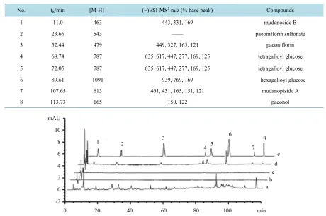
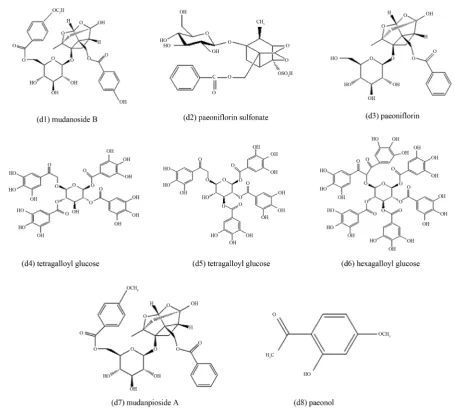
As a powerful technique for identification of molecular, LC/ESI/MS/MS was introduced for the identification of components in the present study. The components in immobilized sample could be directly identified using LC/ESI/MS/MS analysis. The MS spectra of d1-d8 were shown in Figure 5 and the information was listed in Table 1. These components were identified as mudanoside B, paeoniflorin sulfonate, paeoniflorin, tetragalloyl glucose (isomeride), hexagalloyl glucose, mudanopiside A, paeonol according to the MS ion fragments (Figure 6).
Table 1.Characterization of 8 components in MC after incubation with HBZY-1 cells by LC/ESI/MS/MS.
No. tR/min [M-H]− (−)ESI-MS2 m/z (% base peak) Compounds
1 11.0 463 443, 331, 169 mudanoside B
2 23.66 543 —— paeoniflorin sulfonate
3 52.44 479 449, 327, 165, 121 paeoniflorin
4 68.74 787 635, 617, 447, 277, 169, 125 tetragalloyl glucose 5 72.05 787 635, 617, 447, 277, 169, 125 tetragalloyl glucose
6 89.61 1091 939, 769, 169 hexagalloyl glucose
7 107.65 613 461, 431, 165, 151, 121 mudanopiside A
8 113.73 165 150, 122 paeonol
Figure 4. Chromatogram of the incubation of MC detected by HPLC-DAD (a), final washing eluate of biomembrane inte-racted with PBS (b), blank desorption eluate of biomembrane (c), desorption eluate of biomembrane inteinte-racted with MC (d), the mixed standard of (1) mudanoside B; (3) paeoniflorin; (5) tetragalloyl glucose; (6) hexagalloyl glucose; (7) mudanopiside
A; (8) paeonol (e). Sample was detected at 254 nm.
stances [25], the components for peak d3 and d8 were identified as paeoniflorin and paeonol. Peak d1 exhibited an [M-H]-ion at m/z 463 was tentatively identified as mudanoside B in that their ion fragments at m/z 331 (a loss of a pentose (132 Da)) and m/z 169 (losses of a pentose (132 Da) according to previous references [25] [26].
It was well known that there was no components showing the [M-H]-ion at m/z 543 in MC, but d2 was identi-fied as paeoniflorin sulfonate by comparison with the literature data previously [27]. Hence, it might be the product of the processing drug of MC which was changed from paeoniflorin.
It could also be easily seen that peak d4 and d5 had similar behavior displaying [M-H]-ions at m/z 787 in the ESI mass spectra, so it is very possible that they were isomeride. Their MS/MS spectra showed ion fragments at m/z 635, 617, 447, 277 169 and 125 with the losses of three gallic acids and a galloyl radical. Thus, they were proposed to be tetragalloyl glucose.
Peak d6 was identified as hexagalloyl glucose due to its [M-H]-ions at m/z 1091 in the full mass spectra. The greater 304 molecular weight of d6 compound than d4 and d5 was consistent with additional substitution of a galloyl group. The ion fragments of d6 at m/z 939 in that the losses of a gallic acid. For peak d8, it exhibited a [M-H]-ion at m/z 613. Its ion fragments at 151 and 121 attributed to [methoxybenzoic acid-H] and [benzoic ac-id-H]. The ion fragment at m/z 165 was reasonably identified as the pinane skeleton. Hence, compound d8 was assigned to be mudanpioside A.
Figure 5. MS spectra of d1-d8 screened by cell membrane immobilized chromatography. (d1) mudanoside B; (d2) paeoniflorin sulfonate; (d3) paeoniflorin; (d4) tetragalloyl glucose; (d5)
Figure 6. The structures of d1-d8 screened by cell membrane immobilized chromatography.
4. Conclusion
In the present study, CMC which relied on the living HBZY-1 cells incubated in the culture flask associated with the DN pathogenesis through the relevant passageway by biologic membrane immobilization part and LC/ ESI/MS/MS part was presented for screening potential active components of MC. Finally, eight potential active components were obtained in MC binding to HBZY-1 cells. In conclusion, CMC might be a beneficial method in searching potential active components in complicated TCMs.
Acknowledgements
The authors gratefully acknowledge the financial support of the National Natural Science of China (81202906), Natural Science Foundation of Jiangsu (BK2012491), Major changes in the central level support projects (2060302) and National Natural Science Foundation of China (81130070).
Conflict of Interests
Authors’ Contributions
JFG, MHZ, JRY, BJZ, LZ, XBJ and YSW contributed to the study design, analysis, and interpretation of data. LF and LQH participated in statistical analysis and drafted the manuscript. All authors approved the final ma-nuscript.
References
[1] Fox, C.S., Larson, M.G., Leip, E.P., Meigs, J.B., Wilson, P.W. and Levy, D. (2005) Glycemic Status and Development of Kidney Disease: The Framingham Heart Study. Diabetes Care, 28, 2436-2440.
http://dx.doi.org/10.2337/diacare.28.10.2436
[2] Choudhury, D., Tuncel, M. and Levi, M. (2010) Diabetic Nephropathy—A Multifaceted Target of New Therapies. Discovery Medicine, 10, 406-415.
[3] Poon, T.Y., Ong, K.L. and Cheung, B.M. (2011) Review of the Effects of the Traditional Chinese Medicine Rehman-nia Six Formula on Diabetes Mellitus and Its Complications. Journal of Diabetes, 3, 184-200.
http://dx.doi.org/10.1111/j.1753-0407.2011.00130.x
[4] Ha, D.T., Tuan, D.T., Thu, N.B., Nhiem, N.X., Ngoc, T.M., Yim, N. and Bae, K. (2009) Palbinone and Triterpenes from Moutan Cortex (Paeonia suffruticosa, Paeoniaceae) Stimulate Glucose Uptake and Glycogen Synthesis via Acti-vation of AMPK in Insulin-Resistant Human HepG2 Cells. Bioorganic & Medicinal Chemistry Letters, 19, 5556-5559.
http://dx.doi.org/10.1016/j.bmcl.2009.08.048
[5] Wu, M. and Gu, Z. (2009) Screening of Bioactive Compounds from Moutan Cortex and Their Anti-Inflammatory Ac-tivities in Rat Synoviocytes. Evidence-Based Complementary and Alternative Medicine, 6, 57-63.
http://dx.doi.org/10.1093/ecam/nem066
[6] Rho, S., Chung, H.S., Kang, M., Lee, E., Cho, C., Kim, H., Park, S., Kim, H.Y., Hong, M., Shin, M. and Bae, H. (2005) Inhibition of Production of Reactive Oxygen Species and Gene Expression Profile by Treatment of Ethanol Extract of Moutan Cortex Radicis in Oxidative Stressed PC12 Cells. Biological and Pharmaceutical Bulletin, 28, 661-666.
http://dx.doi.org/10.1248/bpb.28.661
[7] Chen, G., Zhang, L. and Zhu, Y. (2006) Determination of Glycosides and Sugars in Moutan Cortex by Capillary Elec-trophoresis with Electrochemical Detection. Journal of Pharmaceutical and Biomedical Analysis, 41, 129-134.
http://dx.doi.org/10.1016/j.jpba.2005.11.001
[8] Dong, Z.B., Li, S.P., Hong, M. and Zhu, Q. (2005) Hypothesis of Potential Active Components in Angelica Sinensis by Using Biomembrane Extraction and High Performance Liquid Chromatography. Journal of Pharmaceutical and Biomedical Analysis, 38, 664-669. http://dx.doi.org/10.1016/j.jpba.2005.02.008
[9] Ha, D.T., Ngoc, T.M., Lee, I., Lee, Y.M., Kim, J.S., Jung, H., Lee, S., Na, M. and Bae, K. (2009) Inhibitors of Aldose Reductase and Formation of Advanced Glycation End-Products in Moutan Cortex (Paeonia suffruticosa). Journal of Natural Products, 72, 1465-1470. http://dx.doi.org/10.1021/np9002004
[10] Lau, C.H., Chan, C.M., Chan, Y.W., Lau, K.M., Lau, T.W., Lam, F.C., Law, W.T., Che, C.T., Leung, P.C., Fung, K.P., Ho, Y.Y. and Lau, C.B. (2007) Pharmacological Investigations of the Anti-Diabetic Effect of Cortex Moutan and Its Active Component Paeonol. Phytomedicine, 14, 778-784. http://dx.doi.org/10.1016/j.phymed.2007.01.007
[11] Ha, D.T., Trung, T.N., Hien, T.T., Dao, T.T., Yim, N., Ngoc, T.M., Oh, W.K. and Bae, K. (2010) Selected Compounds Derived from Moutan Cortex Stimulated Glucose Uptake and Glycogen Synthesis via AMPK Activation in Human HepG2 Cells. Journal of Ethnopharmacology, 131, 417-424. http://dx.doi.org/10.1016/j.jep.2010.07.010
[12] Koo, Y.K., Kim, J.M., Koo, J.Y., Kang, S.S., Bae, K., Kim, Y.S., Chung, J.H. and Yun-Choi, H.S. (2010) Platelet Anti-Aggregatory and Blood Anti-Coagulant Effects of Compounds Isolated from Paeonia lactiflora and Paeonia suffruticosa. Pharmazie, 65, 624-628.
[13] Zhang, M.H., Feng, L., Zhu, M.M., Gu, J.F., Wu, C. and Jia, X.B. (2013) Antioxidative and Anti-Inflammatory Activities of Paeoniflorin and Oxypaeoniflora on AGEs-Induced Mesangial Cell Damage. Planta Medica, 79, 1319- 1323. http://dx.doi.org/10.1055/s-0033-1350649
[14] Huang, X., Kong, L., Li, X., Chen, X., Guo, M. and Zou, H. (2004) Strategy for Analysis and Screening of Bioactive Compounds in Traditional Chinese Medicines. Journal of Chromatography B, 812, 71-84.
http://dx.doi.org/10.1016/j.jchromb.2004.06.046
[15] Liu, Y., Liu, J., Yin, P., Gao, M., Deng, C. and Zhang, X. (2011) High Throughput Identification of Components from Traditional Chinese Medicine Herbs by Utilizing Graphene or Graphene Oxide as MALDI-TOF-MS Matrix. Journal of Mass Spectrometry, 46, 804-815. http://dx.doi.org/10.1002/jms.1952
http://dx.doi.org/10.2174/156802612801319034
[17] Yu, L., Zhao, J., Zhu, Q. and Li, S.P. (2007) Macrophage Biospecific Extraction and High Performance Liquid Chro-matography for Hypothesis of Immunological Active Components in Cordyceps sinensis. Journal of Pharmaceutical and Biomedical Analysis, 44, 439-443. http://dx.doi.org/10.1016/j.jpba.2007.01.003
[18] Li, Y., Liu, S., Zhang, Z., Xu, Q., Xie, F., Wang, J., Ping, S., Li, C., Wang, Z., Zhang, M., Huang, J., Chen, D., Hu, L. and Li, C. (2012) RAGE Mediates Accelerated Diabetic Vein Graft Atherosclerosis Induced by Combined Mechanical Stress and AGEs via Synergistic ERK Activation. PLoS ONE, 7, e35016.
http://dx.doi.org/10.1371/journal.pone.0035016
[19] Kim, J.Y., Park, H.K., Yoon, J.S., Kim, S.J., Kim, E.S., Ahn, K.S., Kim, D.S., Yoon, S.S., Kim, B.K. and Lee, Y.Y. (2008) Advanced Glycation End Product (AGE)-Induced Proliferation of HEL Cells via Receptor for AGE-Related Signal Pathways. International Journal of Oncology, 33, 493-501.
[20] Zhang, M.H., Feng, L., Zhu, M.M., Gu, J.F., Jiang, J., Cheng, X.D., Ding, S.M., Wu, C. and Jia, X.B. (2014) The Anti-Inflammation Effect of Moutan Cortex on Advanced Glycation End Products-Induced Rat Mesangial Cells Dysfunction and High-Glucose—Fat Diet and Streptozotocin-Induced Diabetic Nephropathy Rats. Journal of Ethnopharmacology, 151, 591-600. http://dx.doi.org/10.1016/j.jep.2013.11.015
[21] Yu, L., Li, Y., Fan, H., Duan, J., Zhu, Q. and Li, S. (2011) Analysis of Marker Compounds with Anti-Platelet Aggregation Effects in Mailuoning Injection Using Platelet Binding Assay Combined with HPLC-DAD-ESI-MS and Solid-Phase Extraction Technique. Phytochemical Analysis, 22, 87-93. http://dx.doi.org/10.1002/pca.1260
[22] Schnaper, H.W., Hayashida, T., Hubchak, S.C. and Poncelet, A.C. (2003) TGF-Beta Signal Transduction and Mesangial Cell Fibrogenesis. American Journal of Physiology—Renal Physiology, 284, F243-F252.
http://dx.doi.org/10.1152/ajprenal.00300.2002
[23] Fukami, K., Yamagishi, S., Ueda, S. and Okuda, S. (2008) Role of AGEs in Diabetic Nephropathy. Current Pharma-ceutical Design, 14, 946-952. http://dx.doi.org/10.2174/138161208784139710
[24] Yamagishi, S., Nakamura, K. and Matsui, T. (2009) Regulation of Advanced Glycation End Product (AGE)-Receptor (RAGE) System by PPAR-Gamma Agonists and Its Implication in Cardiovascular Disease. Pharmacological Research,
60, 174-178. http://dx.doi.org/10.1016/j.phrs.2009.01.006
[25] Xu, S.J., Yang, L., Zeng, X., Zhang, M. and Wang, Z.T. (2006) Characterization of Compounds in the Chinese Herbal Drug Mu-Dan-Pi by Liquid Chromatography Coupled to Electrospray Ionization Mass Spectrometry. Rapid Commu-nications in Mass Spectrometry, 20, 3275-3288. http://dx.doi.org/10.1002/rcm.2717
[26] Lin, H.C., Ding, H.Y. and Wu, Y.C. (1998) Two Novel Compounds from Paeonia suffructicosa. Journal of Natural Products, 61, 343-346. http://dx.doi.org/10.1021/np9704258
[27] Wu, J., Shen, H., Xu, J., Zhu, L.Y., Jia, X.B. and Li, S. (2012) Detection of Sulfur-Fumigated Paeoniae Alba Radix in Complex Preparations by High Performance Liquid Chromatography Tandem Mass Spectrometry. Molecules, 17, 8938-8954. http://dx.doi.org/10.3390/molecules17088938
[28] Liu, J.P., Wang, S., Feng, L., Ma, D., Fu, Q., Song, Y., Jia, X.B. and Ma, S. (2013) Hypoglycemic and Antioxidant Activities of Paeonol and Its Beneficial Effect on Diabetic Encephalopathy in Streptozotocin-Induced Diabetic Rats. Journal of Medicinal Food, 16, 577-586. http://dx.doi.org/10.1089/jmf.2012.2654