Susceptibility Testing for the
Polymyxins: Two Steps Back, Three Steps
Forward?
Shawn Vasoo
Department of Infectious Diseases and Infectious Diseases Research Laboratory, Institute of Infectious Diseases and Epidemiology, Tan Tock Seng Hospital, Singapore; Lee Kong Chian School of Medicine, Nanyang Technological University, Singapore
ABSTRACT Optimizing and standardizing susceptibility testing for the polymyxins have become pressing issues, given the rise in multidrug-resistant Gram-negative bacilli. Recently, both the CLSI and EUCAST have recommended broth microdilution (BMD) (without polysorbate) as the reference method for polymyxin susceptibility
testing. In this issue, K. L. Chew et al. (J Clin Microbiol 55:2609 –2616, 2017,https://doi
.org/10.1128/JCM.00268-17) compare the performances of three commercial BMD
pan-els and the Etest to the reference, BMD, for polymyxin B and colistin, using 76 Enterobacteriaceae isolates (21 of which weremcr-1positive). Although none of the commercial BMD panels strictly met FDA performance standards in this evaluation, possibly because of the small number isolates tested, the Sensititre panel achieved
⬎90% categorical agreement for both polymyxin compounds. These results also
reaf-firm CLSI and EUCAST guidance that gradient diffusion testing, which had unacceptable error rates, should be abandoned. In a simulated analysis with lowered breakpoints
(susceptible,ⱕ1 mg/liter; intermediate, 2 mg/liter; resistant,ⱖ4 mg/liter), error rates
and agreement were improved across the various methods and the rate of detection of mcr-1-positive isolates improved. These observations, taken together with recent phar-macokinetic data on optimizing target attainment for the polymyxins, suggest that more-stringent (lower) breakpoints may be reasonable, although such an approach may be limited by the inherent reliability of current testing methodologies and a lack of robust clinical correlative data, which are sorely needed.
S
usceptibility testing for the polymyxins has been beleaguered over the years. Thepolymyxins were first isolated fromPaenibacillus polymyxain 1947 by two
inde-pendent American groups (1, 2), and there has been a therapeutic renaissance in the use of both polymyxin B and colistin (polymyxin E) over the last decade, given the rise
of multidrug-resistant (MDR) Gram-negative bacilli, such as MDR Acinetobacter and
Pseudomonasand carbapenemase-producingEnterobacteriaceae.
Despite susceptibility testing being available for a long time, clarity has been lacking for appropriate methods of testing and optimal dosing for the polymyxins. Polymyxins are cationic polypeptides comprised of a heptapeptide ring, an exocyclic chain, and a fatty acid tail with positively charged residues which interact with and disrupt the Gram-negative lipopolysaccharide membrane; polymyxin B and colistin differ by only one amino acid in the heptapeptide ring (3, 4). Several reasons have accounted for difficulties with susceptibility testing for the polymyxins, including their cationic nature, their poor diffusion in agar due to their large molecular size, concerns over drug powder composition, and their heteroresistance. Further, the complex pharmacokinet-ics (PK) and pharmacodynampharmacokinet-ics (PD) of these compounds and the paucity of data correlating MIC data, drug concentration, and clinical outcomes have made setting clinical breakpoints challenging. The mechanisms underlying polymyxin resistance are
Accepted manuscript posted online19 July 2017
CitationVasoo S. 2017. Susceptibility testing for the polymyxins: two steps back, three steps forward? J Clin Microbiol 55:2573–2582. https://doi.org/10.1128/JCM.00888-17.
EditorErik Munson, Marquette University Copyright© 2017 American Society for Microbiology.All Rights Reserved.
Address correspondence to [email protected].
For the article discussed, seehttps://doi.org/10 .1128/JCM.00268-17
The views expressed in this Commentary do not necessarily reflect the views of the journal or of ASM.
crossm
on May 16, 2020 by guest
http://jcm.asm.org/
complex, and correlation between these has been comprehensively reviewed recently (4). Complicating this has been the discovery of plasmid-mediated resistance due to mcr-1and -2(5, 6).
Polymyxin susceptibility testing guidance by various professional bodies has varied over the years (e.g., there have been different breakpoints and disk antimicrobial contents for diffusion testing) (7), although efforts have recently been made to har-monize these. In 2016, the joint CLSI-EUCAST polymyxin breakpoint working group agreed that the ISO-20776 standard broth microdilution method (BMD) (which is the same method outlined in CLSI document M07-A010 [8]) should be used for colistin MIC determination and be performed with sulfate salts of colistin in plain polystyrene trays without additives like polysorbate-80 (P-80) (9) and that diffusion methods should be abandoned. P-80 had at one point been recommended by the CLSI as a supplement for colistin BMD quality control testing (10, 11) to mitigate the cationic properties of the polymyxins, which cause them to adhere to the negatively charged polystyrene surface. However, P-80 in itself has some antibacterial activity and may act synergistically with polymyxins to spuriously lower MICs, especially for organisms for which MICs are in the
low range, i.e., near the breakpoints and/or epidemiologic cutoff values of 1 to 2g/ml
(13, 14). This led to the removal of P-80 from BMD for the polymyxins in CLSI document M100-S27 (15, 16). Manual BMD, however, is laborious and not performed in many routine clinical microbiology laboratories, which often rely on diffusion or automated systems for susceptibility testing. Moreover, and somewhat paradoxically in view of the drugs’ poor diffusion in agar, diffusion methods (both disk and gradient strips) have been found to have unacceptably high levels of false susceptibility (or very major errors [VMEs]) in multiple studies (12, 17–28) (Table 1). Both the CLSI and EUCAST have now advised against diffusion methods for the polymyxins, removing disk diffusion inter-pretive criteria for them from their guidance documents (9, 15, 22).
Parsing the literature for the comparative performances of the various susceptibility testing methods for the polymyxins may be somewhat confusing, with studies seem-ingly giving contradictory results; Table 1 attempts to summarize data published from 2001 to date. Several important reasons accounting for these disparities should be borne in mind when evaluating studies. These include different susceptibility break-points used and various proportions of isolates studied for which the MICs are near the breakpoint. With regard to the number of isolates near susceptibility breakpoints, studies often lack a sizeable number of resistant isolates (especially resistant isolates for which the polymyxin MICs are low [4 to 8 mg/liter]), which may obscure an accurate estimation of VME rates. These MICs are also of clinical relevance given what we currently know about the pharmacokinetics of polymyxin B and colistin, dose optimi-zation, and target attainment (29, 30). Indeed, VMEs or major errors (MEs) may be underreported if the MICs for the isolates studied fall into extremes, with either very high MICs (high-level resistance) or very low MICs (very susceptible isolates). VME rates should also be calculated with the number of resistant isolates as the denominator and ME rates with susceptible isolates as the denominator, rather than expressed as a percentage of the total number of isolates (12). Unfortunately, the literature has not been consistent, and VMEs and MEs have often been expressed as percentages of the
number of isolates tested (resistant and susceptible) (Table 1, footnotec). Studies may
also vary depending on the relative proportions ofEnterobacteriaceaeand/or
nonfer-menting Gram-negative bacilli, and heteroresistance has been described in some
species, such as Enterobacter, Pseudomonas aeruginosa, and Acinetobacter spp. The
most important reason for the variation of reported results in the literature for poly-myxin antimicrobial susceptibility testing (AST), however, has been the lack of consen-sus in what constitutes a “gold standard” comparator. Until further data are available, per CLSI-EUCAST guidance, BMD with no supplementation should be the standard comparator.
In this issue, Chew et al. (31) pragmatically evaluate four commercial polymyxin B and colistin AST methods commonly used and/or more easily implemented in routine clinical microbiology laboratories than BMD performed by the reference ISO-20776
on May 16, 2020 by guest
http://jcm.asm.org/
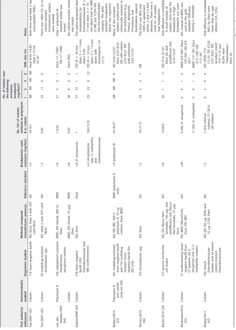
TABLE 1 Summary of published comparative susceptibility studies for polymyxin B and colistin from 2001 to 2017 a First author/yr (reference) Antimicrobial(s) studied Organisms studied Methods compared (manufacturer, potency) Reference standard Breakpoint(s) used, resistance (mg/liter) No. (%) of isolates resistant to polymyxin B or colistin No. of isolates near the following
resistance breakpoint (mg/liter)
b: VME rate (%) Notes 124 8 Gales/2001 (17 ) Polymyxin B 200 Gram-negative bacilli BMD (PML Microbiologics), DD (BD/Difco, 300 U polymyxin) BMD ⱖ 4 30 (15) NR 31 1 7 DD (with S indicated by a ⱖ 8-mm diam) 12/30 (40) c Disk diffusion is unreliable. Only 14 resistant isolates were studied for AD (1 isolate each was near the resistance breakpoints of 4 and 8 mg/liter). Colistin BMD (PML Microbiologics), DD (BD, 10 g colistin), AD (performed only on a subset of 35 isolates) ⱖ 4 30 (15) NR 8 2 2 AD (0); DD (with S indicated by a ⬎ 8-mm diam) 12/30 (40) c;D D (wth S indicated by a ⱖ 14-mm diam) 7/30 (23.3) c Hogardt/2004 (38 ) Polymyxin B and colistin 401 P. aeruginosa , 50 Achromobacter xylosoxidans , and 50 Stenotrophomonas maltophilia isolates from CF patients and 100 P. aeruginosa isolates from non-CF patients AD for polymyxin B, BMD (Merlin Diagnostika) for colistin Unclear, AD (polymyxin B) was compared against BMD (colistin) ⬎ 4 (DIN), ⬎ 8 (BSAC), ⬎ 2 (SFM) 29 (5.2) of 401 P. aeruginosa isolates by BMD (colistin) 111 46 33 12 NR Different compounds were tested against each other using different methods. Nicodemo/2004 (18 ) Polymyxin B 70 S. maltophilia isolates (66 isolates were tested for colistin and polymyxin) c AD, DD (Oxoid, 300 IU polymyxin B), Etest AD ⱖ 4 15 (22.7) NR NR NR NR DD 12/15 (80) c; Etest 8/15 (54.3) c Disk diffusion and Etest results had unacceptable VMEs. Colistin AD, DD (Oxoid, 10 g colistin), Etest ⱖ 4 16 (24.3) NR NR NR NR DD 15/16 (93.7) c; Etest 6/16 (37.5) c Arroyo/2005 (39 ) Colistin 115 A. baumannii isolates Etest, BMD BMD ⱖ 4 22 (19.1) 3 0 0 2 2/22 (9.1) The EA was 16.5%. The authors state that the worst agreement for strains was at 0.06 to 0.25 mg/liter and at 64 to ⬎ 1,024 mg/liter by the reference method, but few isolates studied were near resistance breakpoints. Tan/2006 (19 ) Colistin 228 Acinetobacter species, Pseudomonas aeruginosa , and Enterobacteriaceae isolates AD, DD (BD, 10 g [CLSI]; Oxoid, 25 g [BSAC]; Oxoid, 50 g [SFM]) AD ⱖ 4 (CLSI), ⬎ 4 (BSAC), ⬎ 2 (SFM) 27 (11.8) 92 107 13 4 CLSI DD 22/27 (81.5) c; BSAC DD 11/14 (78.6) c; SFM DD 24/27 (88.9) c Disk diffusion is unreliable. Goldstein/2007 (40 ) Colistin 170 clinical Gram-negative bacilli and 22 in vitro -selected mutants AD, Etest AD ⬎ 4 31 (18) (clinical isolates) 31 9 43 13 7/12 (58.3) for P. aeruginosa Etest MICs were 2-fold and 4-to 8-fold lower than AD MICs for 37.3% and 6.5% of isolates, respectively. Lo-Ten-Foe/2007 (41 ) Colistin and polymyxin B (DD) 102 Gram-negative bacilli (70 Enterobacteriaceae and 32 nonfermenters) AD with MH, AD with Iso-Sensitest agar, BMD, DD (Rosco, 150 g polymyxin B, 10 g colistin), Vitek 2 with AST card N038, Etest with MH, Etest with Iso-Sensitest agar BMD ⱖ 4 42 (41.2) 3 6 0 9 NR There was a high error rate and low reproducibility with DD. Authors report high levels of agreement with AD and Vitek 2 and relatively high levels of agreement with Etest, especially if Iso-Sensitest agar was used. (Continued on next page)
on May 16, 2020 by guest
http://jcm.asm.org/
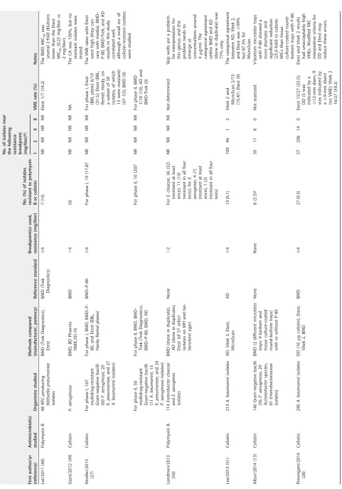
TABLE 1 (Continued) First author/yr (reference) Antimicrobial(s) studied Organisms studied Methods compared (manufacturer, potency) Reference standard Breakpoint(s) used, resistance (mg/liter) No. (%) of isolates resistant to polymyxin B or colistin No. of isolates near the following
resistance breakpoint (mg/liter)
b: VME rate (%) Notes 124 8 Tan/2007 (42 ) Colistin 172 Gram-negative bacilli AD, Etest, Vitek 2 with AST card N032 AD ⱖ 4 54 (31) NR NR NR NR Etest 8/54 (14.8) c; Vitek 2 31/54 (57.4) c Both Etest and Vitek 2 had unacceptable VMEs. Tan/2007 (43 ) Colistin 44 multidrug-resistant Acinetobacter spp. AD, Vitek 2 with AST card N032 AD ⬎ 2 0 (0) 33 11 0 0 There was 100% CA, as no colistin-resistant Acinetobacter isolates were studied. van der Heijden/2007 (44 ) Polymyxin B 109 carbapenem-resistant Pseudomonas aeruginosa isolates BMD, DD (Oxoid, 300 U), Etest BMD ⱖ 8 1 (0.9) 27 4 0 1 Etest 1/1 (100) c; DD 1/1 (100) c Only 1 polymyxin B-resistant isolate was tested. Colistin BMD, DD (Oxoid, 10 g), Etest BMD ⱖ 8 0 (0) 38 8 0 0 Etest (NA) No colistin-resistant isolate was tested. Galani/2008 (45 ) Colistin 778 Gram-negative bacilli (392 Enterobacteriaceae and 386 nonfermenters) DD, Etest Etest ⱖ 8( P. aeruginosa ), 7 5 73 11 1 D D (R ⱕ 10-mm diam, S ⱖ 11-mm diam) 0 (0) The author-proposed diam interpretations for Enterobacteriaceae (R ⱕ 11-mm diam, S ⱖ 14-mm diam) had no VME or ME, but 15.6% fell into the intermediate category. The gold standard comparator (Etest) was imperfect. ⱖ 4( Acinetobacter spp., S. maltophilia , and Enterobacteriaceae ) 104 (15.9) 29 22 8 22 DD (R ⱕ 12-mm diam, S ⱖ 13-mm diam) with Acinetobacter 1/9 (11.1) Behera/2010 (23 ) Polymyxin B and colistin (only for DD) 281 multidrug-resistant Gram-negative bacilli and 723 multidrug-resistant Gram-negative bacilli (for DD only) AD, DD (BD, 300 U polymyxin B, 10 g colistin), Etest, BMD BMD (polymyxin B only) ⬎ 4 (polymyxin B) 24 (8.5) d NR NR NR 8 AD 3/24 (12.5) c; Etest 2/24 (8.3) c; DD with colistin 2/24 (8.3) c;D D with polymyxin 3/24 (12.5) c Many of the resistant isolates studied were intrinsically resistant to the polymyxins (e.g., Proteus , Morganella , Providencia , and Burkholderia cepacia ). Nemec/2010 (46 ) Colistin 154 Acinetobacter spp. AD, Etest AD ⬎ 2 18 (11.7) 18 3 4 1 NR The Etest was 88% and 100% in agreement within 2 and 3 2-fold dilutions. There was a small no. of isolates near breakpoints. Boyen/2010 (24 ) Colistin 157 porcine Escherichia coli strains AD, DD (Rosco Neo-Sensitabs, 150 g), disk prediffusion test (Rosco Neo-Sensitabs, 10 g), Etest AD ⱖ 8 14 (8.9) 20 1 1 11 DD 3/14 (21.4) c; Etest (0); disk prediffusion test 2/14 (14.2) c Disk diffusion is unreliable. A small number of isolates were near the breakpoint. A high breakpoint was used. Moskowitz/2010 (25 ) Colistin 37 nonfermenting Gram-negative bacilli from CF patients (25 P. aeruginosa and 12 S. maltophilia isolates) AD, BMD (MicroTech), Etest, DD (BD) AD ⱖ 8 e 9 (36) ( P. aeruginosa ) 6 4 1 0 BMD 4/9 (44) c; Etest 5/9 (56) c;D D6 /9 (67) c AD, instead of BMD, was used as the gold standard. Diffusion testing is unreliable. 11 (92) ( S. maltophilia ) 0 0 1 0 BMD 1/11 (9) c; Etest 2/11 (18) c;D D 1/11 (9) c Maalej/2011 (47 ) Colistin 150 clinical Enterobacteriaceae isolates and 50 known colistin-resistant Enterobacteriaceae AD, DD (50 g, SFM, and 10 g, CLSI), Etest AD ⬎ 2 7 (4.6) (clinical isolates), 57 (28.5) (all isolates) 8 2 0 6 DD (SFM) 7/57 (12.3) c; DD (CLSI) 5/57 (8.8) c;D D (product insert) 1/57 (1.7) cbut 92.9% were “intermediate”; Etest (0) Disk diffusion is unreliable. MICs by Etest were lower than by agar dilution (CA, 33%). (Continued on next page)
on May 16, 2020 by guest
http://jcm.asm.org/
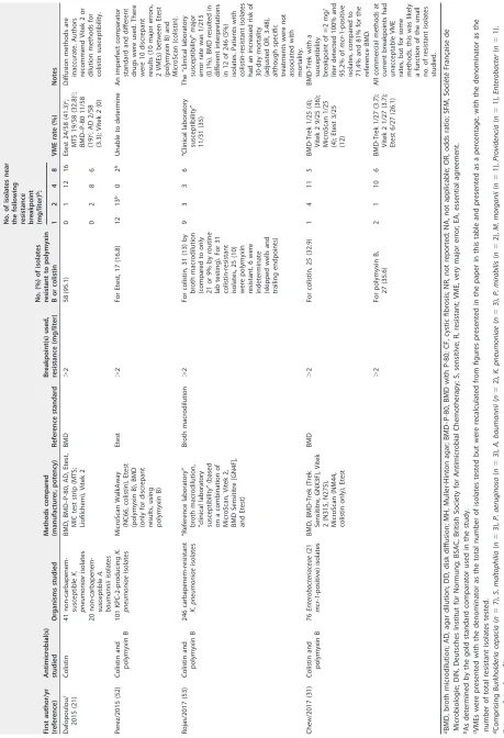
[image:4.585.51.531.75.744.2]TABLE 1 (Continued) First author/yr (reference) Antimicrobial(s) studied Organisms studied Methods compared (manufacturer, potency) Reference standard Breakpoint(s) used, resistance (mg/liter) No. (%) of isolates resistant to polymyxin B or colistin No. of isolates near the following
resistance breakpoint (mg/liter)
b: VME rate (%) Notes 124 8 Lat/2011 (48 ) Polymyxin B 48 KPC-producing Klebsiella pneumoniae isolates BMD (Trek Diagnostics), Etest BMD (Trek Diagnostics) ⱖ 4 7 (15) NR NR NR NR Etest 1/7 (14.2) The BMD MIC 50 was several 2-fold dilutions lower than the Etest MIC 50 (0.25 mg/liter vs 2 mg/liter). Giani/2012 (49 ) Colistin P. aeruginosa BMD, BD Phoenix NMIC/ID-76 BMD ⬎ 4 (0) NR NR NR NR NA The CA was 100%, but no resistant isolates were tested. Hindler/2013 (27 ) Colistin For phase I, 107 multidrug-resistant Gram-negative bacilli (60 P. aeruginosa ,2 0 K. pneumoniae , and 27 A. baumannii isolates) For phase I, BMD, BMD–P-80, and Etest (BBL, Hardy Remel plates) BMD–P-80 ⱖ 4 For phase I, 19 (17.8) f NR NR NR NR For phase I, Etest (BBL plate) 6/19 (31.5); Etest (BBL, Remel, Hardy, on a subset of 50 isolates, of which 15 were resistant) (47–53); BMD (0) The VME rates with Etest were high (they can vary with media). BMD– P-80, BMD-Trek, and AD results in this study correlated well, although a small no. of colistin-resistant isolates were studied. For phase II, 50 multidrug-resistant Gram-negative bacilli (11 A. baumannii ,1 5 K. pneumoniae , and 24 P. aeruginosa isolates) For phase II, BMD, BMD-Trek (Trek Diagnostics), BMD–P-80, BMD, AD For phase II, 10 (20) f NR NR NR NR For phase II, BMD 1/10 (10); AD and BMD-Trek (0) Landman/2013 (50 ) Polymyxin B 114 Enterobacter cloacae and E. aerogenes isolates BMD (done in duplicate), AD (done in duplicate), Etest (of 57 select isolates on MH and Iso-Sensitest agar) None ⬎ 2 For E. cloacae , 36 (32) (resistant at least once); 11 (10) (resistant in all four tests); for E. aerogenes , 4 (7) (resistant at least once); 1 (2) (resistant in all four tests) NR NR NR NR Not determined Skip wells are a problem for interpretation for this genus, and the problem tends to emerge at concentrations around 4 g/ml. The categorical agreement within BMD and AD (done in duplicate) was 77% only. Lee/2013 (51 ) Colistin 213 A. baumannii isolates AD, Vitek 2, Etest, MicroScan AD ⱖ 4 13 (6.1) 109 46 1 2 Vitek 2 and MicroScan 2/13 (15.4) c; Etest (0) The categorical agreement between AD, Vitek 2, and Etest was ⱖ 99% but 87.3% for MicroScan. Albur/2014 (13 ) Colistin 146 Gram-negative bacilli (56 P. aeruginosa ,2 9 Acinetobacter species, 61 Enterobacteriaceae isolates) BMD (2 different microtiter trays: V-bottom and tissue culture-coated round-bottom trays) with or without P-80 None None 8 (5.5) g 50 11 8 0 Not assessed V-bottom microtiter trays with P-80 showed a more dramatic and significant reduction (25.6-fold) in colistin MICs than tissue culture-coated round-bottom trays with P-80. Piewngam/2014 (28 ) Colistin 290 A. baumannii isolates DD (10 g colistin), Etest, Vitek 2, BMD BMD ⱖ 4 27 (9.3) 57 206 14 0 Etest 15/27 (55.5); DD (S was indicated by a ⱖ 12-mm diam; R was indicated by a ⱕ 9-mm diam) (no VME); Vitek 2 16/27 (59.2) Etest and Vitek 2 results had unacceptably high VMEs. Modified MIC interpretive criteria for DD and Etest may reduce these errors. (Continued on next page)
on May 16, 2020 by guest
http://jcm.asm.org/
[image:5.585.53.534.57.745.2]TABLE 1 (Continued) First author/yr (reference) Antimicrobial(s) studied Organisms studied Methods compared (manufacturer, potency) Reference standard Breakpoint(s) used, resistance (mg/liter) No. (%) of isolates resistant to polymyxin B or colistin No. of isolates near the following
resistance breakpoint (mg/liter)
b: VME rate (%) Notes 124 8 Dafopoulou/ 2015 (21 ) Colistin 41 non-carbapenem- susceptible K. pneumoniae isolates BMD, BMD–P-80, AD, Etest, MIC test strip (MTS; Liofilchem), Vitek 2 BMD ⬎ 2 58 (95.1) 0 1 12 16 Etest 24/58 (41.3) c; MTS 19/58 (32.8) c; BMD–P-80 11/58 (19) c; AD 2/58 (3.3); Vitek 2 (0) Diffusion methods are inaccurate. Authors recommend Vitek 2 or dilution methods for colistin susceptibility. 20 non-carbapenem- susceptible A. baumannii isolates 028 6 Perez/2015 (52 ) Colistin and polymyxin B 101 KPC-2-producing K. pneumoniae isolates MicroScan WalkAway (NC66, colistin), Etest (polymyxin B), BMD (only for discrepant results, using polymyxin B) Etest ⬎ 2 For Etest, 17 (16.8) 12 13 h 02 h Unable to determine An imperfect comparator standard and different drugs were used. There were 10 discrepant results (10 major errors, 2 VMEs) between Etest (polymyxin B) and MicroScan (colistin). Rojas/2017 (53 ) Colistin and polymyxin B 246 carbapenem-resistant K. pneumoniae isolates “Reference laboratory” broth macrodilution, “clinical laboratory susceptibility” (based on a combination of MicroScan, Vitek 2, BMD Sensititre [GN4F], and Etest) Broth macrodilution ⬎ 2 For colistin, 31 (13) by broth macrodilution (compared to only 21 or 9% by routine lab testing). For 31 colistin-resistant isolates, 25 (10) were polymyxin resistant, 6 were indeterminate (skipped wells and trailing endpoints) 9 3 3 6 “Clinical laboratory susceptibility” 11/31 (35) The “clinical laboratory susceptibility” major error rate was 1/215 (0.1%). BMD resulted in different interpretations in 12 of 246 (5%) isolates. Patients with colistin-resistant isolates had an increased risk of 30-day mortality (adjusted OR, 3.48), although specific treatments were not associated with mortality. Chew/2017 (31 ) Colistin and polymyxin B 76 Enterobacteriaceae (21 mcr-1 -positive) isolates BMD, BMD-Trek (Trek Sensititre, GNX3F), Vitek 2 (N315, N275), MicroScan (NM44, colistin only), Etest BMD ⬎ 2 For colistin, 25 (32.9) 1 4 11 5 BMD-Trek 1/25 (4); Vitek 2 9/25 (36); MicroScan 1/25 (4); Etest 3/25 (12) BMD-Trek with a susceptibility breakpoint of ⱕ 2 mg/ liter detected 100% and 95.2% of mcr -1-positive isolates, compared to 71.4% and 81% for the reference BMD. ⬎ 2 For polymyxin B, 27 (35.6) 2 1 10 6 BMD-Trek 1/27 (3.7); Vitek 2 1/27 (3.7); Etest 6/27 (26.1) All commercial methods at current breakpoints had unacceptable VME rates, but for some methods, this was likely a function of the small no. of resistant isolates studied. aBMD, broth microdilution; AD, agar dilution; DD, disk diffusion; MH, Muller-Hinton agar; BMD–P-80, BMD with P-80; CF, cystic fibrosis; NR, not report ed; NA, not applicable; OR, odds ratio; SFM, Société Française de Microbiologie; DIN, Deutsches Institut für Normung; BSAC, British Society for Antimicrobial Chemotherapy; S, sensitive; R, resistant; VME, very m ajor error; EA, essential agreement. bAs determined by the gold standard comparator used in the study. cVMEs were presented with the denominator as the total number of isolates tested but were recalculated from figures presented in the paper in this table a nd presented as a percentage, with the denominator as the number of total resistant isolates tested. dComprising Burkholderia cepacia ( n ⫽ 7), S. maltophilia ( n ⫽ 3), P. aeruginosa ( n ⫽ 3), A. baumannii ( n ⫽ 2), K. pneumoniae ( n ⫽ 3), P. mirabilis ( n ⫽ 2), M. morganii ( n ⫽ 1), Providencia ( n ⫽ 1), Enterobacter ( n ⫽ 1). eIntermediate, 4 mg/liter. fBy BMD–P-80. gThe MIC was ⬎ 4 mg/liter with V-bottom trays without P-80. hMICs were determined by Etest. For 13 isolates, the MIC was 1.5 mg/liter, and for 2 isolates, the MIC was 6 mg/liter (rounded up in the table to the next hig her dilution).
on May 16, 2020 by guest
http://jcm.asm.org/
[image:6.585.53.512.70.742.2]standard (8, 9). These comprise a commercial BMD system (Sensititre), gradient diffusion (Etest), and two automated AST systems: Vitek 2 and MicroScan (colistin only). A total
of 76Enterobacteriaceaewere studied, including 21 isolates harboringmcr-1. Using the
EUCAST colistin resistance breakpoint of⬎2 mg/liter forEnterobacteriaceaeand
apply-ing the same to polymyxin B, the authors found high levels of essential and categorical agreement (EA and CA, respectively) for polymyxin B with Sensititre and Vitek 2 (both
⬎90%); for colistin, EA was 89.5% for Sensititre and 93.4% for Vitek 2. Due to limited
dilutions, EA was not assessed for MicroScan. CA for colistin was 88.2% for MicroScan and Vitek 2 and 90.1% for Sensititre. VME rates for all commercial systems assessed, however, were in excess of the 1.5% recommended by the FDA (12), although in this comparison, Sensititre and MicroScan achieved the lowest VME rate for colistin (VME rate, 4%), and Sensititre and Vitek 2 achieved the lowest VME rate for polymyxin B (VME rate, 3.7%) (only 1 VME for both compounds with these systems). Using FDA require-ments, the Etest did not meet acceptance criteria for EA, CA, VMEs, and MEs for polymyxin B and met CA criteria (92.1%) for colistin only. As the authors point out, the
VME rates of⬎1.5% may be in part due to the relatively small (although comparable
to those of previous studies) number of resistant isolates in their study. Larger studies with more resistant isolates may allow a more comprehensive assessment.
Overall, the study by Chew et al. (31) found Sensititre to be generally reliable for polymyxin B and colistin, although it tended to overcall resistance, and it found Vitek 2 to have an unacceptable false-susceptibility (VME) rate with colistin, although inter-estingly, it performed similarly to Sensititre with polymyxin B. MicroScan with colistin (despite having limited dilutions) performed similarly to Sensititre. The findings with the Sensititre panel are in keeping with previously published data (27) and a recent evaluation by EUCAST, which also examined other commercial BMD methods (e.g., Micronaut-S and Micronaut MIC Strip, SensiTest [Liofilchem], and UMIC [Biocentric]) (32, 33). It should be noted that not all commercial BMD systems perform alike and that few data are available for some systems, like the BD Phoenix; unpublished data from one evaluation with this system found an unacceptable VME rate of 15% (4).
The strengths of Chew et al.’s study include the utilization of the ISO-20776 reference BMD as the gold standard and the multiple commercial AST methods studied, which will be of interest to clinical laboratories seeking to evaluate a method for AST of the polymyxins. This is also the largest multimethod comparison, to date, on mcr-1-positive Enterobacteriaceae. Interestingly, the authors note that, if a colistin
susceptibility breakpoint ofⱕ2 mg/liter is used, the Sensititre and MicroScan systems
would detect 100% of themcr-1-positive isolates included in their study (compared to
only 71.4% by the reference method, BMD) and that the Sensititre and Vitek systems would detect 95.2% (compared to 81% by BMD) using the same breakpoint with
polymyxin B. By reference BMD, six and fourmcr-1-positive isolates, respectively, had
colistin and polymyxin B MICs of ⱕ2 mg/liter. In an analysis with lower simulated
breakpoints for polymyxin B and colistin (susceptible [S],ⱕ1 mg/liter; intermediate [I],
2 mg/liter; resistant [R], ⱖ4 mg/liter), the authors found reductions in MEs for all
methods and VMEs for Vitek 2 (colistin) and Etest (colistin and polymyxin B), although again, FDA acceptance criteria were not fully met.
Currently, the CLSI and EUCAST share harmonized colistin breakpoints for
Pseu-domonas aeruginosaandAcinetobacterspp. (susceptible,ⱕ2 mg/liter). For the Entero-bacteriaceae, EUCAST adopts the same colistin breakpoints, but the CLSI has yet to set these, although it adopts 2 mg/liter as the epidemiologic cutoff value (ECOFF) for Escherichia coli,Klebsiella pneumoniae,Enterobacter aerogenes,Enterobacter cloacae, and Raoultella ornithinolytica. Clinical breakpoints are meaningful if they delineate an MIC for which there is a high probability of clinical response to properly dosed antibiotics. However, robust data correlating MICs, drug concentrations, and clinical outcomes of the polymyxins are very limited, and a clear correlation has not always been demon-strated (34). Retrospective correlative studies are also difficult to perform because polymyxin resistance may be lost upon subculture and storage (4). Less than 40% of patients with normal or augmented renal function, however, are able to achieve a
on May 16, 2020 by guest
http://jcm.asm.org/
steady-state plasma colistin concentration of 2 mg/liter, which has been proposed as a surrogate target for the optimization of the area under the curve of free or unbound
drug at the MIC for the bacterial pathogen (fAUC/MIC), the PK/PD parameter that
predicts treatment success (29). PK data lend support to lowering the current colistin breakpoints. The simulated lowered breakpoint analyses (with the introduction of an “intermediate” category) by Chew et al. was also found to improve CA and diminished VME and ME rates. Lowering breakpoints would bring about some technical challenges,
however, as these would fall within the wild-type MIC distribution of isolates (ⱕ2
mg/liter), and there remain concerns over the reliability of current testing systems to parse out S and R adequately. Perhaps, as the authors suggest, adding an intermediate category would help reflect these uncertainties in testing and also address the concerns over current dosing strategies and target attainment. More importantly, actual clinical correlative data are sorely needed.
With polymyxin B, there are even fewer comparative data for AST and PK, although
the CLSI has breakpoints for Acinetobacter and P. aeruginosa. This lack of data is
somewhat historic, as traditionally, colistin has been more widely used in North America and Europe, with correspondingly more studies performed, despite the fact it is administered as a prodrug, colistimethate, of which only about up to a quarter is
converted to active colistinin vivo(4). In contrast, polymyxin B is administered in its
active form, and target concentrations are more easily achieved, with less interpatient variability, and its PK is independent of renal function (30), making it a more attractive option for clinical use than colistin. Although polymyxin B MICs generally trend within
⫾1 dilution with colistin (10, 31, 35), they are different drugs, and AST should ideally be
performed individually. Besides considering the format of the assay adopted, individual laboratories should carefully weigh, with clinician input, the preferred polymyxin at their institutions when introducing susceptibility testing for this class of antibiotics.
The study by Chew et al. included 21mcr-1-positive isolates, for a number of which
strains MICs wereⱕ2 mg/liter. Laboratories performing surveillance should note that
surveillance criteria by current breakpoints may not always identify all isolates with mcr-1. The implications of polymyxin therapy for infections caused by the isolates for which MICs are low are uncertain, as there are currently no clinical outcome data. While
the promoter sequences upstream ofmcr-1in the current study were examined and
found to be intact, the actual level of mcr-1expression was not determined in their
study, and it is plausible that phenotypic resistance may become apparent only after
polymyxin exposure. A recent study found that for transformants with mcr-1, the
colistin MICs were usually 16-fold or higher (except for Pseudomonas aeruginosa, for
which MICs were only modestly increased) (36). The possibility thatmcr-1isolates for
which polymyxin MICs are low may remain undetected and silently spread is somewhat
disconcerting given that mcr-1 and -2 are plasmid borne and may be shared by
horizontal gene transfer. A lower breakpoint was found to increase the rate of detection ofmcr-1-positive isolates in the authors’ study across all methods, although the utility
of such an approach would need to be examined with furtherin vitroand clinical data,
which will hopefully also help shed light on the extent and clinical significance of this phenomenon.
The study performed by Chew et al. (31) indicates that commercial and automated BMD systems, which are in reach of most routine microbiology laboratories, may provide results fairly comparable to those of the reference, BMD. Future assessments will be strengthened by including larger numbers of isolates, especially those with polymyxin resistance and MICs straddling existing or putative breakpoints. It is also anticipated that impending regulatory changes will soon lead to FDA-cleared tests for the polymyxins (16). Future studies should focus on correlating microbiologic, PK, and clinical outcome data. For this to happen, quality assurance standards utilized in determining the potencies of clinically administered drugs also need to be reassessed and updated, given that these still rely on derivatives of diffusion-based methods (37). Nevertheless, the adoption of a standard reference methodology and continued har-monization of breakpoints by the CLSI and EUCAST, along with work such as that by
on May 16, 2020 by guest
http://jcm.asm.org/
Chew et al. (31) and other groups, are steps in the right direction toward further clarity in susceptibility testing for the polymyxins.
REFERENCES
1. Stansly P, Schlosser M. 1947. Studies on polymyxin: isolation and
iden-tification ofBacillus polymyxaand differentiation of polymyxin from
certain known antibiotics. J Bacteriol 56:549 –556.
2. Benedict RG, Langlykke AF. 1947. Antibiotic activity ofBacillus polymyxa.
J Bacteriol 54:24.
3. Gallardo-Godoy A, Muldoon C, Becker B, Elliott AG, Lash LH, Huang JX, Butler MS, Pelingon R, Kavanagh AM, Ramu S, Phetsang W, Blaskovich MA, Cooper MA. 2016. Activity and predicted nephrotoxicity of synthetic
antibiotics based on polymyxin B. J Med Chem 59:1068 –1077.https://
doi.org/10.1021/acs.jmedchem.5b01593.
4. Poirel L, Jayol A, Nordmann P. 2017. Polymyxins: antibacterial activity, susceptibility testing, and resistance mechanisms encoded by plasmids
or chromosomes. Clin Microbiol Rev 30:557–596. https://doi.org/10
.1128/CMR.00064-16.
5. Xavier BB, Lammens C, Ruhal R, Butaye P, Goossens H, Malhotra-Kumar S. 2016. Identification of a novel plasmid-mediated colistin-resistance
gene,MCR-2, inEscherichia coli, Belgium, June 2016. Euro Surveill 21(27):
30280.https://doi.org/10.2807/1560-7917.ES.2016.21.27.30280.
6. Liu Y, Wang Y, Walsh TR, Yi LX, Zhang R, Spencer J, Doi Y, Tian G, Dong B, Huang X, Yu LF, Gu D, Ren H, Chen X, Lv L, He D, Zhou H, Liang Z, Liu JH, Shen J. 2016. Emergence of plasmid-mediated colistin resistance mechanism MCR-1 in animals and human beings in China: a microbio-logical and molecular biomicrobio-logical study. Lancet Infect Dis 16:161–168.
https://doi.org/10.1016/S1473-3099(15)00424-7.
7. Landman D, Georgescu C, Martin DA, Quale J. 2008. Polymyxins
revis-ited. Clin Microbiol Rev 21:449 – 465.https://doi.org/10.1128/CMR.00006
-08.
8. CLSI. 2015. Methods for dilution antimicrobial susceptibility tests for bacteria that grow aerobically. Approved standard, 10th ed. CLSI docu-ment M07-A010. CLSI, Wayne, PA.
9. CLSI-EUCAST Polymyxin Breakpoints Working Group. 2016. Recommen-dations for MIC determination of colistin (polymyxin E) as recommended by the joint CLSI-EUCAST Polymyxin Breakpoints Working Group.
E U C A S T h t t p : / / w w w . e u c a s t . o r g / fi l e a d m i n / s r c / m e d i a / P D F s /
EUCAST_files/General_documents/Recommendations_for_MIC_ determination_of_colistin_March_2016.pdf. Accessed 4 July 2017. 10. Sader HS, Rhomberg PR, Flamm RK, Jones RN. 2012. Use of a surfactant
(polysorbate 80) to improve MIC susceptibility testing results for
poly-myxin B and colistin. Diagn Microbiol Infect Dis 2012:412– 414.https://
doi.org/10.1016/j.diagmicrobio.2012.08.025.
11. CLSI. 2014. Performance standards for antimicrobial testing. CLSI docu-ment M100-S24. CLSI, Wayne, PA.
12. CLSI. 2015. Verification of commercial microbial identification and anti-microbial susceptibility testing systems, 1st ed. CLSI guideline M52. CLSI, Wayne, PA.
13. Albur M, Noel A, Bowker K, Macgowan A. 2014. Colistin susceptibility:
time for a review. J Antimicrob Chemother 69:1432–1434.https://doi
.org/10.1093/jac/dkt503.
14. Humphries RM. 2015. Susceptibility testing of the polymyxins: where are
we now? Pharmacotherapy 35:22–27.https://doi.org/10.1002/phar.1505.
15. CLSI. 2017. Performance standards for antimicrobial testing. CLSI docu-ment M100-S27. CLSI, Wayne, PA.
16. Humphries RM. 2017. Colistin testing—are we whacked out? ASM
Clinical Microbiology: Bugs & Drugs Blog.https://www.asm.org/index
.php/clinmicro-blog/item/6453-colistin-testing-are-we-whacked-out. Ac-cessed 2 July 2017.
17. Gales AC, Reis AO, Jones RN. 2001. Contemporary assessment of anti-microbial susceptibility testing methods for polymyxin B and colistin: review of available interpretative criteria and quality control guidelines.
J Clin Microbiol 39:183–190.https://doi.org/10.1128/JCM.39.1.183-190
.2001.
18. Nicodemo AC, Araujo MRE, Ruiz AS, Gales AC. 2004. In vitro susceptibility
ofStenotrophomonas maltophiliaisolates: comparison of disc diffusion,
Etest and agar dilution methods. J Antimicrob Chemother 53:604 – 608.
https://doi.org/10.1093/jac/dkh128.
19. Tan TY, Ng LS. 2006. Comparison of three standardized disc
susceptibil-ity testing methods for colistin. J Antimicrob Chemother 58:864 – 867.
https://doi.org/10.1093/jac/dkl330.
20. Burns JL, Saiman L, Whittier S, Larone D, Krzewinski J, Liu Z, Marshall SA, Jones RN. 2000. Comparison of agar diffusion methodologies for anti-microbial susceptibility testing of Pseudomonas aeruginosa isolates from cystic fibrosis patients. J Clin Microbiol 38:1818 –1822.
21. Dafopoulou K, Zarkotou O, Dimitroulia E, Hadjichristodoulou C, Genni-mata V, Pournaras S, Tsakris A. 2015. Comparative evaluation of colistin
susceptibility testing methods among carbapenem-nonsusceptible
Kleb-siella pneumoniaeandAcinetobacter baumanniiclinical isolates.
Antimi-crob Agents Chemother 59:4625– 4630. https://doi.org/10.1128/AAC
.00868-15.
22. EUCAST. 2017. Clinical breakpoints version 7.1 (update 2017-03-13).
EUCAST http://www.eucast.org/clinical_breakpoints/. Accessed 4 July
2017.
23. Behera B, Mathur P, Das A, Kapil A, Gupta B, Bhoi S, Farooque K, Sharma V, Misra MC. 2010. Evaluation of susceptibility testing methods for
polymyxin. Int J Infect Dis 14:e596 – e601.https://doi.org/10.1016/j.ijid
.2009.09.001.
24. Boyen F, Vangroenweghe F, Butaye P, De Graef E, Castryck F, Heylen P, Vanrobaeys M, Haesebrouck F. 2010. Disk prediffusion is a reliable method for testing colistin susceptibility in porcine E. coli strains. Vet
Microbiol 144:359 –362.https://doi.org/10.1016/j.vetmic.2010.01.010.
25. Moskowitz SM, Garber E, Chen Y, Tabibi S, Miller AK, Doctor M, Saiman L. 2010. Colistin susceptibility testing: evaluation of reliability for cystic fibrosis isolates of Pseudomonas aeruginosa and Stenotrophomonas
maltophilia. J Antimicrob Chemother 65:1416 –1423.https://doi.org/10
.1093/jac/dkq131.
26. Hermes DM, Pitt CP, Lutz L, Teixeira AB, Ribeiro VB, Netto B, Martins AF, Zavascki AP, Barth AL. 2013. Evaluation of heteroresistance to polymyxin B among carbapenem-susceptible and -resistant Pseudomonas
aerugi-nosa. J Med Microbiol 62:1184 –1189. https://doi.org/10.1099/jmm.0
.059220-0.
27. Hindler JA, Humphries RM. 2013. Colistin MIC variability by method for contemporary clinical isolates of multidrug-resistant Gram-negative
ba-cilli. J Clin Microbiol 51:1678 –1684.https://doi.org/10.1128/JCM.03385
-12.
28. Piewngam P, Kiratisin P. 2014. Comparative assessment of antimicrobial susceptibility testing for tigecycline and colistin against Acinetobacter baumannii clinical isolates, including multidrug-resistant isolates. Int J
Antimicrob Agents 44:396 – 401. https://doi.org/10.1016/j.ijantimicag
.2014.06.014.
29. Nation RL, Garonzik SM, Thamlikitkul V, Giamarellos-Bourboulis EJ, For-rest A, Paterson DL, Li J, Silveira FP. 2017. Dosing guidance for
intrave-nous colistin in critically-ill patients. Clin Infect Dis 64:565–571.https://
doi.org/10.1093/cid/ciw839.
30. Sandri AM, Landersdorfer CB, Jacob J, Boniatti MM, Dalarosa MG, Falci DR, Behle TF, Bordinhão RC, Wang J, Forrest A, Nation RL, Li J, Zavascki AP. 2013. Population pharmacokinetics of intravenous polymyxin B in critically ill patients: implications for selection of dosage regimens. Clin
Infect Dis 57:524 –531.https://doi.org/10.1093/cid/cit334.
31. Chew KL, La MV, Lin RTP, Teo JWP. 2017. Colistin and polymyxin B
susceptibility testing for carbapenem-resistant and mcr-positive
Enterobacteriaceae: comparison of Sensititre, MicroScan, Vitek 2, and
Etest with broth microdilution. J Clin Microbiol. 55:2609 –2616.https://
doi.org/10.1128/JCM.00268-17.
32. Matuschek E, Åhman J, Webster C, Kahlmeter G. 2017. Evaluation of five commercial MIC methods for colistin antimicrobial susceptibility testing for Gram-negative bacteria. Poster P0161. 27th Eur Congr Clin Microbiol Infect Dis, Vienna, Austria, 22 to 25 April 2017.
33. EUCAST. 2017. Warnings concerning antimicrobial susceptibility testing products or procedures. Antimicrobial susceptibility testing of colistin— problems detected with several commercially available products.
EUCAST http://www.eucast.org/ast_of_bacteria/warnings/. Accessed 4
July 2017.
34. Sorlí L, Luque S, Segura C, Campillo N, Montero M, Esteve E, Herrera S, Benito N, Alvarez-Lerma F, Grau S, Horcajada JP. 2017. Impact of colistin
on May 16, 2020 by guest
http://jcm.asm.org/
plasma levels on the clinical outcome of patients with infections caused by extremely drug-resistant Pseudomonas aeruginosa. BMC Infect Dis
17:11.https://doi.org/10.1186/s12879-016-2117-7.
35. Sader HS, Rhomberg PR, Farrell DJ, Jones RN. 2015. Differences in potency and categorical agreement between colistin and polymyxin B when testing 15,377 clinical strains collected worldwide. Diagn
Micro-biol Infect Dis 83:379 –381.https://doi.org/10.1016/j.diagmicrobio.2015
.08.013.
36. Liu YY, Chandler CE, Leung LM, McElheny CL, Mettus RT, Shanks RMQ, Liu JH, Goodlett DR, Ernst RK, Doi Y. 2017. Structural modification of
lipopolysaccharide conferred bymcr-1in Gram-negative ESKAPE
patho-gens. Antimicrob Agents Chemother 61:e00580 –17.https://doi.org/10
.1128/AAC.00580-17.
37. Kassamali Z, Rotschafer JC, Jones RN, Prince RA, Danziger LH. 2013. Polymyxins: wisdom does not always come with age. Clin Infect Dis
57:877– 883.https://doi.org/10.1093/cid/cit367.
38. Hogardt M, Schmoldt S, Götzfried M, Adler K, Heesemann J. 2004. Pitfalls
of polymyxin antimicrobial susceptibility testing ofPseudomonas
aerugi-nosa isolated from cystic fibrosis patients. J Antimicrob Chemother
54:1057–1061.https://doi.org/10.1093/jac/dkh470.
39. Arroyo LA, García-Curiel A, Pachón-Ibañez ME, Llanos AC, Ruiz M, Pachón J, Aznar J. 2005. Reliability of the E-test method for detection of colistin
resistance in clinical isolates ofAcinetobacter baumannii. J Clin Microbiol
43:903–905.https://doi.org/10.1128/JCM.43.2.903-905.2005.
40. Goldstein FW, Ly A, Kitzis MD. 2007. Comparison of Etest with agar dilution for testing the susceptibility of Pseudomonas aeruginosa and other multidrug-resistant bacteria to colistin. J Antimicrob Chemother
59:1039 –1040.https://doi.org/10.1093/jac/dkm046.
41. Lo-Ten-Foe JR, de Smet AM, Diederen BM, Kluytmans JA, van Keulen PH. 2007. Comparative evaluation of the Vitek 2, disk diffusion, Etest, broth microdilution, and agar dilution susceptibility testing methods for colis-tin in clinical isolates, including heteroresistant Enterobacter cloacae and Acinetobacter baumannii strains. Antimicrob Agents Chemother
51:3726 –3730.https://doi.org/10.1128/AAC.01406-06.
42. Tan TY, Ng SY. 2007. Comparison of Etest, Vitek and agar dilution for
susceptibility testing of colistin. Clin Microbiol Infect 13:541–544.https://
doi.org/10.1111/j.1469-0691.2007.01708.x.
43. Tan TY, Siew L, Ng Y, Poh K. 2007. Susceptibility testing of unconven-tional antibiotics against multiresistant Acinetobacter spp. by agar
dilu-tion and Vitek 2. Diagn Microbiol Infect Dis 58:357–361.https://doi.org/
10.1016/j.diagmicrobio.2007.02.008.
44. van der Heijden IM, Levin AS, De Pedri EH, Fung L, Rossi F, Duboc G, Barone AA, Costa SF. 2007. Comparison of disc diffusion, Etest and broth
microdilution for testing susceptibility of carbapenem-resistant P.
aeruginosato polymyxins. Ann Clin Microbiol Antimicrob 6:8.https://doi
.org/10.1186/1476-0711-6-8.
45. Galani I, Kontopidou F, Souli M, Rekatsina PD, Koratzanis E, Deliolanis J, Giamarellou H. 2008. Colistin susceptibility testing by Etest and disk
diffusion methods. Int J Antimicrob Agents 31:434 – 439.https://doi.org/
10.1016/j.ijantimicag.2008.01.011.
46. Nemec A, Dijkshoorn L. 2010. Variations in colistin susceptibility among different species of the genus Acinetobacter. J Antimicrob Chemother
65:367–369.https://doi.org/10.1093/jac/dkp440.
47. Maalej SM, Meziou MR, Rhimi FM, Hammami A. 2011. Comparison of disc diffusion, Etest and agar dilution for susceptibility testing of colistin
against Enterobacteriaceae. Lett Appl Microbiol 53:546 –551.https://doi
.org/10.1111/j.1472-765X.2011.03145.x.
48. Lat A, Clock SA, Wu F, Whittier S, Della-Latta P, Fauntleroy K, Jenkins SG, Saiman L, Kubin CJ. 2011. Comparison of polymyxin B, tigecycline, cefepime, and meropenem MICs for KPC-producing Klebsiella pneu-moniae by broth microdilution, Vitek 2, and Etest. J Clin Microbiol
49:1795–1798.https://doi.org/10.1128/JCM.02534-10.
49. Giani T, Morosini MI, D’Andrea MM, García-Castillo M, Rossolini GM,
Cantón R. 2012. Assessment of the Phoenix™automated system and
EUCAST breakpoints for antimicrobial susceptibility testing against iso-lates expressing clinically relevant resistance mechanisms. Clin Microbiol
Infect 18:E452–E458.https://doi.org/10.1111/j.1469-0691.2012.03980.x.
50. Landman D, Salamera J, Quale J. 2013. Irreproducible and uninterpre-table polymyxin B MICs for Enterobacter cloacae and Enterobacter
aerogenes. J Clin Microbiol 51:4106 – 4111.https://doi.org/10.1128/JCM
.02129-13.
51. Lee SY, Shin JH, Lee K, Joo MY, Park KH, Shin MG, Suh SP, Ryang DW, Kim SH. 2013. Comparison of the Vitek 2, MicroScan, and Etest methods with the agar dilution method in assessing colistin susceptibility of blood-stream isolates of Acinetobacter species from a Korean university
hos-pital. J Clin Microbiol 51:1924 –1926.https://doi.org/10.1128/JCM.00427
-13.
52. Perez LR. 2015. Evaluation of polymyxin susceptibility profile among KPC-producing Klebsiella pneumoniae using Etest and MicroScan
Walk-Away automated system. APMIS 123:951–954.https://doi.org/10.1111/
apm.12438.
53. Rojas LJ, Salim M, Cober E, Richter SS, Perez F, Salata RA, Kalayjian RC, Watkins RR, Marshall S, Rudin SD, Domitrovic TN, Hujer AM, Hujer KM, Doi Y, Kaye KS, Evans S, Fowler VG, Jr, Bonomo RA, van Duin D, Antibacterial Resistance Leadership Group. 2017. Colistin resistance in carbapenem-resistant Klebsiella pneumoniae: laboratory detection and
impact on mortality. Clin Infect Dis 64:711–718.https://doi.org/10.1093/
cid/ciw805.