R E S E A R C H
Open Access
FOXM1 binds directly to non-consensus
sequences in the human genome
Deborah A. Sanders
1,4, Michael V. Gormally
1, Giovanni Marsico
1, Dario Beraldi
1, David Tannahill
1and Shankar Balasubramanian
1,2,3*Abstract
Background:The Forkhead (FKH) transcription factor FOXM1 is a key regulator of the cell cycle and is
overexpressed in most types of cancer. FOXM1, similar to other FKH factors, binds to a canonical FKH motifin vitro. However, genome-wide mapping studies in different cell lines have shown a lack of enrichment of the FKH motif, suggesting an alternative mode of chromatin recruitment. We have investigated the role of direct versus indirect DNA binding in FOXM1 recruitment by performing ChIP-seq with wild-type and DNA binding deficient FOXM1. Results:Anin vitrofluorescence polarization assay identified point mutations in the DNA binding domain of FOXM1 that inhibit binding to a FKH consensus sequence. Cell lines expressing either wild-type or DNA binding deficient GFP-tagged FOXM1 were used for genome-wide mapping studies comparing the distribution of the DNA binding deficient protein to the wild-type. This shows that interaction of the FOXM1 DNA binding domain with target DNA is essential for recruitment. Moreover, analysis of the protein interactome of wild-type versus DNA binding deficient FOXM1 shows that the reduced recruitment is not due to inhibition of protein-protein interactions.
Conclusions:A functional DNA binding domain is essential for FOXM1 chromatin recruitment. Even in FOXM1 mutants with almost complete loss of binding, the protein-protein interactions and pattern of phosphorylation are largely unaffected. These results strongly support a model whereby FOXM1 is specifically recruited to chromatin through co-factor interactions by binding directly to non-canonical DNA sequences.
Background
FOXM1 is a member of the Forkhead family of tran-scription factors and is a master regulator of the cell cycle [1–3]. There is growing evidence that FOXM1 is also involved in the regulation of many other cellular processes through the activation of specific transcrip-tional pathways. FOXM1 is frequently overexpressed in cancer [4–6] and is linked with many processes involved in oncogenesis, such as metastasis [7, 8], cancer stem cell proliferation [9, 10], and angiogenesis [11, 12]. Dys-regulated FOXM1 expression is an early initiating event in cancer [13, 14] and as such it represents a novel therapeutic target.
FOXM1-regulated processes are mediated through the transactivation of key genes by FOXM1 protein binding
to target sequences in gene promoters [2]. In common with other members of the Forkhead family, FOXM1 contains a highly conserved DNA binding domain (DBD) [15], whichin vitrobinds to DNA sequences con-taining a canonical FKH motif (RYAAAYA) [16, 17]. In humans there are over 40 different Forkhead family members with diverse biological functions [18] and it is currently unclear how different Forkhead factors are re-cruited to specific genomic sites to regulate distinctly different transcriptional responses.
A number of genome-wide studies have mapped FOXM1 binding to the FKH target motif, while others have mapped the indirect binding of FOXM1 through its interaction with B-Myb or LIN9, a component of the MuvB complex [19]. These studies present conflicting models of FOXM1 recruitment to chromatin binding sites. For example, Sadasivam et al. [19] using HeLa cells, found that the FKH motif was enriched in genomic sites bound by LIN9 and B-Myb, which were predomin-antly located within cell cycle promoters. These data * Correspondence:[email protected]
1
Cancer Research UK, Cambridge Research Institute, Li Ka Shing Center, Robinson Way, Cambridge CB2 0RE, UK
2
Department of Chemistry, University of Cambridge, Lensfield Road, Cambridge CB2 1EW, UK
Full list of author information is available at the end of the article
suggest that FOXM1 directly binds to the FKH consen-sus and is co-bound by the MuvB complex and B-Myb. In contrast, Chenet al.[20] found no enrichment of the FKH consensus at FOXM1 binding sites in U2OS cells. This latter study identified just 270 sites total, again lo-cated primarily in promoter regions associated with cell cycle genes. The majority of these sites overlapped with the LIN9/B-Myb binding sites uncovered by Sadasivam
et al. [19]. This study suggested an alternative mechan-ism of FOXM1 recruitment to chromatin whereby FOXM1 protein directly interacts with the MuvB/B-Myb complex rather than at FKH sequences. Our previous ChIP-seq analysis of FOXM1 binding [21] in two breast cancer cell lines appears to support existence of both modes of recruitment. For both MCF7 and MDA-MB-231 cells, while only 35 and 15 % of peaks respectively contained the FKH consensus motif these were signifi-cantly enriched over background.
Conventional recruitment of transcription factors to genomic binding sites is based on a model of direct binding at high affinity consensus motif sequences in
cis-regulatory regions. However, there are many exam-ples of non-canonical modes of transcription factor binding, including tethering, where a transcription factor is recruited by other protein complexes previously as-sembled at the DNA target site. For example, genome-wide binding studies of ERα, have identified sites lacking an ERE (estrogen response element), in which binding is mediated by tethering to a large repertoire of pre-assembled DNA-binding transcription factors including AP-1 [22]. Another common mechanism that may be used together or independently of tethering, is the rec-ognition by a transcription factor of lower affinity non-consensus binding sites.
Additionally, some transcription factors show different modes of recruitment to chromatin at specific sub-sets of genomic binding sites. For example, the ETS family member ELK1 [23] has two distinct types of binding modes, either binding redundantly with other ETS fac-tors at regulatory sites or uniquely to different binding sites leading to the regulation of different transcriptional programs. Similarly, ERα was recently shown to have two modes of binding, one present in shared sites across multiple cell lines with high-affinity ERE sequences and the other cell line-specific that is defined by the lack of ERE consensus motifs and co-occurrence of a distinct set of transcription factors [24]. A similar mechanism may also apply to FOXM1 recruitment, thereby explain-ing the presence of sites without a consensus FKH bind-ing site. Such alternative mechanisms of transcription factor recruitment are thought to increase regulatory flexibility through the recognition of a wider repertoire of sites mediated by combinations of different sets of co-factors [22].
Deciphering the precise mechanism of FOXM1 re-cruitment to genomic binding sites is of key importance to understand howin vivobinding specificity is achieved. This has become of great interest due to our recent proof-of-principle for the potential therapeutic of target-ing the DBD of FOXM1 by small molecules to prevent chromatin recruitment and transactivation [25, 26], thus it is important to elucidate which binding sites represent direct binding versus indirect events. The aim of this study was to elucidate the details of FOXM1 binding genome-wide, by exploring the role of direct versus indirect FOXM1 recruitment and the mechanism of binding at sites lacking a FKH consensus sequence. Additionally, we have examined whether unique FOXM1 binding modes are characterized by any distinctive affinity binding motifs or the presence of specific protein co-factors.
Results
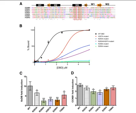
The FOXM1 DBD is essential for DNA bindingin vitro The Forkhead DBD [27] adopts a structure consisting of three α helices, three βsheets, and two wings with the main contact points with the DNA major groove located in helix H3. Amino acid residues involved in the base-specific contacts are highly conserved among all Fork-head members [28]. To investigate the importance of direct interaction of the FOXM1 DBD with the FKH consensus onin vitrobinding, four highly conserved H3 amino acids were chosen to generate mutations that are predicted to interfere with DNA binding. (Fig. 1a; H3 residues selected for mutation are indicated with red box). Four point mutations, N283A, R286A, H287A, S290A. and one double mutant N283A/H287A (Fig. 1a) were engineered and used to generate FOXM1 DBD-GST-tagged proteins.
The DNA binding activity of the wild-type (WT) and mutated FOXM1 proteins was assessed by fluorescence polarization (FP) binding assays using a carboxyfluores-cein (6FAM) -tagged dsDNA oligonucleotide containing the FKH consensus sequence (5′AAACAAACAAA-CAATC). Figure 1b shows that all mutations signifi-cantly decrease the binding affinity compared to the WT FOXM1 protein. The Kd for WT FOXM1 for the
con-sensus DNA is 1.10 ± 0.02μM, compared to 3.04 ± 0.10 μM for H287A. For both the N283A and double mutant (N283A/H287A) binding did not reach saturation (Kd>5
μM). No significant binding was observed for the R286A or S290A mutants. While double mutation (N283A/ H287A) appeared to show an additive inhibitory effect compared to either individual mutations, it still was not as effective as R286A and S290A, which led to complete loss of DNA binding.
reporter system. Identical mutations were therefore gen-erated in the DBD of the full-length FOXM1B protein, as this isoform is the predominantly overexpressed iso-form in cancer [4, 29]. Transient transfections were per-formed using inducible FOXM1B constructs and the level of transactivation from the FOXM1 DBD mutations com-pared to WT, following addition of doxycycline. An add-itional control was performed based on a T596A mutation in the transactivation domain of FOXM1 that has been previously shown to reduce transcriptional activity [17].
Constructs were co-transfected into HeLa TRex cells with a firefly luciferase (FLuc) reporter (6xDB) plasmid con-taining six copies of the FKH consensus sequence (AAA CAAACAAAC) and a renilla luciferase reporter (RLuc) plasmid to control for transfection efficiency. Figure 1c shows that all mutated FOXM1 proteins had a significant reduction in transcriptional activity compared to the WT FOXM1B construct (approximately 25-75 % lower), which had approximately 10-fold activation on induction with doxycycline.
Fig. 1Mutation of the FOXM1 DBD inhibits DNA binding.aSequence alignments of the DBD for a number of Forkhead family members with the secondary structure shown schematically above. The residues used to generate point mutations are outlined in red. (*) conserved amino acids. H1-3 areα-helices, the orange arrows areβstrands, and W1-2 are winged domains.bPlot showing relative change of polarization of a fluorescently-labeled (6FAM) dsDNA FKH consensus oligonucleotide upon addition of increasing concentrations of GST-FOXM1 WT or mutant DBD proteins. The FP assay provides a quantitative method and non-disruptive method to determine FOXM1 affinity for target by measuring the fluorescence polarization signals from the FAM-labeled FKH consensus (see Materials and methods). Data are plotted as % binding and show mean ± SD of triplicate experiments. (WT Kd= 1.10 ± 0.02μM, H287A Kd= 3.04 ± 0.10μM).cPlots showing relative luciferase activity of a 6X
[image:3.595.59.536.89.485.2]To confirm that the FOXM1 DBD mutations reduce transcriptional activity in the context of endogenous genomic binding sites rather than the 6x FKH consensus alone, the cell reporter assay was repeated using pro-moter regions (approximately 200 bp) from known FOXM1 target genes,CCNB1andPLK1[30, 31], neither of which contain a FKH consensus motif. Following doxycycline treatment (1 μg/mL), the level of induction by the FOXM1 WT construct was approximately three-fold lower for expression driven by both theCCNB1and
PLK1reporters than the 6x FKH consensus (Fig. 1d and Additional file 1: Figure S1). Results for both of these promoter constructs showed significantly reduced tran-scriptional activation following doxycycline induction with three of the FOXM1 DBD mutations compared to the WT (R286A, H267A, and S290A). The degree of re-duction was lower compared to the artificial 6xDB, how-ever the activity of these promoters is not exclusively regulated by FOXM1, for exampleCCNB1expression is known to be regulated by an NF-Y binding site con-tained within this 200 bp region [32] and indeed both of these natural promoter regions have a higher level of basal activity than the artificial FKH domain leading to lower levels of induction with doxycycline. In contrast, there was no significant difference observed when the assay was performed using the non-FOXM1 responsive promoters, CYP1B1 and SV40 (Additional file 1: Figure S1). These results therefore confirm that specific point mutations in the DBD can reduce the transcriptional ac-tivity of FOXM1 at an endogenous binding site.
Generation of epitope tagged FOXM1 cell lines for ChIP-seq
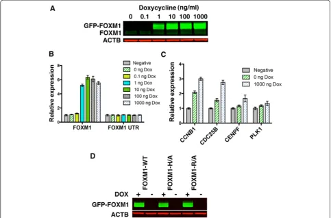
To compare the genome-wide distribution of binding sites for WT and DBD mutants of FOXM1, we gener-ated stable cell lines expressing inducible FOXM1B (WT or DBD mutants) for use in ChIP-seq experiments. The FOXM1B construct was tagged with an N-terminus GFP tag to enable ChIP-seq using an anti-GFP antibody to distinguish the expressed protein from the endogenous FOXM1. Transient transfection of FOXM1B or a GFP only control in HeLa TRex cells confirmed efficient in-duction of protein expression following doxycycline treatment (1 μg/mL) (Additional file 1: Figure S2A). ChIP-qPCR was then performed on five FOXM1 target genes to assess pull-down efficiency using an anti-GFP antibody. These results showed the enrichment of GFP-FOXM1 at the target genes with no detectable binding of GFP alone (Additional file 1: Figure S2B).
The Flp-In system (Invitrogen) was next used to gen-erate stable cell lines expressing GFP-FOXM1 WT by the targeted insertion of an expression construct at a single transcriptionally active genomic site. This system was utilized to ensure that the WT and mutant FOXM1
proteins were expressed at equivalent levels following in-duction. HEK293Flp-In cells stably expressing the Tet repressor at high levels were first generated by transfec-tion of a TetR plasmid under the control of a CMV pro-moter. These cells were transfected with WT or mutant FOXM1-GFP plasmids together with Flp recombinase plasmid to generate inducible cell lines. In the absence of doxycycline, no detectable expression of the GFP-FOXM1 protein or transcript was observed (Fig. 2a and b), while expression was induced after addition of doxy-cycline at concentrations above 1 ng/mL, giving approxi-mately 50-fold higher levels of total FOXM1 protein (1000 ng/mL) compared to the uninduced cells. There was no significant change in the level of the endogenous FOXM1 protein or mRNA following overexpression of the GFP-FOXM1 as shown by western blotting and qPCR for the FOXM1 UTR [33]. This result contrasts the hypothesis, proposed by Halasi et al. [33], that FOXM1 expression is primarily regulated by a positive auto-regulatory loop. Isoform specific qPCR (Additional file 1: Figure S3) showed that only GFP-FOXM1B pro-tein was significantly upregulated following doxycycline induction.
Doxycycline induction of WT GFP-FOXM1 led to a small but significant increase in transcript levels for a number of known FOXM1 target genes as assessed by qPCR (Fig. 2c).CCNB1andCDC25Blevels increased by approximately 2.8 fold, while CENPF showed a smaller increase of approximately 1.3-fold when compared to the parental HEK293 TetR cell line (negative) and there was no significant change in levels of PLK1. When ex-pression is compared before and after the addition of doxycycline all four genes showed a slight increase in ex-pression compared to the parental cell line. The generally low level of induction observed is similar to that described in other FOXM1 overexpression studies [3, 31].
nuclear localization of the WT and mutant GFP-FOXM1 proteins in the majority of cells (Additional file 1: Figure S5).
GFP-FOXM1 binding recapitulates endogenous FOXM1 binding
To confirm that the genomic distribution of GFP-FOXM1 expressed protein matched that of endogenous FOXM1, ChIP-seq data were compared between HEK 239 Flp-In and HEK293 GFP-FOXM1 cells using with a FOXM1 or GFP antibody, respectively, following induc-tion with doxycycline (1 μg/mL). In addition, the gen-omic binding of GFP in GFP-only expressing cells was tested, this was done by ChIP-qPCR as it was not pos-sible to generate a sequencing library due to very low levels of enrichment following pull-down. No enrich-ment of 10 known FOXM1 binding sites was observed in the GFP-only expressing cell line compared to the GFP-FOXM1 WT cells (Additional file 1: Figure S6). This confirms that binding of GFP-FOXM1 is due to the
specific interaction of FOXM1 with target DNA rather than non-specific GFP recruitment.
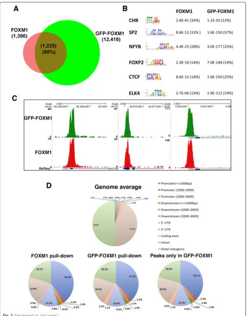
[image:5.595.56.540.90.408.2]sites shared between endogenous FOXM1 and the GFP-FOXM1 increased to 3,173 representing approximately 26 % of the total GFP-FOXM1 binding sites. The ap-proximately 10-fold increase in high confidence peaks called in the GFP-FOXM1 samples compared to en-dogenous FOXM1 is possibly due to the higher affinity of the GFP antibody and the relatively poor performance of commercial FOXM1 antibodies in ChIP-seq or the in-creased level of GFP-FOXM1 compared to endogenous FOXM1 protein in these cell lines.
Motif analysis using MEME (Multiple Em for Motif Elicitation) [35] of the FOXM1 and GFP-FOXM1 da-tasets revealed similar sets of enriched sequences sug-gesting that the presence of the GFP tag does not significantly affect the binding specificity of FOXM1 (Additional file 1: Table S2). The six highest enriched motifs (ranked by FDR) in the endogenous FOXM1 pull-down are shown in Fig. 3b and the P values from the GFP-FOXM1 are shown for comparison. Both sets contained the FKH motif as well as the CHR and CCAAT (NFYA) box motifs that have also previously been shown to be associated with FOXM1 binding [19, 20]. The percentage of peaks containing the FKH motif was similar in both sets (approximately 14 %) and critically when the peaks present only in the GFP-FOXM1 dataset were analyzed, these also showed a similar significant enrichment of the FKH motif (1.73E-152) in approximately 14 % peaks (Additional file 1: Table S2). Although the FKH motif is enriched, the rela-tive number of peaks containing the consensus sequences is lower than that reported for some other transcription factors such as POU5F1, where approximately 70 % of peaks match the POU5F1 PWM motif [36]. This therefore highlights the observation that the majority of FOXM1 peaks, both in the endogenous and epitope tagged ChIP-seq, do not contain a consensus FKH motif.
In both the FOXM1 and GFP-FOXM1 datasets, peaks were identified in the promoter regions of known FOXM1 target genes including CDK1 [30], FZR1 [20], andKNSTRN[20] (Fig. 3c), confirming that the GFP tag does not affect FOXM1 recruitment in vivo. Manual inspection of some of the unique GFP-FOXM1 peaks revealed small peaks at the same location in the en-dogenous FOXM1 dataset. This suggests that additional peaks present in the GFP-FOXM1 dataset are low
affinity endogenous sites, in which the endogenous FOXM1 ChIP-seq shows insufficient enrichment over the input to be considered significant by the MACS peak caller (Additional file 1: Figure S7A). Furthermore, analysis of read counts in all the 12,418 regions iden-tified as peaks in the GFP-FOXM1 shows high cor-relation (r = 0.70-0.75) with endogenous FOXM1 ChIP-seq peaks (Additional file 1: Figure S7B). This indicates that even when the endogenous FOXM1 signal is insufficient to be called as a peak, it still shows a significant correlation to the GFP-FOXM1 signal and is not present in input controls. This lends fur-ther weight to our contention that the signal detected by FOXM1-GFP ChIP-Seq is not spurious.
Comparison of the distribution of peaks across the genome using the Cis-regulatory Element Annotation System (CEAS) tool [37] also shows a very similar pat-tern between endogenous FOXM1 and GFP-FOXM1 (Fig. 3d), with the greatest proportion in promoter/5′ UTR regions (approximately 53 % and approximately 56 % for GFP-FOXM1 and endogenous FOXM1 re-spectively compared with 2.8 % for the genome average distribution). Furthermore, peaks present only in the GFP-FOXM1 and not the endogenous FOXM1 pull-down, showed a similar genomic distribution to the en-dogenous FOXM1, with the majority of events found in gene promoter regions. The reproducibility, distri-bution, and motif analysis of the additional peaks present in the GFP-FOXM1 pull-down strongly sug-gests that these are not non-specific and are genuine endogenous FOXM1 sites, which are beyond the level detectable with the FOXM1 antibody. A similar phenomenon has been previously observed for other transcription factors in which improved methodology using higher affinity antibodies leads to an increased number of binding sites.
Gene ontology analysis using the Reduce and Visualize Gene ontology tool (ReViGo) [38] tool of overrepre-sented GO terms associated with genes located within 50 kb of FOXM1 and GFP-FOXM1 binding peaks high-lights an enrichment in the known functional roles for FOXM1 including cell cycle, chromosome segregation and mitotic spindle formation processes (Additional file 1: Figure S8). In addition, processes related to methylation were also identified, which is in concordance with (See figure on previous page.)
previous studies linking FOXM1 to promoter hyperme-thylation [39, 40]. Analysis of enriched GO terms associ-ated with peaks identified only in the GFP-FOXM1 dataset highlighted processes associated with S phase and M/G1 transition of the cell cycle, both of which are known functions of FOXM1 [41, 42] in addition to its major role in the G2/M transition (Additional file 1: Table S3). This functional categorization of peaks suggests that that the GFP-FOXM1 binding pattern reflects that of endogenous FOXM1.
Overall, these analyses demonstrate that the inducible GFP-FOXM1 expression system in HEK293 cells is a suitable model for FOXM1 binding studies. Recruitment occurs at biologically relevant genomic locations that are representative of endogenous FOXM1 binding, thus GFP-FOXM1 expression is able to compete with en-dogenous FOXM1.
Comparison of genomic binding sites of WT GFP-FOXM1 to DBD mutants
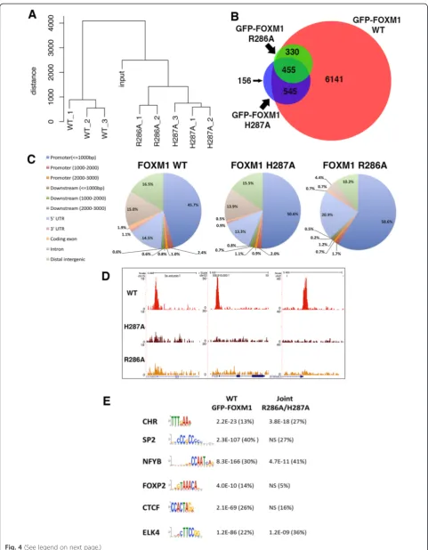
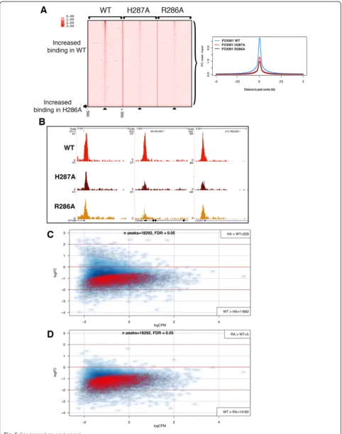
ChIP-seq was next performed to compare the genomic binding sites of GFP-tagged WT FOXM1 to the two GFP-tagged DBD mutant versions of FOXM1. Three biological replicates were performed for the WT and H287A and two for the R286A cell line. All cells were treated for 24 h with doxycycline (1 μg/mL) prior to chromatin extraction.
Each replicate for each cell line resulted in 25–30 million reads (Additional file 1: Table S1). For each construct, peaks in common between each replicate showed a good level of concordance. WT, H287A and R286A replicates showed 7,473 (72 % overlap), 1,169 (60 % overlap) and 804 shared binding events (81 % overlap), respectively (Additional file 2). Comparing this GFP-FOXM1 dataset with that in the preceding section again showed a high level of overlap with 6,443 shared peaks (86 % concordance), further con-firming the high degree of reproducibility for the GFP-FOXM1 ChIP-seq. Furthermore, hierarchical clustering (Fig. 4a) showed that the replicate samples grouped to-gether indicating a reproducible dataset. Overlapping the peaks from the WT and DBD FOXM1 mutants (Fig. 4b) showed that the majority of H287A (86.5 %) and R286A (98.5 %) peaks are a subset of WT GFP-FOXM1 binding sites. For H287A, although 156 peaks did not overlap with the WT or R286A peaks, most overlapped with at least one WT replicate, suggesting that they are not novel binding sites. The majority of R286A peaks are also shared by H287A (approximately 57 %). Of particular note is the observation that both FOXM1 DBD mutations significantly reduce the over-all level of genomic binding. The R286A mutation was more effective at reducing binding compared to H287A, which is consistent with the in vitro DBD FP
analysis showing that R286A has significantly lower binding than H287A (Fig. 1c) for the FKH consensus.
To identify any differences in the genomic distribution of the binding regions, CEAS was used to analyze the lo-cation of the binding peaks in the WT and DBD FOXM1 mutants (Fig. 4c). Confirming earlier results (Fig. 3d), the majority of WT FOXM1 peaks were observed to be in promoter/5′UTR regions (63.6 % compared to gen-ome average of 2.8 %). In both the H287A and R286A mutants, there was an increase in peaks located in the promoter/5′ UTR regions (66.8 and 81.9 %, respect-ively), which was more evident in the binding sites com-mon to both mutants (86 %). These data suggest that the DBD mutant binding is preferentially retained in these regions and is also more effectively competed at other sites by endogenous WT FOXM1 protein. It is notable that the majority (80–90 %) of WT FOXM1 peaks are almost completely absent in the DBD mutant binding peaks. Figure 4d shows the ChIP-seq enrich-ment profiles for WT, R286A and H287A FOXM1 at three representative known FOXM1 promoter binding sites with binding peaks only being detected in the WT sample.
To determine whether any significantly enriched mo-tifs are present in the peaks where DBD mutant FOXM1 binding is retained, MEME analysis was performed (Fig. 4e; Additional file 1: Table S4). For WT GFP-FOXM1, enriched motifs were similar to the earlier dataset (Fig. 3b) including the FKH and CCAAT box motifs. When the enriched motifs present uniquely in the WT GFP-FOXM1 were compared with those retained in the H287A and H286A mutants, it was found that the CCAAT box was common to all, while the FKH motif was only enriched in WT. This con-firmed that the binding peaks retained in both the FOXM1 DBD mutants are not regions enriched in the FKH consensus. Overall, these data indicate that FOXM1 binding requires a functional DBD but the targets are in fact quite divergent, encompassing both canonical FKH and non-consensus DNA sites.
both DBD mutant ChIP-seq datasets, although with an overall loss in the average signal density. We also con-firmed this observation by ChIP-PCR for three known promoter binding sites of FOXM1 (Additional file 1: Figure S9). Figure 5b illustrates three examples of differ-ential binding of the DBD mutant proteins compared to the GFP-WT FOXM1 at high-affinity genomic binding sites.
To investigate whether any particular functional pro-cesses are associated with binding regions retained in the mutant DBD FOXM1 samples, the web-based tool GREAT (Genomic Regions of Enrichment Annota-tions) [43] was next employed. Output from GREAT identified a significant enrichment in cell cycle pro-cesses, particularly those related to M phase control (Additional file 1: Table S5), which correlates with the established functions of FOXM1 in cell cycle regula-tion [2]. A similar analysis on regions bound only by WT GFP-FOXM1 and not the DBD mutants showed that there was enrichment of processes associated with nucleosome organization and chromatin structure, which is consistent with the identified roles of Fork-head family members in chromatin remodeling [44]. Enrichment of processes associated with epigenetic regulation of gene expression was also revealed, which is consistent with the known link of FOXM1 to the indirect regulation of methylation through DNMT3b recruitment [39] or via HELLS expression [40]. Several other enriched processes related to translation were also identified in the GFP-FOXM1 WT dataset (Additional file 1: Table S5).
These data suggest that high affinity FOXM1 binding sites are retained in the DBD mutants and are associated with its key functions as a regulator of the cell cycle, whereas the sites where binding is lost from the DBD mutants reflect regions associated with other roles such as chromatin remodeling and epigenetic regulation. The absence of FKH motif enrichment in the DBD mutants provides strong evidence for alternative mechanisms of FOXM1 recruitment possibly mediated by indirect tethering.
To identify any statistically significant regions of differential binding between FOXM1 WT and DBD
mutants, differential binding analysis (DBA, see Materials and methods) was performed. For this analysis all peaks identified in each stable cell line were first used to generate a consensus dataset containing 18,292 peaks. In the H287A mutant compared to the WT, 11,682 peaks were identified with decreased binding, and 229 peaks with increased binding (FDR <0.05) (Fig. 5c and Additional file 3). A similar analysis of the R286A mutant identified 14,160 regions with de-creased binding compared to the WT and only five with increased binding (Fig. 5d and Additional file 3). No enrichment of biological processes associated with the 229 peaks with increased binding was found. These data further confirm that the majority of FOXM1 gen-omic binding sites are dependent on the presence of a functional DBD. The R286A mutation, which showed no discernable DNA binding in the FP assays (Fig. 1b), showed the greatest reduction in binding, (77 % of peaks significantly reduced, FDR <0.05). In keeping with the requirement of the DBD for binding, less peaks (64 %) showed decreased binding in the H287A mutant, which is consistent with the residual DNA binding activity determined by FP.
Overall, our data support the observations of Chen
et al. [20] who also identified a set of FOXM1 binding sites lacking the FKH consensus motif, where recruit-ment was mediated through binding to the MuvB com-plex. However, our study additionally demonstrates that conserved amino acids of helix H3 in the DBD are none-theless required for DNA binding even at non-FKH con-taining sites. For example, Fig. 5a illustrates that binding intensity is reduced in both DBD mutants compared to the WT even at highest intensity sites in the WT. Taken together our observations support two concurrent mech-anisms for FOXM1 binding: the first being direct inter-action of FOXM1 with target DNA occurring at the majority genomic binding sites, and second being recruit-ment or stabilization by chromatin bound proteins such as MuvB. As peaks missing in the DBD mutants include both FKH and divergent motifs, this strongly suggests that FOXM1 binding does not occur exclusively at consensus FKH sites. We further discuss these motifs further below.
(See figure on previous page.)
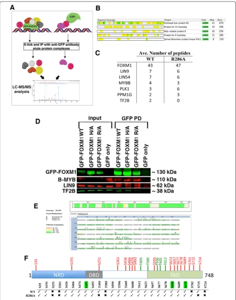
RIME proteomic analysis of WT and DBD mutant GFP-FOXM1
To investigate whether the reduced DNA binding of the DBD mutant proteins is due to perturbation of protein-protein interactions involved in the recruitment, prote-omic analysis of the FOXM1 interacting proteins was performed by RIME (Rapid immunoprecipitation mass spectrometry of endogenous proteins) [45] for both the WT GFP-FOXM1 and the R286A DBD mutant. Formaldehyde-fixed chromatin was isolated from HEK293 cells expressing either GFP only, GFP-FOXM1 WT, or GFP-FOXM1 R286A and immunoprecipitation was performed with an anti-GFP antibody followed by LC/MS-MS (Fig. 6), to identify associated proteins. To eliminate any non-specific interactions with GFP, which can occur through cross-linking of the GFP to highly abundant proteins such as keratins and riboso-mal sub-units, proteins identified in the GFP only ex-pressing cell line were not considered in the WT or R286A samples. High confidence interacting proteins were then identified using the Scaffold proteomic ana-lysis tool [46], details of the proteins identified are shown in Additional file 4. In the WT, several proteins previously shown to interact with FOXM1 were identi-fied, including components of the MuvB complex [19] (LIN9, LIN54), B-MYB [47] and PLK1 [30]. Figure 6b shows the sequence coverage from Scaffold for the top five high confidence proteins. These results show that the presence of the GFP epitope tag does not signifi-cantly affect the interaction of the GFP-FOXM1 fusion protein with known partners of endogenous FOXM1. Overlap of proteins identified in the WT and R286A replicates (identified by unique peptides present in at least three out of the four samples) revealed 21 common to both, while eight were present in the WT only and 15 in the R286A mutant (Additional file 1: Table S6 and Figure S10).
When the highest confidence interacting proteins identified in the WT were compared with those in the R286A DBD mutant (Fig. 6c), it was of note that the FOXM1 DBD mutant was still able to interact with MuvB components (LIN9, LIN54), PLK1 and B-MYB. This interaction was further confirmed by co-IP for GFP-FOXM1 with B-MYB and LIN9 (Fig. 6d). We ob-served that the general transcription factor TF2B, which is known to directly interact with FOXM1 [48], was
identified in all the WT FOXM1 RIME samples, how-ever for the DBD mutants T2FB was seen in only one of four replicates of the R286A mutant. It is possible that the inability of the DBD mutant to bind DNA impairs the assembly of active transcriptional complexes despite preserving the direct binding of core FOXM1 interacting proteins.
In both the WT and DBD mutant RIME samples, other components of the MuvB complex [19], LIN37 and LIN54 were also identified, albeit at lower confi-dence (Additional file 1: Table S6). The identification of these known FOXM1 binding proteins in the R286A mutant, which show reduced chromatin recruitment, could be due to interaction with nuclear non-chromatin bound FOXM1. Other proteins identified include the serine/threonine phosphatases PPM1G and PP1G; and it is of note that a subunit of the related phosphatase PP2A is known to interact with FOXM1 [49], while PPM1G is thought to have a key role in cell cycle regula-tion and in the DNA damage response [50]. Analysis of the biological pathways associated with the WT GFP-FOXM1 interacting proteins using the GeneGo Meta-core tool (Additional file 1: Figure S11), showed a range of processes that correlate with known FOXM1 roles including cell cycle, DNA damage and nucleosome assembly.
FOXM1 activity is regulated by a series of CDK-cyclin mediated phosphorylation steps [17, 51], which are es-sential for the formation and nuclear localization of the transcriptionally active protein at the correct stage of the cell cycle [17, 52]. We therefore compared serine and threonine phosphorylation modifications present in both the WT and R286A DBD mutant FOXM1 proteins isolated by GFP pull down and subjected to RIME. Ana-lysis using the Proteome Discoverer with the PhosphoRS algorithm [53] revealed the presence of many previously described phosphorylation sites that are required for FOXM1 transactivation (Fig. 6e and f; Additional file 1: Figure S12 and Table S7; Additional file 5). These data also show that the R286A mutant protein under-goes a similar pattern of phosphorylation as the WT protein. Our analysis identified one of two known PLK1-mediated serine phosphorylation sites (S715 but not S724) in the transactivation domain (TAD) of FOXM1 [30]. It is unclear why this phosphorylation event was only seen in the WT and not the R286A mutant and this (See figure on previous page.)
might suggest that phosphorylation happens after FOXM1 has bound chromatin. In addition, this analysis identified several novel serine phosphorylation sites present in both WT and mutant FOXM1, including S608 and S680 (Additional file 1: Table S7), however their biological relevance is unclear at present.
Overall, RIME has confirmed previously known pro-tein interactions and identified novel FOXM1 propro-tein interactions and post-translational modifications. The majority of known and novel phosphorylation marks were found in both mutant and WT protein, with the notable exception of PLK1 target S715. Furthermore, the significant reduction in DNA binding for the R286A mu-tant protein shown in the ChIP-seq analysis is not due to this mutation affecting protein-protein interactions or phosphorylation, as overall similar profiles to the WT were observed.
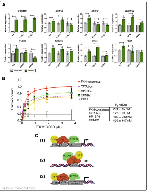
FOXM1 DBD mutations inhibit expression of FOXM1-regulated genes
To investigate the effect of the DBD mutations on FOXM1-regulated gene expression, qPCR was used to measure the expression levels of several known target genes (Fig. 7a). Transcript levels were compared in the WT and DBD mutant cells (H287A and R286A) between ± doxycycline. The level of GFP-FOXM1B induction was first measured to ensure comparable expression of the WT and DBD mutants. Indeed by qPCR, the addition of doxycycline addition led to an approximately 13-fold in-crease in GFP-FOXM1 expression in both the WT and DBD mutant cell lines compared to the non-induced WT GFP-FOXM1 cells (Fig. 7a). Next, the expression of seven known FOXM1 target genes was measured;
AURKB, CENPF, KNSTRN, CCNB1, CDC25B, NEK2, and PLK1.In each case, a significant increase in expres-sion was observed in the GFP WT-FOXM1 cell line fol-lowing induction by doxycycline (P<0.05). Although the relative increase in expression was small (in the range of 1.3-fold forAURKB to 1.8-fold for NEK2), this is in the context of endogenous FOXM1 and comparable to pre-vious studies [3, 31]. In contrast, for the R286A DBD mutant there was no significant difference in transcript levels following induction, whereas for the H287A DBD
mutant, three transcripts (CENPF, CCNB1, and PLK1) showed no significant change on induction This latter re-sult may reflect a higher residual level of DNA binding ac-tivity that is observed in the FP assay and by ChIP-seq when compared to R286A. This is supported by inspec-tion of the ChIP-seq data for three transcripts (KNSTRN,
CDC25B, andNEK2) that show different responses in the H287A compared to the R286A mutant (Additional file 1: Figure S13). In each case the peak in the promoter region of the gene is significantly smaller in the R286A sample.
These data show that mutations in the DBD, which significantly impair the DNA binding interaction of FOXM1, reduce the induction of downstream target genes. The reduction in transcription seen in the R286A DBD mutant correlates with RIME analysis (Fig. 6c and d) showing a decreased association with the general transcription factor TF2B, which forms part of the RNA pol II pre-initiation complex [54]. The R286A DBD mutant reduces expression to levels seen in the uninduced cells, which suggests that it is unable to block the transcription mediated by endogenous FOXM1 protein binding.
FOXM1 binds to lower affinity consensus sequences Our ChIP-seq results show that overall FOXM1 binding is significantly depleted when the DNA binding affinity is reduced, demonstrating the requirement of a func-tional DBD. However, only approximately 14 % of the total binding peaks in the WT FOXM1 contain a canon-ical FKH consensus sequence suggesting that FOXM1 DBD must also be required for recruitment to non-consensus motifs. FOXM1 binding to alternative motifs has also been supported by high-throughput studies which show that Forkhead factors (including mouse Foxm1) bound two distinctly different DNA sequence motifs [55]. Furthermore, when the Systematic Evolution of Ligands by EXponential Enrichment (SELEX) method was combined with ChIP-seq, a greater diversity of FKH binding sites was also identified [56]. To confirm whether FOXM1 binds directly to non-consensus se-quences identified by ChIP-seq, FP assays were used to determine the in vitro binding affinity of GST-FOXM1 for non-canonical DNA sequences (Fig. 7b). Regions (See figure on previous page.)
highly enriched in FOXM1 binding at gene promoters were selected from the ChIP-seq dataset, using se-quences located at the peak center with partial similarity to the FKH motif (enriched in A/T bases). Additionally, a previously reported non-consensus FOXM1 binding site in TATA box of MYC [57] was included. As ex-pected the FOXM1B DBD associates with a positive control FKH consensus sequence in a dose dependent manner (Kdof 224 ± 45 nM). The FOXM1B DBD also
associates with several non-consensus sequences tested including CCNB2 (Kd= 436 ± 147 nM), HP1BP3 (Kd=
489 ± 233 nM), and the MYC TATA box (Kd= 177 ± 79
nM), but only weakly with the others such as PLK1 where a Kd value could not be determined. Similar
re-sults were observed by Electrophoretic Mobility Shift Assay (EMSA) (Additional file 1: Figure S14) although the apparent binding affinities measured were lower (Kd
of 346 ± 24 nM for FKH consensus).
Collectively, these results together with previousin vitro binding studies [55, 58] demonstrate that FOXM1 can bind to DNA sequences with significant sequence diver-gence from the canonical FKH motif. Furthermore, our results using ChIP-seq and expression analysis with FOXM1 DBD mutants show that recruitment to non-consensus sites is also critical for transcriptional activity. Overall, our data support a mixed model of direct FOXM1 recruitment to chromatin at lower affinity non-consensus sequences mediated by protein-protein interactions.
Discussion
The aim of this study was to investigate the mechanisms regulating FOXM1 DNA binding in a cellular context. FOXM1 has previously been demonstrated to bind to the FKH consensus motif in vitro. In common with other Forkhead factors, this interaction is mediated by the DNA binding domain [57], although with lower af-finity compared to other FKH factors [59]. Since FOXM1 has also been suggested to bind at non-FKH consensus sequences mediated by indirect protein-protein interactions, we examined the role of direct ver-sus indirect DNA binding in FOXM1 recruitment using GFP-tagged FOXM1 expressed in HEK293. Exogenous expression of a GFP epitope-tagged protein enabled the
generation of cell lines with specific point mutations in the DBD of FOXM1 for use in ChIP-seq analysis while avoiding the known phenotypic responses caused by re-duced FOXM1 transactivation [60], such as inhibition of the cell cycle and mitotic catastrophe.
Results for GFP-FOXM1 WT showed that <14 % peaks contained a consensus FKH motif, which is consistent with previous results investigating endogenous FOXM1 binding in U2OS cells [20]. However, in contrast to the prior study, our results provide strong evidence that DNA recognition by the FOXM1 DBD remains critical for recruitment to non FKH-consensus genomic binding sites as opposed to an indirect mechanism mediated solely by protein-protein interactions. Our results sug-gest that FOXM1 binding occurs by a process of assisted recruitment, as proposed by Rabinovich et al. [61] for the E2F transcription factor, in which most binding sites lack a consensus motif with binding mediated by add-itional transcription factors at lower affinity DNA target sites. Indeed, genome-wide studies have highlighted sev-eral transcription factors that show low enrichment of the consensus sequence in their binding sites [61], sug-gesting that this model of assisted recruitment is a more general mechanism of transcription factor binding.
Two models previously proposed for the assembly of FOXM1 with the MuvB complex and B-Myb protein on cell cycle-regulating promoters are shown in Fig. 7c. The first (1) shows FOXM1 binding to FKH consensus se-quences in concert with recruitment of the MuvB com-plex present at cell cycle homology (CHR) sequences and B-Myb bound at a MYB binding site in close proximity [19]. In the second (2), FOXM1 is indirectly recruited by the MuvB complex at CHR sites together with B-Myb, without contribution of DBD-DNA interactions [20]. A third model (3) that is inferred by our results shows an alternative, mixed mode for FOXM1 recruitment. This mechanism involves a degree of DNA binding at lower af-finity, non-canonical sites that is also facilitated by protein-protein interactions. This latter model would account for the lack of FKH motifs in the peaks enriched in FOXM1/FOXM1-GFP pull-down, and is supported by our data showing that mutations of the DBD in FOXM1 significantly reduce genome-wide binding at both (See figure on previous page.)
Fig. 7FOXM1 transcriptional activity requires direct chromatin interaction involving recruitment to non-consensus sequences.aqPCR analysis of the mRNA transcript levels in GFP-FOXM1 WT or mutant (H287A or R286A) cell lines treated ± doxycycline (1μg/mL) for 24 h showing the relative change in the levels ofFOXM1B,AURKB,CCNB1,CDC25B,CENPF, andPLK1. In each case the data are normalized to the minus doxycycline control. bBinding curves measured by fluorescence polarization analysis (assay details in the Materials and methods section), showing binding affinity of GST-FOXM1B DBD for 16-mer [FAM]dsDNA sequences present in FOXM1 binding peaks from the ChIP-seq dataset compared to the FKH consensus. The plot shows the fraction bound with increasing protein concentration. The table shows theKdvalues ± SD determined for each sequence.
consensus and non-consensus sites without affecting either the protein-protein interactions or the phosphoryl-ation status of FOXM1.
Considering ours and previous biophysical binding data, it is probable that FOXM1 exhibits some degree of DNA binding to the consensus FKH motif. Indeed, we observe that while FOXM1/FOXM1-GFP ChIP-seq re-veals a majority of peaks at non-FKH motifs, the FKH motif is still apparent present in a small but significantly enriched set (14 %,P= 10−144). Future experiments such as genetic deletion of discrete motifs by CRISPR would be needed to unambiguously establish which FKH or other motifs FOXM1 directly associates. Nonetheless, our data support the previously proposed model 1 as well as that suggested by model 3, namely that FOXM1 binding in chromatin operates through a mechanism dependent on a functional DBD assisted by local protein-protein recruitment regardless of sequence content. In support of this, mutant FOXM1 is unable to induce trans-activation of known FOXM1 target genes, all of which lack a canonical FKH consensus within the FOXM1 bind-ing site. It is also notable that bindbind-ing studies with WT FOXM1 confirmed that the protein additionally binds non-consensus sequences, further supporting an assisted model of FOXM1 binding whereby protein recruitment stabilizes the association.
Besides an additional model of FOXM1 chromatin re-cruitment, our work has also revealed novel protein-protein interactions of FOXM1 by use of the RIME methodology [45]. Newly identified FOXM1-interacting proteins include two phosphatases, PPM1G and PP1G, which may act in a similar manner to the PP2A phos-phatase that regulates FOXM1 activity during the cell cycle by controlling dephosphorylation to prevent pre-mature transcriptional activity [49]. The E3 Ubiquitin-Protein Ligase, UHRF1 was identified in all the WT GFP-FOXM1 samples. UHRF1 is known to regulate gene expression and the cell cycle and is overexpressed in many cancers [62], and it is of note that UHFR1 binds inverted CCAAT motifs [63] perhaps enabling require-ment of FOXM1 at these specific genomic binding sites.
The proposed model of assisted recruitment for FOXM1 (Fig. 7c, 3) could be applied more widely to in-clude binding sites other than those at cell-cycle related promoters, particularly at known sites of MuvB complex and B-Myb binding [19, 20, 47]. This is exemplified by the binding of FOXM1 in the COX2 promoter, which is mediated by interaction with the SP1 transcription factor at the SP1 binding site in the absence of a canonical FKH motif [64]. This further suggests that binding to non-consensus sequences at intronic and intergenic sites could be facilitated by other transcription factors.
The importance of the Forkhead DBD for direct inter-action with target DNA sites has been shown for a
number of other Forkhead factors, with disease-causing mutations having been identified in the FKH DBD domain [65, 66]. Both DBD mutations examined in this study are known naturally occurring missense mutations associated with the loss-of-function of the Forkhead factor. For ex-ample, the R553H mutation in FOXP2 (equivalent to FOXM1 R286) is associated with development of a severe speech language disorder [67], while a mutation of the same arginine residue in FOXC2 (R121C) is associated with a developmental disorder affecting the lymphatic vas-culature system. In the case of FOXM1, the fact that no similar disease-causing mutations have been associated with the DBD supports the critical importance of direct DNA binding for FOXM1 function as are likely to com-promise cell viability. Indeed, Korveret al. [41], suggests that any loss-of-function mutations in the FOXM1 DBD would be embryonic lethal and therefore are not repre-sented within the general population.
In fact, it is overexpression of WT FOXM1 that is asso-ciated with disease. In many human cancers FOXM1 over-expression promotes aberrant activation of FOXM1 target genes, contributing to oncogenesis and facilitating inva-sion, metastasis, and therapeutic resistance [6]. Evidence from this study along with results from our previous work [25, 26], showing inhibition of FOXM1 DNA binding by direct interaction with both a small molecule, FDI6 and with the natural product thiostrepton, suggests that target-ing the DNA bindtarget-ing domain of FOXM1 might provide a particularly effective means to ameliorate such a disease phenotype. We are currently developing chemical tools to test this hypothesis in future experiments.
Conclusions
Overall, we have demonstrated that the DNA binding domain of FOXM1, in common with other Forkhead factors is necessary for recruitment to DNA at consen-sus and non-consenconsen-sus FKH genomic binding sites and activation of down-stream transcriptional activation. Furthermore, we found that FOXM1 DBD mutants are unable to bind DNA yet maintain similar protein-protein interactions to the WT protein-protein. Finally, we iden-tified novel FOXM1 phosphorylation sites and found that the majority of all phosphorylation events are un-affected by DBD mutation. The mechanism of inter-action of FOXM1 with DNA is of critical importance to support any development of novel therapeutics designed to specifically target the DBD of FOXM1, and thereby reducing transactivation of FOXM1-regulated genes caused by overexpression in cancer.
Materials and methods Cell culture
Human HeLa TRex and HEK293 Flp-In cells were
supplemented with 10 % tetracycline free FBS and 5 μg/ mL blasticidin (Sigma) or DMEM supplemented with 10 % tetracycline free FBS and 100 μg/mL zeocin (Invitrogen), respectively.
Generation of FOXM1 expressing cell line
The EGFP-FOXM1B fusion plasmid was a gift from Dr M. Teh (Queen Mary University of London) and was cloned into the pcDNA4/TO plasmid (Invitrogen) for transient expression in HeLa TRex cells or into the pcDNA5FRT (Addgene) to generate stable cell lines. HEK293 Flp-In cells (Invitrogen) were first transfected with a pcDNA6/TR plasmid (Invitrogen) and selected with 5μg/mL blasticidin to generate a stable HEK293tetR Flp-In cell line. This cell line was co-transfected with the GFP-FOXM1 and pOG44 (Flp recombinase vector) and selected with 100μg/mL hygromycin (Invitrogen).
Fluorescence polarization assays
Fluorescence polarization (FP) assays were performed to assess the binding of the FOXM1 mutant DBD compared to the WT, using a 16mer dsDNA forkhead consensus se-quence (AAACAAACAAACAATC) labeled with carboxy-fluorescein at the 5′position on one strand. Assays were performed with serial dilutions of DBD proteins from 5 μM to 80 nM in FP-binding buffer (50 mM Tris–HCl pH 7.5, 5 mM MgCl2, 1 mM DTT, and 2 % glycerol).
Fluores-cence was measured using 485 nm excitation and 520 nm emission filters. Binding plots were generating after ex-pressing the data as the fraction bound over protein con-centration, with fraction bound defined as:
%bound¼ P−P0 P100−P0
(Where P0 is the polarization value at 0 % saturation,
P100is the polarization value at 100 % saturation, and P
is the observed fluorescence polarization (FP) at each concentration point.)
Luciferase reporter assays
The pGL4.26 plasmid (Promega) containing a minimum promoter upstream of the firefly luciferase gene was used to generate a reporter containing six copies of the FKH binding consensus as described by Major et al. [17]. For the 6X FKH consensus, a dsDNA sequence containing 72 bp with 5′phosphate groups was ordered from IDT (Integrated DNA technologies). For the CCNB1 and PLK1 reporters, 200 bp sequences from the CCNB1 and PLK1 promoters were synthesized and inserted into the promoter-less pGL3 luciferase vector (Promega). Sanger sequencing was performed commercially to con-firm the constructs (ATGC Inc.). The renilla control plas-mid, pRL-TKL, (Promega) was used to normalize for
transfection efficiency. Luciferase assays were performed using the Dual Luciferase Kit (Promega) as described by the manufacturer. Briefly, HeLa TRex cells were plated at 2 × 10⁴ cells/well in 96 well plates in antibiotic free media containing 10 % tetracycline free FCS and cultured overnight. Transfections were performed using lipofecta-mine 2000 following the manufacturer’s protocol. For each pcDNA4/TO pEGFP FOXM1 construct replicates of 10 were prepared; each transfection contained: 50 ng/well pEGFP-FOXM1, 50 ng/well pGL4.26 (6XBD), or pGL3 CCNB1/PLK1 and 10 ng/well pRL-TK. Plates were cul-tured overnight, then for each condition, five wells had 100 μL culture media added and five had media plus doxycycline (2μg/mL). These were incubated for an add-itional 24 h period. Luciferase activity was measured using the Dual Luciferase Kit (Promega). Luminescence readings were taken after the addition of each reagent and relative activity calculated by obtaining the ratio of firefly lucifer-ase to renilla to account for transfection efficiency, then the ratio of plus doxycycline to minus to give the relative induction. Four independent experiments were run with five replicates for each condition. The control non-FOXM1 responsive promoter for SV40 in pGL4.10 was obtained from Promega and the human CYP1B1 pro-moter was cloned from human gDNA using primers con-tainingAcc65I/EcoRVrestriction sites generating a 600 bp product:
Fw: GACTGGTACCGGATTCCTGATCTCGCCGCA AGAACTGG
Rv: GACTGATATCCGTTGAGATTGAGACTGGGG GTCGG
The PCR product was digested and ligated into the pGL4.10 vector (Promega).
Western blots
Western blots were performed using antibodies for anti-FOXM1 (sc-502) and anti-HA (sc-543) and anti-β-actin (ab6276) purchased from Abcam. Fluorescent imaging of GFP-FOXM1 was performed using an anti-GFP antibody (ab290) purchased from Abcam. Cell lysates were pre-pared using RIPA buffer (20 mM Tris–HCl, pH 7.5, 150 mM NaCl, 1 mM Na2EDTA, 1 mM EGTA, 1 % NP-40, 1
% sodium deoxycholate, 2.5 mM sodium pyrophosphate, 1 mM ß-glycerophosphate, 1 mM Na3VO4) with
room temperature and probed with FOXM1, HA, or GFP antibody 1:1,000 andβ-actin 1:5,000 overnight at 4 °C. For detection, the blot was incubated with LiCor IRDye sec-ondary antibodies; 680LT goat anti-rabbit IgG and 800LT goat anti-mouse IgG both at 1:10,000 and visualized using an Odyssey scanner.
Immunofluorescence
Cells were plated 4 × 10⁴/well in Ibidi treated 8-well cul-ture slides (Ibidi) and left to adhere overnight. Following doxycycline treatment for 24 h the cells were washed 1X with PBS and fixed with 4 % methanol-free formalde-hyde at room temperature for 15 min. After washing 3X with PBS, cells were permeabilized and blocked using blocking buffer (PBS/5 % normal goat serum/0.3 % Triton-X 100) for 1 h at room temperature. GFP anti-body was diluted in antianti-body blocking buffer (PBS/1 % BSA/0.3 % Triton-X 100) FOXM1 (1:500) and added to appropriate wells leaving control wells with buffer only. Culture wells were incubated at 4 °C overnight followed by washing 3X with PBS. Secondary antibodies were di-luted 1:2,000 in antibody dilution buffer, using either goat anti-Rabbit Alexa Fluor 488 (Invitrogen) and added for 1 h in the dark at room temperature. Culture wells were washed 3X with PBS, with one wash containing DAPI (1 μg/mL) for nuclear staining. Liquid was removed from wells and anti-fade mounting media (Ibidi) added to each well. Plates were stored in the dark at 4 °C and vi-sualized using an inverted Leica DMI6000B microscope.
Quantitative real-time PCR analysis
RNA was collected after the indicated time-points and qPCR was performed using Power Sybr mix (ABI) on a CFX96 Real-time thermal cycler (Bio-Rad). Total RNA was extracted using the RNeasy mini kit (Qiagen) fol-lowing the manufacturer’s protocol. cDNA was prepared from 1 μg RNA using Maxima reverse transcriptase (Fermentas) following the manufacturer’s protocol. qPCR was performed in triplicate in 10 μL reactions with Power sybr mix (ABI) using Qiagen quantitect primers for B2M, ACTB, CCNB1, CDC25B, and FOXM1 and additional primers shown below. ACTB and B2M were used as housekeeping genes for normalization of the data. PCR conditions were: 95 °C 10 min, 40 cycles of 95 °C for 15 s, and 60 °C for 30 s followed by a dis-sociation curve (60–95 °C). Relative expression levels were calculated using the delta delta CT method [68].
Chromatin immunoprecipitation
ChIP experiments were performed as previously described [69] using the following antibodies: anti-FOXM1:Genetex (GTX1000276), Genetex (GTX102170), and anti-GFP (Abcam ab-290). Experimental details and primer se-quences are listed below.
Primer sequences:
Primer name Forward Reverse
AURKB TACGGCCGACAGACGGCTCCA AGCGGCTCATGAGGACAAGTGC
CENPF CGGCTGCGGGCAGTTTGAAT AAATAAACTTGCTCTCGGGGACG
FOXM1A TGGGGAACAGGTGGTGTTTGG GCTAGCAGCACTGATAAACAAAG
FOXM1B CCAGGTGTTTAAGCAGCAGA TCCTCAGCTAGCAGCACCTTG
FOXM1C CAATTGCCCGAGCACTTGGAATCA TCCTCAGCTAGCAGCACCTTG
FOXM1 UTR TCCCTGCTGCCTGATTATGC TCACCATTGCCTTTGTTGTTC
KNSTRN CCCTGGCATCACGACAAGAA TCCAAGCAATCTGTAACTCCTCC
NEK2 GTCTCCTGAACAAATGAATCGC CTCATACAGCAAGCAGCCCA
PLK1 TTCCCAAGCACATCAACCCCGT AATGGTTGGGCGGGCAGTGG
qPCR primers
Primer name Forward Reverse
AURKB promoter
GGGGTCCAAGGCACTGCTAC GGGGCGGGAGATTTGAAAAG
CCNB1 promoter
CGCGATCGCCCTGGAAACGCA CCCAGCAGAAACCAACAGCCGT
CDC20 promoter
TCTCGTGATAGCTGAGACTTTCC CTATTGGCTCCTTCAAAATCCA
CDC25B promoter
AAGAGCCCATCAGTTCCGCTTG CCCATTTTACAGACCTGGACGC
CDK1 promoter
TAGCCGCCCTTTCCTCTTTC CAAAGCAGCCAATCAGCGA
CDKN3 promoter
AGCCAATCAACGTCAACACAG GACTCGGCCTCTAATCGCTG
CENPF promoter
CACCTCCAGTAGAGGGGCTTG TACCTCCACGCCTATTGGTC
KIF20A promoter
TCTGATTGGCCGAACGAACG TACTCACACCTAGTCGGCGA
NEK1 promoter
GTTTGGAAGGGCAAAGGAAT GTCACAGAGAGGTTTGGGAGTAA
PLK1 promoter
CCAGAGGGAGAAGATGTCCA GTCGTTGTCCTCGAAAAAGC
TOP2A promoter
CGGAAAGCTTGGAAGAGATG AGATTGGCAGTTCCTGGAGA
Actin control AGCGCGGCTACAGCTTCA CGTAGCACAGCTTCTCCTTAATGT
Cyclin D1 Control
TGCCACACACCAGTGACTTT ACAGCCAGAAGCTCCAAAAA
ChIP-qPCR
Primer name Top strand Bottom strand
Consensus [6FAM]AAACAAACAAACAATC GATTGTTTGTTTGTTT
HP1BP3 [6FAM]CCTCAGCCAATCGGGG CCCCGATTGGCTGAGG
PLK1 [6FAM]TCGGGAGCATGAGTGC GCACTCATGCTCCCGA
CCNB2 [6FAM]ACGCGGTATTTGAATC GATTCAAATACCGCGT
CCNB1 [6FAM]GAACCTTTTGAAAAAG CTTTTTGAAAAGGTTC
MYC P2 TATA box [6FAM]TGAGTATAAAAGCCGG CCGGCTTTTATACTCA
CDK1 [6FAM]GCTGCTTTGAAAGTCT AGACTTTCAAAGCAGC
ChIP-sequencing experiments
Initial ChIP-Seq experiments were performed using two biological replicates for both endogenous and GFP-tagged FOXM1 in HEK293 cells. To analyze the WT GFP-FOXM1 versus DBD mutants, three repli-cates were performed for the WT and H287A and two for the R286A HEK293 stable cell lines (Additional file 1: Table S1). ChIP DNA was processed for Illumina sequencing as previously described [69]. Further details are given in Additional file 3. Data are available through the NCBI’s Gene Expression Omnibus [70] using GEO Series accession number GSE60032. Single end 36-bp ChIP-seq data were gen-erated by the Illumina analysis pipeline CASAVA 1.7 and OLB 1.9.4. Reads were aligned to the Human Reference Genome (assembly hg19, NCBI Build 37, February 2009) using bwa 0.6.1 [71] with default settings and reads that could not be confidently assigned to a unique genome position (that is, with mapping quality mapq <15) were removed (Additional file 1: Table S1). In addition, reads overlapping re-gions known to accumulate unusually large number of reads in a non-specific manner were excluded (excluded regions obtained from [72]). Read-enriched regions (that is, binding sites) were identified with MACS 1.4.1 [34] using as control file a genomic input prepared from the same cell lines as the ChIP libraries.
RIME analysis
RIME analysis was performed as previously described [45] using the anti-GFP antibody (ab290, Abcam). Samples were processed for LC/MS-MS and analyzed by the Proteomics Core facility at the CRUK Cam-bridge Research Institute. The RIME protocol (Rapid Im-munoprecipitation Mass Spectrometry of Endogenous proteins) developed by Mohammed and Carroll [73] was used to identify FOXM1 interacting proteins. Preparation of nuclear fraction is similar to that described for ChIP samples in with minor modification: cells from 4 × 15 cm2 dishes were cross-linked using 1 % methanol-free formal-dehyde (Pierce) for 7 min and quenched with 2.5 M gly-cine (final concentration 0.2 M). After scraping the plates the cells were combined a 15 mL tube and lysed with 10 mL LB1, 10 mL LB2, then resuspended in 1,200μL LB3 and split into 4 × 1.5 mL tubes for sonication. Protein G Dynal beads (Invitrogen) were used for the IP and after overnight incubation with the cleared lysate the beads were washed 10X with RIPA buffer followed by 2X with 100 mM ammonium hydrogen carbonate solu-tion. Following the first wash, the beads were trans-ferred to new tubes. Antibody used for pull-down was GFP (Abcam; ab290). Experiments were performed in triplicate.
Samples were processed and analyzed by LC-MS/MS by the proteomics core at the CRUK Cambridge Insti-tute. In brief, all samples were analyzed using MASCOT (Matrix Science, London, UK; version 1.3.0.339) and X! Tandem (The GPM, thegpm.org; version CYCLONE (2010.12.01.1)) protein identification software. Mascot was set up to search Mascot5_SwissProt_Homo sapiens (human) (unknown version, 20,284 entries) assuming the digestion enzyme trypsin. X! Tandem was set up to search a subset of the SwissProt_2013_05 database also assuming trypsin digestion. Mascot and X! Tandem were searched with a fragment ion mass tolerance of 0.80 Da and a parent ion tolerance of 10.0 PPM. Deami-dation of asparagine and glutamine and oxiDeami-dation of methionine were specified in Mascot as variable modifi-cations. Glu- > pyro-Glu of the n-terminus, ammonia-loss of the terminus, gl > pyro-Glu of the n-terminus, deamidation of asparagine and glutamine and oxidation of methionine were specified in X! Tandem as variable modifications.
Protein identifications were accepted if they could be established at greater than 99.0 % probability based on the peptide coverage and contained at least one unique peptide. Protein probabilities were assigned by the Pro-tein Prophet algorithm [74]. Phosphorylation sites were identified with Proteome discoverer (Thermo Fisher Scientific).
Analysis of differential binding
To identify regions of differential FOXM1 binding between the WT and DBD mutant samples, a gen-eral linear model was fitted to each putative binding site to test for the difference in read count between treatments. Model fitting and testing was performed using the Bioconductor library edgeR [75] using the function estimateGLMTagwiseDisp for estimating the dispersion parameter of the negative binomial distri-bution and glmFit and glmLRT for fitting and testing the difference of treatment of each binding site [76]. The heatmaps were prepared with R. Hierarchical clustering was performed using the hclust package in R.
Where $infasta is the FASTA file of sequences spanning each ChIP-Seq peak. The significance thresh-old for motif detection was set to the default value of 0.05.
Percentages for the enriched motifs were calculated by matching peak sequences to the following regular ex-pressions, where letters are according to the IUPAC nu-cleotide code: TTTRAAW (CHR); GGGMGGGR (SP2); CCAATSR (NFYB); RTAAAYA (FOXP2); AGRDGGCG (CTCF); YTTCCGG (ELK4).
GO pathway analysis
GO pathway enrichment was performed using GeneGo metacore (MetaCore from Thomson Reuters) and visualized with REViGO (Reduce and Visualize Gene Ontology) [38].
Co-immunoprecipitation
Experiments were performed using the nuclear co-IP kit from Active motif with pull-down using a FOXM1 anti-body (Santa Cruz sc-502) following the manufacturer’s protocol with immunoprecipitation carried out using the low buffer provided supplemented with 1X protease in-hibitor cocktail). Detection was performed by western blotting using B-Myb (Santa Cruz sc-724), Lin9 (Abcam ab-71887), and TFIIB (Santa Cruz sc-225) with LiCor IRDye secondary antibodies (800LT goat anti-mouse, 680LT goat anti-rabbit and 680LT donkey anti-goat). Cells were harvested at 70 % confluence in 15 cm2 dishes using PBS with phosphatase inhibitors at 4 °C and spun at 1,500 rpm for 5 min at 4 °C. Nuclear lysates were prepared by resuspending the cell pellets in 1X hypotonic buffer and incubating for 15 min on ice, following detergent addition the supernatant was centri-fuged at 14,000 ×g for 30 s to pellet the nuclear fraction. The nuclear fraction was digested using complete diges-tion buffer with addidiges-tion of the enzymatic shearing cock-tail at 4 °C for 2 h with end-to-end rotation. EDTA was added to give a final concentration of 10 mM and the cleared supernatant collected following centrifugation at 14,000 ×g for 10 min at 4 °C. Protein concentration was measured by BCA (Pierce). Immunoprecipitation (IP) was performed using 400 μg protein per reaction with either 4 μg FOXM1 antibody (sc-502) or Rabbit IgG (Cell Signaling) using either the supplied low or high IP
buffer supplemented with X1 protease inhibitor cocktail and in some samples 1 mM DTT, in a final volume of 500 μL. Incubation was carried out overnight at 4 °C with end-to-end rotation. Pull-down was performed by the addition of 50 μL pre-washed protein A magnetic beads (Invitrogen) for 1 h followed by X6 washes with IP buffer. After the final wash, beads were collected by centrifugation and resuspended in 15 μL of X1 Novex sample buffer (Invitrogen) and heated at 70 °C for 10 min to release the bound proteins. Western blotting was per-formed as detailed above using antibodies for: FOXM1 (sc-502) 1:1,000, GFP (ab290) 1:5,000, LIN9 (ab71887), B-Myb (sc-724) 1:1,000 and TF2B (sc-225) 1:1,000, with LiCor IRDye secondary antibodies; 680LT goat anti-rabbit IgG and 800LT goat anti-mouse IgG both at 1:10,000 and 680LT donkey anti-goat IgG at 1:15,000 and visualized using an Odyssey scanner.
Statistical analysis
All statistical analyses not described above were per-formed with GraphPad prism software or R [80]. The tests for difference between means were performed using the two-tailed Student’s t-test. If not otherwise stated,Pvalue <0.05 were considered statistically signifi-cant. Error bars represent standard deviations.
Additional files
Additional file 1:Supplementary Methods, Tables and Figures as mentioned in the text.
Additional file 2:FOXM1 binding peaks.Excel spreadsheet containing location of FOXM1 binding peaks identified using MACS in replicate ChIP-seq experiments performed with endogenous FOXM1 and WT and DBD mutated GFP-FOXM1 in HEK293 cell lines.
Additional file 3:Differential binding data.Excel spreadsheet with output from DBA. Differential bound peaks (FDR <0.05) were identified using edgeR.
Additional file 4:RIME proteomics data.Excel spreadsheet with details of proteins identified following RIME analysis of FOXM1 interacting proteins. Experiment was performed using four biological replicates analyzed in two batches shown on sheets 1 and 2.
Additional file 5:Phosphorylation sites.Excel spreadsheet with details of phosphorylation sites identified following RIME analysis in four replicates of WT GFP-FOXM1 or R286A DBD mutant GFP-FOXM1.
Abbreviations