Genomics and the evolution, pathogenesis, and
diagnosis of tuberculosis
Joel D. Ernst, … , Giraldina Trevejo-Nuñez, Niaz Banaiee
J Clin Invest.
2007;
117(7)
:1738-1745.
https://doi.org/10.1172/JCI31810
.
Tuberculosis kills nearly 2 million people annually, and current approaches to tuberculosis
control are expensive, have limited efficacy, and are vulnerable to being overcome by
extensively drug-resistant strains of
Mycobacterium tuberculosis
. Determination of the
genome sequence of
M. tuberculosis
has revolutionized tuberculosis research, contributed
to major advances in the understanding of the evolution and pathogenesis of
M.
tuberculosis
, and facilitated development of new diagnostic tests with increased specificity
for tuberculosis. In this review, we describe some of the major progress in tuberculosis
research that has resulted from knowledge of the genome sequence and note some of the
problems that remain unsolved.
Science in Medicine
Find the latest version:
Evolution of the Mycobacterium tuberculosis complex
The Mycobacterium tuberculosis complex presently consists of seven species and subspecies that include M. tuberculosis, M. canettii, M. afri-canum, M. bovis, M. microti, M. pinnipedii, and M. caprae. This number is likely to increase as new genetic differences between strains of the existing members are identified. Although each species of the
M. tuberculosis complex behaves as an ecotype, with preferred mam- malian hosts (1), they are remarkable for their paucity of interspe-cies genetic variation. Members of the M. tuberculosis complex are characterized by more than 99.9% identity at the nucleotide level and an identical 16S rRNA sequence (2–6). Furthermore, there is little evidence for ongoing horizontal gene transfer between con-temporary members of the M. tuberculosis complex (7–10). Taken together, these findings suggest that members of the M. tubercu-losis complex are relatively young pathogens that are the clonal progeny of an ancestral strain that underwent an evolutionary bottleneck 20,000–35,000 years ago (3, 5, 6, 11). More recently, sequence analysis of six housekeeping genes revealed that human isolates of M. canettii from East Africa represent extant progenies of an ancestral species (termed M. prototuberculosis) from which the
M. tuberculosis complex evolved (12). M. canettii housekeeping genes are mosaic, and their individual segments can be found in the other members of the M. tuberculosis complex, thus providing evi-dence for horizontal gene transfer among the ancestral strains. By analysis of synonymous nucleotide variation, it has now been esti-mated that ancestral tubercle bacilli may have existed as far back as 3 million years ago (13, 14). The same studies also suggest that the geographic restriction of M. canettii strains to East Africa indicates that tubercle bacilli originated in Africa and thus afflicted human ancestors much earlier than previously believed.
Such long-lasting association of tubercle bacilli with the homi-nids has undoubtedly shaped the evolution of M. tuberculosis. The
outcome is a pathogen that is transmitted efficiently and is capa- ble of causing disease, yet it causes a latent infection in the major-ity of the individuals it infects. We speculate that the population structure of early humans provided selection pressure for these phenotypes. It was likely essential for M. tuberculosis to transmit efficiently when human tribes came in transient contact with each other. However, only those strains that caused chronic disease survived and continue to afflict us, as strains with a high rate of reactivation or acute virulence likely eliminated susceptible hosts within the population and caused their own extinction. While the oldest genetic lineage of M. tuberculosis, principal genetic group 1 (6), has retained a WT genotype at the polyketide synthase 15/1 (pks 15/1) locus, more recent lineages have undergone a 7-bp dele-tion in this locus and appear to be less virulent, as indicated by population studies and experiments in animal models (15–18).
M. tuberculosis continues to adapt to selective pressures, as indicat-ed by the ease of development of drug resistance.
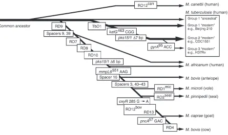
Several studies have taken advantage of irreversible genetic events such as chromosomal deletions (also known as large sequence polymorphisms [LSPs]), together with analysis of single nucleotide polymorphisms (SNPs) and direct repeat content (“spoligotype”) patterns, to decipher the phylogeny of the M. tuberculosis complex (8, 11, 19, 20). In this approach, the successive and unidirectional loss of DNA in representative strains reveals the order in which members of the complex descended from their ancient common ancestor (Figure 1) (11). The extant representatives of the com-mon ancestor of the M. tuberculosis complex include the ancestral and modern strains of M. tuberculosis. The latter are characterized by the deletion of a 2.1-kb fragment termed TbD1, account for most current tuberculosis (TB) cases, and include the Beijing fam-ily of strains that are prevalent in Asia and have caused outbreaks worldwide (21). A separate lineage, marked by deletion of region of difference 9 (RD9), diverged from an ancestral M. tuberculosis and sequentially gave rise to M. africanum, M. microti, M. pinnipedii,
M. caprae, and M. bovis. It is thought that M. bovis, which has the greatest number of deletions, is the most recent member of this lineage (10). These results clearly refute the popular belief that
M. tuberculosis evolved from M. bovis through the adaptation of a bovine strain to the human host (22). Since M. tuberculosis contains chromosomal segments that have been deleted from M. bovis, M. tuberculosis could not have evolved from M. bovis. In fact, these data
losis
has revolutionized tuberculosis research, contributed to major advances in the understanding
of the evolution and pathogenesis of
M. tuberculosis
, and facilitated development of new diagnostic
tests with increased specificity for tuberculosis. In this review, we describe some of the major
prog-ress in tuberculosis research that has resulted from knowledge of the genome sequence and note
some of the problems that remain unsolved.
Nonstandard abbreviations used: BCG, bacille Calmette-Guérin; CFP-10, culture filtrate protein 10 kDa; ESAT-6, early secreted antigen 6 kDa; ESX-1, ESAT-6 secretion system 1; IGRA, IFN-γ release assay; INH, isoniazid; LSP, large sequence polymor-phism; MODS, microscopic-observation drug susceptibility; RD9, region of difference 9; TB, tuberculosis.
science in medicine
raise the converse possibility that humans transmitted tubercle bacilli to animals and those bacilli subsequently evolved into M. bovis. Although this is possible, the isolation of M. tuberculosis com-plex DNA from a 17,000-year-old bison skeleton in North America suggests that wild animals were infected prior to domestication of livestock 8,000–10,000 years ago (23, 24).
Modern evolution of M. tuberculosis
Several recent studies have used synonymous SNPs and/or LSPs to construct deep phylogenetic lineages for M. tuberculosis iso- lates from around the globe (3, 6, 7, 25–27). These studies uni-formly show that distinct geographic regions of the world have variable distributions of lineages, with certain regions having a major lineage that is a minor contributor elsewhere. Moreover, these studies have identified a clonal population structure for
M. tuberculosis and confirmed the absence of ongoing horizontal gene transfer between strains. It is hypothesized that the genetic population structure of M. tuberculosis identified by these studies will yield an association between different mycobacterial lineages and important traits such as clinical phenotypes, transmissibility, likelihood of drug resistance, and adaptation to distinct human populations. However, the relative importance of SNPs versus LSPs as the molecular determinants of trait variability is debat-ed. Studies indicate that each strain has roughly 200 intergenic SNPs that can potentially alter gene expression, 500 genes that are affected by least one nonsynonymous SNP, and 100 genes that are lost or inactivated due to LSPs (2, 28, 29). Given that the
effect of SNPs is usually smaller than that of the loss of an entire gene, it is suggested that LSPs are the main source of phenotypic variability between strains (29). However, while the majority of drug-resistant mutations are due to SNPs (30–33), the best-char-acterized example of variability in virulence is due to an LSP in the pks 15/1 locus (15, 17). Thus both SNPs and LSPs contribute to strain variability, and it is possible that specific selective pres-sures favor one over the other.
Several studies have found associations between mycobacterial genotypes and phenotypes in humans. First, in an urban cosmo-politan setting, the transmission of phylogeographic lineages of M. tuberculosis is nonrandomly associated with various ethnic populations (7, 26, 34). For example, an individual of Chinese ethnicity in San Francisco is more likely to acquire and develop TB due to a strain of M. tuberculosis of the East Asian lineage than any other phylogeographic lineage. These results suggest that over human evolutionary times, specific phylogeographic lin- eages have adapted to (or coevolved with) distinct human popu-lations and that such host-pathogen compatibility operates even when transmission takes place outside the region of origin (26). The extent to which these host-pathogen associations are driven by sociologic and epidemiologic factors versus biological deter-minants is not yet known. However, if biological determinants make a major contribution, then the greatest diversity among
[image:3.585.63.530.82.358.2]M. tuberculosis lineages should be observed in Africa, where the genetic diversity among human populations is greater than any-where else in the world (35).
Figure 1
Similar host-pathogen associations have been documented between the phylogeographic lineage of M. tuberculosis and the type of mutations (i.e., KatG[S315T] or a C-to-T substitution in inhA promoter–15) that renders strains resistant to the first-line anti-tuberculosis drug isoniazid (INH) (34, 36). These findings suggest that the lineage of M. tuberculosis influences the mechanism of INH resistance, which in turn affects bacterial fitness and the transmis-sion of specific INH-resistant strains (36).
RD1/ESX-1 and the pathogenesis of TB
Determination of the genome sequence of M. tuberculosis (37) has also facilitated discovery of bacterial determinants of virulence. One particularly productive line of investigation has revealed the molecular basis of attenuation of the bacille Calmette-Guérin (BCG) vaccine strain(s) of M. bovis. M. bovis BCG, the most widely used human vaccine in history (more than 3.5 billion doses have been administered), was derived by in vitro passage of an isolate of M. bovis . A spontaneous change in colony morphology was fol- lowed by discovery that the bacteria with altered colony morphol-ogy had lost their virulence in experimental animals (38). Since its first use as a human vaccine against TB in 1921, BCG has shown a high degree of safety, indicating that it clearly lost elements that are essential to cause disease in immunocompetent humans.
Initial insight into the mode of attenuation of BCG was obtained when subtractive hybridization identified three RDs that were present in M. tuberculosis and M. bovis but absent from BCG (39). Subsequent analysis using whole-genome microarrays or bacterial artificial chromosome arrays revealed additional RDs, but RD1, a 9.5-kb deletion, is the only region of difference
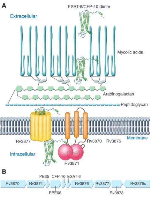
that is absent from all strains of BCG (n = 13) and present in all of the hundreds of strains of virulent M. tuberculosis analyzed to date (38, 40, 41). Compared with M. tuberculosis , BCG RD1 encom-passes seven genes and truncations of two additional genes (in the original annotated genome sequence, these genes are termed
Rv3870–Rv3879c; ref. 37 and Figure 2). M. microti, which has also been used as a human vaccine, lacks an overlapping set of genes:
Rv3864–Rv3876 (42). Therefore, comparative genomic analysis of mycobacteria that have been proven to be attenuated in humans has pointed to a specific chromosomal region implicated in viru-lence. It is interesting to note that another human mycobacterial pathogen, M. ulcerans, lacks an intact RD1 (43) and causes disease as an extracellular, not an intracellular, pathogen that expresses a characteristic mycolactone toxin (44, 45).
There is now strong experimental evidence that genes present in RD1 are essential for virulence of members of the M. tuberculo-sis complex. BCG complemented with a cosmid that spans RD1 increased growth of the bacteria in the lungs and spleen of mice compared with the parental BCG, although RD1-complemented BCG remained less virulent than virulent M. tuberculosis (H37Rv) (46). Complementation with RD1 increased virulence of BCG more markedly in SCID mice than in WT mice, implying that the effects of RD1 genes in vivo include roles in evasion of innate immunity. In addition, deletion of RD1 from virulent M. tuberculosis H37Rv resulted in attenuation of the ability to grow intracellularly in human macrophages, to cause macrophage cytotoxicity, and to grow and cause pathology in the lungs and disseminate to the spleen of immunocompetent mice after aerosol infection; deletion of RD1 from M. tuberculosis yielded a strain whose virulence was similar to that of BCG Russia (47). Deletion of RD1 also attenuat-ed other strains of virulent mycobacteria, including M. tuberculosis H37Rv, Erdman, and CDC1551, and M. bovis Ravenel, and viru-lence of the mutants was attenuated in SCID mice, as well as in immunocompetent mice (48). Together, these results indicate that genes within RD1 are essential for complete virulence of M. tuber-culosis and that deletion of RD1 was likely the original mutation attenuating virulence that occurred during derivation of BCG.
RD1 genes contribute to the virulence of M. tuberculosis by encod-ing a secretory apparatus and its substrates (48–50) (together termed early secreted antigen 6 kDa [ESAT-6] secretion system 1 [ESX-1]) or secretion in mycobacteria (SNM). Two of the genes,
Rv3874 and Rv3875 , encode two previously identified secreted pro-teins, ESAT-6 (51) and culture filtrate protein 10 kDa (CFP-10)
[image:4.585.42.286.79.399.2]science in medicine
(52), which are targets of cellular immune responses in mice and humans infected with M. tuberculosis. While these proteins are detected in supernatants of M. tuberculosis cultures, it was unclear how they were secreted, since they both lack recognizable signal sequences. The observation that other RD1 genes encode predict-ed membrane proteins suggested that they encode a novel system for protein secretion, and this hypothesis has been sustained by strong evidence. In M. tuberculosis, mutation of Rv3870, Rv3871,
Rv3876, and Rv3877 results in lack of secretion, but not lack of synthesis, of ESAT-6 and CFP-10 (48, 50, 53). ESAT-6 (Rv3875) and CFP-10 (Rv3874) interact with each other with high affinity (≤1 × 10⁻⁸ M) to form 1:1 heterodimers (54, 55), and their secretion
appears to be mutually dependent, at least in M. tuberculosis and
M. bovis (48, 49, 53, 56, 57). The ESAT-6/CFP-10 complex interacts directly with the C-terminal domain of Rv3871 (50) through the disordered carboxyterminal tail of CFP-10 (58). Addition of the carboxyterminal domain of CFP-10 to yeast ubiquitin caused the heterologous protein to be secreted from M. tuberculosis, implying that this domain serves as a signal sequence for the ESX-1 sys-tem (Figure 2). In contrast to ESAT-6 and CFP-10, Rv3873 (also known as PPE68) is neither secreted itself nor essential for secre-tion of ESAT-6/CFP-10 (56, 59). Rv3872 (also known as PE35) has been detected as a secreted protein (57), but its role in the secretion of ESAT-6 is unknown, although it may be needed for synthesis of ESAT-6 (56).
Secretion of ESAT-6 requires the genes in RD1 and at least 3 additional genes at a different locus. A signature-tagged mutagen-esis screen identified Rv3614c and Rv3615c (60), while a proteomic screen identified the product of Rv3616c (termed EspA) in superna-tants of WT, but not RD1-deficient, M. tuberculosis (57). Secretion of EspA required both ESAT-6 and CFP-10, and secretion of ESAT-6 and CFP-10 required EspA. Yeast two-hybrid analysis revealed that the product of Rv3615c interacted stably with itself but not other identified components of the secretion system and the product of Rv3614c interacted with the product of Rv3882c (a gene near
the RD1 locus), while EspA exhibited no detectable interactions with other screened proteins (60). It is likely that other proteins, in addition to ESAT-6, CFP-10, PE35, and EspA, are secreted by the RD1/ESX-1 system, and the identity of the secreted effector(s) of virulence remains to be established.
M. marinum, which causes TB in fish and amphibians, contains an RD1 locus with an organization identical to that of M. tuberculo-sis, and the RD1 locus is required for M. marinum virulence (61, 62). Moreover, mutation of the M. marinum homologs of the M. tuber-culosis RD1 genes Rv3876, Rv3878,and Rv3879c resulted in loss of virulence and loss of secretion of ESAT-6. Secretion and virulence were also lost with mutations in the homologs of genes flanking RD1, including Rv3866, Rv3867, Rv3868,and Rv3881c, indicating that the corresponding M. tuberculosis genes should be examined for roles in secretion and virulence. While the existing evidence in
M. tuberculosis, M. bovis, and M. microti indicates that secretion of ESAT-6 and CFP-10 may be absolutely interdependent, mutations in the M. marinum homologs of Rv3876, Rv3878, and Rv3879c allowed secretion of CFP-10 without secretion of ESAT-6 (61). The possibility that dissociation of ESAT-6 and CFP-10 secretion may occur in human TB is suggested by the recent observation that immune responses to ESAT-6, but not to CFP-10, are attenu-ated in people infected with M. africanum, in which the homolog of Rv3879c is a pseudogene (63). Analysis of protein secretion by
M. africanum will provide information as to whether defective secre-tion of ESAT-6 accounts for the attenuated immune responses to ESAT-6 in people infected with M. africanum , and complementa-tion with a functional copy of Rv3879c will determine whether the inactivation of this gene accounts for the observation.
[image:5.585.47.540.82.251.2]What are the roles of the RD1/ESX-1 system in virulence? ESX-1– replete mycobacteria exhibit higher cytotoxicity for cultured human and mouse macrophages and for a human alveolar epithe-lial cell line than do ESX-1–deficient mycobacteria (47, 48, 53, 61). Since RD1-containing pathogenic mycobacteria are facultative intracellular pathogens, the fact they cause death of their resident
Figure 3
there was complete concordance between loss of cytolytic activ-ity and loss of spreading activity in all of the single-gene mutants examined. In vivo studies also indicate that ESX-1 contributes to cell-to-cell spread. In adult zebrafish inoculated with M. mari-num in the peritoneum, dissemination to the liver was markedly decreased in fish inoculated with ESX-1–deficient bacteria (61). In a real-time assay using GFP-expressing M. marinum and trans-parent zebrafish embryos, RD1-deficient bacteria were defective in causing aggregation of macrophages to initiate granuloma formation, and subsequent to macrophage aggregation, RD1-deficient bacteria were defective in their ability to spread from initially infected macrophages to their newly recruited neighbors (62). Studies on M. marinum expressing red or green fluorescent protein to differentiate between RD1-deficient and WT bacteria revealed that macrophages infected with RD1-deficient bacteria could traffic to aggregates of macrophages infected with WT bac-teria, but macrophages infected with RD1-deficient bacteria did not cause recruitment of macrophages infected with WT bacteria (62). These results indicate that one or more components of ESX-1 are required for a signal for recruitment and aggregation of macro- phages in order to bring additional target cells into close apposi-tion to promote spread of mycobacteria within the host (Figure 3). That this mechanism is likely to occur in lungs of animals infected with M. tuberculosis is supported by the observation that infection with ESX-1–deficient bacteria results in aggregates of cells imme-diately adjacent to the airways but not to more distal portions of the lung parenchyma (47, 53). Additional activities attributed to the ESX-1 secretion system include suppression of proinflamma- tory cytokine production (50, 60), arrest of phagosome matura-tion (64), and induction of IFN-β (65).
The mechanisms whereby proteins exported by the ESX-1 sys- tem exert their effects on host cells have not yet been fully eluci-dated. The observation that ESAT-6 alone or complexes of ESAT-6 and CFP-10 can cause membrane permeabilization (48) has been challenged (54), and the solution structure of the complex does not reveal hydrophobic domains or patches of charged residues that would provide a structural basis for perturbation of a phos-pholipid bilayer. In the solution structure of ESAT-6/CFP-10 dimers, the complexes are composed of a 4-helix core (2 helices contributed by each protein), with the amino and carboxyl termini of both proteins unstructured (54). The dimers bind selectively to mononuclear phagocytes, and deletion of the carboxyl terminus of CFP-10, but not ESAT-6, markedly diminished binding, indicating that this unstructured domain may specifically interact with a sur-face receptor to modulate macrophage activities. While this may be a lead to understanding the mechanisms of ESX-1–dependent
Diagnosis of TB
Improved control of TB depends on development and application of better means of diagnosing it. While nucleic acid amplification would seem an obvious choice for the detection of M. tuberculosis, this approach has so far been successfully applied only to confirm the identity of an acid-fast smear–positive or culture-positive sam-ple as a member of the M. tuberculosis complex and for sequence-based detection of drug-resistant mutations.
One significant advance that has benefited from M. tuberculosis genomics is the development of new in vitro diagnostic assays that distinguish immune responses due to infection with M. tuber-culosis from responses due to BCG vaccination. For more than 4 decades, tuberculin skin testing with purified protein derivative (PPD; which is hardly pure by modern standards, but is purer than its predecessors) has been the major means of diagnosing latent infection with M. tuberculosis. However, since BCG vaccination also causes tuberculin skin test reactivity, the diagnostic value of tuberculin skin testing is largely abrogated in individuals that have received BCG. The discoveries that ESAT-6 and CFP-10 are present in all strains of virulent M. tuberculosis, and absent from all strains of BCG, together with the knowledge that ESAT-6 and CFP-10 are immunogenic in humans (reviewed in ref. 66), has led to develop-ment of diagnostic assays that use these proteins as antigens for detection of M. tuberculosis–specific immune responses.
science in medicine
the information from comparative genomics of M. tuberculosis and BCG has led to the development of these new diagnostic assays, developed based on the information from comparative genomics of M. tuberculosis and BCG, are likely to replace tuberculin skin test-ing for detection of latent TB infection.
Unfortunately, diagnosis and treatment of latent TB infection is a luxury restricted to areas of low TB prevalence. The greater urgent need is the development and application of improved diagnostic techniques for active TB, to identify individuals who need treat-ment, and to keep them from transmitting TB. Current TB control programs in nonindustrialized countries depend on microscopic examination of stained sputum smears, a procedure with less than 50% sensitivity for active pulmonary TB. While recent initiatives have stimulated pursuit of a wide variety of approaches to diag-nose active TB and detect drug resistance, the most promising new assay available so far is an adaptation of long-established broth culture techniques. The microscopic-observation drug suscepti-bility (MODS) assay, developed in Peru, combines broth culture in 24-well tissue culture plates with early detection of growth by microscopic examination of cultures to detect characteristic cord formation of M. tuberculosis (69). A recent study in Lima, Peru, found that the sensitivity of MODS exceeded that of automated mycobacterial culture or culture on Löwenstein-Jensen solid medi-um (70). Moreover, MODS provided positive results in a median of 7 days, compared with 13 days for automated mycobacterial culture, and 26 days for culture on Löwenstein-Jensen medium, and MODS provided similar advantages in the time to obtain drug susceptibility results. While MODS shows clear promise for higher-sensitivity detection of cases of TB, further innovations will be required to provide even more rapid diagnosis of active TB in resource-poor settings.
Unsolved questions and problems
TB is an enormous health problem worldwide, and the spread of extensively drug-resistant M. tuberculosis threatens to overcome the available tools of TB control. Despite this, TB has not yet attracted celebrity advocates in the manner of HIV and malar-ia. Thanks in part to the availability of the genome sequences of multiple strains of M. tuberculosis, the pathogenesis of TB is increasingly amenable to study, but many important unsolved biological problems remain.
Among the unsolved biological problems is the lack of under-standing of the mechanisms of the characteristic slow growth of
M. tuberculosis. The doubling time of M. tuberculosis in rich broth media or during logarithmic growth in vivo is approximately 24 hours, but the rate-limiting determinants of this growth rate are not known. In addition, the advantages to the bacteria of such slow replication are unclear. Does slow growth specifically contribute to stealth mechanisms that minimize the efficacy of innate and adaptive immune responses, or is slow growth an effect of evolution of the mechanisms that allow M. tuberculosis to remain latent for decades within a healthy host? Perhaps the greatest benefit to the bacteria is the maximization of transmis- sion by slow growth during reactivation. By promoting an insidi- ous onset and slow progression without immobilizing or caus-ing rapid death of the host, the bacteria optimize the chances of being spread to additional susceptible individuals. While com-parative genomic studies have revealed clear divisions between slowly growing and rapidly growing mycobacteria, further studies are needed to elucidate the mechanisms that determine the rate
of growth and to identify the specific aspects of M. tuberculosis biology that benefit from slow growth.
Another unsolved biological problem is the identification of the breadth and thorough characterization of the ecological niches of
M. tuberculosis in vivo. M. tuberculosis is a facultative intracellular bacterium, but the diversity of the cells in which M. tuberculosis resides, replicates, and traffics through the host during the course of infection is incompletely defined. For example, it is widely believed that macrophages are the predominant resident cells of
M. tuberculosis in vivo, though dendritic cells have also been observed to be infected. What is the quantitative distribution of
M. tuberculosis in cells of distinct phenotypes? Do distinct cell types differ as sanctuaries from innate or adaptive immune responses? It is also unclear whether, or at what stages of infection, M. tuberculo-sis exists extracellularly, other than at the terminal stages of a pul- monary cavity. It is similarly unclear how the bacteria are specifi-cally adapted to the lungs in preference to other tissues and how the bacteria survive and grow within phagosomes of macrophages and other cells. Even when M. tuberculosis is injected intravenously into mice, subsequent replication in the lungs far exceeds that in other tissues (71, 72). Is this solely due to factors that are enriched in the lungs (such as oxygen) that benefit the bacteria, or are the lungs immunologically disadvantaged in specific ways that allow
M. tuberculosis to thrive? There is no question that M. tuberculosis transmission is enhanced by its adaptation to the lungs, but the mechanisms that are essential for the bacteria to selectively thrive there still need to be defined, and the success of drug and vaccine development may depend on understanding and overcoming those mechanisms. The availability of the genome sequences of several strains of M. tuberculosis has already facilitated discovery of determinants of certain aspects of virulence, and application of that knowledge in novel genetic screens offers considerable prom-ise for the discovery of unique aspects of M. tuberculosis biology.
One of the most important unsolved biological problems is that
1. Smith, N.H., et al. 2006. Ecotypes of the Myco-bacterium tuberculosis complex. J. Theor. Biol. 239:220–225.
2. Fleischmann, R.D., et al. 2002. Whole-genome com-parison of Mycobacterium tuberculosis clinical and laboratory strains. J. Bacteriol. 184:5479–5490.
3. Gutacker, M.M., et al. 2002. Genome-wide analy-sis of synonymous single nucleotide polymor-phisms in Mycobacterium tuberculosis complex organisms: resolution of genetic relationships among closely related microbial strains. Genetics. 162:1533–1543.
4. Huard, R.C., et al. 2006. Novel genetic polymor-phisms that further delineate the phylogeny of the Mycobacterium tuberculosis complex. J. Bacteriol. 188:4271–4287.
5. Hughes, A.L., Friedman, R., and Murray, M. 2002. Genomewide pattern of synonymous nucleotide substitution in two complete genomes of Mycobac-terium tuberculosis. Emerg. Infect. Dis. 8:1342–1346. 6. Sreevatsan, S., et al. 1997. Restricted structural
gene polymorphism in the Mycobacterium tuber-culosis complex indicates evolutionarily recent global dissemination. Proc. Natl. Acad. Sci. U. S. A. 94:9869–9874.
7. Hirsh, A.E., Tsolaki, A.G., DeRiemer, K., Feldman, M.W., and Small, P.M. 2004. Stable association between strains of Mycobacterium tuberculosis and their human host populations. Proc. Natl. Acad. Sci. U. S. A. 101:4871–4876.
8. Smith, N.H., et al. 2003. The population structure of Mycobacterium bovis in Great Britain: clonal expan-sion. Proc. Natl. Acad. Sci. U. S. A. 100:15271–15275. 9. Supply, P., et al. 2003. Linkage disequilibrium
between minisatellite loci supports clonal evo-lution of Mycobacterium tuberculosis in a high tuberculosis incidence area. Mol. Microbiol. 47:529–538.
10. Smith, N.H., Gordon, S.V., de la Rua-Domenech, R., Clifton-Hadley, R.S., and Hewinson, R.G. 2006. Bottlenecks and broomsticks: the molecular evolu-tion of Mycobacterium bovis. Nat. Rev. Microbiol. 4:670–681.
11. Brosch, R., et al. 2002. A new evolutionary scenario for the Mycobacterium tuberculosis complex. Proc. Natl. Acad. Sci. U. S. A. 99:3684–3689.
12. Gutierrez, M.C., et al. 2005. Ancient origin and gene mosaicism of the progenitor of Mycobacte-rium tuberculosis. PLoS Pathog. 1:e5.
13. Smith, N.H. 2006. A re-evaluation of M. prototu-berculosis. PLoS Pathog. 2:e98.
14. Brisse, S., Supply, P., Brosch, R., Vincent, V., and Gutierrez, M.C. 2006. “A re-evaluation of M. proto-tuberculosis”: continuing the debate. PLoS Pathog. 2:e95. 15. Constant, P., et al. 2002. Role of the pks15/1 gene in the biosynthesis of phenolglycolipids in the Mycobacterium tuberculosis complex. Evidence that all strains synthesize glycosylated p-hydroxy-benzoic methly esters and that strains devoid of phenolglycolipids harbor a frameshift mutation in the pks15/1 gene. J. Biol. Chem. 277:38148–38158.
16. Marmiesse, M., et al. 2004. Macro-array and bio-informatic analyses reveal mycobacterial “core” genes, variation in the ESAT-6 gene family and new phylogenetic markers for the Mycobacterium tuberculosis complex. Microbiology. 150:483–496.
17. Reed, M.B., et al. 2004. A glycolipid of hyperviru-lent tuberculosis strains that inhibits the innate immune response. Nature. 431:84–87.
18. Tsolaki, A.G., et al. 2005. Genomic deletions classi-fy the Beijing/W strains as a distinct genetic lineage of Mycobacterium tuberculosis. J. Clin. Microbiol. 43:3185–3191.
19. Kamerbeek, J., et al. 1997. Simultaneous detec-tion and strain differentiation of Mycobacterium tuberculosis for diagnosis and epidemiology.
J. Clin. Microbiol. 35:907–914.
20. Mostowy, S., Cousins, D., Brinkman, J., Aranaz, A., and Behr, M.A. 2002. Genomic deletions suggest a phylogeny for the Mycobacterium tuberculosis complex. J. Infect. Dis. 186:74–80.
21. Glynn, J.R., Whiteley, J., Bifani, P.J., Kremer, K., and van Soolingen, D. 2002. Worldwide occurrence of Beijing/W strains of Mycobacterium tuberculosis: a systematic review. Emerg. Infect. Dis. 8:843–849. 22. Stead, W.W., et al. 1995. When did Mycobacterium
tuberculosis infection first occur in the New World? An important question with public health implica-tions. Am. J. Respir. Crit. Care Med. 151:1267–1268. 23. Bruford, M.W., Bradley, D.G., and Luikart, G. 2003.
DNA markers reveal the complexity of livestock domestication. Nat. Rev. Genet. 4:900–910. 24. Rothschild, B.M., et al. 2001. Mycobacterium
tuberculosis complex DNA from an extinct bison dated 17,000 years before the present. Clin. Infect. Dis. 33:305–311.
25. Gutacker, M.M., et al. 2006. Single-nucleotide polymorphism-based population genetic analysis of Mycobacterium tuberculosis strains from 4 geo-graphic sites. J. Infect. Dis. 193:121–128.
26. Gagneux, S., et al. 2006. Variable host-pathogen compatibility in Mycobacterium tuberculosis. Proc. Natl. Acad. Sci. U. S. A. 103:2869–2873.
27. Filliol, I., et al. 2006. Global phylogeny of Myco-bacterium tuberculosis based on single nucleotide polymorphism (SNP) analysis: insights into tuber-culosis evolution, phylogenetic accuracy of other DNA fingerprinting systems, and recommenda-tions for a minimal standard SNP set. J. Bacteriol. 188:759–772.
28. Musser, J.M., Amin, A., and Ramaswamy, S. 2000. Negligible genetic diversity of mycobacterium tuberculosis host immune system protein targets: evidence of limited selective pressure. Genetics. 155:7–16.
29. Tsolaki, A.G., et al. 2004. Functional and evolu-tionary genomics of Mycobacterium tuberculosis: insights from genomic deletions in 100 strains.
Proc. Natl. Acad. Sci. U. S. A. 101:4865–4870.
30. Cavusoglu, C., Hilmioglu, S., Guneri, S., and Bilgic, A. 2002. Characterization of rpoB mutations in rifampin-resistant clinical isolates of Myco-bacterium tuberculosis from Turkey by DNA sequencing and line probe assay. J. Clin. Microbiol. 40:4435–4438. 31. Finken, M., Kirschner, P., Meier, A., Wrede, A., and Bottger, E.C. 1993. Molecular basis of streptomycin resistance in Mycobacterium tuberculosis: altera-tions of the ribosomal protein S12 gene and point mutations within a functional 16S ribosomal RNA pseudoknot. Mol. Microbiol. 9:1239–1246.
32. Gagneux, S., et al. 2006. The competitive cost of anti-biotic resistance in Mycobacterium tuberculosis.
Science. 312:1944–1946.
33. Ramaswamy, S.V., et al. 2000. Molecular genetic analysis of nucleotide polymorphisms associated with ethambutol resistance in human isolates of Mycobacterium tuberculosis. Antimicrob. Agents Chemother. 44:326–336.
34. Baker, L., Brown, T., Maiden, M.C., and Drobniews-ki, F. 2004. Silent nucleotide polymorphisms and a phylogeny for Mycobacterium tuberculosis. Emerg. Infect. Dis. 10:1568–1577.
35. Ramachandran, S., et al. 2005. Support from the relationship of genetic and geographic distance in human populations for a serial founder effect originating in Africa. Proc. Natl. Acad. Sci. U. S. A. 102:15942–15947.
36. Gagneux, S., et al. 2006. Impact of bacterial genet-ics on the transmission of isoniazid-resistant Mycobacterium tuberculosis. PLoS Pathog. 2:e61. 37. Cole, S.T., et al. 1998. Deciphering the biology of
Mycobacterium tuberculosis from the complete genome sequence. Nature. 393:537–544. 38. Behr, M.A., and Small, P.M. 1999. A historical
and molecular phylogeny of BCG strains. Vaccine. 17:915–922.
39. Mahairas, G.G., et al. 1996. Molecular analysis of genetic differences between Mycobacterium bovis BCG and virulent M. bovis. J. Bacteriol. 178:1274–1282.
40. Behr, M.A., et al. 1999. Comparative genomics of BCG vaccines by whole-genome DNA microarray.
Science. 284:1520–1523.
41. Gordon, S.V., et al. 1999. Identification of variable regions in the genomes of tubercle bacilli using bac-terial artificial chromosome arrays. Mol. Microbiol. 32:643–655.
42. Brodin, P., et al. 2002. Bacterial artificial chromo- some-based comparative genomic analysis identi-fies Mycobacterium microti as a natural ESAT-6 deletion mutant. Infect. Immun. 70:5568–5578. 43. Stinear, T.P., et al. 2007. Reductive evolution and
niche adaptation inferred from the genome of Mycobacterium ulcerans, the causative agent of Buruli ulcer. Genome Res. 17:192–200.
science in medicine
in vitro and in vivo. Cell. Microbiol. 7:1295–1304. 45. Coutanceau, E., et al. 2005. Modulation of the host
immune response by a transient intracellular stage of Mycobacterium ulcerans: the contribution of endogenous mycolactone toxin. Cell. Microbiol. 7:1187–1196.
46. Pym, A.S., Brodin, P., Brosch, R., Huerre, M., and Cole, S.T. 2002. Loss of RD1 contributed to the attenuation of the live tuberculosis vaccines Myco-bacterium bovis BCG and Mycobacterium microti.
Mol. Microbiol. 46:709–717.
47. Lewis, K.N., et al. 2003. Deletion of RD1 from Myco- bacterium tuberculosis mimics bacille Calmette-Guerin attenuation. J. Infect. Dis. 187:117–123. 48. Hsu, T., et al. 2003. The primary mechanism of
attenuation of bacillus Calmette-Guerin is a loss of secreted lytic function required for invasion of lung interstitial tissue. Proc. Natl. Acad. Sci. U. S. A. 100:12420–12425.
49. Pym, A.S., et al. 2003. Recombinant BCG exporting ESAT-6 confers enhanced protection against tuber-culosis. Nat. Med. 9:533–539.
50. Stanley, S.A., Raghavan, S., Hwang, W.W., and Cox, J.S. 2003. Acute infection and macrophage subver- sion by Mycobacterium tuberculosis require a spe-cialized secretion system. Proc. Natl. Acad. Sci. U. S. A. 100:13001–13006.
51. Sorensen, A.L., Nagai, S., Houen, G., Andersen, P., and Andersen, A.B. 1995. Purification and charac-terization of a low-molecular-mass T-cell antigen secreted by Mycobacterium tuberculosis. Infect. Immun. 63:1710–1717.
52. Berthet, F.X., Rasmussen, P.B., Rosenkrands, I., Andersen, P., and Gicquel, B. 1998. A Mycobacte-rium tuberculosis operon encoding ESAT-6 and a novel low-molecular-mass culture filtrate protein (CFP-10). Microbiology. 144:3195–3203.
53. Guinn, K.M., et al. 2004. Individual RD1-region genes are required for export of ESAT-6/CFP-10 and for virulence of Mycobacterium tuberculosis.
Mol. Microbiol. 51:359–370.
54. Renshaw, P.S., et al. 2005. Structure and func-tion of the complex formed by the tuberculosis virulence factors CFP-10 and ESAT-6. EMBO J. 24:2491–2498.
55. Renshaw, P.S., et al. 2002. Conclusive evidence that the major T-cell antigens of the Mycobacterium tuberculosis complex ESAT-6 and CFP-10 form
a tight, 1:1 complex and characterization of the structural properties of ESAT-6, CFP-10, and the ESAT-6*CFP-10 complex. Implications for pathogen-esis and virulence. J. Biol. Chem. 277:21598–21603.
56. Brodin, P., et al. 2006. Dissection of ESAT-6 sys-tem 1 of Mycobacterium tuberculosis and impact on immunogenicity and virulence. Infect. Immun. 74:88–98.
57. Fortune, S.M., et al. 2005. Mutually dependent secre-tion of proteins required for mycobacterial virulence.
Proc. Natl. Acad. Sci. U. S. A. 102:10676–10681. 58. Champion, P.A., Stanley, S.A., Champion, M.M.,
Brown, E.J., and Cox, J.S. 2006. C-terminal signal sequence promotes virulence factor secretion in Myco-bacterium tuberculosis. Science. 313:1632–1636. 59. Demangel, C., et al. 2004. Cell envelope protein
PPE68 contributes to Mycobacterium tuberculosis RD1 immunogenicity independently of a 10-kilo-dalton culture filtrate protein and ESAT-6. Infect. Immun. 72:2170–2176.
60. MacGurn, J.A., Raghavan, S., Stanley, S.A., and Cox, J.S. 2005. A non-RD1 gene cluster is required for Snm secretion in Mycobacterium tuberculosis. Mol. Microbiol. 57:1653–1663.
61. Gao, L.Y., et al. 2004. A mycobacterial virulence gene cluster extending RD1 is required for cytoly-sis, bacterial spreading and ESAT-6 secretion. Mol. Microbiol. 53:1677–1693.
62. Volkman, H.E., et al. 2004. Tuberculous granuloma formation is enhanced by a mycobacterium viru-lence determinant. PLoS Biol. 2:e367.
63. de Jong, B.C., et al. 2006. Mycobacterium africa-num elicits an attenuated T cell response to early secreted antigenic target, 6 kDa, in patients with tuberculosis and their household contacts. J. Infect. Dis. 193:1279–1286.
64. Tan, T., Lee, W.L., Alexander, D.C., Grinstein, S., and Liu, J. 2006. The ESAT-6/CFP-10 secretion system of Mycobacterium marinum modulates phago-some maturation. Cell. Microbiol. 8:1417–1429. 65. Stanley, S.A., Johndrow, J.E., Manzanillo, P., and
Cox, J.S. 2007. The type I IFN response to infection with Mycobacterium tuberculosis requires ESX-1-mediated secretion and contributes to pathogenesis.
J. Immunol. 178:3143–3152.
66. Andersen, P., Munk, M.E., Pollock, J.M., and Doherty, T.M. 2000. Specific immune-based diag-nosis of tuberculosis. Lancet. 356:1099–1104.
67. Johnson, P.D., et al. 1999. Tuberculin-purified pro-tein derivative-, MPT-64-, and ESAT-6-stimulated gamma interferon responses in medical students before and after Mycobacterium bovis BCG vac-cination and in patients with tuberculosis. Clin. Diagn. Lab. Immunol. 6:934–937.
68. Dewan, P.K., Grinsdale, J., and Kawamura, L.M. 2007. Low sensitivity of a whole-blood interferon-gamma release assay for detection of active tuberculosis.
Clin. Infect. Dis. 44:69–73.
69. Moore, D.A., et al. 2004. Microscopic observation drug susceptibility assay, a rapid, reliable diag-nostic test for multidrug-resistant tuberculosis suitable for use in resource-poor settings. J. Clin. Microbiol. 42:4432–4437.
70. Moore, D.A., et al. 2006. Microscopic-observation drug-susceptibility assay for the diagnosis of TB.
N. Engl. J. Med. 355:1539–1550.
71. Cox, J.S., Chen, B., McNeil, M., and Jacobs, W.R., Jr. 1999. Complex lipid determines tissue-specific replication of Mycobacterium tuberculosis in mice.
Nature. 402:79–83.
72. Peters, W., et al. 2001. Chemokine receptor 2 serves an early and essential role in resistance to Myco-bacterium tuberculosis. Proc. Natl. Acad. Sci. U. S. A. 98:7958–7963.
73. Shi, L., North, R., and Gennaro, M.L. 2004. Effect of growth state on transcription levels of genes encoding major secreted antigens of Mycobacteri-um tuberculosis in the mouse lung. Infect. Immun. 72:2420–2424. 74. Banaiee, N., Kincaid, E.Z., Buchwald, U., Jacobs, W.R., Jr., and Ernst, J.D. 2006. Potent inhibition of macrophage responses to IFN-gamma by live viru-lent Mycobacterium tuberculosis is independent of mature mycobacterial lipoproteins but dependent on TLR2. J. Immunol. 176:3019–3027.
75. Pai, R.K., Convery, M., Hamilton, T.A., Boom, W.H., and Harding, C.V. 2003. Inhibition of IFN-gamma- induced class II transactivator expression by a 19- kDa lipoprotein from Mycobacterium tuberculo-sis: a potential mechanism for immune evasion.
J. Immunol. 171:175–184.