CELL TYPE AND TISSUE SPECIFIC FUNCTIONS OF CD73
Marquet Minor
A thesis submitted to the faculty at the University of North Carolina at Chapel Hill in partial fulfillment of the requirements for the degree of Master of Science in the Curriculum of Cell Biology and Physiology
in the School of Medicine.
Chapel Hill 2018 Approved by: A. Sidney Barritt Temitope Keku Lori O’Brien Scott Randell Natasha Snider
ii © 2018 Marquet Minor ALL RIGHTS RESERVED
iii ABSTRACT
Marquet Minor: Cell Type and Tissue Specific Functions of CD73 (Under the direction of Natasha Snider)
CD73 is a ubiquitously expressed glycosylphosphatidylinositol (GPI)-anchored
glycoprotein that converts extracellular adenosine 5'-monophosphate (AMP) to adenosine. CD73
couples closely with ecto-apyrase (CD39), which supplies the AMP substrate via sequential
dephosphorylation of extracellular ATP. CD73-generated adenosine functions in an autocrine
and paracrine manner to control numerous physiological responses by activating one of four
subtypes of G-protein-coupled adenosine receptors: A1R, A2AR, A2BR, and A3R. Missense
mutations in the CD73-encoding gene NT5E cause the rare, adult-onset vascular disease named
‘arterial calcifications due to deficiency of CD73’ (ACDC). Aside from direct human disease
involvement, cellular and animal model studies have revealed key functions of CD73 in tissue
homeostasis and pathophysiologic responses in the cardiovascular and central nervous system, as
well as epithelial tissues, including the lung, kidney and liver. This review covers CD73
iv
TABLE OF CONTENTS
LIST OF FIGURES ……… vii
LIST OF ABBREVATIONS ………. viii
CHAPTER 1: INTRODUCTION ……….. 1
Structural and molecular features of CD73 ………... 2
Evidence for non-enzymatic CD73 functions ……… 2
Physiological and disease relevance of CD73 ………... 3
CHAPTER 2: CD73 FUNCTIONS IN THE CARDIOVASCULAR SYSTEM ……….. 4
CD73 expression and distribution in the cardiovascular system ………... 4
Cell type-dependent roles of CD73 after myocardial infarction (MI) ………... 5
CHAPTER 3: CD73 FUNCTIONS IN THE CENTRAL NERVOUS SYSTEM ………. 7
CD73 expression and distribution in the central nervous system (CNS) ………... 7
CD73 controls locomotion ………. 8
v
CD73 on tissue-resident cells regulates CNS inflammation ……….. 9
Neuronal CD73 regulates nociception ………. 10
CHAPTER 4: CD73 FUNCTIONS IN THE LUNG ………... 12
CD73 expression and distribution in the lung ……….. 12
CD73 promotes maintenance of tissue barrier function in the lungs during hypoxia and hyperoxia ……… CD73 protects against lung infection, inflammation and fibrosis ………... 13
CD73 may contribute to asthma pathogenesis ………. 13
CHAPTER 5: CD73 FUNCTIONS IN THE KIDNEY ………... 15
CD73 expression and distribution in the kidney ……….. 15
CD73 regulates renal ischemia-reperfusion (I/R) injury ………. 15
CD73 regulates hypertension-associated renal injury ……….. 15
Other functions of CD73 in the aging and injured kidney ………... 16
CHAPTER 6: CD73 FUCNTIONS IN THE LIVER ……….. 17
CD73 expression and distribution in the liver ………. 17
CD73 regulates hepatic fibrosis ………... 17
CD73 regulates hepatic steatosis ………. 18
CD73 regulates hepatic fibrosis ………... 18
CD73 regulates hepatocyte injury ……… 18 12
vi
CHAPTER 7: CD73 FUNCTIONS IN THE IMMUNE SYSTEM ………. 20
CD73 expression on immune cells ……….. 20
CD73 regulates immune cell migration ………... 20
CD73 regulates chronic inflammation ………. 21
CHAPTER 8: CD73 FUNCTIONS IN OTHER TISSUES AND CELL TYPES ………... 22
Gastrointestinal system ……… 22
Bone ………. 22
Inflammation of the eye ………... 23
Reproductive tissues ……… 23
Skin and Muscle ………... 23
vii
LIST OF FIGURES
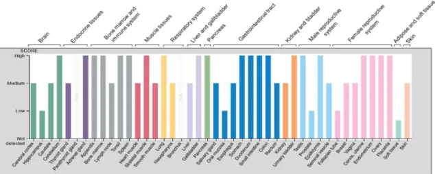
Figure 1 - Relative levels of CD73 protein expression across human
organ systems and tissues ……… 25
viii
LIST OF ABBREVIATIONS AND SYMBOLS
Γδ Gamma-delta
A1R Adenosine receptor A1
A2AR Adenosine receptor A2A
A2BR Adenosine receptor A2B
A3R Adenosine receptor A3
A1R-/- Adenosine receptor 1 knockout mouse model
A2AR-/- Adenosine receptor 2A knockout mouse model
A2BR-/- Adenosine receptor 2B knockout mouse model
ACDC Arterial calcifications due to deficiency of CD73
AMP Adenosine 5'-monophosphate
AMPASE Class of enzymes that catalyze the dephosphorylation of AMP into a nucleoside and a free phosphate ion
APCP Adenosine 5′-(α,β-methylene)diphosphate ARDS Acute respiratory distress syndrome
ATP Adenosine triphosphate
BAL Bronchoalveolar lavage
CAMP Cyclic adenosine monophosphate
ix CD39 Ecto-apyrase
CD4+ Cluster of differentiation 4 expressing
CD73 Ecto-5’-nucleotidase
CGRP Calcitonin gene-related peptide
CNS Central nervous system
EAE Experimental autoimmune encephalomyelitis
EAU Experimental autoimmune uveitis
ELF2 E74-like factor 2
ENT Equilibrative nucleoside transporter
EPDC Epicardium-derived cell
FP-1201 Recombinant human interferon beta-1a
GPI Glycosylphosphatidylinositol
GVHD Graft vs host disease
IB4 Isolectin B4
IFN-β1 Interferon beta IFN-γ Interferon gamma IGG3 Immunoglobulin G 3
x IL1-β Interleukin 1 beta
IL-6 Interleukin 6 IL-11 Interleukin 11 IL17A Interleukin-17A IL-17 Interleukin 17 I/R Ischemia-reperfusion K8/K18 Keratins 8 and 18 L3-5 Lumbar vertebrae 3-5 LPS Lipopolysaccharide M1 Pro-inflammatory macrophages
M2 Macrophages that are anti-inflammatory
MC3T3-E1 Mouse osteoblastic cell line
MDB Mallory-Denk bodies
MI Myocardial infarction
MRGPRD Mas-related G-protein coupled receptor member D
MSC Mesenchymal stem cell
NAD Nicotinamide adenine dinucleotide
xi NT5E Ecto-5’-nucleotidase-encoding gene
NT5E-/- NT5E knockout mouse model
P62 Nucleoporin 62
PAP Prostatic acid phosphatase
PH Measure used to determine the acidity or basicity of a solution
SP1 Specificity protein 1
TAA Thioacetamide
TGFβ Transforming growth factor beta TGF-β1 Transforming growth factor beta 1 TH1 Type 1 helper T cells
TH17 Pro-inflammatory helper T-cells that produce interleukin 17
TNAP Tissue Non-specific Alkaline Phosphatase
TNF-α Tumor necrosis factor alpha
TRPV1 Transient receptor potential cation channel subfamily V member 1
VEGF Vascular endothelial growth factor
1
CHAPTER 1: INTRODUCTION
The enzymatic dephosphorylation of nucleotides, such as adenosine
5´-monophosphate (AMP) is widespread across mammalian systems and represents a key step in
purine salvage pathways to modulate purinergic signaling (Ipata & Balestri, 2013; Zimmermann,
Zebisch, & Strater, 2012). This catalysis occurs inside the cell or in the extracellular space. The
major enzyme catalyzing the formation of extracellular adenosine from AMP is
ecto-5´-nucleotidase, encoded by the NT5E gene (Zimmermann et al., 2012). Ecto-5’-nucleotidase was
named CD73 in 1989 after it was shown that its engagement by specific antibodies induced T
cell activation (Thompson, Ruedi, Glass, Low, & Lucas, 1989). In this review, we refer to the
protein as CD73 and the gene as NT5E. Extracellular adenosine produced by CD73 acts on
adenosine receptors (A1R, A2AR, A2BR, and A3R) to activate downstream signaling (Antonioli,
Blandizzi, Pacher, & Hasko, 2013; Chen, Eltzschig, & Fredholm, 2013; Eltzschig, 2009;
Fredholm, 2007). Through its function as the major extracellular source of adenosine, CD73 is a
key regulator of tissue homeostasis and pathophysiologic responses related to immunity,
inflammation, and cancer, covered in recent reviews (Antonioli, Pacher, Vizi, & Hasko, 2013;
Beavis, Stagg, Darcy, & Smyth, 2012; Colgan, Eltzschig, Eckle, & Thompson, 2006; Roberts,
Lu, Rajakumar, Cowan, & Dwyer, 2013). The focus of this review is to highlight known and
emerging tissue-specific functions of CD73 in the brain and spinal cord, the heart, and epithelial
2
Structural and molecular features of CD73. Molecular and structural studies have revealed that CD73 is glycosylphosphatidylinositol (GPI)-anchored protein that is functionally
coupled with another enzyme, ecto-apyrase (CD39), which supplies the AMP substrate for
CD73(Antonioli, Pacher, et al., 2013). Characterization of the human CD73 protein using X-ray
crystallography has revealed that human CD73 is a dimer and that its dimerization interface is
formed via the C-terminal domain (K. Knapp et al., 2012). The crystal structure of CD73 from
the T. thermophiles bacteria species differs from the human structure at the N-terminal metal
ion-binding domain and the C-terminal substrate-ion-binding domain. While structurally the bacterial
and human proteins are different, the ecto-nucleotidase function is evolutionarily conserved
(Knapp, Zebisch, & Strater, 2012).
Evidence for non-enzymatic CD73 functions. CD73 function and regulation differ between cell types with respect to phospholipase sensitivity, shedding from the cell membrane,
and ability to trigger intracellular signals in response to antibody stimulation, which suggests a
potential signaling function independent of adenosine (Airas et al., 1997). Indeed, several studies
highlight important functions of CD73 that are independent of its activity as an AMPase,
including: (i) T-cell activation via protein interactions to deliver a co-stimulatory signal (Resta &
Thompson, 1997); (ii) promoting adhesion of lymphocytes to the endothelium (Airas et al.,
1995) by inducing integrin clustering; (Airas, Niemela, & Jalkanen, 2000) (iii) conferring
resistance to apoptosis of leukemic cells via GPI-anchor-dependent mechanisms (Mikhailov et
al., 2008); (iv) inducing phosphorylation of endothelial and lymphocyte proteins in response to
antibody ligation (Airas et al., 1997; Dianzani et al., 1993); and inhibition metastasis of breast
3
known whether these observations are linked to a common function, such as CD73 potentially
functioning as a receptor for a putative ligand, which has yet to be defined.
Physiological and disease relevance of CD73. CD73 is ubiquitously expressed in humans, but its expression pattern varies across tissues and cells types (Figure 1).
CD73-generated adenosine plays a pivotal role in several physiological functions including: epithelial
ion and fluid transport, tissue barrier maintenance, hypoxia, ischemic preconditioning, and
inflammation (Colgan et al., 2006). Mutations in NT5E leading to catalytically nonfunctional
CD73 cause ACDC, a disease that manifests with symptomatic arterial and joint calcifications in
humans (St Hilaire et al., 2011). ACDC has an autosomal recessive pattern of inheritance and is
adult onset.
The mouse Nt5e-/- knockout model recapitulates some, but not the full spectrum, of the
human pathology (Li, Price, Sundberg, & Uitto, 2014). Part of the mechanism by which
mutations of the human NT5E gene contribute to disease is attributed in part to defective
intracellular trafficking of CD73 (Fausther, Lavoie, Goree, Baldini, & Dranoff, 2014). Global
deletion of the mouse gene (Nt5e) leads to hypoxia-induced vascular leakage in multiple tissues,
most profoundly in the lung (Thompson et al., 2004). The Nt5e-/- mice, which were generated
and first described by Linda Thompson and her colleagues fifteen years ago (Thompson et al.,
2004) have been used in numerous studies since then to uncover a number of phenotypes, as
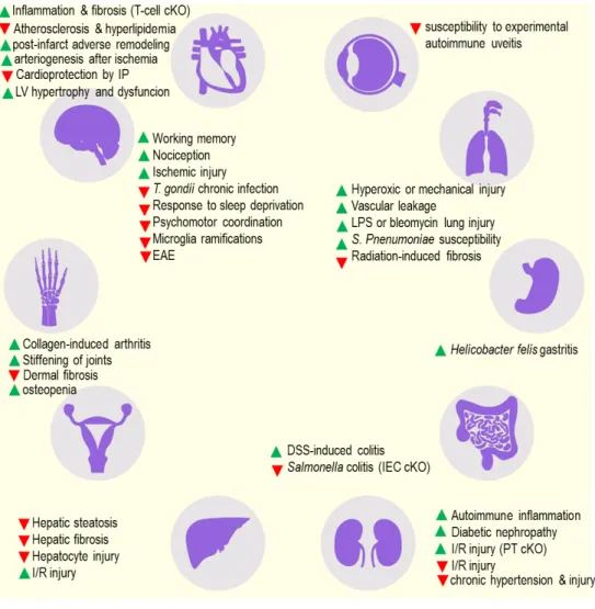
described in Figure 2. The initial characterization of the Nt5e-/- mice provided the foundational
knowledge to further examine CD73 in various pathophysiological states. This review will
address the current understanding of CD73 function and regulation landscape at the cell and
4
CHAPTER 2: CD73 FUNCTIONS IN THE CARDIOVASCULAR SYSTEM CD73 expression and distribution in the cardiovascular system. Robert Berne showed 55 years ago that adenosine production in the heart is stimulated in response to hypoxia and
proposed that adenosine mediates local metabolic control of coronary blood flow (the adenosine
hypothesis) (Berne, 1963). A brief historical account of some of the studies (Eltzschig et al., 2003; Koszalka et al., 2004) leading to the recognition of CD73 as a key player in the
cardiovascular system was provided by Ray Olsson in 2004 (Olsson, 2004). Other relevant work
on CD73 and purinergic signaling in the heart was highlighted in a review by Burnstock and
Pelleg (Burnstock & Pelleg, 2015). Here we focus on the most recent work regarding cell type
specific functions of CD73 in the cardiovascular system.
In the cardiovascular system CD73 is expressed on smooth muscle cells (Yang et al.,
2015), endothelial cells (M. Ohta et al., 2013; Pluskota et al., 2013), and resident lymphocytes
(Bonner, Borg, Burghoff, & Schrader, 2012). The Human Protein Atlas reports moderate levels
of CD73 protein expression on normal human smooth muscle cells, endothelial cells, and
cardiomyocytes (Uhlen et al., 2015), whereas Bonner et al. noted lack of CD73 on these cell
types in mouse heart (Bonner et al., 2013). It is possible that disruption of the tissue architecture
during digestion and processing for cell sorting activated mechanical signaling to trigger
downregulation of surface CD73 from non-immune cells in the latter study (Bonner et al., 2013).
For example, hepatocytes in vivo express CD73, but this expression is dramatically
5
potential molecular mechanism may be through the actions of the mechanosensitive cytoskeletal
protein kindlin-2, a modulator of integrin signaling in endothelial cells and cardiomyocytes
(Dowling et al., 2008) that regulates CD73 trafficking to the membrane (Pluskota et al., 2013). In
the Bonner et al. study, the resident leukocyte population, in particular T cells had the highest
level of CD73 per cell and highest proportion of CD73-positive cells. However, ~50% of the
resident T cells expressed CD73, in contrast to almost ~90% of T cells in the circulation that
were CD73 positive. Therefore, assessing functional contributions to CD73 on different cell
types in the heart and vasculature will require additional studies utilizing cell type-specific
deletion of CD73 in cells and in mice. The key functions of CD73 reported relate to supporting
recovery after myocardial infarction (MI), protection during in heat failure (Quast, Alter, Ding,
Borg, & Schrader, 2017), promoting atherosclerosis (Yang et al., 2015), supporting aortic valve
function (Zukowska et al., 2017) and blocking arteriogenesis (Boring et al., 2013). We will
highlight recent work related to the cell-specific roles of CD73 in MI below.
Cell type-dependent roles of CD73 after myocardial infarction (MI). Healing and recovery of tissue function following myocardial infarction is dependent on T cell-expressed
CD73, which decreases inflammation through the generation of adenosine (Borg et al., 2017).
Specifically, circulating T cells invade the injured heart after infarction and upregulate
expression of hydrolyzing enzymes that act on ATP, cAMP, and NAD, culminating in adenosine
production via CD73. Activation of A2AR and A2BR leads to reduction in the release of
inflammatory mediators. T cells from Nt5e-/- mice are skewed toward Th1 and Th17 types,
resulting in increased levels of their respective pro-inflammatory cytokine products IFN-γ and IL-17 (Borg et al., 2017). When co-cultured with mesenchymal stem cells, monocytes upregulate
6
This promotes an anti-inflammatory state and implicates CD73 in the healing functions of
mesenchymal stem cells.
Interestingly, a functionally significant upregulation of CD73 on epicardium-derived cells
(EPDCs) following MI promotes the release of pro-inflammatory cytokines and pro-fibrogenic
matrix proteins (Hesse et al., 2017). EPDCs, which are normally quiescent in the adult heart, are
activated and give rise to multiple cell types following ischemic heart injury. The increased
production of CD73-generated adenosine from EPDCs leads to A2BR activation, which
stimulates the release of pro-inflammatory cytokines (IL-6, IL-11, and VEGF) (Hesse et al.,
2017). Although the overall in vivo evidence thus far suggests a protective, anti-inflammatory
function of CD73, a critical understanding of the cell-specific mechanisms for how CD73
regulates cardiac remodeling or healing after MI may promote development of potential
7
CHAPTER 3: CD73 FUNCTIONS IN THE CENTRAL NERVOUS SYSTEM CD73 expression and distribution in the central nervous system (CNS).
Immunohistochemical localization of CD73 in mouse brain performed in two independent
studies revealed intense staining in the striatum (Augusto et al., 2013; Kulesskaya et al., 2013) as
well as global pallidus, choroid plexus and meninges (Kulesskaya et al., 2013). Of note, these
structures did not show any positive signal with an anti-CD73 antibody in brain sections from
Nt5e-/- mice, establishing specificity of the assay (Kulesskaya et al., 2013). Biochemically CD73
appears to contribute ~90% of the 5’-nucleotidase activity across the brain, but the spatial
distribution and the identity of the predominant cell types that exhibit CD73 activity in the
mouse brain was not determined (Kulesskaya et al., 2013). The Human Protein Atlas reports
highest expression of CD73 in the cerebellum (Purkinje and granular cell layer) and cortical
endothelial cells (Uhlen et al., 2015), which is in stark contrast to previous work in mouse brain
reporting complete lack of 5’-nucleotidase activity in the granular layer of the cerebellum and
blood vessels (Langer et al., 2008). Several potential factors may account for this discrepancy,
including assay sensitivity (e.g. antibody detection versus enzymatic activity) and differences in
CD73 distribution between mouse and human brain. The cell type specific distribution of CD73
in the mouse spinal cord was characterized with the use of specific neuronal subtype markers,
showing strong expression on L3-L5 dorsal root ganglion neuron membranes, particularly on the
subset of neurons that express nociceptive neuron markers (IB4, Mrgprd, CGRP, and TRPV1)
8
CD73, along with another ecto-nucleotidase, prostatic acid phosphatase (PAP). Similar to CD73,
PAP hydrolyzes AMP to produce adenosine and both proteins and their corresponding activities
decrease in response to nerve injury (Sowa, Taylor-Blake, et al., 2010). Since the activities of
CD73 and PAP enzymes are sensitive to the pH, Sowa et al. proposed that functional
predominance of one enzyme may be relevant under certain conditions, such as during
inflammation or tissue acidosis (Sowa, Taylor-Blake, et al., 2010). Multiple studies to date have
implicated CD73 in major CNS functions, including locomotion and behavior (Augusto et al.,
2013; Kulesskaya et al., 2013), memory functions and plasticity (Blundon et al., 2017;
Zlomuzica, Burghoff, Schrader, & Dere, 2013), sleep regulation (Zielinski, Taishi, Clinton, &
Krueger, 2012), thermoregulation (Muzzi et al., 2013), host-pathogen interactions during brain
infection (Mahamed, Mills, Egan, Denkers, & Bynoe, 2012), inflammation (Mills et al., 2008;
Petrovic-Djergovic et al., 2012; Xu et al., 2018), and nociception. We highlight several studies
where the mechanisms of CD73 have been well described.
CD73 controls locomotion. In the striatum CD73 is closely associated with the A2AR in the post-synaptic compartment, and this interaction appears to be important for controlling
locomotion because Nt5e-/- mice have decreased locomotor activity compared to WT mice after
repeated amphetamine administration, phenocopying A2AR knockout mice (Augusto et al.,
2013). In contrast, baseline locomotion in the Nt5e-/- mice is elevated when measured in the
elevated plus maze, open field test, circadian activity test, and monitoring in the housing cages,
although the mechanisms behind these observations were not investigated (Kulesskaya et al.,
2013). Therefore, the effect of CD73 on locomotion appears to be context specific, and likely
mediated by the spatiotemporal dynamics of the signaling pathways over which CD73 exerts
9
Thalamic CD73 tunes the plasticity of the auditory cortex. The auditory cortex of adults, unlike newborns, lacks the plasticity required to tune neural circuitry upon passive exposure to
auditory inputs from the environment (Kehayas & Holtmaat, 2017). Recently, Blundon et al.
(Blundon et al., 2017) identified CD73-generated adenosine and subsequent A1R activation to be
a key mechanism for age-dependent decline in auditory cortex plasticity. Genetic deletion of the
A1Rs from the auditory thalamus of mature mice promoted plasticity of the auditory cortex after
passive tone exposure (Blundon et al., 2017). Compared to neonates, thalamic expression of
CD73 in mature mice was significantly elevated, which paralleled increased adenosine
production (Blundon et al., 2017). In mature Nt5e–/– mice exposed to a pure tone there was
induction in auditory cortex plasticity, and tone-exposed Nt5e–/– mice distinguished frequencies
better than tone-naive Nt5e–/– mice (Blundon et al., 2017). These results may have implications
for restoring cortical plasticity via CD73 manipulations in learning and other contexts, such as
recovery after stroke.
CD73 on tissue-resident cells regulates CNS inflammation. Given the central role of adenosine as an immunomodulator, several studies have addressed the function of CD73 in brain
inflammation (Mills et al., 2008; Petrovic-Djergovic et al., 2012; Xu et al., 2018). Using genetic
and pharmacological approaches, Petrovic-Djergovic and colleagues demonstrated a protective
role of CD73 in neuroinflammation due to ischemic stroke (Petrovic-Djergovic et al., 2012).
Specifically, Nt5e–/– mice were more susceptible to ischemic stroke injury, and the ischemic
tissue had increased influx and activation of macrophages and pro-inflammatory markers, such as IL1-β, IL6, and TNF-α (Petrovic-Djergovic et al., 2012). This effect was reversed by
administration of soluble CD73, suggesting that the effect was adenosine-mediated
10
that the protective effect of CD73 stemmed from tissue-resident cells, as opposed to CD73 on
circulating immune cells that infiltrated after the injury (Petrovic-Djergovic et al., 2012). While
the specific role of CD73 on astrocytes has not been examined, it is possible that astrocytes play
an key role in this model, since astrocytes contribute to CD73-generated adenosine (Chu, Xiong,
& Parkinson, 2014) and control neuronal injury following ischemic stroke (Takano, Oberheim,
Cotrina, & Nedergaard, 2009).
Surprisingly, CD73 is pro-inflammatory in a mouse model of experimental autoimmune
encephalomyelitis (EAE), which mimics inflammation associated with multiple sclerosis (Mills
et al., 2008). Whereas WT mice displayed weak tail and partial hind limb paralysis and weak tail
by 3 weeks of disease onset, the Nt5e-/- mice only had a weak tail and the disease did not worsen
over time (Mills et al., 2008). Lymphocyte infiltration into the brain was significantly blunted in
the Nt5e-/- mice, implicating CD73 as a facilitator for the entry of pathogenic T cells into the
CNS (Mills et al., 2008). Similar to the stroke model, adoptive transfer studies demonstrated a
role for CD73 on non-hematopoietic cells, potentially choroid plexus epithelial cells, which
expressed CD73 in the WT mice (Mills et al., 2008). Modulation of blood-brain barrier function
via CD73-generated adenosine and activation of A1R and A2AR receptors in one potential
mechanism behind the increased lymphocyte infiltration and inflammation in the EAE model
(Carman, Mills, Krenz, Kim, & Bynoe, 2011). Combined, these two studies demonstrate that
CD73 can exert pro- or anti-inflammatory effects in the brain, depending on the specific
inflammatory condition and the cell types involved, which is an important consideration for
potential therapeutic applications of CD73 modulators in CNS inflammation.
Neuronal CD73 regulates nociception. CD73 and two additional nucleotidases (PAP and tissue non-specific alkaline phosphatase) generate extracellular adenosine in the spinal cord to
11
regulate the function of pain sensing neurons (Street et al., 2013; Street & Zylka, 2011). A series
of elegant studies on Nt5e-/- mice, utilizing supplementation with soluble mouse CD73 enzyme,
demonstrated that CD73 plays a key role in regulating nociception in the mouse spinal cord
(Sowa, Voss, & Zylka, 2010; Xu et al., 2018). Intrathecal administration of soluble mouse CD73
protein elicited dose-dependent and long-lasting (2 days) antinociceptive effects in response to
heat-induced pain (Sowa, Voss, et al., 2010). Similarly, soluble CD73 had antinociceptive effects
in inflammatory and neuropathic pain models (Sowa, Voss, et al., 2010). At the molecular level,
these effects were the result of adenosine-dependent A1R activation, since soluble CD73 did not
produce antinociceptive effects in A1R-/- mice (Sowa, Voss, et al., 2010). The relative
contribution of CD73 on hematopoietic cells versus neurons in the inflammatory pain models is
not presently clear, because studies using bone marrow chimera have not been performed.
However, upon spinal cord injury, CD73 promotes polarization of macrophages and microglia to
the M2 anti-inflammatory phenotype, suggesting that immune cells may play at least a partial
12
CHAPTER 4: CD73 FUNCTIONS IN THE LUNG
CD73 expression and distribution in the lung. Together with tissue non-specific alkaline phosphatase, CD73 is known to be the major regulator of adenosine production on airway
surfaces (Picher, Burch, Hirsh, Spychala, & Boucher, 2003). Human Protein Atlas reports high
CD73 expression on human pneumocytes, which exhibit both cytoplasmic and membrane
distribution. CD73 is also expressed and enzymatically active on primary human nasal and
bronchial epithelial cells where it controls proper cilia beating frequency and ion transport, thus
demonstrating its role in mucociliary clearance which is protective in the development of
infectious lung diseases (Picher et al., 2003).
CD73 promotes maintenance of tissue barrier function in the lungs during hypoxia and hyperoxia. One of the earliest phenotypes reported using the global CD73-null mice showed vascular leakage in response to normobaric hypoxia in multiple tissues, which was most
pronounced in the lung (Thompson et al., 2004). Hypoxia-induced vascular leakage was only
partially reversed by adenosine receptor agonists and administration of soluble CD73, leaving
open the possibility for additional non-enzymatic functions of CD73 that may be compromised
in the Nt5e-/- mice. In humans, acute respiratory distress syndrome (ARDS) leads to pulmonary
vascular leakage and clinical trials utilizing recombinant IFN-β1 (FP-1201) as a treatment strategy have revealed the potential to induce CD73 expression in the lungs and reduce mortality
13
demonstrated that CD73 was significantly upregulated ex vivo in peri-bronchiolar vessels of
cultured human lung tissue in response to IFN-β1 treatment, and that IFN-β1 attenuated 28-day mortality (8% of IFN-β1 treated patients died, versus 32% in the control group) (Bellingan et al., 2014). CD73 is highly upregulated in response to hyperoxia, and Nt5e-/- mice develop more
severe pulmonary edema during hyperoxic lung injury. The latter effect, which is also
phenocopied in A2AR-/- mice, is attributed to loss of barrier function in the setting of decreased
adenosine production (Davies et al., 2014).
CD73 protects against lung infection, inflammation and fibrosis. CD73-null mice exhibit increased weight loss and inflammation in response to intratracheal administration of
lipopolysaccharide (LPS) (Ehrentraut et al., 2013) concomitant with significant transcriptional upregulation of TNFα, IL-1β, and IL-6. These effects were attributed to adenosine generation by regulatory T cells and the phenotypes of the Nt5e-/- mice were partially rescued by
administration of soluble CD73. Deletion of CD73 does not affect baseline adenosine levels in
the lungs, but it prevents adenosine production in response to bleomycin treatment (Volmer,
Thompson, & Blackburn, 2006). Consequently, bleomycin treated Nt5e-/- mice exhibit enhanced
inflammation, collagen production, and more severe fibrosis, which were attenuated by
intranasal administration of exogenous nucleotidase. Treatment with the CD73 inhibitor APCP
or the non-selective adenosine receptor antagonist CGS 15943 caused increased susceptibility of
mice to S. pneumoniae lung challenge, although the effect was more pronounced upon CD73
inhibition (Bou Ghanem et al., 2015).
CD73 may contribute to asthma pathogenesis. Although CD73 function is known to be critical for regulating airway diseases, its involvement in asthma is poorly understood. CD73
14
single study reported that Nt5e-/- mice are protected against ovalbumin-induced airway
hyperresponsiveness and inflammation (Schreiber, Castrop, & Kunzelmann, 2008). To our
knowledge, CD73 expression and activity in asthma patients has not been examined, with the
exception of demonstration that BAL fluid from a patient with asthma contained a subpopulation
of CD73-expressing cells that were thought to represent a mesenchymal progenitor cell type able
to differentiate into fat, bone or cartilage (Bentley et al., 2010). Whether CD73 is simply a
marker of these cells, or an active player in MSC-mediated regulation of inflammatory and
15
CHAPTER 5: CD73 FUNCTIONS IN THE KIDNEY
CD73 expression and distribution in the kidney. The enzymatic activity of CD73 in the kidneys is nearly highest of all the tissues in the body (Colgan et al., 2006; Thompson et al.,
2004). In rat kidneys CD73 decorates the apical membranes of the proximal convoluted tubule,
intercalated cells of the distal nephron, and is also expressed in the peritubular space (Shirley,
Vekaria, & Sevigny, 2009). Similar to other tissues, renovascular function is under the control
of adenosine produced by both CD73 and TNAP (Jackson, Cheng, Verrier, Janesko-Feldman, &
Kochanek, 2014).
CD73 regulates renal ischemia-reperfusion (I/R) injury. Adenosine receptor signaling promotes renal I/R injury, based on findings that A2BR-/- are protected in I/R (Grenz et al., 2008).
Additionally, genetic deletion or pharmacological inhibition of CD73, CD39, or both in mice
revealed that CD73 and CD39 exacerbate I/R injury (Rajakumar et al., 2010; Roberts et al.,
2013). Proximal tubular CD73 in particular is important for regulating renal I/R injury (Sung et
al., 2017). In contrast to this reported injury-promoting role, CD73 plays a protective role in the
context of isoflurane-mediated protection against renal I/R injury. Mice pretreated with a CD73
inhibitor or an adenosine receptor antagonist are insenstitive to this isofluorane-mediated
protection (Kim et al., 2013).
CD73 regulates hypertension-associated renal injury. CD73 is upregulated and
generates adenosine in a model of hypertensive nephropathy induced by angiotensin II (Zhang et
al., 2013). CD73-generated adenosine promotes hypertension associated with chronic kidney
16
contrast to this hypertension promoting role, CD73 is protective through A2BR signaling in the
context of diabetic neuropathy in the mouse kidney (Tak et al., 2014). CD73 is also protective in
the rat kidney in a diabetic neuropathy model induced viastreptozotocin (Taskiran et al., 2016).
Furthermore, pharmacological antagonists for A3R alleviate fibrosis driven by diabetic
neuropathy in rat kidneys (Kretschmar et al., 2016).
Other functions of CD73 in the aging and injured kidney. CD73 knockout mice exhibit spontaneous proteinuria and renal functional deterioration as they age because of an autoimmune
inflammation affecting the glomerular endothelium (Blume et al., 2012). Pharmacological
inhibition or genetic deletion of CD73 in mice leads to more severe kidney injury in sepsis
models (via cecal ligation) compared to WT mice (Hasko et al., 2011). Polymorphisms in NT5E
have been implicated in the development of calcific uremic arteriolopathy in dialysis patients,
17
CHAPTER 6: CD73 FUNCTIONS IN THE LIVER
CD73 expression and distribution in the liver. In the normal human liver CD73 is expressed on the apical membrane of hepatocytes as well as endothelial cells, which express
CD73 at lower levels compared to hepatocytes (Matsuura, Eto, Kato, & Tashiro, 1984). During
myofibroblast differentiation of activated hepatic stellate cells in culture there is transcriptional
upregulation of NT5E (Fausther, Sheung, Saiman, Bansal, & Dranoff, 2012), but this is in
contrast to significant downregulation observed in human liver fibrosis, regardless of etiology or
severity (Snider et al., 2013). CD73 recently has emerged as a critical regulator of hepatocyte
responses to different forms of injury (Hart et al., 2008; Peng et al., 2009; Peng et al., 2008;
Snider et al., 2013), illuminating common disease mechanisms that may be exploited
therapeutically.
CD73 regulates hepatic fibrosis. Nt5e-/- mice were previously reported to be resistant to
hepatic fibrosis (Peng et al., 2008) induced by carbon tetrachloride (CCl4) or thioacetamide
(TAA), which is surprising because genetic or pharmacologic inhibition of adenosine A2AR
causes exacerbated liver injury in inflammation and fibrosis injury models (Chan et al., 2006; A.
Ohta & Sitkovsky, 2001). If adenosine has a protective role in hepatic fibrosis, then removal of
the major enzymatic source of extracellular adenosine (CD73) should exacerbate the pathology,
but the opposite is seen. One of several possible interpretations is that constitutive Nt5e deletion
eliminated an adenosine-independent function of CD73 that promotes hepatic fibrosis. If that is
the case, it may also help explain the resistance of CD73-/- mice to ethanol-induced steatosis
18
CD73 regulates hepatic steatosis. An early phenotype of ethanol consumption that damages the liver is the development of hepatic steatosis, which is the accumulation of fat in the
liver. WT mice develop hepatic steatosis while mice lacking CD73 or adenosine A1 or A2B
receptors are protected after alcohol treatment (Peng et al., 2009). Additionally, in vitro studies
using cultured murine hepatocyte cell lines supported these findings, suggesting adenosine
generated by ethanol metabolism plays an important role in ethanol-induced hepatic steatosis
(Peng et al., 2009). These data demonstrate that CD73 is important in the role acute liver
diseases, and it would be beneficial to determine if CD73 can mediate the progression into
chronic liver diseases.
CD73 regulates hepatic fibrosis. Several studies have shown the importance of CD73 in chronic liver disease models, such as liver fibrosis. For example, studies utilizing chemical
injury (carbon tetrachloride or thioacetamide treatment) for the induction of liver fibrosis in mice
illustrated that Nt5e-/- mice were protected from the development of liver fibrosis or cirrhosis,
while WT mice developed fibrosis and cirrhosis(Peng et al., 2008). Liver fibrosis is
accompanied by the activation of liver myofibroblasts, that typically originate from hepatic
stellate cells and portal fibroblasts (Iwaisako et al., 2014). Although CD73 is expressed in
murine liver fibrosis models and liver myofibroblasts, NT5E is transcriptionally down-regulated
via mediation by Elf2-like transcription factors in mouse-activated liver myofibroblasts
(Fausther, Lavoie, Goree, & Dranoff, 2017). Conversely, other studies have shown NT5E
transcriptional up-regulation through promoter response elements for SP1 and SMAD
transcription factors (Fausther et al., 2012).
CD73 regulates hepatocyte injury. In the context of hepatic ischemia, CD73 is induced at the transcriptional and protein level (Hart et al., 2008) and serves a protective function. Nt5e
-/-19
mice develop more severe injury, which can be reversed by administration of soluble enzyme
(Hart et al., 2008). Based on this a proposed method to elicit hepatoprotection during hepatic
ischemia/reperfusion would be to inhibit adenosine uptake (Zimmerman et al., 2013).
Equilibrative nucleoside transporters (ENTs) are responsible for the uptake of adenosine, and
transcriptional downregulation of ENTs are correlated with ischemia and reperfusion during
human liver transplantation and during murine liver ischemia and reperfusion (Zimmerman et al.,
2013). Ultimately, these studies provide the framework for potential therapies to manage
ischemia and reperfusion by maintaining extracellular adenosine levels.
The formation of hepatocyte Mallory-Denk bodies (MDBs), which are aggregates of
keratins 8 and 18 (K8/K18), ubiquitin, and the ubiquitin-binding protein p62, are generally
associated with the development of chronic liver diseases (Snider et al., 2013). We previously
have demonstrated that CD73 modulates the formation of MDBs in a strain-related manner
(Snider et al., 2013). In mice livers that lacked CD73, chemical-induced liver injury (in the form
of MDBs) was prevented, but this protection was not observed in WT mice under the same
20
CHAPTER 7: CD73 FUNCTIONS IN THE IMMUNE SYSTEM
CD73 expression on immune cells. The role of CD73 has been most extensively studied in context of the immune system (Antonioli, Pacher, et al., 2013; Resta, Yamashita, &
Thompson, 1998). CD73 is expressed in macrophages, monocytes, dendritic cells, and
neutrophils (Antonioli, Pacher, et al., 2013; Resta & Thompson, 1997; Resta et al., 1998; Saze et
al., 2013). It is highly expressed on lymphocytes, particularly T cells and B cells (Conter, Song,
Shlomchik, & Tomayko, 2014; Kaku, Cheng, Al-Abed, & Rothstein, 2014; Saze et al., 2013). In
T cells, thymic γδ lineage commitment relies on T cell receptor ligand-induction of CD73 expression.
CD73 regulates immune cell migration. The expression of CD73 on the cell surface has been shown to play a key role in immune cell migration in response to inflammatory stimuli.
Upon LPS treatment in mice, CD73 on endothelial cells restricts the migration of lymphocytes
into the draining lymph nodes through the activation of A2Rs (Takedachi et al., 2008; Thompson
et al., 2008). Mills et al showed that the infiltration of T cells into the central nervous system
during experimental autoimmune encephalomyelitis in mice is regulated by CD73 that is highly
expressed in the choroid plexus (Mills et al., 2008). In the absence of CD73 in the immune cells,
the number of infiltrating lymphocytes increases and further exacerbates the inflammation or
condition. CD73 KO mice that were infected with Toxoplasma gondii showed a higher
propensity for neutrophil and T cell infiltration, inducing an immune-mediated pathology
(Mahamed, Toussaint, & Bynoe, 2015). The administration of the adenosine receptor agonist,
adenosine-21
generating function of CD73. Similarly, CD4+ T cells that lack CD73 produce a stronger immune
response against bacterial infection, and generate pro-inflammatory cytokines like TNFα, IFN γ, IL17A, and keratinocyte chemoattractant.
CD73 regulates chronic inflammation. In the germinal center, follicular dendritic cells interact with B cells via CD73 and induce maturation (Airas & Jalkanen, 1996). Interestingly, B1
cells, which are a part of the humoral response, express a 5-fold increase in CD73 compared to
the adaptive immune B2 cells (Conter et al., 2014). Allard et al further showed that the lack of
CD73 and subsequent adenosine signaling in B1 cells delayed isotype switching from the
primary response IgM antibody to the pro-inflammatory IgG3 antibody (Allard, Charlebois,
Gilbert, Stagg, & Chrobak, 2018). However, this delay in isotype switching did not alter the
protective response to infection. In a chronic state of inflammation, CD73-expressing T follicular
helper cells regulate the differentiation and maintenance of B cells in to plasma cells, suggesting
an important role of CD73 in establishing humoral immunity (Conter et al., 2014). While
macrophages and monocytes express cell surface CD73 in both mice and humans, their
polarization towards a pro-inflammatory M1 or anti-inflammatory M2 phenotype is independent
of CD73 (Eichin, Laurila, Jalkanen, & Salmi, 2015).
Genetic deletion of CD73 promotes T cell expansion, and production of pro-inflammatory cytokines IFNγ and IL-6 in a mice model of graft vs host disease (GvHD) (Tsukamoto et al., 2012). Specifically, blocking the enzymatic activity of CD73 induced a
stronger alloreactive T cell activity, suggesting that the adenosine-generating function is
important in the progression of GvHD (Wang et al., 2013). Thus, these suggest an important
function of CD73 in the immune cells in attenuating the severity of inflammation or condition
22
CHAPTER 8: CD73 FUNCTIONS IN OTHER TISSUES AND CELL TYPES Gastrointestinal system. CD4+ T cells that lack CD73 produce a stronger immune
response against bacterial infection, and generate pro-inflammatory cytokines like TNFα, IFN-γ, IL17A, and keratinocyte chemoattractant. This type of cytokine milieu resulted in more severe
Helicobacter felis-induced gastritis or murine salmonellosis compared to the WT mice due to the failure of T regulatory cells lacking CD73 to attenuate inflammation. However, there was a
significant enhancement of bacterial clearance in CD73 KO mice that were infected with H. felis
or Salmonella (Alam et al., 2014; Alam et al., 2009). CD73 KO mice also exhibited a more
severe condition of experimental inflammatory colitis compared to WT mice (Kaku et al., 2014).
Upon adoptive transfer of CD73-expressing B1 cells from WT mice to CD73 KO mice, the
disease severity was reduced (Kaku et al., 2014).
Bone. The role of adenosine signaling in the development and differentiation of bone tissue, osteoblasts, and osteoclasts has been implicated in several studies. For example, a study
demonstrated that adenosine receptor activation induced mitogenesis and triggered a protective
response in regard to cell death (Fatokun, Stone, & Smith, 2006; Shimegi, 1998). Additionally,
in human primary osteoblast cells it was shown that pharmacological activation of adenosine
receptors promoted proliferation, and proliferation was halted with antagonism (Costa et al.,
2011). CD73 is a known marker of osteochondroprogenitor cells, which readily form bone after
transplantation (Singh et al., 2015) and CD73-null mice develop bone deficiencies, such as
osteopenia (Takedachi et al., 2012). Furthermore, in vitro studies using MC3T3-E1 cells
23
modulate osteoblast differentiation. Recently, it was demonstrated that CD73-generated
adenosine in human amniotic fluid stem cells enhances osteogenic potential via TGFβ induction (Hau et al., 2017). Overall, CD73 and CD73-generated adenosine are important in the process of
bone development and growth. In terms of pathologic conditions, CD73-deficient mice are
significantly more susceptible to developing collagen-induced arthritis compared to wild-type
mice (Chrobak et al., 2015). In the absence of CD73, there is enhanced production of
proinflammatory cytokines in the joints and augmented Th1 T cell responses, leading to joint
destruction. Importantly, using bone marrow transplants it was shown that the protective activity
of CD73 originated from nonhematopoietic cells (Chrobak et al., 2015).
Inflammation of the eye. CD73 marks an intermediate stage in γδ T cells from which their ability to express effector genes IFNγ or IL-17 is resolved (Coffey et al., 2014). Related to this function, it was reported that CD73 expression on γδ T cells is dynamically regulated during experimental autoimmune uveitis (EAU) and that low CD73 expression on γδ T cells correlated with enhanced Th17 response-promoting activity. Mice lacking CD73 showed decreased
susceptibility to developing EAU (Liang et al., 2016).
Reproductive tissues. In CD73-deficient mice, it was demonstrated that CD73-generated adenosine is important to initiate and maintain penile erections (Wen et al., 2011). In humans,
the expression of ecto-nucleotidases, such as CD73, fluctuates in concert with the menstrual
cycle and changes after menopause in the endometrium (Aliagas et al., 2013).
Skin and Muscle. Nt5e-/- mice have lower susceptibility to developing
bleomycin-induced skin fibrosis (Fernandez et al., 2013). This response, also seen in the CD39-deficient mice, was associated with reduced expression of the profibrotic mediators, TGF-β1 and
24
2013). In Nt5e-/- mice that were fed a high fat diet, CD73 plays a protective role in the
development of dyslipidemia and intramyocellular lipid accumulation in the muscle of mice
(Burghoff et al., 2013). Ultimately, CD73 function is critical in various tissues and processes in
the body, therefore, it is essential to potentially exploit CD73 and adenosine signaling in the
25
Figure 1. Relative levels of CD73 protein expression across human organ systems and tissues. The expression data on CD73 (Gene: NT5E) were collected from the Human Protein Atlas Project (https://www.proteinatlas.org/). Note that CD73 is ubiquitous and abundantly expressed in most tissue types.
26
Figure 2. Reported phenotypes of the CD73 knockout mice. Unless otherwise specified, all studies utilized the whole body knockout model. Green upward pointing arrow denotes that the response was elevated in the CD73 knockout mouse; red downward arrow denotes that the response was decreased in the CD73 knockout mouse. In cases where conditional knockouts (cKO) were used, this is specified in the parenthesis.
27 REFERENCES
Airas, L., Hellman, J., Salmi, M., Bono, P., Puurunen, T., Smith, D. J., & Jalkanen, S. (1995). CD73 is involved in lymphocyte binding to the endothelium: characterization of lymphocyte-vascular adhesion protein 2 identifies it as CD73. J Exp Med, 182(5), 1603-1608.
Airas, L., & Jalkanen, S. (1996). CD73 mediates adhesion of B cells to follicular dendritic cells. Blood, 88(5), 1755-1764.
Airas, L., Niemela, J., & Jalkanen, S. (2000). CD73 engagement promotes lymphocyte binding to endothelial cells via a lymphocyte function-associated antigen-1-dependent mechanism. J Immunol, 165(10), 5411-5417.
Airas, L., Niemela, J., Salmi, M., Puurunen, T., Smith, D. J., & Jalkanen, S. (1997). Differential regulation and function of CD73, a glycosyl-phosphatidylinositol-linked 70-kD adhesion molecule, on lymphocytes and endothelial cells. J Cell Biol, 136(2), 421-431.
Alam, M. S., Kuo, J. L., Ernst, P. B., Derr-Castillo, V., Pereira, M., Gaines, D., . . . Williams, K. (2014). Ecto-5'-nucleotidase (CD73) regulates host inflammatory responses and exacerbates murine salmonellosis. Sci Rep, 4, 4486. doi:10.1038/srep04486
Alam, M. S., Kurtz, C. C., Rowlett, R. M., Reuter, B. K., Wiznerowicz, E., Das, S., . . . Ernst, P. B. (2009). CD73 is expressed by human regulatory T helper cells and suppresses proinflammatory cytokine production and Helicobacter felis-induced gastritis in mice. J Infect Dis, 199(4), 494-504. doi:10.1086/596205
Aliagas, E., Vidal, A., Torrejon-Escribano, B., Taco Mdel, R., Ponce, J., de Aranda, I. G., . . . Martin-Satue, M. (2013). Ecto-nucleotidases distribution in human cyclic and postmenopausic endometrium. Purinergic Signal, 9(2), 227-237. doi:10.1007/s11302-012-9345-0
Allard, D., Charlebois, R., Gilbert, L., Stagg, J., & Chrobak, P. (2018). CD73-A2a adenosine receptor axis promotes innate B cell antibody responses to pneumococcal polysaccharide vaccination. PLoS One, 13(1), e0191973. doi:10.1371/journal.pone.0191973
Antonioli, L., Blandizzi, C., Pacher, P., & Hasko, G. (2013). Immunity, inflammation and cancer: a leading role for adenosine. Nat Rev Cancer, 13(12), 842-857. doi:10.1038/nrc3613
Antonioli, L., Pacher, P., Vizi, E. S., & Hasko, G. (2013). CD39 and CD73 in immunity and inflammation. Trends Mol Med, 19(6), 355-367. doi:10.1016/j.molmed.2013.03.005
Augusto, E., Matos, M., Sevigny, J., El-Tayeb, A., Bynoe, M. S., Muller, C. E., . . . Chen, J. F. (2013). Ecto-5'-nucleotidase (CD73)-mediated formation of adenosine is critical for the striatal adenosine A2A receptor functions. J Neurosci, 33(28), 11390-11399. doi:10.1523/JNEUROSCI.5817-12.2013
Beavis, P. A., Stagg, J., Darcy, P. K., & Smyth, M. J. (2012). CD73: a potent suppressor of antitumor immune responses. Trends Immunol, 33(5), 231-237. doi:10.1016/j.it.2012.02.009
Bellingan, G., Brealey, D., Mancebo, J., Mercat, A., Patroniti, N., Pettila, V., . . . Ranieri, V. M. (2017). Comparison of the efficacy and safety of FP-1201-lyo (intravenously administered recombinant human interferon beta-1a) and placebo in the treatment of patients with moderate or severe acute
28
respiratory distress syndrome: study protocol for a randomized controlled trial. Trials, 18(1), 536. doi:10.1186/s13063-017-2234-7
Bellingan, G., Maksimow, M., Howell, D. C., Stotz, M., Beale, R., Beatty, M., . . . Jalkanen, S. (2014). The effect of intravenous interferon-beta-1a (FP-1201) on lung CD73 expression and on acute respiratory distress syndrome mortality: an open-label study. Lancet Respir Med, 2(2), 98-107. doi:10.1016/S2213-2600(13)70259-5
Bentley, J. K., Popova, A. P., Bozyk, P. D., Linn, M. J., Baek, A. E., Lei, J., . . . Hershenson, M. B. (2010). Ovalbumin sensitization and challenge increases the number of lung cells possessing a mesenchymal stromal cell phenotype. Respir Res, 11, 127. doi:10.1186/1465-9921-11-127 Berne, R. M. (1963). Cardiac nucleotides in hypoxia: possible role in regulation of coronary blood flow.
Am J Physiol, 204, 317-322. doi:10.1152/ajplegacy.1963.204.2.317
Blume, C., Felix, A., Shushakova, N., Gueler, F., Falk, C. S., Haller, H., & Schrader, J. (2012).
Autoimmunity in CD73/Ecto-5'-nucleotidase deficient mice induces renal injury. PLoS One, 7(5), e37100. doi:10.1371/journal.pone.0037100
Blundon, J. A., Roy, N. C., Teubner, B. J. W., Yu, J., Eom, T. Y., Sample, K. J., . . . Zakharenko, S. S. (2017). Restoring auditory cortex plasticity in adult mice by restricting thalamic adenosine signaling. Science, 356(6345), 1352-1356. doi:10.1126/science.aaf4612
Bonner, F., Borg, N., Burghoff, S., & Schrader, J. (2012). Resident cardiac immune cells and expression of the ectonucleotidase enzymes CD39 and CD73 after ischemic injury. PLoS One, 7(4), e34730. doi:10.1371/journal.pone.0034730
Bonner, F., Borg, N., Jacoby, C., Temme, S., Ding, Z., Flogel, U., & Schrader, J. (2013). Ecto-5'-nucleotidase on immune cells protects from adverse cardiac remodeling. Circ Res, 113(3), 301-312. doi:10.1161/CIRCRESAHA.113.300180
Borg, N., Alter, C., Gorldt, N., Jacoby, C., Ding, Z., Steckel, B., . . . Schrader, J. (2017). CD73 on T Cells Orchestrates Cardiac Wound Healing After Myocardial Infarction by Purinergic Metabolic Reprogramming. Circulation, 136(3), 297-313. doi:10.1161/CIRCULATIONAHA.116.023365 Boring, Y. C., Flogel, U., Jacoby, C., Heil, M., Schaper, W., & Schrader, J. (2013). Lack of
ecto-5'-nucleotidase (CD73) promotes arteriogenesis. Cardiovasc Res, 97(1), 88-96. doi:10.1093/cvr/cvs286
Bou Ghanem, E. N., Clark, S., Roggensack, S. E., McIver, S. R., Alcaide, P., Haydon, P. G., & Leong, J. M. (2015). Extracellular Adenosine Protects against Streptococcus pneumoniae Lung Infection by Regulating Pulmonary Neutrophil Recruitment. PLoS Pathog, 11(8), e1005126.
doi:10.1371/journal.ppat.1005126
Burghoff, S., Flogel, U., Bongardt, S., Burkart, V., Sell, H., Tucci, S., . . . Schrader, J. (2013). Deletion of CD73 promotes dyslipidemia and intramyocellular lipid accumulation in muscle of mice. Arch Physiol Biochem, 119(2), 39-51. doi:10.3109/13813455.2012.755547
Burnstock, G., & Pelleg, A. (2015). Cardiac purinergic signalling in health and disease. Purinergic Signal, 11(1), 1-46. doi:10.1007/s11302-014-9436-1
29
Carman, A. J., Mills, J. H., Krenz, A., Kim, D. G., & Bynoe, M. S. (2011). Adenosine receptor signaling modulates permeability of the blood-brain barrier. J Neurosci, 31(37), 13272-13280.
doi:10.1523/JNEUROSCI.3337-11.2011
Chan, E. S., Montesinos, M. C., Fernandez, P., Desai, A., Delano, D. L., Yee, H., . . . Cronstein, B. N. (2006). Adenosine A(2A) receptors play a role in the pathogenesis of hepatic cirrhosis. Br J Pharmacol, 148(8), 1144-1155. doi:10.1038/sj.bjp.0706812
Chen, J. F., Eltzschig, H. K., & Fredholm, B. B. (2013). Adenosine receptors as drug targets--what are the challenges? Nat Rev Drug Discov, 12(4), 265-286. doi:10.1038/nrd3955
Chrobak, P., Charlebois, R., Rejtar, P., El Bikai, R., Allard, B., & Stagg, J. (2015). CD73 plays a protective role in collagen-induced arthritis. J Immunol, 194(6), 2487-2492.
doi:10.4049/jimmunol.1401416
Chu, S., Xiong, W., & Parkinson, F. E. (2014). Effect of ecto-5'-nucleotidase (eN) in astrocytes on adenosine and inosine formation. Purinergic Signal, 10(4), 603-609. doi:10.1007/s11302-014-9421-8
Coffey, F., Lee, S. Y., Buus, T. B., Lauritsen, J. P., Wong, G. W., Joachims, M. L., . . . Wiest, D. L. (2014). The TCR ligand-inducible expression of CD73 marks gammadelta lineage commitment and a metastable intermediate in effector specification. J Exp Med, 211(2), 329-343.
doi:10.1084/jem.20131540
Colgan, S. P., Eltzschig, H. K., Eckle, T., & Thompson, L. F. (2006). Physiological roles for ecto-5'-nucleotidase (CD73). Purinergic Signal, 2(2), 351-360. doi:10.1007/s11302-005-5302-5 Conter, L. J., Song, E., Shlomchik, M. J., & Tomayko, M. M. (2014). CD73 expression is dynamically
regulated in the germinal center and bone marrow plasma cells are diminished in its absence. PLoS One, 9(3), e92009. doi:10.1371/journal.pone.0092009
Costa, M. A., Barbosa, A., Neto, E., Sa-e-Sousa, A., Freitas, R., Neves, J. M., . . . Correia-de-Sa, P. (2011). On the role of subtype selective adenosine receptor agonists during proliferation and osteogenic differentiation of human primary bone marrow stromal cells. J Cell Physiol, 226(5), 1353-1366. doi:10.1002/jcp.22458
Davies, J., Karmouty-Quintana, H., Le, T. T., Chen, N. Y., Weng, T., Luo, F., . . . Blackburn, M. R. (2014). Adenosine promotes vascular barrier function in hyperoxic lung injury. Physiol Rep, 2(9). doi:10.14814/phy2.12155
Dianzani, U., Redoglia, V., Bragardo, M., Attisano, C., Bianchi, A., Di Franco, D., . . . et al. (1993). Co-stimulatory signal delivered by CD73 molecule to human CD45RAhiCD45ROlo (naive) CD8+ T lymphocytes. J Immunol, 151(8), 3961-3970.
Dowling, J. J., Gibbs, E., Russell, M., Goldman, D., Minarcik, J., Golden, J. A., & Feldman, E. L. (2008). Kindlin-2 is an essential component of intercalated discs and is required for vertebrate cardiac structure and function. Circ Res, 102(4), 423-431. doi:10.1161/CIRCRESAHA.107.161489 Ehrentraut, H., Clambey, E. T., McNamee, E. N., Brodsky, K. S., Ehrentraut, S. F., Poth, J. M., . . .
Eltzschig, H. K. (2013). CD73+ regulatory T cells contribute to adenosine-mediated resolution of acute lung injury. FASEB J, 27(6), 2207-2219. doi:10.1096/fj.12-225201
30
Eichin, D., Laurila, J. P., Jalkanen, S., & Salmi, M. (2015). CD73 Activity is Dispensable for the
Polarization of M2 Macrophages. PLoS One, 10(8), e0134721. doi:10.1371/journal.pone.0134721 Eltzschig, H. K. (2009). Adenosine: an old drug newly discovered. Anesthesiology, 111(4), 904-915.
doi:10.1097/ALN.0b013e3181b060f2
Eltzschig, H. K., Ibla, J. C., Furuta, G. T., Leonard, M. O., Jacobson, K. A., Enjyoji, K., . . . Colgan, S. P. (2003). Coordinated adenine nucleotide phosphohydrolysis and nucleoside signaling in
posthypoxic endothelium: role of ectonucleotidases and adenosine A2B receptors. J Exp Med, 198(5), 783-796. doi:10.1084/jem.20030891
Fatokun, A. A., Stone, T. W., & Smith, R. A. (2006). Hydrogen peroxide-induced oxidative stress in MC3T3-E1 cells: The effects of glutamate and protection by purines. Bone, 39(3), 542-551. doi:10.1016/j.bone.2006.02.062
Fausther, M., Lavoie, E. G., Goree, J. R., Baldini, G., & Dranoff, J. A. (2014). NT5E mutations that cause human disease are associated with intracellular mistrafficking of NT5E protein. PLoS One, 9(6), e98568. doi:10.1371/journal.pone.0098568
Fausther, M., Lavoie, E. G., Goree, J. R., & Dranoff, J. A. (2017). An Elf2-like transcription factor acts as repressor of the mouse ecto-5'-nucleotidase gene expression in hepatic myofibroblasts. Purinergic Signal, 13(4), 417-428. doi:10.1007/s11302-017-9570-7
Fausther, M., Sheung, N., Saiman, Y., Bansal, M. B., & Dranoff, J. A. (2012). Activated hepatic stellate cells upregulate transcription of ecto-5'-nucleotidase/CD73 via specific SP1 and SMAD promoter elements. Am J Physiol Gastrointest Liver Physiol, 303(8), G904-914.
doi:10.1152/ajpgi.00015.2012
Fernandez, P., Perez-Aso, M., Smith, G., Wilder, T., Trzaska, S., Chiriboga, L., . . . Chan, E. S. L. (2013). Extracellular generation of adenosine by the ectonucleotidases CD39 and CD73 promotes dermal fibrosis. Am J Pathol, 183(6), 1740-1746. doi:10.1016/j.ajpath.2013.08.024
Fredholm, B. B. (2007). Adenosine, an endogenous distress signal, modulates tissue damage and repair. Cell Death Differ, 14(7), 1315-1323. doi:10.1038/sj.cdd.4402132
Grenz, A., Osswald, H., Eckle, T., Yang, D., Zhang, H., Tran, Z. V., . . . Eltzschig, H. K. (2008). The reno-vascular A2B adenosine receptor protects the kidney from ischemia. PLoS Med, 5(6), e137. doi:10.1371/journal.pmed.0050137
Hart, M. L., Much, C., Gorzolla, I. C., Schittenhelm, J., Kloor, D., Stahl, G. L., & Eltzschig, H. K. (2008). Extracellular adenosine production by ecto-5'-nucleotidase protects during murine hepatic
ischemic preconditioning. Gastroenterology, 135(5), 1739-1750 e1733. doi:10.1053/j.gastro.2008.07.064
Hasko, G., Csoka, B., Koscso, B., Chandra, R., Pacher, P., Thompson, L. F., . . . Nemeth, Z. H. (2011). Ecto-5'-nucleotidase (CD73) decreases mortality and organ injury in sepsis. J Immunol, 187(8), 4256-4267. doi:10.4049/jimmunol.1003379
31
Hau, K. L., Ranzoni, A. M., Vlahova, F., Hawkins, K., De Coppi, P., David, A. L., & Guillot, P. V. (2017). TGFbeta-induced osteogenic potential of human amniotic fluid stem cells via CD73-generated adenosine production. Sci Rep, 7(1), 6601. doi:10.1038/s41598-017-06780-1 Hesse, J., Leberling, S., Boden, E., Friebe, D., Schmidt, T., Ding, Z., . . . Schrader, J. (2017).
CD73-derived adenosine and tenascin-C control cytokine production by epicardium-CD73-derived cells formed after myocardial infarction. FASEB J, 31(7), 3040-3053. doi:10.1096/fj.201601307R Ipata, P. L., & Balestri, F. (2013). The functional logic of cytosolic 5'-nucleotidases. Curr Med Chem,
20(34), 4205-4216.
Iwaisako, K., Jiang, C., Zhang, M., Cong, M., Moore-Morris, T. J., Park, T. J., . . . Kisseleva, T. (2014). Origin of myofibroblasts in the fibrotic liver in mice. Proc Natl Acad Sci U S A, 111(32), E3297-3305. doi:10.1073/pnas.1400062111
Jackson, E. K., Cheng, D., Verrier, J. D., Janesko-Feldman, K., & Kochanek, P. M. (2014). Interactive roles of CD73 and tissue nonspecific alkaline phosphatase in the renal vascular metabolism of 5'-AMP. Am J Physiol Renal Physiol, 307(6), F680-685. doi:10.1152/ajprenal.00312.2014
Kaku, H., Cheng, K. F., Al-Abed, Y., & Rothstein, T. L. (2014). A novel mechanism of B cell-mediated immune suppression through CD73 expression and adenosine production. J Immunol, 193(12), 5904-5913. doi:10.4049/jimmunol.1400336
Kehayas, V., & Holtmaat, A. (2017). Rejuvenating brain plasticity. Science, 356(6345), 1335-1336. doi:10.1126/science.aan8374
Kim, M., Ham, A., Kim, J. Y., Brown, K. M., D'Agati, V. D., & Lee, H. T. (2013). The volatile anesthetic isoflurane induces ecto-5'-nucleotidase (CD73) to protect against renal ischemia and reperfusion injury. Kidney Int, 84(1), 90-103. doi:10.1038/ki.2013.43
Knapp, K., Zebisch, M., Pippel, J., El-Tayeb, A., Muller, C. E., & Strater, N. (2012). Crystal structure of the human ecto-5'-nucleotidase (CD73): insights into the regulation of purinergic signaling. Structure, 20(12), 2161-2173. doi:10.1016/j.str.2012.10.001
Knapp, K. M., Zebisch, M., & Strater, N. (2012). Crystallization and preliminary X-ray analysis of the open form of human ecto-5'-nucleotidase (CD73). Acta Crystallogr Sect F Struct Biol Cryst Commun, 68(Pt 12), 1545-1549. doi:10.1107/S1744309112045447
Koszalka, P., Ozuyaman, B., Huo, Y., Zernecke, A., Flogel, U., Braun, N., . . . Schrader, J. (2004). Targeted disruption of cd73/ecto-5'-nucleotidase alters thromboregulation and augments vascular inflammatory response. Circ Res, 95(8), 814-821. doi:10.1161/01.RES.0000144796.82787.6f Kretschmar, C., Oyarzun, C., Villablanca, C., Jaramillo, C., Alarcon, S., Perez, G., . . . San Martin, R. (2016). Reduced Adenosine Uptake and Its Contribution to Signaling that Mediates Profibrotic Activation in Renal Tubular Epithelial Cells: Implication in Diabetic Nephropathy. PLoS One, 11(1), e0147430. doi:10.1371/journal.pone.0147430
Kulesskaya, N., Voikar, V., Peltola, M., Yegutkin, G. G., Salmi, M., Jalkanen, S., & Rauvala, H. (2013). CD73 is a major regulator of adenosinergic signalling in mouse brain. PLoS One, 8(6), e66896. doi:10.1371/journal.pone.0066896
32
Langer, D., Hammer, K., Koszalka, P., Schrader, J., Robson, S., & Zimmermann, H. (2008). Distribution of ectonucleotidases in the rodent brain revisited. Cell Tissue Res, 334(2), 199-217.
doi:10.1007/s00441-008-0681-x
Li, Q., Price, T. P., Sundberg, J. P., & Uitto, J. (2014). Juxta-articular joint-capsule mineralization in CD73 deficient mice: similarities to patients with NT5E mutations. Cell Cycle, 13(16), 2609-2615. doi:10.4161/15384101.2014.943567
Liang, D., Zuo, A., Zhao, R., Shao, H., Born, W. K., O'Brien, R. L., . . . Sun, D. (2016). CD73 Expressed on gammadelta T Cells Shapes Their Regulatory Effect in Experimental Autoimmune Uveitis. PLoS One, 11(2), e0150078. doi:10.1371/journal.pone.0150078
Mahamed, D. A., Mills, J. H., Egan, C. E., Denkers, E. Y., & Bynoe, M. S. (2012). CD73-generated adenosine facilitates Toxoplasma gondii differentiation to long-lived tissue cysts in the central nervous system. Proc Natl Acad Sci U S A, 109(40), 16312-16317. doi:10.1073/pnas.1205589109 Mahamed, D. A., Toussaint, L. E., & Bynoe, M. S. (2015). CD73-generated adenosine is critical for
immune regulation during Toxoplasma gondii infection. Infect Immun, 83(2), 721-729. doi:10.1128/IAI.02536-14
Matsuura, S., Eto, S., Kato, K., & Tashiro, Y. (1984). Ferritin immunoelectron microscopic localization of 5'-nucleotidase on rat liver cell surface. J Cell Biol, 99(1 Pt 1), 166-173.
Mikhailov, A., Sokolovskaya, A., Yegutkin, G. G., Amdahl, H., West, A., Yagita, H., . . . Eriksson, J. E. (2008). CD73 participates in cellular multiresistance program and protects against TRAIL-induced apoptosis. J Immunol, 181(1), 464-475.
Mills, J. H., Thompson, L. F., Mueller, C., Waickman, A. T., Jalkanen, S., Niemela, J., . . . Bynoe, M. S. (2008). CD73 is required for efficient entry of lymphocytes into the central nervous system during experimental autoimmune encephalomyelitis. Proc Natl Acad Sci U S A, 105(27), 9325-9330. doi:10.1073/pnas.0711175105
Monguio-Tortajada, M., Roura, S., Galvez-Monton, C., Franquesa, M., Bayes-Genis, A., & Borras, F. E. (2017). Mesenchymal Stem Cells Induce Expression of CD73 in Human Monocytes In Vitro and in a Swine Model of Myocardial Infarction In Vivo. Front Immunol, 8, 1577.
doi:10.3389/fimmu.2017.01577
Muzzi, M., Blasi, F., Masi, A., Coppi, E., Traini, C., Felici, R., . . . Chiarugi, A. (2013). Neurological basis of AMP-dependent thermoregulation and its relevance to central and peripheral
hyperthermia. J Cereb Blood Flow Metab, 33(2), 183-190. doi:10.1038/jcbfm.2012.157 Ohta, A., & Sitkovsky, M. (2001). Role of G-protein-coupled adenosine receptors in downregulation of
inflammation and protection from tissue damage. Nature, 414(6866), 916-920. doi:10.1038/414916a
Ohta, M., Toyama, K., Gutterman, D. D., Campbell, W. B., Lemaitre, V., Teraoka, R., & Miura, H. (2013). Ecto-5'-nucleotidase, CD73, is an endothelium-derived hyperpolarizing factor synthase. Arterioscler Thromb Vasc Biol, 33(3), 629-636. doi:10.1161/ATVBAHA.112.300600
Olsson, R. A. (2004). Cardiovascular ecto-5'-nucleotidase: an end to 40 years in the wilderness? Circ Res, 95(8), 752-753. doi:10.1161/01.RES.0000146278.94064.4b
33
Peng, Z., Borea, P. A., Varani, K., Wilder, T., Yee, H., Chiriboga, L., . . . Cronstein, B. N. (2009). Adenosine signaling contributes to ethanol-induced fatty liver in mice. J Clin Invest, 119(3), 582-594. doi:10.1172/JCI37409
Peng, Z., Fernandez, P., Wilder, T., Yee, H., Chiriboga, L., Chan, E. S., & Cronstein, B. N. (2008). Ecto-5'-nucleotidase (CD73) -mediated extracellular adenosine production plays a critical role in hepatic fibrosis. FASEB J, 22(7), 2263-2272. doi:10.1096/fj.07-100685
Petrovic-Djergovic, D., Hyman, M. C., Ray, J. J., Bouis, D., Visovatti, S. H., Hayasaki, T., & Pinsky, D. J. (2012). Tissue-resident ecto-5' nucleotidase (CD73) regulates leukocyte trafficking in the ischemic brain. J Immunol, 188(5), 2387-2398. doi:10.4049/jimmunol.1003671
Picher, M., Burch, L. H., Hirsh, A. J., Spychala, J., & Boucher, R. C. (2003). Ecto 5'-nucleotidase and nonspecific alkaline phosphatase. Two AMP-hydrolyzing ectoenzymes with distinct roles in human airways. J Biol Chem, 278(15), 13468-13479. doi:10.1074/jbc.M300569200
Pluskota, E., Ma, Y., Bledzka, K. M., Bialkowska, K., Soloviev, D. A., Szpak, D., . . . Plow, E. F. (2013). Kindlin-2 regulates hemostasis by controlling endothelial cell-surface expression of ADP/AMP catabolic enzymes via a clathrin-dependent mechanism. Blood, 122(14), 2491-2499.
doi:10.1182/blood-2013-04-497669
Quast, C., Alter, C., Ding, Z., Borg, N., & Schrader, J. (2017). Adenosine Formed by CD73 on T Cells Inhibits Cardiac Inflammation and Fibrosis and Preserves Contractile Function in Transverse Aortic Constriction-Induced Heart Failure. Circ Heart Fail, 10(4).
doi:10.1161/CIRCHEARTFAILURE.116.003346
Rajakumar, S. V., Lu, B., Crikis, S., Robson, S. C., d'Apice, A. J., Cowan, P. J., & Dwyer, K. M. (2010). Deficiency or inhibition of CD73 protects in mild kidney ischemia-reperfusion injury.
Transplantation, 90(12), 1260-1264. doi:10.1097/TP.0b013e3182003d9b
Resta, R., & Thompson, L. F. (1997). T cell signalling through CD73. Cell Signal, 9(2), 131-139.
Resta, R., Yamashita, Y., & Thompson, L. F. (1998). Ecto-enzyme and signaling functions of lymphocyte CD73. Immunol Rev, 161, 95-109.
Roberts, V., Lu, B., Rajakumar, S., Cowan, P. J., & Dwyer, K. M. (2013). The CD39-adenosinergic axis in the pathogenesis of renal ischemia-reperfusion injury. Purinergic Signal, 9(2), 135-143. doi:10.1007/s11302-012-9342-3
Rothe, H., Brandenburg, V., Haun, M., Kollerits, B., Kronenberg, F., Ketteler, M., & Wanner, C. (2017). Ecto-5' -Nucleotidase CD73 (NT5E), vitamin D receptor and FGF23 gene polymorphisms may play a role in the development of calcific uremic arteriolopathy in dialysis patients - Data from the German Calciphylaxis Registry. PLoS One, 12(2), e0172407.
doi:10.1371/journal.pone.0172407
Saze, Z., Schuler, P. J., Hong, C. S., Cheng, D., Jackson, E. K., & Whiteside, T. L. (2013). Adenosine production by human B cells and B cell-mediated suppression of activated T cells. Blood, 122(1), 9-18. doi:10.1182/blood-2013-02-482406