Article
1
MS-based approaches enable the structural
2
characterization of transcription factor / DNA
3
response element complex
4
Lukáš Slavata1, 2, Josef Chmelík1, 2, Daniel Kavan1, 2, Růžena Filandrová1, 2, Jan Fiala1, 2, Michal
5
Rosůlek1,2, Hynek Mrázek1, Zdeněk Kukačka1, Karel Vališ1, Petr Man1, 2, Michael Miller3, William
6
McIntyre3, Daniele Fabris3, Petr Novák1, 2, *
7
1 Institute of Microbiology, The Czech Academy of Sciences, Prague, 14220, Czech Republic
8
2 Faculty of Science, Charles University, Prague, 12843, Czech Republic
9
3 RNA Institute, University at Albany, State University of New York, Albany, NY 12222, U.S.A.
10
11
* Correspondence: [email protected]; Tel.: +42-0325-873-610
12
13
Abstract: The limited information available on the structure of complexes involving transcription
14
factors and cognate DNA response elements represents a major obstacle in the quest to understand
15
their mechanism of action at the molecular level. We implemented a concerted structural
16
proteomics approach, which combined hydrogen-deuterium exchange (HDX), quantitative
17
protein-protein and protein-nucleic acid cross-linking (XL), and homology analysis, to model the
18
structure of the complex between the full-length DNA binding domain (DBD) of FOXO4 and its
19
DNA binding element (DBE). The results confirmed that FOXO4-DBD assumes the characteristic
20
forkhead topology shared by these types of transcription factors, but its binding mode differs
21
significantly from those of other members of the family. The results showed that the binding
22
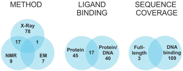
interaction stabilized regions that were rather flexible and disordered in the unbound form.
23
Surprisingly, the conformational effects were not limited only to the interface between bound
24
components but extended also to distal regions that may be essential to recruiting additional
25
factors to the transcription machinery. In addition to providing valuable new insights into the
26
binding mechanism, this project provided an excellent evaluation of the merits of structural
27
proteomics approaches in the investigation of systems that arematerial not directly amenable to
28
traditional high-resolution techniques.
29
Keywords: transcription factor, protein, DNA, protein-nucleic acid cross-linking, cross-linking,
30
transplatin, trans-dichlorodiamineplatinum(II), hydrogen-deuterium exchange, FOXO4, molecular
31
modeling
32
33
1. Introduction
34
The central dogma of biology rationalizes the flow of information leading from the sequence
35
encoded by a gene to the biosynthesis of the corresponding protein. This flow must be finely
36
regulated to coordinate the proper development and functioning of any living organism. A key role
37
in the regulatory machinery is covered by transcription factors (TFs), proteins that recognize target
38
DNA sequences called response elements and establish specific interactions with additional factors
39
to activate or inhibit the transcription process [1–4]. The species involved in the process have been
40
unambiguously identified [5,6], but significant information is still lacking on the effects of
41
structure/dynamics on specific recognition and mechanism of action. The Protein Data Bank
42
contains the high-resolution structures of at least 483 TFs from different species [7], which include
43
less than 10% of all predicted human TFs [6]. Of such structures, only one third include also the
44
cognate DNA response element and only one fifth are available in both bound and unbound states
45
(Figure 1). Due to the size and complexity of such systems, the vast majority of solved structures do
46
not cover the entire TF sequence, but consist almost exclusively of the DNA binding domain (DBD).
47
This fact reflects the modular organization of TFs, which includes discrete domains acting in rather
48
independent manner [8,9]. While DBDs tend to be highly structured, other regions responsible for
49
either modulating transcription activity, or supporting facultative ligand interactions, are rather
50
flexible and assume well-defined conformations only upon binding to the intended factor [10]. The
51
unstructured nature of these regions poses many challenges to conventional high-resolution
52
approaches, which require adequate stability and homogeneity. In most cases, the natural
53
interactions established in vivo, which are responsible for stabilizing well-defined functional
54
conformations, cannot be properly replicated in vitro. These challenges explain the chronic lack of
55
comprehensive information on full-fledged TF structures, which still hampers the elucidation of
56
their mechanism of action at the molecular level.
57
58
Figure 1: Transcription factors structures - current state: Statistics on high-resolution structures
59
deposited in the Protein Data Bank (80), with consideration of selected methodology, presence of
60
interaction partner, and sequence coverage. To date, the structure of only 112 human transcription
61
factor (TF) have been solved out of a total predicted to be in the 1300 - 1900 range [6].
62
Powered by the development of new experimental strategies and mass spectrometric (MS)
63
instrumentation, structural proteomics has rapidly become an essential approach for gathering
64
valuable structural information for species that are not directly amenable to conventional
65
high-resolution techniques [11–14]. In addition to hydrogen-deuterium exchange (HDX) [15], a
66
combination of chemical and biochemical techniques broadly known as MS3D [16–18] have been
67
effectively utilized to identify the regions of contact between bound biomolecules and reveal their
68
mutual spatial organization. In HDX experiments, the exchange rate of backbone amide hydrogens,
69
which is affected by solvent accessibility and possible involvement in hydrogen bonding, can be
70
directly determined by MS analysis. This technique has been broadly employed to study
71
conformational changes [19,20], protein folding [21,22] and protein-protein interactions [23,24], as
72
well as nucleic acid-protein complexes [25–30]. Among the MS3D techniques, chemical and
73
photo-activated cross-linking (XL) are employed to generate stable covalent bridges between
74
contiguous functional groups, which can reveal their mutual placement in the targeted assembly
75
[13,16]. A variety of bifunctional reagents with different spacing between reactive groups have been
76
developed to determine the distance between susceptible residues. In this way, the sequence
77
position of cross-linked residues and the length of the respective cross-linker provide valid
78
constraints for building accurate molecular models through established computational methods
79
[17,18,31,32]. The excellent versatility of these approaches has prompted the development of
80
reagents capable of targeting functional groups present on protein as well as nucleic acid substrates
81
[33–39]. Over time, capture tags and isotopic labels have been included in the cross-linker design to
82
facilitate the isolation and analysis of cross-linked products[40,41]. Isotope-labelling, in particular,
83
has provided a valuable tool for highlighting the presence of different conformational states and
84
quantifying their partitioning on the basis of cross-linking probability [13].
In this study, we evaluated the concerted application of complementary structural proteomics
86
techniques to overcome the challenges posed by full-fledged complexes between TFs and respective
87
DNA response elements. We selected a model system consisting of the human transcription factor
88
FOXO4 and its cognate Daf-16 family member-binding element (DBE) [42]. FOXO4 is part of the “O”
89
subfamily of the forkhead box (FOX) class of transcription factors [42–44]. The DBD regions of this
90
class are characterized by winged helix structures, which are formed by approximately 100 amino
91
acids folded into helix-turn-helix motifs and β-sheet-bordered loops that make them resemble
92
butterfly wings [45]. FOXO4 is involved in cell signaling pathways responsible for oxidative stress
93
response [44,46], proliferation regulation [47,48], and angiogenesis [49]. Different mechanisms have
94
been described to explain its activity and localization [50], which involve lysine acetylation [51–54],
95
serine and threonine phosphorylation, as well as mono- [55] and poly-ubiquitination [56]. Enzymes
96
responsible for FOXO4 phosphorylation are primary effectors in two important regulatory
97
networks: the phosphatidylinositol-3-kinase [57–59] and Ras-like GTPase/c-Jun N-terminal kinase
98
pathways [46]. The first high-resolution structure of FOXO4-DBD, which was obtained by NMR
99
spectroscopy, confirmed the presence of a typical forkhead, winged helix fold [60]. At the same time,
100
however, the report indicated that the N- and C- terminal regions of the DBD displayed chemical
101
shifts consistent with highly flexible, disordered structures. The more recent identification of
102
consensus sequences for the FOXO family [61] enabled the crystallization of a complex comprising a
103
selected DBE duplex and a FOXO4-DBD construct that lacked the C-terminal region to facilitate
104
crystal formation [62]. This high-resolution structure provided valuable details on the protein-DNA
105
interaction, but revealed also numerous discrepancies with the binding modes exhibited by other
106
members of the FOXO family [44], which were attributed to possible crystal-packing issues [62]. For
107
these reasons, the FOXO4-DBD•DBE system offered an excellent opportunity for testing the ability
108
of structural proteomics to probe the conformational effects of binding, which would help rectify or
109
corroborate the observed discrepancies. On the other hand, it also afforded sufficient structural
110
information to determine the validity of the new experimental constraints and evaluate the merits of
111
the selected approaches.
112
The experimental strategies were selected for their ability to provide specific information on a
113
typical protein-DNA complex. For instance, HDX was applied to recognize the regions of the protein
114
affected by DNA binding, either through direct protection of the contact interface, or through
115
allosteric conformational changes involving distal regions of the protein. Quantitative XL was used
116
instead to identify possible variations between free and DNA-bound DBD structures, which would
117
help elucidate the effects of the interaction on overall structure topology. In the case of the DNA
118
component, the fast rate of back-exchange characteristic of nucleic acid hydrogens prevented the
119
application of HDX to recognize the surface of the DBE duplex in direct contact with the DBD. As a
120
possible alternative, we explored the application of transplatin (trans-dichlorodiamineplatinum(II),
121
tPt) to generate protein-DNA cross-links that would help locate the mutual positions of interacting
122
structural features [63]. The spatial constraints afforded by these determinations were combined to
123
guide model-building operations and obtain a full-fledged structure for the complex. The results
124
were compared to the available high-resolution structures to assess possible discrepancies and
125
highlight the new information afforded by the selected techniques. It is necessary to point out here,
126
that the FOXO4-DBD protein construct used in this study was identical to protein constructs used in
127
previous high-resolution structural studies [60,62]. The outcome clearly demonstrated the benefits
128
of structural proteomics to tackle the elucidation of structure and dynamics in systems that elude
129
established high-resolution techniques.
130
2. Materials and Methods
131
2.1. Materials
132
Non-labelled and isotope-labelled cross-linkers di(N-succinimidyl) glutarate (DSGd0/DSGd4)
133
and di(N-succinimidyl) suberate (DSSd0/DSSd4) were purchased form ProteoChem (USA).
134
Modified protease trypsin (Gold, mass spectrometry grade) was purchased from Promega (USA).
Nuclease Bal-31 was obtained from New England BioLabs (USA). Liquid chromatography solvents
136
of LC/MS grade were purchased from Thermo Fisher Scientific (USA). Other chemicals (highest
137
available purity) were obtained from Sigma-Aldrich. All other chemicals, solvents and buffers for
138
SDS-PAGE were obtained from Bio-Rad Laboratories, Inc. (USA). The pET-15b plasmid carrying
139
His-tag, thrombin cleavage site, and FOXO4-DBD (Uniprot ID: P98177-1; residues 86 - 211)
140
sequences was obtained from Prof. Obsil.
141
2.2. Sample preparation
142
Full-length DBD (residues 82 – 207 of the entire FOXO4 sequence) was expressed with an
143
N-terminal His-tag from an appropriate pET-15b plasmid, and then affinity captured on a TALON
144
Superflow Resin (Clontech Laboratories, USA) charged with Co2+. The captured protein was
145
submitted to thrombin digestion to eliminate the tag, followed by gel permeation chromatography.
146
A more detailed description of all experimental procedures is included in the Appendix (Methods in
147
detail) section. The identity, integrity, and purity of the final sample were verified by MS analysis
148
(vide infra). A duplex DNA construct containing one of the DBE consensus sequences (i.e., TTG TTT
149
AC) [42,64] was obtained by annealing complementary oligonucleotides (i.e., 5’-TTG GGT AAA
150
CAA G-3’ forward and 5’-CTT GTT TAC CCA A-3’ reverse). Equimolar amounts were mixed and
151
then heated to 95°C for 1 min. Finally, the sample was let cool to room temperature to form the 13 bp
152
duplex DNA. The desired FOXO4-DBD•DBE complex was obtained by mixing equimolar amounts
153
of protein and DNA samples in 10mM HEPES buffer with 50mM NaCl (pH adjusted to 7.4) to a final
154
33.7µ M concentration, and incubating the mixture at 18°C for 1 hr.
155
2.3. Product characterization
156
Initial stocks of FOXO4-DBD, DBE construct, and FOXO4-DBD•DBE were analyzed to assess
157
sample purity after expression/purification, annealing of the duplex structure, and proper complex
158
formation. The products of XL reaction were also analyzed in the same fashion to assess the
159
incidence of modification. Briefly, each stock was diluted to a final 10-µ M concentration by adding a
160
7.5 mM solution of ammonium acetate (AA) with 50% MeOH (pH 6.85), and then analyzed on a
161
Bruker Daltonics (Billerica, MA) 15T-Solarix XR Fourier transform ion cyclotron resonance (FTICR)
162
mass spectrometer. FOXO4-DBD, as well as FOXO4-DBD•DBE, were analyzed in positive ion mode.
163
Each sample was loaded onto a syringe and introduced into the electrospray ionization (ESI) source
164
at a 2 µ L/min flow rate. The FTICR analyzer was calibrated by using a solution of sodium
165
trifluoroacetate (NaTFA), which afforded a typical 1 ppm accuracy. Mass spectra were acquired over
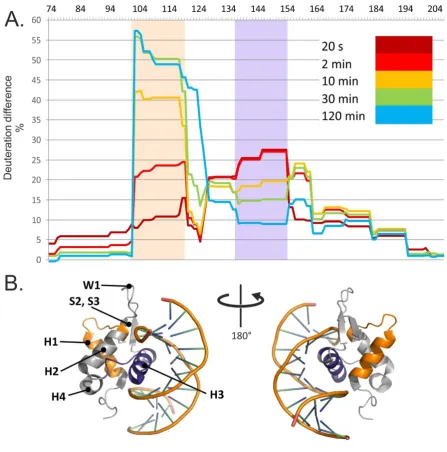
166
a 250 – 4000 m/z range for 3 min.
167
2.4. Hydrogen-deuterium exchange
168
HDX reactions were performed on 20µ M solutions of either FOXO4-DBD or FOXO4-DBD•DBE
169
complex prepared in an H2O-based buffer (pH 7.4) containing 10mM HEPES and 50mM NaCl. After
170
pre-incubation for an hour at 20 °C, the exchange was initiated by diluting each sample 10-fold into a
171
D2O-based buffer (pD 7.4) containing 10mM HEPES and 50mM NaCl. The reaction was allowed to
172
proceed at 20 °C, while small aliquots containing 100pmol of protein were taken at predetermined
173
intervals (i.e., 0.33, 2, 5, 10, 30, 60, 180, and 300 min). Quenching was achieved by immediately
174
mixing the aliquot with a 1M glycine/HCl buffer with pH 2.35, and then rapidly freezing the
175
solution in liquid nitrogen. Samples were stored at -80°C. The analysis was performed by using
176
columns with different immobilized proteases followed by liquid chromatography-mass
177
spectrometry (LC-MS) according to ref. [65]. All experiments were performed as triplicate. A
178
complete description of these procedures is included in the Appendix (Methods in detail) section.
179
2.5. Quantitative protein-protein cross-linking
180
Samples containing 20µ M of either FOXO4-DBD or FOXO4-DBD•DBE complex prepared in
181
10mM HEPES buffer (pH 7.4) with 50mM NaCl were pre-incubated for an hour at 20°C before
introducing the cross-linking reagent. Separate samples of FOXO4-DBD were treated with either
183
DSGd0 or DSSd0 in their regular, non-labelled form, whereas FOXO4-DBD•DBE samples were
184
reacted with the deuterium-labelled DSGd4 and DSSd4 versions. The reagents were dissolved in
185
dimethyl-sulfoxide (DMSO) to 6.74 mM concentrations and then added to each substrate to achieve
186
a 10:1 molar ratio. The cross-linking reaction was allowed to proceed undisturbed for 2 hrs, after
187
which corresponding regular and deuterium-labelled samples (e.g., treated with DSSd0 and DSSd4)
188
were mixed in a 1:1 ratio to enable quantification. In parallel, control samples were also examined,
189
which were treated with pure DMSO lacking cross-linker, or matching cross-linker mixtures with a
190
1:1 ratio of either DSGd0/DSGd4 or DSSd0/DSSd4. All reactions solutions were analyzed by
191
SDS-PAGE to check whether cross-linking had induced any unwanted formation of non-specific
192
higher-order aggregates, or had prevented a sufficient degree of digestion necessary to enable
193
subsequent analysis. Characterization of cross-linked conjugates was achieved according to a
194
bottom up approach that employed trypsin digestion followed by LC-MS analysis [13]. All
195
experiments were performed as triplicate. A complete description is included in the Appendix
196
(Methods in detail) section.
197
2.6. Protein-DNA cross-linking
198
Samples containing 25µ M of either FOXO4-DBD or FOXO4-DBD•DBE complex in 150 mM
199
ammonium acetate (pH 6.85) were treated with a 1mM solution of trans-platinum(II)diammine
200
dichloride (transplatin, tPt), which had been pre-incubated for an hour at 18°C. Reaction mixtures
201
containing a final 200µ M concentration of transplatin and 20µ M of protein/complex were incubated
202
at 18°C for 14 hr. Control samples devoid of transplatin were prepared at the same time in the same
203
manner. Reaction and control samples were analyzed by native and denaturing DNA
204
polyacrylamide gel electrophoresis, SDS polyacrylamide gel electrophoresis, and mass
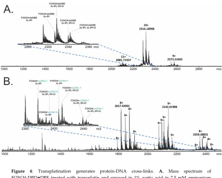
205
spectrometry. Characterization of cross-linked conjugates was achieved according to a bottom up
206
approach that involved treatment with Bal-31 nuclease and trypsin to digest DNA and protein
207
components, respectively, followed by LC-MS/MS analysis with data-independent acquisition with
208
broad isolation window. A complete description is included in the Appendix (Methods in detail)
209
section.
210
2.7. Data processing and interpretation
211
The SNAP 2.0 algorithm of the DataAnalysis 4.2 (Bruker Daltonics) software package was
212
utilized to generate deconvoluted spectra and lists of monoisotopic masses from the acquired MS
213
data. MASCOT 2.2 search engine was used to search MS/MS data and achieve the identification of
214
on-line digest products from a theoretical library of digestion products. Deuteration rate was
215
determined by using the home-built Deutex software (unpublished). The home-built LinX software
216
(available online) and Stavrox (v. 3.6.0.1 by Michael Götze) software was then used to compare the
217
experimental data with a library of theoretical cross-linking products to correctly identify the
218
sought-after conjugates. The proportion of labelled versus un-labelled species was determined by
219
applying mMass 5.4.1 [66] to the signals of such conjugates. In the case of peptide-DNA conjugates,
220
deconvoluted spectra and monoisotopic masses were calculated by using a constant unit to mimic
221
the presence of a certain oligonucleotide cross-linked by a transplatin equivalent and searched by
222
LinX. A complete description of these procedures is included in the Appendix (Methods in detail)
223
section.
224
2.8. Molecular modeling
225
The program Modeller [67] was employed to generate models of FOXO4-DBD and
226
FOXO4-DBD•DBE according to the spatial constraints afforded by the HDX and cross-linking
227
experiments. Initially, six different structures available in the Protein Data Bank were utilized as
228
possible templates to guide homology modeling, which displayed different levels of sequence
229
identity (s. i.) with our target. For instance, the 1E17 (100% s. i.), 2K86 (83% s. i.), and 2KIU (51% s. i.)
structures contain only the apo-protein with no bound DNA, whereas 3L2C (100% s. i.), 2UZK (83%
231
s. i.), and 2A07 (48% s. i.) include also the latter. These structures, however, covered only the 101-176
232
residues of the DBD sequence, whereas our intended target covered the 82-207 section to include the
233
additional flanking sequences that had eluded structural elucidation. The missing regions spanning
234
G74-Q100 at the N-terminus and N177-A207 at the C-terminus of the DBD sequence were generated
235
directly in Modeller by using distance restraints derived from our XL data. The online server make-na
236
(http://structure.usc.edu/make-na/) was employed to generate the initial structure of DBE duplex in
237
an ideal B-DNA conformation.
238
The types of cross-linking restraints informing these operations included Cα - Cα distances that
239
were set to 20.5 ± 3.0 Å for DSG and 24.2 ± 3.0 Å for DSS, respectively. The maximal LYS Cα- LYS Cα
240
cross-link distance was calculated as a sum of the spacer arm length [68] and the distances between
241
Cα and Nζ atoms (for both LYS involved in the cross-link) where all χ side-chain torsion angles are
242
set to the trans conformation. The same approach was used for the calculation of the maximal Cα-Cα
243
cross-link distances for other amino acids. For cross-links involving the N-terminal residue, the Nterm
244
- Cα distance was set to 14.1 ± 3.0 Å for DSG and 17.8 ± 3.0 Å for DSS, respectively. A distance of 5.5 ±
245
2.5 Å [69,70] was assigned between atoms bridged by the transplatin reagent. For all distance
246
restraints, a 3-Å standard deviation was used to account for the intrinsic flexibility of linker spacers
247
and side chains involved in the conjugate. The initial structures were submitted to limited molecular
248
dynamics (MD) simulations in the Modeler package to eliminate any possible angle strains and
249
steric clashes. Subsequently, this process a simulated annealing protocol in the torsion angle space
250
was accomplished in CNS [71]. An ensemble of 50 structures was calculated for each starting model.
251
During simulations, the coordinates afforded by the initial PDB templates and DBE structure, as well
252
as the XL distances, were kept fixed. The resulting models were visualized by using Pymol [72].
253
The HADDOCK [73] program was utilized to perform docking experiments between
254
FOXO4-DBD and DBE substrate. The process designated the D139-L154 stretch as active residues on
255
the basis of the results of HDX experiments. The THY16-ADE21 region was designated as active
256
because of its high degree of conservation in the observed consensus sequences. The passive
257
residues were automatically defined around the active ones. The WeNMR/WestLife infrastructure
258
[74] was used to carry out the computationally intensive docking calculations. The first structure of
259
the cluster of the FOXO4-DBD•DBE complex, which displayed the best HADDOCK score for each
260
run, was used for the subsequent modeling operations.
261
2.9. Data availability and software
262
MSTools package – available at http://peterslab.org/MSTools
263
LinX - available at http://peterslab.org/MSTools
264
Stavrox (v. 3.6.0.1 by Michael Götze) – available at http://www.stavrox.com/
265
DeutEx - In house developed program DeutEx is based on a Tcl macro. It requires protein
266
sequence, list of identified peptides from search engines such as MASCOT or PEAKS. Basic
267
overview of the workflow shown on unrelated example data can be found here
268
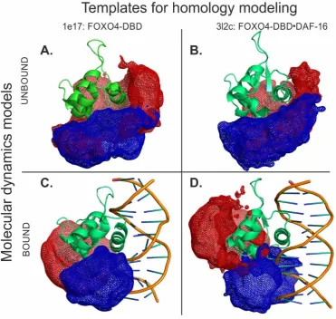
- http://peterslab.org/downloads/SW/DeutEx.mp4
269
Mass spectrometry data available at https://www.ebi.ac.uk/pride/archive/ (Project accession:
270
PXD013969)
271
3. Results and Discussion
272
The crystal structure available for the FOXO4-DBD•DBE complex does not cover the entire
273
sequence of the DNA binding domain [62], which spans the 82 - 207 section of FOXO4, but omits
274
instead flanking regions hypothesized to promote the recruiting of additional components of the
275
transcription machinery. At the same time, the NMR structure of full-length FOXO4-DBD provides
276
limited information on the G138 - A144, E166 - K170, and the N- and C-terminal regions, which were
277
described as rather flexible and disordered in solution [60]. Although DNA binding has been
278
credited for stabilizing at least some of these regions, the structure of the bound form still displayed
279
significant discrepancies with those of homologous members of the FOXO family [44], which were
possibly caused by crystal packing [62]. For this reason, we investigated such discrepancies by
281
implementing biochemical approaches to probe the effects of ligand binding directly in solution. The
282
study employed recombinant full-length DBD (i.e., residues G82 - A207) and a duplex DNA construct
283
containing the 5’-TAC CCA A-3’ consensus sequence, which was obtained by annealing commercial
284
oligo-deoxyribonucleotides. As shown in Figure S-1A and Figure S-2 of Supporting Information,
285
mixing equimolar amounts of protein and duplex DNA provided the expected 1:1 species
286
corresponding to the desired FOXO4-DBD•DBE complex. These samples were submitted to
287
hydrogen-deuterium exchange, quantitative protein-protein cross-linking, protein-DNA
288
cross-linking, and docking experiments to obtain complementary information on their mutual
289
interactions and spatial arrangement. The results were compared to those obtained from the
290
individual FOXO4-DBD protein to investigate the effects induced by specific DNA binding.
291
3.1. Combined online digestion by pepsin and nepenthesin I improves the HDX resolution
292
We initially pursued the identification of the regions of FOXO4-DBD, which were making direct
293
contact with the DBE duplex, or were subjected to detectable microenvironment variations upon
294
binding. Hydrogen-deuterium exchange (HDX) was performed on both free and bound forms of
295
full-length FOXO4-DBD. The determinations followed a well-established protocol in which the
296
exchange process was stopped at predetermined intervals to monitor the rate of exchange. Since,
297
hydrogen-deuterium exchange has not yet been used to study the complex of protein and duplex
298
DNA, we first tuned conditions for on-line digestion in order to obtain the best spatial resolution. It
299
was achieved using the combination of nepenthesin I and pepsin where rather small and
300
overlapping peptides were observed. The robustness of the setup was approved by multiple
301
injections. The final analysis were carried out at low pH and temperature to minimize
302
back-exchange and included protein digestion in consecutive on-line columns containing
303
immobilized proteases (i.e., pepsin and nepenthesin-1), followed by LC-MS and LC-MS/MS analysis
304
of digested peptides (see Materials and Methods and Appendix (Methods in detail) section) [75].
305
The ensuing peptide map demonstrated that the procedure afforded full coverage of the
306
FOXO4-DBD sequence (Figure 2).
307
308
Figure 2: Combined online digestion by pepsin and nepenthesin I improve the HDX resolution.
309
Comparison of hydrolytic products obtained by on-line digestion in columns derivatized with
310
nepentesin-1, pepsin, or pepsin/nepenthesin-1 combination. The peptides are mapped along
311
sequence and topological information. The peptides afforded 100% coverage of the protein sequence.
312
Note that the G74 - P87 region of the construct was contributed by the recombinant-production vector
313
and, thus, was not part of the wild-type FOXO4 sequence (see also Figure S-33 of Supporting
314
Information).
316
3.2. HDX identified the interaction interface and long-distance stabilization of protein structure
317
For each digestion product, a relative deuteration rate was calculated by considering the
318
number of hydrogens exchanged with deuterium atoms against the total number of exchangeable
319
amide hydrogens in the peptide. The relative deuteration rates versus exchange time were
320
calculated at both the peptide and amino acid levels to recognize possible variations between the
321
free and bound FOXO4-DBD (see Figure S-3 to S-6 of Supporting Information, respectively). This
322
task was facilitated by calculating actual differences for each amino acid in the sequence, which were
323
visualized onto a 3D model of FOXO4-DBD•DBE by using an appropriate color palette (Figure S-7
324
of Supporting Information).
325
The results were summarized in HDX difference plot that provided a comprehensive view of
326
the variations of solvent accessibility induced by the specific interactions between FOXO4-DBD and
327
its cognate DBE duplex (Figure 3). Starting from the N-terminus, the G74 - Y102 region displayed
328
relatively high levels of deuteration regardless of reaction time, with no significant differences
329
between free and bound form. These observations indicated that this region was rather exposed and
330
capable of exchanging freely with the solvent in both forms. In contrast, the next section spanning
331
the A103 - T130 residues displayed the most extensive differences in deuteration rates, which increased
332
significantly as a function of time. This sequence folds helix H1 and H2, strand S1, and intervening
333
loops (see topology annotation in Figure 2). According to the crystal structure, none of these
334
distinctive features is supposed to make direct contact with the duplex DNA [62], which would help
335
explain the drop of deuteration by invoking a simple protection effect. In the absence of direct
336
contact, the observed loss of solvent accessibility must be attributed to indirect conformational
337
effects induced by binding. The fact that the difference in deuteration levels increased gradually
338
with time and stabilized after 60 min suggests that, in the free form, this set of secondary structures
339
may undergo slow mutual dynamics that delay the exchange of susceptible hydrogens. In the bound
340
form, such dynamics may be stabilized by interactions with contiguous structures that, in turn, make
341
direct contact with the DNA ligand. Like falling dominoes, a series of relatively minor
342
conformational variations linked together may ultimately induce observable inhibition of the
343
exchange reaction. This long-distance effect is clearly evident, for example, in the relative
344
deuteration plot of peptide A103 - L118 (2-3), which shows increasing uptake in the free FOXO4-DBD
345
as a function of time, but constant low-level deuteration in the bound form (Figure S-3 of Supporting
346
Information).
348
Figure 3: HDX identified the interaction interface and long-distance stabilization of protein structure.
349
A. Relative deuteration differences [DR(FOXO4-DBD) – DR(FOXO4-DBD•DBE)] plotted along the
350
FOXO4-DBD sequence and their evolution in time. B. FOXO4-DBD•DBE structure with highlighted
351
regions showing significant differences in deuteration levels. Note that the G74 - P87 region of the
352
construct was contributed by the recombinant-production vector and, thus, was not part of the
353
wild-type FOXO4 sequence (see also Figure S-3 to Figure S-7 of Supporting Information).
354
The next region spanning the V131 - K159 residues also manifested significant differences
355
between free and bound forms, but their time-dependence displayed a rapid increase at shorter
356
intervals, followed by a decline near initial levels at longer reaction times (Figure 3A). In particular,
357
the residues forming helix H3, which the crystal structure places directly in the major groove of the
358
DBE duplex [62], experienced the largest differences. For this reason, direct steric protection induced
359
by bound DNA could explain the uptake inhibition observed for such residues. In contrast, the
360
outcome observed for the contiguous helix H4 and intervening loop could indirectly result from the
361
stabilization of the H3 conformation, which could constrain the placement of such residues and
362
restrict their solvent accessibility. The crystal structure identified also a handful of contacts that were
363
mediated by water molecules trapped in the binding interface. While these interactions have been
suggested to further stabilize the dynamics of helix H3 and flanking regions, it is not clear how
365
trapping D2O versus H2O present in the solvent may affect the observed deuteration rates. It should
366
be also pointed out that FOXO4 differs from other FOXO homologues by the insertion of five amino
367
acids (K137 - N141) in the H4-H3 loop (Figure 2). It is not clear whether this insertion may be
368
responsible for the unusual conformation assumed by helix H3 in the bound form, which differs
369
from those assumed in other members of the family [62]. However, the observed inhibition pattern
370
supports a role in stabilizing the fold of FOXO4-DBD and reducing its overall flexibility upon
371
complex formation.
372
The F160 - D195 section corresponds to sequences located on the C-terminal side of helix H3,
373
which manifested smaller but still perceptible differences of deuteration rates. This region contains
374
strands S2 and S3, as well as the W1 and W2 wings, which the crystal structure placed far removed
375
from the DNA binding interface. Also, in this case, an overall decrease of structural flexibility upon
376
binding could explain the reduced deuterium uptake. It should be noted that, in addition to
377
conferring FOXO4-DBD its winged look and fine tuning of the interaction with DNA [61], W1 and
378
W2 could contribute to constitute possible regions of contact for auxiliary components of the
379
transcription complex. In this context, the DNA binding could represent a possible mechanism for
380
modulating the recruitment of such components. The final section covered by HDX determinations
381
consisted of the S196 - A207 sequence and displayed marginal deuteration differences. This observation
382
indicated the absence of any protection or conformational effects induced by DNA binding.
383
3.3. Protein-DNA cross-linking revealed the mutual placement of protein and DNA components
384
The HDX experiments identified the regions affected directly and indirectly by DNA binding,
385
which experienced clearly detectable variations of solvent accessibility. However, these types of
386
determinations could not identify the structures responsible for limiting the access of solvent to a
387
specific region. In other words, these experiments could not reveal the mutual spatial relationships
388
between such structures, nor assess the effects of binding on such relationships. For this reason, we
389
employed different types of cross-linking strategies to probe the organization of the various
390
structures and recognize their mutual placement in the overall fold. The first approach employed
391
transplatin to generate putative protein-DNA conjugates that may be capable of constraining the
392
position of the DBE ligand onto the FOXO4-DBD substrate. The reactivity of platinum compounds
393
towards specific functional groups of nucleic acids is well documented [76] and involves the
394
preferential attack of the N7 position of guanine base [77]. Although the characteristics of their
395
reactivity towards protein residues are still unclear [63], amino acids with electron-rich S, N and O
396
atoms, such as Cys, Met, His, and Thr, have been described as preferred targets [78]. We treated
397
samples of FOXO4-DBD•DBE complex, as well as free FOXO4-DBD and DBD, with a 10:1
398
transplatin to substrate molar ratio in 150 mM ammonium acetate (pH 6.85) and incubated at 18 °C
399
for 14 hr (see Materials and Methods and Appendix (Methods in detail) section). The mixtures were
400
analyzed by both ESI-MS and SDS-PAGE to assess the distribution of transplatin adducts and
401
estimate the proportion of sought-after intermolecular cross-links. The representative data in Figure
402
4A provides a view of the typical product distributions obtained from these probing reactions,
403
which included monofunctional “dangling” adducts displaying a still unreacted chloride function
404
(i.e., marked as tPt-Cl adducts), as well as bifunctional conjugates in which both functions had
405
effectively reacted (i.e., marked as tPt adducts).
407
408
Figure 4: Transplatination generates protein-DNA cross-links. A. Mass spectrum of
409
FOXO4-DBD•DBE treated with transplatin and sprayed in 1% acetic acid in 7.5 mM ammonium
410
acetate solution. B. Mass spectrum of FOXO4-DBD•DBE treated with transplatin and sprayed in 1%
411
acetic acid and 50% methanol in 7.5 mM ammonium acetate solution. Abbreviations: tPt –
412
bifunctionally bound transplatin cross-linker (Pt(NH2)2); tPt-Cl – monofunctionally bound
413
transplatin cross-linker (Pt(NH2)2Cl); dsDBD – duplex DNA; ssDBD-F – forward oligonucleotide
414
strand; ssDBD-R – reverse oligonucleotide strand. Species denoted here as FOXO4 correspond to the
415
form of FOXO4-DBD shortened by three C-terminal amino acids (see also Figure S-1, Figure S-2,
416
Figure S-8, Figure S-9 of Supporting Information.
417
Based on mass alone, it is not typically possible to distinguish desired intermolecular
418
conjugates from intramolecular crosslinks, which share the same elemental composition. For this
419
reason, ESI-MS analysis was repeated in the presence of 50% methanol to achieve mild denaturing
420
conditions. In this way, products stabilized by bridging bifunctional cross-links were still detected
421
intact by virtue of their covalent nature, such as the conjugates containing FOXO4-DBD and
422
individual DBE-F or DBE-R strands produced by dissociation of the DBE duplex. In contrast, no
423
signal was observed for complexes devoid of any intermolecular conjugation, whereas adducts of
424
their free unbound components were individually detected, such as those of FOXO4-DBD protein,
425
DBE-F, and DBE-R strand (Figure 4B). In analogous fashion, the reaction mixtures were also
426
analyzed by PAGE under both native and denaturing conditions (Figure S-1 of Supporting
427
Information). Direct data comparison enabled the identification of bands that eluded dissociation by
428
elevated concentrations of SDS or urea and, thus, could be attributed to the presence of bridging
429
bifunctional crosslinks. These data enabled us to estimate that the desired intermolecular cross-links
430
amounted to less than 5% of typical transplatin reactions mixtures.
431
A classic bottom-up strategy was carried out to complete the characterization of cross-linked
432
products, which included digesting the material with protein- and nucleic acid-specific enzymes to
433
obtain samples amenable to LC-MS and LC-MS/MS analysis. In particular, reaction mixtures were
434
treated with trypsin to map the position of peptides conjugated to DNA strands (see Materials and
Methods and Appendix (Methods in detail) section). In subsequent experiments, the size of the
436
oligonucleotide moieties was reduced by treatment with Bal-31 nuclease to facilitate analysis. The
437
representative data in (Figure S-8 of Supporting Information) illustrates the challenges faced by the
438
MS/MS analysis of these types of hetero-conjugates. Upon gas-phase activation, a precursor ion
439
consisting of G74 - R88 cross-linked to the DBE_R strand underwent dissociation around the bridging
440
Pt atom, rather than along the backbones of the bridged moieties. The absence of sequence
441
information afforded by this type of fragmentation prevented the identification of the actual
442
residues involved in the cross-linking reaction. Nevertheless, the identity of the conjugated
443
components still represented valuable information on the mutual relationships between contiguous
444
regions (summarized in Figure S-9 of Supporting Information).
445
The detected peptide-oligonucleotide conjugates were examined in the context of the results
446
afforded by the HDX determinations and other structural information available for the system. For
447
instance, peptide N148 - K159 spanning helix H3 was found conjugated to the forward strand of DBE,
448
consistent with the placement of H3 directly into the major groove of the DNA duplex in the crystal
449
structure [62]. This finding agreed also with the prominent protection effects observed in this region
450
during HDX experiments (Figure 3A). Residues S149 and H152, in particular, should represent
451
excellent conjugation sites by virtue of their susceptibility to transplatin reaction and favorable
452
orientation facing the duplex’s major groove. Peptide F160 - K170 covering the end of S2 and beginning
453
of W1 was also cross-linked to the forward strand of DBE, in spite of the absence of any direct
454
contact in the crystal structure. In this peptide, reactivity and orientation considerations would point
455
towards H164 and T168 as possible conjugation sites, if their distances from susceptible DNA
456
structures were sufficiently favorable. In this direction, the HDX data indicated that this region
457
experienced a detectable decline in deuterium uptake consistent with the adoption of a rather
458
constrained conformation upon DNA binding (Figure 3A). The new conformation could place
459
susceptible groups within mutual striking distance, thus promoting the formation of the observed
460
cross-linked product. A similar explanation is applicable also to the S171 - K182 peptide spanning the
461
end of W1, beginning of W2, and intervening S3 strand, which formed cross-links with both forward
462
and reverse strands of DBE. Also, this region experienced a significant decrease of deuterium
463
exchange upon binding, which was not explainable by direct steric protection, but rather by indirect
464
conformational effects transmitted through contiguous structures. The crystal structure orients S171
465
and S172 to face the minor groove of the duplex construct, which would represent prime positions for
466
promoting conjugation with either strand. Also, in this case, the respective functional groups could
467
be placed within striking distance by the more constrained conformation revealed by HDX
468
experiments. The remaining products consisted of the G74 - R88 peptide cross-linked to either the
469
forward or reverse strand. These products are a testament to the flexibility of the N-terminal loop,
470
which was supported by the lack of any significant variation of deuteration patterns reported by the
471
HDX experiments.
472
3.4. DNA binding induced significant effects on protein conformation
473
The observed protein-DNA cross-links provided valuable information not only on the
474
reciprocal positions of protein and DNA components, but also on the significant changes induced by
475
binding on the initial protein conformation. We employed protein-specific reagents to evaluate the
476
extent of such variations and enable a better appreciation of indirect conformational effects. Our
477
quantitative crosslinking approach involved the concerted application of the homobifunctional
478
reagents DSGd0/d4 and DSSd0/d4 [di(N-succinimidyl) suberate and di(N-succinimidyl) glutarate]
479
to bridge susceptible amino or hydroxy groups that may be placed within 20.5 ± 3.0 or 24.2 ± 3.0 Å ,
480
respectively, from one another (see Appendix (Methods in detail) section). The utilization of
481
reagents with different bridging spans offered the ability to determine an average distance between
482
residues. At the same time, the isotopic labels facilitated the identification of cross-linked products
483
in complex digestion mixtures from their characteristic 4-Da spacing and enabled the acquisition of
484
unbiased quantitative data on the incidence of cross-linking in the free or bound FOXO4-DBD.
485
Initially, separate samples were treated with 1:1 mixtures of matching unlabelled/labelled reagents
of same length to complete a survey of the regions susceptible to cross-linking (see Materials and
487
Methods and Supporting Information). A total of 39 conjugates were identified, which bridged
488
lysine and serine residues, as well as the N-terminal amino group (Table S-1 and Figure 10 to Figure
489
26 of Supporting Information). The majority of them were detected in matching pairs generated by
490
reagents of either length, and were observed in both free and bound samples. However, a small
491
portion was unique for just one form and/or cross-linker length. Next, individual aliquots of free
492
FOXO4-DBD were treated with either DSGd0 or DSSd0, whereas those of FOXO4-DBD•DBE
493
complex were separately treated with either DSGd4 or DSSd4 (see Materials and Methods and
494
Appendix (Methods in detail) section). Corresponding samples treated with the same
495
unlabelled/labelled reagent were mixed in a 1:1 molar ratio prior to protease digestion and analysis
496
to compare the incidence of each conjugate in either free or bound samples. The proportion of each
497
of the 39 conjugates identified earlier was determined for at least one of the cross-linker lengths, as
498
summarized in (Table S-2 of Supporting Information).
499
A close examination of the results revealed distinctive cross-linking patterns associated with the
500
presence of bound DNA. In particular, the incidence of some conjugates decreased significantly
501
upon binding, while others increased. Among the former, the DGS conjugates bridging K147 to
502
either K170 or N-term dropped from 95.9% and 92.8% to 4.1% and 7.2%, respectively (Table S-2 of
503
Supporting Information). The cross-linking inhibition manifested by K147 cannot be merely ascribed
504
to its location on helix H3, in direct contact with the duplex construct, because this residue was still
505
capable of supporting conjugation with both K135 and K137. A more plausible explanation could be
506
that DNA binding forced K147 out of the reach of either K170 on helix H2, or K162 on the adjacent S2
507
region. The limited nature of such conformational changes was revealed by the fact that K147 was
508
pushed out of N-term’s reach for the shorter DSG reagent, but was still sufficiently close for the
509
longer DSS, with incidence of cross-linking dropping from 92.8 to 7.2% and 72.7 to 27.3%,
510
respectively (see Table S-2 of Supporting Information). The limited extent of these changes was also
511
evident in the subtler cross-linking variations between K147 and either K135 or K137 located on the
512
H3-H4 intervening loop.
513
In other cases, DNA binding increased the incidence of specific conjugates by placing residues
514
within mutual striking distance in the complex, which were marginally susceptible or inert in the
515
free protein. For example, the conjugates bridging residue K182 with N-term, K89, K116, K159, K162, or
516
K182 were greatly enhanced by the presence of DNA duplex (Table S-2 of Supporting Information).
517
For the majority of these positions, the levels of cross-linking observed with the longer DSS reagent
518
displayed more significant variations than those with the shorter DSG. Considering that K182 is
519
located on the W2 wing region, these observations offered further evidence of the long-range
520
conformational effects of DNA binding suggested by the results of HDX and protein-DNA
521
cross-linking experiments. Another example was provided by the numerous conjugates involving
522
the N-term, which suggested that DNA binding had prominent stabilizing effects on a region that
523
was rather flexible in free FOXO4-DBD. Consistent with the HDX data, the variations of
524
cross-linking patterns confirmed that DNA binding induced significant effects on protein
525
conformation not only within the contact interface, but also in rather distal positions.
526
3.5. Structural proteomics could effectively guide model-building operations to produce very high-quality 3D
527
models
528
The HDX and XL experiments provided a wealth of information that was used to guide the
529
molecular modelling of full-fledged FOXO4-DBD and FOXO4-DBD•DBE complex. Our approach
530
took advantage of available high-resolution structures that, although incomplete in their coverage of
531
the protein sequence, still represented excellent templates for homology modelling operations (see
532
Materials and Methods and Appendix (Methods in detail) section). In particular, the templates were
533
used to obtain the coordinates of what could be defined as the structured core of the complex, a
534
region spanning approximately from R93 to N177, which displayed limited discrepancies across the
535
available structures. In contrast, the regions, which were either absent from the templates, or
536
displayed significant variations, or had been predicted to possess a high degree of flexibility by
PSIPRED [79], were modeled according to the HDX and cross-linking information (see Materials
538
and Methods and Appendix (Methods in detail) section). These regions corresponded to the G74 - Q100
539
and N177 - A207 sections located respectively at the N- and C-terminal ends of the DBD sequence.
540
These operations were performed in the Modeler suite [67], which was also used to eliminate
541
possible strains and steric clashes introduced during model building. The program applied the
542
DOPE scoring algorithm to identify the best possible structures that were subsequently employed in
543
docking and simulated annealing procedures. The former was carried out to place the DBE
544
structure, which was created separately by using the make-na server
545
(http://structure.usc.edu/make-na/), onto the putative binding site of the protein. This operation was
546
accomplished in HADDOCK [73] by designating as active those residues that had experienced
547
reduced rates of exchange (Figure 3A) upon DNA binding (see Figure S-27 of Supporting
548
Information). The mutual positioning between the DBE and FOXO4-DBD components was further
549
refined according to the results of the protein-DNA cross-linking experiments, which were
550
introduced by using Modeller software. Finally, the structures of both FOXO4-DBD and
551
FOXO4-DBD•DBE complex were submitted to simulated annealing and energy minimization in
552
CNS [71] to generate the sought-after model ensembles (Figure 5).
553
554
Figure 3: Structural proteomics could effectively guide model-building operations to produce very
555
high-quality 3D models. Models of FOXO4-DBD and FOXO4-DBD•DBE were obtained by
556
combining homology modelling with experimental constraints and molecular dynamics simulations.
557
These models incorporated extensive information from protein-DNA cross-links, quantitative
558
protein-protein cross-links, and hydrogen-deuterium exchange. The green structures show
559
representative models for unbound (A. and B.) and bound (C. and D.) forms based on corresponding
560
1E17 (A. and C.) or 3L2C (B. and D.) high-resolution templates. The mesh areas in blue and red
561
colors represent the spaces occupied by all the models in the ensembles, which provided a measure
562
of the flexibility of the N- and C- terminal regions (see also Figure S-10 to Fig S-32 and Table S-1 and
563
S-2 of Supporting Information).
The structures obtained for FOXO4-DBD and FOXO4-DBD•DBE complex were compared to
565
the corresponding high-resolution structures to assess the robustness of our structural proteomics
566
approach. In the case of individual FOXO4-DBD, the overall topology of the ensemble reflected the
567
typical forkhead structure of the FOXO family and matched very closely that of the NMR structure
568
used as homology template (see Figure 28A and B of Supporting information), thus supporting the
569
validity of the HDX and cross-linking constraints. A more detailed comparison was obtained by
570
calculating the root mean square deviation (RMSD) between the coordinates of corresponding heavy
571
atoms located in the backbone of each ensemble model and the various templates. The
572
representative plot in Figure S-29 of Supporting Information, for example, shows that the model
573
obtained from the 1e17 structure deviated very little from the initial template. The fact that the
574
experimental constraints introduced during modelling did not force any significant variation of the
575
initial coordinates indicates that the probing operations did not cause any perturbation of the
576
substrate’s 3D structure and corroborated the excellent stability of the structured core of
577
FOXO4-DBD. In contrast, larger RMSD values were obtained when models based on other templates
578
were compared with the initial FOXO4 models used in the study (3L2C, 1E17), as expected from the
579
discrepancies between the various NMR and crystal structures available (see Figure S-30 in
580
Supporting Information). Although this type of analysis was not possible for the regions that were
581
absent from the templates, the excellent match manifested by the regions present in both model and
582
template warranted a high level of confidence in the entire structures produced by our approach.
583
The ensemble of the FOXO4-DBD•DBE complex was examined in similar fashion. Also, in this
584
case, the overall topology matched very closely that of the corresponding high-resolution template
585
(i.e., 3l2c), with the DBE component oriented in the proper direction and placed in the correct
586
position onto the FOXO4-DBD’s binding site (see Figure 28C and D of Supporting information).
587
Also, in this case, RMSD comparisons between the model and crystal structure revealed excellent
588
match for the regions present in both, thus ruling out the possibility of inadvertent perturbations
589
introduced by the probing procedures. Additionally, we determined the distances between residues
590
that had been conjugated by the cross-linking reagents, and then compared them with the
591
corresponding distances measured on the crystal structure. The resulting RMSD values revealed
592
excellent agreement across the board, with the sole exception of the distances between the DBE
593
molecule (DBE_F) and specific residues of the H164 - M175 loop (H164, T168, S171, S172 or M175), which were
594
somewhat longer in our model (see Table S-1 in Supporting Information). These discrepancies,
595
however, were consistent with the high degree of flexibility possessed by the loop, which were
596
manifest also in the higher B-factors displayed by this region in the crystal structure. In agreement
597
with the crystal structure, the models showed that helix H3 represents the main interaction interface,
598
as indicated by both HDX and cross-linking data (see Table S-1 in Supporting Information). The
599
DBE structure employed here replicated the consensus binding sequences for all related FOX factors,
600
which contain a general TGTTT motif surrounded by more variable sequences (see Figure S-31 in
601
Supporting Information). Whereas FOXO3 and FOXO6 recognize two nucleotides located after this
602
consensus sequence, FOXP1 only recognize the second nucleotide but not the first one. In contrast,
603
our model indicates that FOXO4 may recognize one nucleotide before and one after the consensus
604
motif, thus affording additional evidence of the uniqueness of the interactions established by this
605
member of the FOXO family. Our results are supported with binding models estimated probability
606
of individual bases at each position in sequence according Position Count Matrix (PCM) values (see
607
Figure S-31 in Supporting Information).
608
A close comparison of the structures of FOXO4-DBD and FOXO4-DBD•DBE complex obtained
609
by our approach allowed us to further explore the effects of binding on protein conformation. The
610
examination confirmed that the N- and C-terminal sequences remained largely unstructured even
611
after DBE binding, as shown by the mesh representation obtained from our models (Figure 5) The
612
main interface region represented by H3 showed limited variations between unbound and bound
613
forms. Similar outcomes were also observed for the contiguous H1 - H2 loop and H2 helix. In
614
contrast, loop H2 – H4 – H3and the S2 and S3 strands of wing W1 showed rather large variations
615
upon binding. Additionally, also the N- and C- terminal regions manifested extensive variations.