Dynamic expression of Endoglin, a TGF-
βββββ
co-receptor,
during pre-circulation vascular development in chick
CANTAS ALEV
1,2,a, BRENDAN A.S. MCINTYRE
1,a,b, KANAKO OTA
1and GUOJUN SHENG*
,11Laboratory for Early Embryogenesis, RIKEN Center for Developmental Biology and
2Vascular Regeneration Research Group, Institute of Biomedical Research and Innovation, Kobe, Japan
ABSTRACT Mutations in the human Endoglin gene, encoding a dimeric TGF-βββββ co-receptor, lead to type 1 hereditary hemorrhagic telangiectasia. Studies in mice have revealed important roles of Endoglin in endothelial cell proliferation, differentiation and integrity. Endoglin-/- mouse embryos die at mid-gestation due to cardiac defects and vessel rupture. Its role during early vasculogenesis is unclear, as the initial phase of vascular endothelial cell formation appears unaffected in Endoglin-/- embryos. In order to understand possible roles of Endoglin in early vascular develop-ment, we used the chick model and analyzed the temporal and spatial expression pattern of Endoglin during vasculogenesis in pre-circulation stage chick embryos. Weak Endoglin expres-sion was detected at HH4 in the node and in the extraembryonic mesoderm. The node-specific expression is transitory and disappears after HH5. Strong up-regulation of Endoglin expression is seen at HH8 in all endothelial progenitors undergoing morphological changes to become endo-thelial cells. Most extraembryonic splanchnopleural vascular endoendo-thelial cells down-regulate Endoglin after their morphological differentiation, whereas lateral plate and cardiac endothelial cells remain positive until HH12, followed by a clear drop after circulation starts at HH13. Progenitors for the pronephric duct are positive from HH10 to HH12, but down-regulate Endoglin after epithelialization of duct cells. Overall, these data reveal a dynamic expression pattern of Endoglin in pre-circulation chick development and indicate that Endoglin may play an important role in the transition from endothelial progenitors to functional endothelial cells during early vascular development.
KEY WORDS: CD105, vasculogenesis, vasculature, endothelial cell, hematopoiesis, progenitor, chicken
Introduction
Endoglin (also known as CD105) is a co-receptor for the transforming growth factor-beta (TGF-β) superfamily (Goumans and Mummery, 2000; Lebrin and Mummery, 2008; ten Dijke et al., 2008). In mammals, it has been reported to be expressed in active endothelial cells and during early extraembryonic mesoderm differentiation (Ema et al., 2006; Jonker and Arthur, 2002). In chick, embryonic circulation starts at stage Hamburger and Hamilton (HH) 13 (Eichmann et al., 2005; Hamburger and Hamilton, 1992). The expression pattern of Endoglin had been studied in chick with respect to cardiac differentiation (Vincent et al., 1998) and in adult lung and post-circulation yolk sac vasculature (Raab
BIOLOGY
www.intjdevbiol.comISSN: Online 1696-3547, Print 0214-6282
© 2009 UBC Press Printed in Spain
Abbreviations used in this paper: EMT, epithelial-mesenchymal transformation; TGF-β, transforming growth factor beta.
et al., 1999). During post-circulation chick development, Endoglin was also found to be expressed intraembryonically in cardiac endothelium and in developing vessels, and was shown to be involved in the process of epithelial-to-mesenchymal transforma-tion (EMT) during cardiac valve formatransforma-tion (Mercado-Pimentel et al., 2007). The Endoglin-/- mouse is embryonic lethal with angio-genic defects in addition to vessel rupturing and lack of cardiac EMT by mid-gestation, but with the vasculature appearing normal before E8.5 (Arthur et al., 2000; Bourdeau et al., 1999; Li et al., 1999). Endoglin+/- mice are viable and have been used to model
*Address correspondence to: Guojun Sheng. Laboratory for Early Embryogenesis, RIKEN Center for Developmental Biology, Kobe, Hyogo 650-0047, Japan. Fax: +81-78-306-3146. e-mail: [email protected]
a Note: These authors contributed equally to this work.
b Current Address: Physiology and Experimental Medicine Program, The Hospital for Sick Children, Toronto, Ontario, Canada
hereditary hemorrhagic telangiectasia type 1 (HHT1; MIM#187300) (ten Dijke et al., 2008), a condition observed in humans with mutations in the Endoglin gene resulting in haploinsufficiency (Abdalla and Letarte, 2006). Although not normally lethal, patients with HHT1 suffer from chronic nosebleeds (epistaxis), telang-iectases, have high incidence of pulmonary and cerebral arterio-venous malformations and appear to be predisposed to develop-ing pulmonary arterial hypertension (Lenato and Guanti, 2006), all of which underscore the important underlying function of Endoglin in the establishment and maintenance of the circulatory system.
Furthermore, it has been suggested that Endoglin is involved in early hematopoietic development, with Endoglin being ex-pressed in mouse embryonic stem cell (mESC)-derived mesoder-mal FLK1+ precursors (Cho et al., 2001) and blast colony-forming
cells (BL-CFCs) (Perlingeiro, 2007). In vitro differentiation assays showed that Endoglin-/- mESCs have reduced myelopoiesis and
erythropoiesis, whereas the formation of lymphoid and vascular precursors appeared unaffected (Cho et al., 2001). Anemia observed in patients with HHT1 and in Endoglin-/- mice is another
indicator for a possible role of Endoglin in erythropoiesis. In the adult setting, in addition to its involvement in angiogenesis and vascular repair (Hayrabedyan et al., 2005; van Laake et al., 2006), Endoglin is expressed in long-term repopulating hematopoietic stem cells from bone marrow (Chen et al., 2002) and was reported to play a role in erythroid lineage differentiation (Moody et al., 2007). Increased levels of Endoglin expression were also found in tumors (Fonsatti et al., 2001), atherosclerotic plaques (Conley et al., 2000) and during inflammation and wound healing (Torsney et al., 2002). Endoglin expression is thus closely correlated with neo-vascular formation and may play a role in the pathogenesis
of vascular diseases. However, in vitro angioblast differentiation from Endoglin-/- mESCs was reported to be unaffected (Perlingeiro,
2007) and the primary endothelial network in Endoglin-/- mouse
embryos appears normal (Li et al., 1999), indicating that Endoglin may be dispensable for early vascular formation.
Results and Discussion
The chick Endoglin gene (AY702002) encodes a 644 amino acid polypeptide, with an overall 34% identity to the human L-Endoglin protein. Searches of EST databases did not indicate the existence of the alternatively spliced S-Endoglin in chick, an isoform up-regulated in senescent endothelial cells in mammals (Bellon et al., 1993; Blanco et al., 2008; Perez-Gomez et al., 2005; Velasco et al., 2008). A high degree of identity between chick and mammalian Endoglins was found in the transmembrane and short cytoplasmic tail domains, with 100% conservation in the last 24 amino acid residues containing the sites that are known to be phosphorylated by activated TβRII, Alk1 and Alk5 and a PDZ motif (Koleva et al., 2006). Its expression pattern in the extraembryonic regions and in pre-circulation development has not been exam-ined. We generated an anti-sense probe corresponding to amino acid residues 111-304 of the full length chick Endoglin (see Materials and Methods), and performed whole-mount in situ hybridization on embryos from stage HH4, when blood/endothe-lial progenitors start to be generated from the posterior primitive streak (Jaffredo et al., 2005; Nakazawa et al., 2006; Shin et al., 2009), to HH13, when circulation is initiated.
At HH4, the earliest time point examined, weak Endoglin expression was observed in the Hensen’s node and the head process (Fig.1A; arrowhead). This is transitory, as no Endoglin
G
B
C
D
E
F
H
A
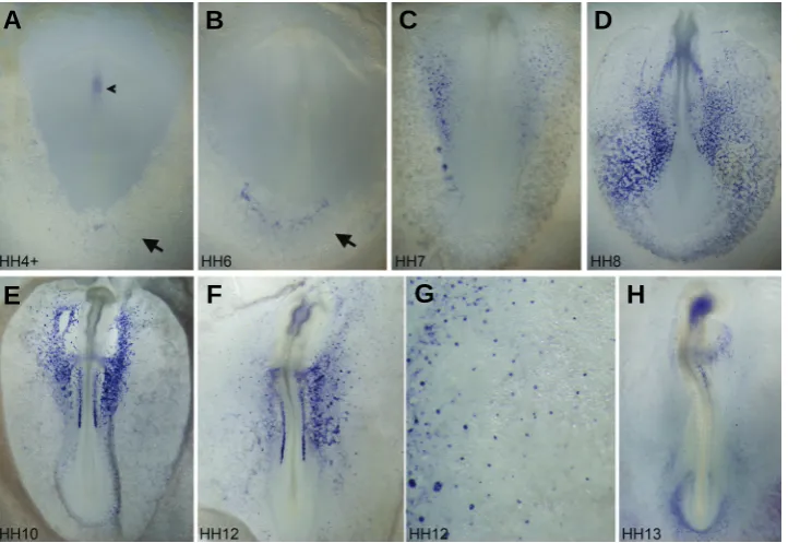
Fig. 1.Whole-mount in situ hybridization analysis of Endoglin from stage HH4 to stage HH13.(A) At HH4+, Endoglin is ex-pressed very weakly in the Hensen’s node and head process (arrowhead) and in the newly formed extraembryonic mesoderm (arrow). (B) At HH6, expression in the node and node-derived tissues is not detected. Broader, yet still very weak, Endoglin ex-pression is observed in the extraembryonic mesoderm (arrow). (C) By HH7, expression in the area vasculosa becomes stronger, especially in the future medial extraembry-onic and lateral plate regions. (D) At HH8, robust expression is observed throughout extraembryonic and intraembryonic vascu-lature. (E) By HH10, extraembryonic vascu-lar expression is reduced significantly, with intraembryonic vasculature (including car-diac and lateral plate vessels) strongly posi-tive for Endoglin. Progenitors for the pro-nephric duct are also strongly positive. (F)expression was detected in the node or node-derived tissues at later stages. Weak expression could also be observed at this stage in the extraembryonic-fated mesoderm territory (Figs. 1A, 2A; arrows). The extraembryonic mesoderm at this stage strongly expresses hemangioblast, hematopoietic and endothelial precur-sor markers such as Gata2, Scl, Lmo2, Ets1 and Vegfr2 (Bollerot et al., 2005; Minko et al., 2003; Nakazawa et al., 2006; Shin et al., 2009), indicating that Endoglin expression does correlate with the initial steps of endothelial precursor specification.
By HH5 and HH6, Endoglin expression in the extraembryonic mesoderm covers a slightly wider area, but remains weak (Fig. 1B; arrow). Stronger and wider expression was observed at HH7 (Fig. 1C). The extraembryonic mesoderm at this stage starts to be segregated morphologically into three cell lineages: the blood, endothelial and smooth muscle cells (Shin et al., 2009). The first two cell lineages come from blood island aggregates and it is at HH7 when hemoglobin gene expression, a marker for terminal differentiation of blood cells, starts to be seen in scattered cells within the blood island population (Alev et al., 2008; Nagai and Sheng, 2007; Nakazawa et al., 2009; Nakazawa et al., 2006; Weng et al., 2007). Endoglin up-regulation at HH7 thus seems to mark the initiation of the morphological differentiation of endothe-lial fated cells. In blood islands located more medially in the embryo, Endoglin expression was seen in most cells (Fig. 2B; right arrow), correlating with the fact that most of medially located blood islands contribute only to endothelial cells (Nakazawa et al.,
2006). More laterally located blood islands have less Endoglin positive cells (Fig. 2B; middle and left arrows; Fig. 2 C,D) and in most cases these positive cells are located at the periphery and on one side of a blood island cluster (Fig. 2D).
At HH8, Endoglin expression becomes very robust (Fig. 1D). Strong expression was detected in all endothelial precursors in the process of undergoing morphological changes to become endothelial cells, including endocardial progenitors (Fig. 2E), dorsal aorta progenitors (Fig. 2F) and all other splanchnopleural-associated vessel progenitors in both non-hemogenic (Fig. 2F) and hemogenic (Fig. 2 G,H) regions. Newly formed vascular endothelial cells are also positive for Endoglin (Fig. 2I). Morpho-logical differentiation of endothelial progenitor cells and formation of the vascular plexus take place actively at HH8 and HH9. Most vascular endothelial cells down-regulate Endoglin expression soon after their morphological differentiation, and by HH10 (Fig. 1E), strong Endoglin expression was only seen in more medially located fusing endocardia (Fig.3 A,B) and lateral plate vessel cells (Fig. 3C), whereas extraembryonic vessels do not express Endoglin at all by this stage (Fig. 3E). Small cell clusters located in the somatopleure express Endoglin strongly (Fig. 3D). These cells, similar to blood island aggregates in the splanchnopleure (our unpublished data), have a slightly delayed differentiation program and Endoglin positive cells there will differentiate into small somatopleural vessels (Fig. 3K).
Circulation in chick embryos starts at about HH12 to HH13.
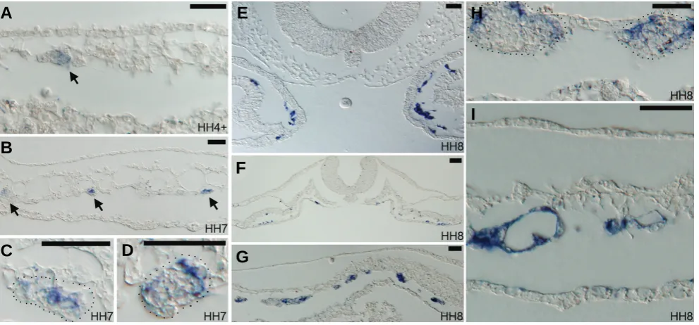
Fig. 2.Paraffin sections of Endoglin expressing embryos from HH4 to HH8.(A) Section through posterior region of the HH4+ embryo shown in Fig.1A. Arrow indicates very weak expression of Endoglin in a small number of extraembryonic mesoderm cells. (B-D) Sections through the posterior part of the HH7 embryo in Fig.1C. Arrows in (B) indicate that some cells within forming blood island clusters are positive for Endoglin. (C,D) Magnified views of lateral blood island cluster (marked by dotted lines), with some cells in the cluster positive for Endoglin. Blood island clusters in this region normally give rise to both blood cells and endothelial cells, whereas blood islands in more medial regions give rise only to endothelial cells. In these lateral blood island clusters, most Endoglin positive cells are located in the periphery of the cluster as shown in (D). Some are located within the cluster.
(E-F) Sections of the HH8 embryo in Fig.1D. (E) A section through the cardiogenic region, with endocardial precursors strongly positive for Endoglin.
(F) A section through mid-level of the embryo, with all embryonic endothelial progenitors positive for Endoglin. (G) A section through the extraembryonic region, with medially located blood islands having most of their constituent cells positive for Endoglin and laterally located blood islands having a few positive cells. (H) A magnified view of lateral extraembryonic blood islands, showing peripheral locations of Endoglin positive cells within blood islands (dotted lines). (I) In many lateral plate vessels, strong Endoglin positive signals remain in morphologically differentiated endothelial cells. Scale bar in all panels: 50 μm.
G
B
C
D
E
F
H
Endoglin expression levels drop in most vessels at peri-circula-tion stages (Fig.1 F,H). In the endocardium, expression is promi-nent at HH12 (Fig.3 F,G), but decreases significantly, although still detectable, at HH13 (Fig. 3L). A similar drop was seen in lateral plate vessels (Fig.3 H,I,M). Endoglin expression in small cell clusters in the somatopleure is still strong at HH12 (Fig.1G; arrows in Fig.3 H,K), but disappears at HH13 (Fig.1H). At HH13, weak Endoglin signals were detected in the contact region be-tween the dorsal aorta and the developing pronephric duct and tubule (Fig. 3N), possibly representing the aortic branch of the developing external glomerulus (Hiruma and Nakamura, 2003). From HH10 to HH12, the precursors for the pronephric duct are strongly positive for Endoglin (Figs.1E,F, 3J). This and the node cells at HH4 are the only two non-endothelial cell types found to express Endoglin in our study.
In summary, we report here that the expression of Endoglin during embryonic pre-circulation stages in the chick is mainly confined to areas of vasculogenesis, being strongest in endothe-lial cells undergoing active vascularization and lower or undetect-able in differentiated vessels. At all stages examined here, vascu-lar smooth muscle cells and their progenitors do not express Endoglin. Our observations support the idea, as proposed in several mouse studies (Carvalho et al., 2004; Ema et al., 2006; Jonker and Arthur, 2002), that Endoglin plays an important role during vasculogenesis. In chick, however, Endoglin does not appear to be involved in early hemangioblast (common blood and endothelial progenitor) specification or hematopoietic develop-ment. During the differentiation of endothelial progenitors, Endoglin expression seems to correlate best with the phase of morphologi-cal changes required for the formation of functional vessel cells,
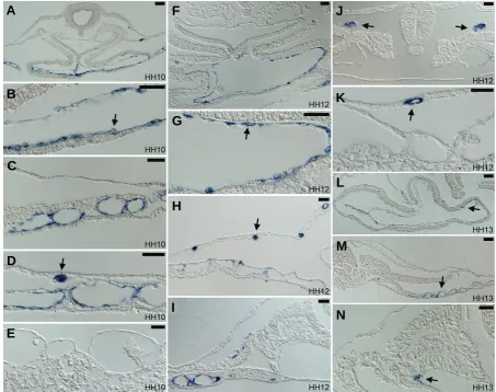
Fig. 3.Paraffin sections of Endoglin expressing embryos from HH10 to HH13.(A-E) Sections of the HH10 embryo shown in Fig.1E. (A) Fusing endocardia are strongly positive for Endoglin (magnified in (B), with arrow indicating a positive endocardial cell). (C) Lateral plate splanchnopleural vessels remain positive for Endoglin. (D) In lateral plate and extraembryonic regions, somatopleural vessel precursors are strongly positive for Endoglin
(arrow). (E) Lateral extraembryonic vessels are negative for Endoglin. Blood cells and blood precursors are always negative for Endoglin. (F-K) Sections of the HH12 embryo shown in Fig.1F. (F) Fused endocardium remains positive for Endoglin (magnified view in (G), with arrow indicating a positive endocardial cell). (H) Somatopleural vessel precursors are strongly positive for Endoglin (arrow), whereas splanchnopleural vessels have much reduced expression. (I) Only in the mid-level of the embryo Endoglin in splanchnopleural vessels persists. (J) Developing pronephric duct progenitors (arrows) are strongly positive for Endoglin. (K) Somatopleural vessels (arrow) remain positive for a short period of time after their formation. (L-N)
Sections of the HH13 embryo shown in Fig.1H. (L) The endocardium remains weakly positive for Endoglin in some cells (arrow). (M) Newly developed vessels in the posterior part of the embryo are weakly positive for Endoglin. (N) A few cells in the aortic branch in contact with the forming pronephron express Endoglin. Scale bar in all panels: 50 μm.
G
B
C
D
E
F
H
I
J
K
L
A
M
and less well with the specification of endothelial progenitor cells or the maintenance of vascular morphology or integrity after their formation. In formed vascular structures undergoing active re-modeling, such as in the endocardium and lateral plate splanchnopleural vessels, Endoglin expression is maintained for a short while after initial morphological differentiation. It is there-fore possible that the Endoglin mediated TGF-β signaling cas-cade regulates the molecular cues involved in the terminal fate choice of endothelial progenitors to adopt a differentiated endo-thelial morphology via the initiation of synthesis of intracellular and extracellular molecular components unique for functional endothelial cells (e.g., endothelial specific cell-cell junctional and extracellular matrix proteins). During chick early development, Endoglin is in our opinion the best molecular marker yet described for the initiation of terminal differentiation of endothelial progenitor cells.
Materials and Methods
Fertilized Gallus gallus domesticus eggs were purchased from
Shiroyama Farm (Kanagawa, Japan) and incubated to desired stages at
38.5°C. The DNA fragment for generating Endoglin in situ probe was
amplified by PCR from stage HH12 cDNA preparation with the following two primers: AGAACCTCCTCATCCACACT-3’ and 5’-GCGATGATGCTGTAGTTCTT-3’. The amplified fragment was confirmed by sequencing and corresponds to nucleotides 332-911 of NCBI #NM_001080887. Whole-mount in situ analysis was carried out as previously described (Nakazawa et al., 2006). All in situ hybridization experiments were performed at least in triplicate for each stage de-scribed. Stained embryos were photographed with an Olympus SZX12 microscope using a DP70 camera, followed by paraffin-embedded
sec-tioning (10 μm). Sections were photographed with an Olympus BX51
microscope. All panels in Figure 1 have stained embryos oriented with the rostral side up and viewed from the ectoderm side. All section panels in Figures 2 and 3 are oriented with the ectoderm side up.
Acknowledgements
The authors would like to thank Dr. Michelle Letarte and Dr. Mira Puri for helpful discussions.
References
ABDALLA, S.A. and LETARTE, M. (2006). Hereditary haemorrhagic telangiectasia:
current views on genetics and mechanisms of disease. J Med Genet 43: 97-110.
ALEV, C., MCINTYRE, B.A., NAGAI, H., SHIN, M., SHINMYOZU, K., JAKT, L.M. and SHENG, G. (2008). BetaA, the major beta globin in definitive red blood cells,
is present from the onset of primitive erythropoiesis in chicken. Dev Dyn 237:
1193-1197.
ARTHUR, H.M., URE, J., SMITH, A.J., RENFORTH, G., WILSON, D.I., TORSNEY,
E., CHARLTON, R., PARUMS, D.V., JOWETT, T., MARCHUK, D.A. et al.
(2000). Endoglin, an ancillary TGFbeta receptor, is required for extraembryonic
angiogenesis and plays a key role in heart development. Dev Biol 217: 42-53.
BELLON, T., CORBI, A., LASTRES, P., CALES, C., CEBRIAN, M., VERA, S., CHEIFETZ, S., MASSAGUE, J., LETARTE, M. and BERNABEU, C. (1993). Identification and expression of two forms of the human transforming growth
factor-beta-binding protein endoglin with distinct cytoplasmic regions. Eur J
Immunol 23: 2340-2345.
BLANCO, F.J., GRANDE, M.T., LANGA, C., OUJO, B., VELASCO, S., RODRIGUEZ-BARBERO, A., PEREZ-GOMEZ, E., QUINTANILLA, M., LOPEZ-NOVOA, J.M. and BERNABEU, C. (2008). S-endoglin expression is induced in senescent
endothelial cells and contributes to vascular pathology. Circ Res 103:
1383-1392.
BOLLEROT, K., ROMERO, S., DUNON, D. and JAFFREDO, T. (2005). Core
binding factor in the early avian embryo: cloning of Cbfbeta and combinatorial
expression patterns with Runx1. Gene Expr Patterns 6: 29-39.
BOURDEAU, A., DUMONT, D.J. and LETARTE, M. (1999). A murine model of
hereditary hemorrhagic telangiectasia. J Clin Invest 104: 1343-1351.
CHEN, C.Z., LI, M., DE GRAAF, D., MONTI, S., GOTTGENS, B., SANCHEZ, M.J., LANDER, E.S., GOLUB, T.R., GREEN, A.R. and LODISH, H.F. (2002). Identi-fication of endoglin as a functional marker that defines long-term repopulating
hematopoietic stem cells. Proc Natl Acad Sci USA 99: 15468-15473.
CHO, S.K., BOURDEAU, A., LETARTE, M. and ZUNIGA-PFLUCKER, J.C. (2001). Expression and function of CD105 during the onset of hematopoiesis from
Flk1(+) precursors. Blood 98: 3635-3642.
CONLEY, B.A., SMITH, J.D., GUERRERO-ESTEO, M., BERNABEU, C. and VARY, C.P. (2000). Endoglin, a TGF-beta receptor-associated protein, is
expressed by smooth muscle cells in human atherosclerotic plaques.
Athero-sclerosis 153: 323-335.
EICHMANN, A., YUAN, L., MOYON, D., LENOBLE, F., PARDANAUD, L. and BREANT, C. (2005). Vascular development: from precursor cells to branched
arterial and venous networks. Int J Dev Biol 49: 259-267.
EMA, M., YOKOMIZO, T., WAKAMATSU, A., TERUNUMA, T., YAMAMOTO, M. and TAKAHASHI, S. (2006). Primitive erythropoiesis from mesodermal precur-sors expressing VE-cadherin, PECAM-1, Tie2, endoglin, and CD34 in the
mouse embryo. Blood 108: 4018-4024.
FONSATTI, E., DEL VECCHIO, L., ALTOMONTE, M., SIGALOTTI, L., NICOTRA, M.R., CORAL, S., NATALI, P.G. and MAIO, M. (2001). Endoglin: An accessory component of the TGF-beta-binding receptor-complex with diagnostic,
prog-nostic, and bioimmunotherapeutic potential in human malignancies. J Cell
Physiol 188: 1-7.
GOUMANS, M.J. and MUMMERY, C. (2000). Functional analysis of the TGFbeta
receptor/Smad pathway through gene ablation in mice. Int J Dev Biol 44:
253-265.
HAMBURGER, V. and HAMILTON, H.L. (1992). A series of normal stages in the
development of the chick embryo. 1951. Dev Dyn 195: 231-272.
HAYRABEDYAN, S., KYURKCHIEV, S. and KEHAYOV, I. (2005). Endoglin (cd105)
and S100A13 as markers of active angiogenesis in endometriosis. Reprod Biol
5: 51-67.
JAFFREDO, T., BOLLEROT, K., SUGIYAMA, D., GAUTIER, R. and DREVON, C. (2005). Tracing the hemangioblast during embryogenesis: developmental
rela-tionships between endothelial and hematopoietic cells. Int J Dev Biol 49:
269-277.
JONKER, L. and ARTHUR, H.M. (2002). Endoglin expression in early development
is associated with vasculogenesis and angiogenesis. Mech Dev 110: 193-196.
KOLEVA, R.I., CONLEY, B.A., ROMERO, D., RILEY, K.S., MARTO, J.A., LUX, A. and VARY, C.P. (2006). Endoglin structure and function: Determinants of
endoglin phosphorylation by transforming growth factor-beta receptors. J Biol
Chem 281: 25110-25123.
LEBRIN, F. and MUMMERY, C.L. (2008). Endoglin-mediated vascular remodeling:
mechanisms underlying hereditary hemorrhagic telangiectasia. Trends
Cardiovasc Med 18: 25-32.
LENATO, G.M. and GUANTI, G. (2006). Hereditary Haemorrhagic Telangiectasia
(HHT): genetic and molecular aspects. Curr Pharm Des 12: 1173-1193.
LI, D.Y., SORENSEN, L.K., BROOKE, B.S., URNESS, L.D., DAVIS, E.C., TAY-LOR, D.G., BOAK, B.B. and WENDEL, D.P. (1999). Defective angiogenesis in
mice lacking endoglin. Science 284: 1534-1537.
MERCADO-PIMENTEL, M.E., HUBBARD, A.D. and RUNYAN, R.B. (2007). Endoglin and Alk5 regulate epithelial-mesenchymal transformation during cardiac valve
formation. Dev Biol 304: 420-432.
MINKO, K., BOLLEROT, K., DREVON, C., HALLAIS, M.F. and JAFFREDO, T. (2003). From mesoderm to blood islands: patterns of key molecules during yolk
sac erythropoiesis. Gene Expr Patterns 3: 261-272.
MOODY, J.L., SINGBRANT, S., KARLSSON, G., BLANK, U., ASPLING, M., FLYGARE, J., BRYDER, D. and KARLSSON, S. (2007). Endoglin is not critical for hematopoietic stem cell engraftment and reconstitution but regulates adult
erythroid development. Stem Cells 25: 2809-2819.
NAGAI, H. and SHENG, G. (2007). Cis-cotranscription of two beta globin genes
NAKAZAWA, F., ALEV, C., SHIN, M., NAKAYA, Y., JAKT, L.M. and SHENG, G. (2009). PBRL, a putative peripheral benzodiazepine receptor, in primitive
erythropoiesis. Gene Expr Patterns 9: 114-121.
NAKAZAWA, F., NAGAI, H., SHIN, M. and SHENG, G. (2006). Negative regulation
of primitive hematopoiesis by the FGF signaling pathway. Blood 108:
3335-3343.
PEREZ-GOMEZ, E., ELENO, N., LOPEZ-NOVOA, J.M., RAMIREZ, J.R., VELASCO, B., LETARTE, M., BERNABEU, C. and QUINTANILLA, M. (2005). Character-ization of murine S-endoglin isoform and its effects on tumor development.
Oncogene 24: 4450-4461.
PERLINGEIRO, R.C. (2007). Endoglin is required for hemangioblast and early
hematopoietic development. Development 134: 3041-3048.
RAAB, U., LASTRES, P., AREVALO, M.A., LOPEZ-NOVOA, J.M., CABANAS, C., DE LA ROSA, E.J. and BERNABEU, C. (1999). Endoglin is expressed in the
chicken vasculature and is involved in angiogenesis. FEBS Lett 459: 249-254.
SHIN, M., NAGAI, H. and SHENG, G. (2009). Notch mediates Wnt and BMP signals in the early separation of smooth muscle progenitors and blood/endothelial
common progenitors. Development 136: 595-603.
TEN DIJKE, P., GOUMANS, M.J. and PARDALI, E. (2008). Endoglin in
angiogen-esis and vascular diseases. Angiogenesis 11: 79-89.
TORSNEY, E., CHARLTON, R., PARUMS, D., COLLIS, M. and ARTHUR, H.M. (2002). Inducible expression of human endoglin during inflammation and wound
healing in vivo. Inflamm Res 51: 464-470.
VAN LAAKE, L.W., VAN DEN DRIESCHE, S., POST, S., FEIJEN, A., JANSEN, M.A., DRIESSENS, M.H., MAGER, J.J., SNIJDER, R.J., WESTERMANN, C.J.,
DOEVENDANS, P.A. et al. (2006). Endoglin has a crucial role in blood
cell-mediated vascular repair. Circulation 114: 2288-2297.
VELASCO, S., ALVAREZ-MUNOZ, P., PERICACHO, M., DIJKE, P.T., BERNABEU, C., LOPEZ-NOVOA, J.M. and RODRIGUEZ-BARBERO, A. (2008). L- and S-endoglin differentially modulate TGFbeta1 signaling mediated by ALK1 and
ALK5 in L6E9 myoblasts. J Cell Sci 121: 913-919.
VINCENT, E.B., RUNYAN, R.B. and WEEKS, D.L. (1998). Production of the transforming growth factor-beta binding protein endoglin is regulated during
chick heart development. Dev Dyn 213: 237-247.
WENG, W., SUKOWATI, E.W. and SHENG, G. (2007). On hemangioblasts in
chicken. PLoS ONE 2: e1228.
Further Related Reading, published previously in the Int. J. Dev. Biol.
See Special Issue Pattern Formation edited by Michael K. Richardson and Cheng-Ming Chuong at: http://www.ijdb.ehu.es/web/contents.php?vol=53&issue=5-6
Insulin-like growth factor-2 regulates early neural and cardiovascular system development in zebrafish embryos
Lori Hartnett, Catherine Glynn, Catherine M. Nolan, Maura Grealy and Lucy Byrnes Int. J. Dev. Biol. (2010) 54: 573-583 (doi: 10.1387/ijdb.092922lh)
Epithelial-Mesenchymal Transitions in development and disease: old views and new perspectives
M. Angela Nieto
Int. J. Dev. Biol. (2009) 53: 1541-1547
A novel role of the glial fate determinant glial cells missing in hematopoiesis
Cécile Jacques, Laurent Soustelle, István Nagy, Céline Diebold and Angela Giangrande Int. J. Dev. Biol. (2009) 53: 1013-1022
Developmentally regulated expression of hemoglobin subunits in avascular tissues
Fiona C. Mansergh, Susan M. Hunter, Jenny C. Geatrell, Miguel Jarrin, Kate Powell, Martin J. Evans and Michael A. Wride
Int. J. Dev. Biol. (2008) 52: 873-886
Vasculogenesis and angiogenesis in the mouse embryo studied using quail/mouse chimeras
Michel Pudliszewski and Luc Pardanaud Int. J. Dev. Biol. (2005) 49: 355-361
Vascular development: from precursor cells to branched arterial and venous networks
Anne Eichmann, Li Yuan, Delphine Moyon, Ferdinand leNoble, Luc Pardanaud and Christiane Bréant
Int. J. Dev. Biol. (2005) 49: 259-267
Development and pathology of the hyaloid, choroidal and retinal vasculature
Magali Saint-Geniez and Patricia A. D’Amore Int. J. Dev. Biol. (2004) 48: 1045-1058
The contribution of the proepicardium to avian cardiovascular development
JM Perez-Pomares, A Phelps, R Munoz-Chapuli, A Wessels Int. J. Dev. Biol. (2001) 45: S155-S156
Functional analysis of the TGFbeta receptor/Smad pathway through gene ablation in mice
M J Goumans and C Mummery Int. J. Dev. Biol. (2000) 44: 253-265