ABSTRACT
BALLARD, THOMAS ERIC, JR. Small Molecule Control of Biological Function. (Under the direction of Dr. Christian Melander).
Attracted by the novel homodimeric glycosylated diazobenzo[b]fluorene natural product
lomaiviticin A and its monomeric cousins the kinamycins, we investigated the ability of simplified diazofluorenes to recapitulate DNA cleaving activity of kinamycin D. Under DTT mediated conditions, we obtained high percentages of DNA nicking by kinamycin D, 1-methoxydiazofluorene and 4-aminodiazofluorene. Upon further examination, the concentrations of both DTT and kinamycin D could be lowered significantly and still retain high DNA cleavage activity. This culminated in the identification that kinamycin D effectively cleaved DNA in a concentration, temperature, and time-dependent fashion by both DTT and glutathione under mild biomimetic conditions.
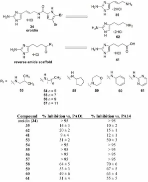
The virulence and persistence of bacterial biofilm infections is responsible for many chronic illnesses as well as increased morbidity and mortality rates in a plethora of infectious diseases. Using the marine natural product oroidin as molecular inspiration, a library of reverse amide (RA) 2-aminoimidazole analogues were synthesized and assayed for anti-biofilm activity. A thorough and detailed structure-activity-relationship (SAR) culminated in the identification of a long linear aliphatic tridecyl RA analogue with sub-micromolar anti-biofilm activity against Pseudomonas aeruginosa and low micromolar activity against Acinetobacter baumannii. Additionally, a sub-class
of aliphatic analogues lacking the amide moiety were synthesized that were very active at inhibiting and dispersing both P. aeruginosa and A. baumannii biofilms.
Small Molecule Control of Biological Function
by
Thomas Eric Ballard, Jr.
A dissertation submitted to the Graduate Faculty of North Carolina State University
in partial fulfillment of the requirements for the Degree of
Doctor of Philosophy
Chemistry
Raleigh, North Carolina 2008
APPROVED BY:
Dr. Daniel L. Comins Dr. Bruce M. Novak
DEDICATION
BIOGRAPHY
The author, Thomas Eric Ballard, Jr., was born in Vacaville, CA on May 20th 1982 to Eric and Kelly Ballard. He spent his early childhood growing up in Germany before moving to Kannapolis, NC where he spent the rest of his years through high school. After high school, Eric attended North Carolina State University where he graduated magna cum laude with honors, receiving his Bachelors of Science in Chemistry in 2004. As an undergraduate, Eric was a resident advisor for three years and received the Central Campus RA of the Year award in 2004. That year, Eric also received the Robert A. Osteryoung Award for Excellence in Teaching for his achievement in teaching general chemistry laboratories.
ACKNOWLEDGEMENTS
The sheer number of people who have affected my life to this point is immeasurable, but I will attempt to thank you, for you have helped achieve what I have today. First, I’d like to extend my most sincere thanks to Dr. Christian Melander. I do not believe my career as a scientist will ever be the same because of you, and I thank you for that. Your guidance, or perhaps freedom, has been decisive in my graduate career and I would not have had the same experience anywhere else. I would also like to thank Dr. Comins and Dr. Novak as they were also instructive in my scientific career. Although not nearly as productive for them when I was an undergraduate, I know that the time I spent in their labs lead me here today. Additionally, my unusual ability to deal with the NMRs in Dabney can only be attributed to Dr. Shultz. However direct or indirect, his entertaining spectroscopy class is unforgettable.
you do. Thanks to all the friends I’ve made during my time here at NCSU, you’ve all impacted my life and I’ll never forget you.
TABLE OF CONTENTS
Page
LIST OF TABLES ………. viii
LIST OF FIGURES……… ix
LIST OF SCHEMES ……… xi
LIST OF ABBREVIATIONS AND TERMS……… xii
PART I CHAPTER 1: Diazofluorenes as Mode-of-Action Probes ……… 1
1.1. Isolation, Properties and Biological Activity ……… 1
1.2. DNA/Mode-of-Action Studies ……… 4
1.2.1. Jebaratnam’s Oxidation Hypothesis ……… 4
1.2.2. Dmitrienko’s Electrophilicity Hypothesis ……… 5
1.2.3. Feldman’s Reductive Activation Hypothesis ……… 7
1.3. Diazofluorene Analogue Design and Synthesis……… 9
1.4. DNA Cleavage with Diazofluorene Analogues ……… 12
1.5. Conclusion ……… 16
1.6. Experimental Section ……… 17
References ……… 38
Appendix ……… 41
CHAPTER 2: Kinamycin-mediated DNA Cleavage……… 97
2.1. Investigation into the Biomimetic Activation of Kinamycin D ………… 97
2.2. Conclusion ……… 101
2.3. Experimental Section ……… 103
References ……… 105
PART II CHAPTER 3: 2-Aminoimidazoles as Modulators of Bacterial Biofilms ……… 106
3.1. Bacterial Biofilms ……… 106
3.1.1. Structure and Properties ……… 107
3.1.2. Influence and Effect on Society ……… 108
3.2. Chemical Controls of Biofilms ……… 109
References ……… 113
CHAPTER 4: A Reverse Amide (RA) Approach to Oroidin Analogues ……… 116
4.1. Synthesis of a Pilot RA Library ……… 117
4.2. Inhibition of Pseudomonas aeruginosa Biofilms ……… 120
4.3. Dispersion of Established P. aeruginosa Biofilms……… 123
4.4. Conclusion ……… 124
4.5. Experimental Section ……… 126
References ……… 145
Appendix ……… 147
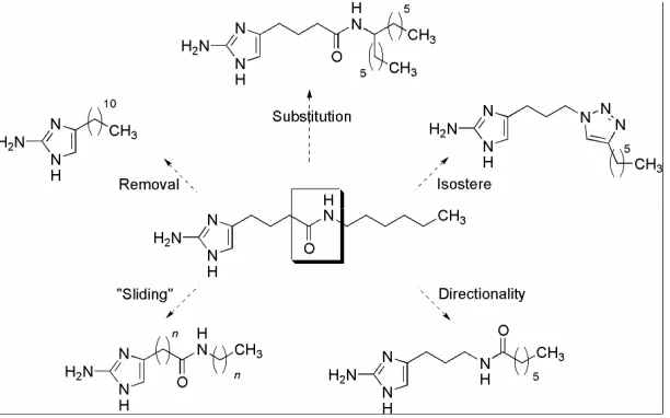
CHAPTER 5: Second Generation RA Library – A Detailed SAR Study ……… 199
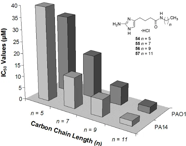
5.1. Reaching the Limit of the Aliphatic Chain Length Effect ……… 200
5.2. Deletion of the Amide Bond ……… 203
5.3. Linker Chain Modification and Sliding of the Amide Bond ……… 206
5.4. Increased Substitution of the Amide Bond……… 210
5.5. Reversal of the Amide Bond to Mimic Oroidin……… 212
5.6. Examination of a Triazole Isostere……… 213
5.7. PA14 Biofilm Inhibition: Activity Profile Comparisons ……… 214
5.8. Acinetobacter baumannii Biofilm Inhibition ……… 217
5.9. Dispersion of P. aeruginosa and A. baumannii Biofilms ……… 219
5.10. Conclusion ……… 221
5.11. Experimental Section ……… 224
References ……… 267
Appendix ……… 269
PART III CHAPTER 6: Synthesis and Anti-HIV Properties of a TAK-779 Analogue …… 408
6.1. Multivalent Displays in Biology……… 408
6.2. Synthesis of TAK-779 Analogue: SDC-1721 ……… 409
6.3. Inhibition of HIV Fusion ……… 410
6.4. Conclusion ……… 413
6.5. Experimental Section ……… 414
References ……… 422
LIST OF TABLES
Page
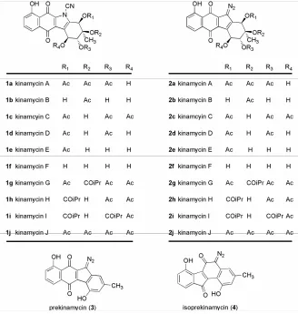
Table 1 Biological Activity of Representative Kinamycins (µM)……… 2
Table 2 Cytotoxicity Data for Lomaiviticin A ……… 3
Table 3 Quantitation of DNA Cleavage (%) ……… 15
Table 4 Completion of the RA Library ……… 120
Table 5 PAO1 and PA14 IC50 Values ……… 122
Table 6 Examining the Aliphatic Chain Length ……… 199
Table 7 Synthesis and Anti-biofilm Activity of 2nd Generation Aliphatic RA Analogues……… 201
Table 8 Anti-biofilm Activity of Amide Deletion Sub-class against PA14 ……… 204
Table 9 Synthesis and Anti-biofilm Activity of Modified Linker Analogues …… 207
Table 10 Synthesis and Anti-biofilm Activity of Phenyl Amine Analogues ……… 208
Table 11 Synthesis and Anti-biofilm Activity of Slider Analogues ……… 209
Table 12 Anti-biofilm Activity of Additionally Substituted RA Analogues against PA14……… 212
Table 13 Synthesis and Anti-biofilm Activity of a Triazole Isostere ……… 214
Table 14 Anti-biofilm Activity against A. baumannii……… 218
LIST OF FIGURES
Page
Figure 1 Representative Kinamycins: Original (left), Revised (right) ……… 1
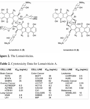
Figure 2 The Lomaiviticins……… 3
Figure 3 Synthetically Derived Diazo Compounds……… 4
Figure 4 Plasmid Cleavage Assay ……… 13
Figure 5 Synthetic Diazofluorene Analogues……… 13
Figure 6 Plasmid Cleavage Assay ……… 14
Figure 7 Plasmid Cleavage Assay ……… 14
Figure 8 Temperature-dependent Plasmid Cleavage Assay ……… 98
Figure 9 Time-dependent Plasmid Cleavage Assay ……… 99
Figure 10 Kinamycin D (model) LUMO ……… 99
Figure 11 Reduced kinamycin D (model) hydroquinone LUMO……… 101
Figure 12 in vitro Development of P. aeruginosa Biofilms……… 108
Figure 13 Molecules Known to Inhibit Biofilm Formation ……… 109
Figure 14 Oroidin as a Scaffold for Anti-biofilm Analogues ……… 111
Figure 15 Retrosynthetic Analysis of Oroidin and the RA Scaffold……… 111
Figure 16 Synthetic Accessibility of RA Analogues ……… 116
Figure 17 Preliminary Inhibition Data at 500 µM ……… 121
Figure 18 Structure Activity Relationship of Aliphatic Chain RA Analogues……… 123
Figure 20 Core Approaches to Turning the RA Scaffold ……… 200
Figure 21 Correlation of Aliphatic RA Analogues ……… 202
Figure 22 AHL vs. 2-AI ……… 205
Figure 23 Sliding the Amide Bond ……… 208
Figure 24 PA14 inhibition IC50 values from selected SAR analogues……… 215
Figure 25 Amide Orientation Comparison ……… 216
Figure 26 TAK-779 and SDC-1721 ……… 410
Figure 27 A-D Multiple-Cycle Inhibition Assays……… 412
LIST OF SCHEMES
Page
Scheme 1 Oxidative Activation ……… 5
Scheme 2 Synthesis of Isoprekinamycin Analogue 9 ……… 6
Scheme 3 Nucleophilic Activation by Increased Diazonium Character ………… 7
Scheme 4 Reductive Activation of Diazoparaquinone Compounds ……… 7
Scheme 5 Evidence for Radical and Orthoquinonemethide Formation……… 9
Scheme 6 Synthesis of Methoxydiazofluorenes and Nitrodiazofluorenes………… 10
Scheme 7 Synthesis of Chlorodiazofluorenes and Aminodiazofluorenes ………… 11
Scheme 8 Synthesis of 1,4-substituted Diazo Analogues ……… 12
Scheme 9 Reduction of kinamycin D ……… 100
Scheme 10 Potential routes for DNA cleavage activity ……… 100
Scheme 11 1st Generation Synthesis of RA Scaffold ……… 117
Scheme 12 2nd Generation Synthesis of RA Scaffold ……… 119
Scheme 13 Deletion of the Amide Bond ……… 203
Scheme 14 Synthesis of Boc-2AI Linker Acids ……… 206
Scheme 15 Synthesis of Additionally Substituted Analogues ……… 211
Scheme 16 A Natural Amide Bond Approach……… 213
LIST OF ABBREVIATIONS AND TERMS 2-AI 2-aminoimidazole
Ac acetyl
AHL N-acylhomoserine lactone AIBN 2,2’-azobisisobutyronitrile aq aqueous
BIA biochemical induction assay
bd broad dublet
Bn benzyl
Boc t-butoxycarboyl
brine saturated aqueous sodium chloride
bs broad singlet
nBu normal butyl
tBu tertiary butyl
CF Cystic Fibrosis
calcd calculated
cm-1 reciprocal centimeters d doublet
dt doublet triplets
dd doublet of doublets
DMSO dimethyl sulfoxide DNA deoxyribonucleic acid dt doublet of triplets
DTT dithiothreitol
EC50 effective concentration: 50%
EDTA ethylene diamine tetraacetic acid EtOAc ethyl acetate
GSH glutathione h hour(s)
HRMS high resolution mass spectrometry Hz hertz
IC30 inhibitory concentration: 30%
IC50 inhibitory concentration: 50%
IR infrared
J coupling constant
LB Luria-Bertani media
LBNS Luria-Bertani no salt media m multiplet MOA mode-of-action MHz megahertz
NADPH β-Nicotinamide adenine dinucleotide 2′-phosphate reduced tetrasodium salt NMR nuclear magnetic resonance spectrometry
OD600(540) optical density at 600 nm (or 540 nm)
Pd/C palladium on carbon ppm parts per million q quartet
QS Quorum Sensing
rt room temperature
s singlet
SAR structure-activity-relationship
TBE Tris/boric acid/EDTA
TFA trifluoroacetic acid TFAA trifluoroacetic anhydride
CHAPTER 1
Diazofluorenes as Mode-of-Action Probes
1.1 Isolation, Properties and Biological Activity
The kinamycins A-D 1a-1d (Figure 1) were first isolated in 1970 by Hata and Ohtani as bright orange crystals from the culture broth of Streptomyces murayamaensis.1 Within a year, the structures of kinamycins A-D 1a-1d had been elucidated and were shown to be 6-6-5-6 ring systems possessing a unique benzo[b]tetrahydrocarbazole skeleton with an N-cyano motif never before seen in nature.2, 3
In addition to their intriguing structure, they were also shown to be biologically active against gram positive, and to a lesser extent, gram negative bacteria (Table 1). Even derivatives that were synthesized from kinamycin C 1c by various protections and deprotections still retained biological activity (1f, 1j).4 In 1994, the structure of the kinamycins was revised to now contain a diazo moiety rather than a carbazole (Figure 1).
In 2001, researchers at Wyeth-Ayerst discovered Lomaiviticins A 9 and B 10, two very potent metabolites from a new species of Micromonospora, LL-37I366, later named M.
lomaivitiensis (Figure 2).5 Both lomaiviticins exhibit potent DNA damage indicated by BIA
(Biochemical Induction Assay), having minimum inhibitory concentrations of ≤ 0.1 ng/spot. Lomaiviticin A 9 is the more potent metabolite and is also the more abundant of the two from the fermentation broth. Lomaiviticin A 9 is a homo-dimeric diazobenzo[b]fluorene glycoside
and is extremely active against gram-positive bacteria, especially S. aureus and E. Faecium
(MIC’s, 6 – 25 ng/spot), and was also shown to be very cytotoxic to a 24-cancer cell line panel with IC50 values ranging from 0.01 to 98 ng/mL (Table 2).5 Lomaiviticin B 10 is also a
homodimeric diazobenzo[b]fluorene glycoside but contains two furanol rings most likely formed by hydrolysis of the tertiary sugar with concomitant cyclization onto the proximal ketone. The cytotoxic profile for lomaiviticin A 9 in the cancer cell lines was unique compared to other DNA-damaging drugs like mitomycin C and andriamycin hinting at a different mode-of-action for the lomaiviticins. Lomaiviticin A 9 was also reported to cleave DNA under reducing conditions but no further experimental data was reported.
1.2 DNA/Mode-of-Action Studies
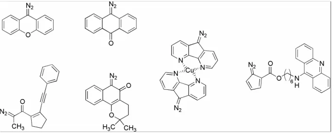
The kinamycins and lomaiviticins have attracted a great deal of interest from the synthetic and biosynthetic community. However, little is known about their mode-of-action (MOA). The diazo moiety, as it is so rare in nature, is the potential driving force for the cytotoxic, antibacterial and antitumor properties. Several diazo containing compounds have been synthesized in the past twenty years and have been reported to cleave DNA (Figure 3).6-11 However, these compounds are activated toward DNA cleavage either photochemically, oxidatively, or with heat, while it has been established that lomaiviticin A cleaved DNA under reducing conditions.
1.2.1 Jebaratnam’s Oxidation Hypothesis
Assuming that the kinamycins work by DNA damage via reaction with the diazo group, Jebaratnam tested this hypothesis using diazofluorene 7 and β-naphthyl phenyl diazomethane 8 against plasmid DNA (pBR322) (Scheme 1).12 Jebaratnam was already interested in activating diazonium compounds with copper salts,13 and he hypothesized that the kinamycins were simply the deprotonated forms of diazonium compounds and protonation at physiological pH might generate an active and unstable diazonium ion.14 To
this end, he submitted pBR322 to DNA cleavage conditions that consisted of 7 with 500 or 1000 µM of the following salts: AgOAc, Tl(OAc)3, Cu(OAc)2, or Hg(OAc)2. Varying the
metal salt and the salt concentration, he obtained single strand nicking of the plasmid DNA with diazofluorene 7 and β-naphthyl phenyl diazomethane 8 under Cu(OAc)2 conditions.
Jebaratnam proposed that the cupric acetate oxidized the diazo moiety, then with loss of N2, yielded the one-electron radical that could then cleave DNA by known radical
pathways.15-18 β-naphthyl phenyl diazomethane 8 also cleaved the plasmid DNA in the presence of cupric acetate while diazofluorene 7 or cupric acetate alone (as controls) did not lead to DNA cleavage.
1.2.2 Dmitrienko’s Electrophilicity Hypothesis
The next MOA proposal for the kinamycins and lomaiviticins was brought forth by Dmitrienko. Dmitrienko believed that the increased diazonium ion character played a large part in the biological activity of these natural products.19 To test his hypothesis he synthesized dideoxyisoprekinamycin derivative 9 (Scheme 2).
Suzuki cross-coupling of the boronate ester of 10 with bromobenzoate 11 afforded biaryl 12. Bromination of biaryl 12 followed by heating in methanesulfonic acid gave the
desired cyclized product 13. Palladium-catalyzed amination with benzylamine followed by hydrogenation and diazotization/demethylation yielded dideoxyisoprekinamycin 9.
Dmitrienko’s mechanistic rationale was based on end product analysis of the reaction of β-naphthol with either isoprekinamycin 4 or isoprekinamycin analogue 9 (Scheme 3). This observation was rationalized by invoking a key hydrogen bond between the phenolic hydroxyl and the ketone alpha to the diazo moiety which produced a more electrophilic diazonium ion. Biologically, this data presents the hypothesis that DNA amino groups (Guanine and Adenine) would add to the diazo group, forming labile triazenes. Subsequent disassociation with concomitant loss of N2, would generate a radical intermediate on the
DNA base and the aryl ring of the natural product. The resulting radicals could then alkylate the DNA base or act independently (i.e. reaction with O2) to cause DNA damage.16, 17
1.2.3 Feldman’s Reductive Activation Hypothesis
In 2005, Feldman proposed a third MOA that was underpinned by the reductive activation by a one-electron reduction of the quinone to generate reactive aryl radical 14. Once generated this radical could abstract a hydrogen atom from DNA inducing DNA strand scission (Scheme 4).20 In addition, once 14 abstracts a hydrogen an orthoquinonemethide is
generated. Orthoquinonemethides are known DNA damaging agents (i.e. mitomycin C, hydroxymethylacylfulvene).21-25
Feldman tested the putative e-/H+ reduction with the use of Bu3SnH/AIBN in
refluxing benzene. In a more recent account,26 Feldman admits to the limitations of the
chosen abiological conditions, but holds fast that the work helps to explore the chemistry of the diazoparaquinone moiety irrespective of the environment. However, there is no biological equivalent for Bu3SnH/AIBN and refluxing benzene is also not biomimetic. It is important to
note the lack of reactivity in non-aromatic solvents (THF, 1,4-dioxane, CCl4, CH3CN, EtOH,
or CHCl3). Feldman attributes this to the stabilization or promoting effect that aromatic
solvents may give to the intermediate radical which, we believe, presents even more evidence that this is not the appropriate means of activation for this class of natural products. The kinamycins and lomaiviticins will not have the benefit of being stabilized by aromatic solvents in vitro or in vivo.
1.3 Diazofluorene Analogue Design and Synthesis
Unable to accept the previous MOA studies as indicative of biomimetic activation, we sought to delineate an appropriate means of testing our own activation hypothesis for the diazofluorene class of compounds. To this end, we synthesized a small library of diazofluorene analogues with varying electronic properties to probe the MOA and assayed against the ability of the analogue library to recapitulate the DNA cleavage activity of kinamycin D.27
It has already been established that the activity of the kinamycins and lomaiviticins is directly related to the presence of the diazo moiety. The hydroxylated naphthoquinone subunit has also been identified as possibly playing an assisting role in the MOA for these
natural products. With these two ideas in mind, the synthetic library would consist of substituted diazofluorenes to explore how the differences in ring electronics would affect the diazo moiety.
1-Aminofluorenone 16 and 4-aminofluorenone 18 were chosen as convenient starting materials to access most of the library through various functionalizations of the amino group (Schemes 6 & 7). The parent diazo compound, 9-diazofluorene 7, was synthesized as reported from fluorenone.28 The methoxyfluorenones were synthesized by diazotization of the aminofluorenone (16/18) at 0 °C with NaNO2/H2SO4 followed by the addition of the
diazonium salt to a solution of refluxing 50% H2SO4.29 The isolated hydroxyfluorenones
were then methylated with NaH/MeI or Me2SO4/NaOH to give the methoxyfluorenones in
high yields. Oxidation of the aminofluorenones (16/18) with TFAA/H2O2 afforded the
nitrofluorenones in moderate yields. Finally, installation of the diazo group was
accomplished by either: hydrazinolysis followed by mercuric oxide oxidation or by condensation with tosyl hydrazine followed by base induced elimination of the tosyl group. The chlorodiazofluorenes were accessed from the known chlorofluorenones under standard diazo installation conditions. Additionally, the aminodiazofluorenes were accessed directly from the aminofluorenones through tosyl hydrazine condensation followed by base induced oxidation (Scheme 7).
The synthesis of two 1,4-substituted fluorenones was also envisioned to more closely mimic the substitution of the natural products. Following the work of Jones, 1,4-dimethoxybenzene 26 was acylated with 2-iodobenzoic acid 27 using TFAA/TFA in 97% yield (Scheme 8). Palladium catalyzed cyclization afforded 1,4-dimethoxyfluorenone 28 in 50% yield.30 Condensation of 1,4-dimethoxyfluorenone 28 with tosyl hydrazine followed by elimination with sodium methoxide yielded the target analogue 1,4-dimethoxydiazofluorene 29 in 15% yield over two steps. The 1,4-diazoquinofluorene 30 was synthesized by quantitative demethylation of 1,4-dimethoxyfluorenone 29 with BBr3, then hydrazinolysis
and Fetizon’s reagent affected the tandem oxidation of the hydroquinone and hydrazone in 42% yield.31
1.4 DNA Cleavage with Diazofluorene Analogues
With the synthesis of the diazofluorene library complete, it was then necessary to assay them for their ability to recapitulate the DNA cleaving ability of the natural products. Since lomaiviticin A was reported to cleave double stranded DNA under reducing conditions,5 we sought to mimic the reducing conditions of the cell in vitro. To this end, we explored the DNA cleaving activity of kinamycin D (2d) under a variety of preliminary activating conditions (Figure 4).32 First we incubated plasmid pBR322 DNA with kinamycin D but no cleavage occurred even after 3 days. Kinamycin D was then incubated with pBR322 in the presense of either dithiothreitol (DTT), nicotinamide adenine dinucleotide phosphate (NADPH), or sodium cyanide (NaCN). Appreciable DNA damage (single strand scission) was observed for the DNA/kinamycin D solution incubated with DTT, while no other significant DNA damage was observed for the other additives. A preliminary time course for optimal cleavage was also conducted for kinamycin D in the presence of DTT and maximal cleavage was obtained after 2 days with 1 mM of kinamycin D and 1 M DTT. It is worth noting that DTT is a free-radical scavenger so the intermediacy of a diffusible free-radical as the active intermediate is not very likely.
Using these optimized conditions for the natural product, we assayed the synthetic diazofluorene analogues for their ability to cleave DNA (Figure 5). The methoxy group (17, 19, 29) was chosen specifically because it mimics the reduced form of the paraquinone natural products, which had already been hypothesized by Feldman as being an active intermediate.
Two compounds assayed, 1-methoxydiazofluorene 17 and 4-aminodiazofluorene 25, cleaved DNA in an efficient manner comparable to kinamycin D 2d under DTT conditions (Figure 6). A control experiment was also conducted; 1-methoxyfluorenone and 4-aminofluorenone were incubated with DNA/DTT as before and showed no cleavage of the DNA clearly indicating that the diazo group is essential for DNA cleavage. To further
Figure 5. Synthetic Diazofluorene Analogues.
explore these as kinamycin mimics, 1-methoxydiazofluroene 17 and 4-aminodiazofluorene 25 were assayed for their ability to cleave DNA in the absence of DTT. Under these conditions, 4-aminodiazofluorene 25 retained DNA cleavage activity, while 1-methoxydiazofluorene 17 displayed minimal activity.
Although strange, these results are thought to show the increased activity of the 4-aminodiazofluorene 25, indicating that it does not need to be activated. A clear analogy would be that the 1-aminodiazofluorene 24 should then be very active and indeed it is so active that it could not be accurately assayed due to its instability even at -80 °C. We also assayed 1-methoxydiazofluorene 17 for its ability to cleave DNA in the presence of NADPH or NaCN but no appreciable amount of cleavage was detected (Figure 7).
We quantitated the extent of DNA cleavage from supercoiled DNA (type I) to nicked DNA (type II) and these results are summarized in Table 3. Most notably, in the presence of DTT, 1-methoxydiazofluorene 17 outperforms kinamycin D 2d, exhibiting 150% of the cleavage activity of the natural product. In the presence of DTT, 4-aminodiazofluorene 25
Figure 7. Plasmid Cleavage Assay, top band is type II nicked DNA, the bottom band is type I supercoiled DNA.
Conditions: all lanes contain 714 ng of plasmid DNA. Final [compound] = 1.0 mM. Lane 1: DNA only, lane 2:
17/DNA, lane 3: 25/DNA, lane 4: 0.5 M NADPH/DNA, lane 5: 17, 0.5 M NADPH/DNA, lane 6: 1.0 M
NaCN/DNA, lane 7: 17, 1.0 M NaCN/DNA.
only exhibits 64% activity in comparison with kinamycin D, but in the absence of DTT, it exhibits 160% of the cleavage activity that kinamycin D exhibited in DTT (or 900% as compared to kinamycin D in the absence of DTT).
In addition to the DNA cleavage assay, we also explored the antiproliferative activity against HeLa cells for 1-methoxydiazofluorene 17, 4-aminodiazofluorene 25 and the diazo parent compound 7. Earlier, diazofluorene 7 was shown to cleave DNA under oxidizing conditions, while 1-methoxydiazofluorene 17 cleaves under reducing thiol conditions and 4-aminodiazofluorene 25 requires no activation. These intrinsic properties allowed us to explore the antiproliferative activity from several avenues to achieve maximal activity. The studies were conducted at 100 nM, the solubility limit of the synthetic compounds in cell media. Although we could not calculate IC50 values due to solubility issues, we did observe
that methoxydiazofluorene 17 had the highest activity of the three compounds tested. 1-Methoxydiazofluorene 17 demonstrated a time-dependent inhibition of HeLa cell proliferation and inhibited cell growth by 35-40% at 12 h. This activity is similar to the antiproliferative activity reported for the most active kinamycins.2
1.5 Conclusion
Using a small library of synthesized diazo derivatives, we have delineated a MOA not precedented in the literature for these compounds. We found that the use of DTT effectively activated kinamycin D 2d and several diazofluorenes for DNA cleavage.27 In total, four major hypotheses have been proposed for the MOA of the kinamycins and lomaiviticins. Interestingly, they have varied tremendously in activating conditions: oxidation, increased electrophilicity, one electron reduction, and thiol-mediated reduction/activation. From these various activating conditions though, a clear trend has emerged that the diazo group is the obvious active site and that loss of N2 forms a reactive site. How this reactive intermediate
1.6 Experimental Section
General Experimental and Procedures
All 1H NMR (400 MHz or 300 MHz) and 13C NMR (100 MHz or 75 MHz) spectra were recorded at 25.0 ºC on a Varian Mercury spectrometer. Chemical shifts (δ) are given in ppm relative to tetramethylsilane or the respective NMR solvent; coupling constants (J) are in hertz. Abbreviations used are s = singlet, bs = broad singlet, d = doublet, dd = doublet of doublets, t = triplet, dt = doublet of triplets, m = multiplet, and br = broad. Infrared spectra (KBr pellet) were taken using JASCO FT-IR-410 spectrophotometer. Wave numbers in cm-1 are reported for characteristic peaks. FAB-MS spectra were measured via high-resolution fast atom bombardment using a matrix of nitrobenzyl alcohol. Silica gel (40 µm average particle size) was used for column chromatography. Toluene was purified by distillation over sodium/benzophenone, and anhydrous MeOH was purified by distillation over magnesium. All other reagents were used as purchased from commercial sources.
9-Fluorenone (p-tosyl)hydrazone (1-i). p-Toluenesulfonylhydrazide (0.98 g, 5.24 mmol) was added to a stirred suspension of 9-fluorenone (0.76 g, 4.2 mmol) in boiling CH3CN (20
mL). The resulting mixture was refluxed for 0.5 h, the yellow crystals obtained upon cooling were filtered. Flash chromatography (2:1 / CH2Cl2:hexanes) afforded 9-fluorenone (p
tosyl)hydrazone (1.20 g, 81%) as yellow crystals. IR (KBr) 3500, 3213, 3065, 1595, 1451, 1384, 1327, 1311, 1152 cm-1; 1H NMR (400 MHz, CDCl3) δ 2.40 (s, 3H), 7.3 (m, 5H), 7.44
(t, 1H, J = 6.8 Hz), 7.53 (d, 1H, J = 7.6 Hz), 7.64 (d, 1H, J = 7.6 Hz), 7.70 (d, 1H, J = 7.6 Hz), 7.86 (d, 1H, J = 7.6 Hz), 7.97 (d, 2H, J = 8.4 Hz), 8.38 (bs, 1H); 13C NMR (100 MHz, CDCl3) δ 21.9, 119.9, 121.1, 122.4, 126.5, 128.4, 128.5, 128.7, 129.9, 130.7, 131.9, 134.9,
136.6, 140.0, 142.7, 144.8; HRMS (FAB) m/z, ([M + H]+, C20H16N2O2S): calcd. 349.1011,
found 349.1032.
9-Diazofluorene (7). A dioxane (4.5 mL) solution of 9-fluorenone (p-tosyl)hydrazone (73 mg g, 0.21 mmol) and aqueous 50% NaOH (2.3 mL) was vigorously stirred at 50 °C for 24 h under Ar. The mixture was then diluted with water (2.5 mL) and extracted quickly with EtOAc (3 x 2.5 mL). The combined organic layer was then washed with water (2.5 mL), dried (MgSO4) and concentrated under reduced pressure. Flash chromatography (2:1 /
CH2Cl2:hexanes) gave 9-diazofluorene 7 (35 mg, 88%) as fine light red crystals. IR (KBr)
3040, 2066, 1439, 752, 722 cm-1; 1H NMR (300 MHz, acetone-d6) δ 7.34 (t, 2H, J = 7.8 Hz),
7.42 (t, 2H, J = 7.8 Hz), 7.67 (d, 2H, J = 7.5 Hz), 8.06 (d, 2H, J = 7.8 Hz); 13C NMR (100 MHz, acetone-d6) δ 119.7, 121.0, 121.1, 124.6, 124.7, 126.5, 126.6; HRMS (FAB) m/z,
([M]+, C
13H8N2): calcd. 192.0687, found 192.0687.
Amino-9-fluorenone (p-tosyl)hydrazone (xiv). A 95% EtOH (7 mL) solution of 1-amino-9-fluorenone 16 (0.32 g, 1.64 mmol), p-toluenesulfonylhydrazide (0.45g, 2.42 mmol) and conc. HCl (280 µL) was refluxed under Ar for 6 h, then cooled and concentrated under reduced pressure. The product was isolated by flash chromatography (CH2Cl2) to give
1-amino-9-fluorenone (p-tosyl)hydrazone (0.45 g, 76%) as yellow crystals. IR (KBr) 3469, 3358, 3195, 1618, 1456, 1311, 1164, 1089, 752 cm-1; 1H NMR (400 MHz, acetone-d6) δ 2.41
(s, 3H), 6.06 (bs, 2H), 6.64 (d, 1H, J = 8.0 Hz), 6.99 (d, 1H, J = 8.0 Hz), 7.13 (t, 1H, J = 8.0 Hz), 7.35 (t, 1H, J = 8.0 Hz), 7.44 (m, 3H), 7.73 (d, 1H, J = 8.0 Hz), 7.94 (m, 2H), 8.16 (d, 1H, J = 8.0 Hz), 9.91 (bs, 1H); 13C NMR (100 MHz, acetone-d6) δ 20.8, 108.7, 116.1, 120.6,
127.6, 127.9, 128.7, 129.8, 130.5, 131.5, 131.8, 135.7, 140.8, 142.6, 144.4, 146.1, 156.6; HRMS (FAB) m/z, ([M + H]+, C20H17N3O2S): calcd. 364.1120, found 364.1113.
1-Amino-9-diazofluorene (24). A MeOH (5 mL) solution of 1-amino-9-fluorenone (p -tosyl)hydrazone (0.10 g, 0.28 mmol) and NaOMe (37 mg, 0.69 mmol) was refluxed for 10 h under Ar. The mixture was then concentrated under reduced pressure. The product was
16 1-xiv
isolated by flash chromatography (1:1 / CH2Cl2:hexanes) to give 24 (15 mg, 26%) as a red
solid. IR (KBr) 3396, 3318, 2923, 2050, 1589, 1435, 1339, 1285, 1152, 789, 714 cm-1; 1H NMR (400 MHz, acetone-d6) δ 6.51 (bs, 2H), 6.80 (d, 1H, J = 8.4 Hz), 7.10 (d, 1H, J = 8.0
Hz), 7.24 (m, 2H), 7.45 (t, 1H, J = 7.2 Hz), 7.77 (d, 1H, J = 8.4 Hz), 8.41 (d, 1H, J = 7.6 Hz); 13C NMR (100 MHz, acetone-d6) δ 109.1, 116.3, 120.6, 128.0, 128.2, 131.5, 132.5;
LRMS (FAB) m/z, ([M]+, C13H9N3): calcd. 207.0796, found 207.1410.
1-Hydroxy-9-fluorenone (1-ii). 1-Amino-9-fluorenone 16 (0.185 g, 0.95 mmol) was suspended in H2O (6 mL) at 0 ºC and dissolved with the addition of conc. H2SO4 (11 mL).
With vigorous stirring, a solution of sodium nitrite (0.091 g, 1.31 mmol) and H2O (4 mL)
was slowly added dropwise. The solution was stirred at 0 ºC for 2 h and then canulated into a boiling solution of conc. H2SO4/H2O (32 mL/32 mL). Following addition, the solution was
boiled for 15 min then cooled to room temperature and allowed to stir for an additional 4 h. The reaction mixture was then extracted with CH2Cl2 (3x50 mL) and the combined organic
layer was washed with H2O (50 mL), dried (Na2SO4), filtered and concentrated. The product
was isolated by flash chromatography (0-15% EtOAc/hexanes) to give 1-hydroxy-9-fluorenone (0.129 g, 70%) as fine yellow needles. IR (KBr) 3368 (br, s), 1690, 1599 (vs) cm
-1; 1H NMR (300 MHz, acetone-d
6) δ 6.78 (dd, 1H, J = 8.2 and 0.6 Hz), 7.22 (dd, 1H, J = 7.2
and 0.6 Hz), 7.37 (dt, 1H, J = 7.5 and 1.2 Hz), 7.44 (dd, 1H, J = 7.2 and 0.9 Hz), 7.57 (m,
2H), 7.70 (dt, 1H, J = 7.5 and 0.9 Hz), 8.76 (bs, 1H); 13C NMR (100 MHz, acetone-d6) δ
113.8, 118.3, 119.3, 122.19, 124.47, 130.2, 135.0, 135.7, 138.4, 144.8, 145.2, 157.9, 195.4; HRMS (FAB) m/z, ([M + H]+, C13H8O2): calcd. 197.0603, found 197.0614.
1-Methoxy-9-fluorenone (1-iii). 1-Hydroxy-9-fluorenone (0.10 g, 0.51 mmol) was dissolved into 3 mL dry DMF under Ar and cooled to 0 ºC with stirring. Then, 60% NaH (0.03 g, 0.76 mmol) was added in portions and the solution was warmed to room temperature for 30 min. Methyl Iodide (0.07 mL, 1.1 mmol) was then added and stirred for 2 h after which the reaction was poured into H2O (10 mL) and extracted with Et2O (3 x 10 mL). The combined
organic layers were then washed with H2O and brine, dried (Na2SO4), filtered and
concentrated. The product was isolated by flash chromatography (0-20% EtOAc/hexanes) to give 1-methoxy-9-fluorenone (0.103 g, 96%) as fine yellow-green needles. IR (KBr) 2837 (w), 1700 (vs), 1592 (vs), 1023 (m) cm-1; 1H NMR (300 MHz, acetone-d6) δ 3.94 (s, 3H),
7.01 (d, 1H, J = 8.7 Hz), 7.31 (dt, 1H, J = 7.2 and 1.2 Hz), 7.36 (dd, 1H, J = 7.2 and 1.2 Hz), 7.53 (m, 3H), 7.69 (dt, 1H, J = 7.2 and 1.2 Hz); 13C NMR (75 MHz, acetone-d6) δ 55.6,
113.2, 113.9, 119.9, 120.7, 123.3, 129.4, 134.2, 134.5, 137.2, 143.3, 146.4, 158.6, 190.4; HRMS (FAB) m/z, ([M + H]+, C14H10O2): calcd. 211.0759, found 211.0759.
Methoxy-9-fluorenone (p-tosyl)hydrazone (iv). A THF (3.0 mL) solution of 1-methoxy-9-fluorenone (86.1 mg, 0.41 mmol), p-toluenesulfonylhydrazide (86.6 mg, 0.47 mmol) and conc. HCl (50 µL) was refluxed under Ar for 12 h, then cooled and dried under reduced pressure. The product was isolated by flash chromatography (0-50% EtOAc/hexanes) to give 1-methoxy-9-fluorenone (p-tosyl)hydrazone (0.131 g, 85%) as bright yellow crystals. IR (KBr) 3117 (br), 2841 (w), 1602, 1578 (s), 1351, 1168 (vs), 1051 (s) cm-1; 1H NMR (300 MHz, CDCl3) δ 2.37 (s, 3H), 4.03 (s, 3H), 6.76 (d, 1H, J = 8.4 Hz),
7.27 (m, 6H), 7.48 (d, 1H, J = 7.5 Hz), 7.76 (d, 1H, J = 7.2 Hz), 7.94 (d, 2H, J = 8.1 Hz), 11.66 (s, 1H); 13C NMR (100 MHz, CDCl3) δ 21.8, 56.8, 111.3, 114.6, 118.1, 119.8, 122.3,
128.0, 128.5, 129.7, 129.9, 133.7, 136.4, 137.6, 138.5, 144.1, 144.7, 145.5, 153.0; HRMS (FAB) m/z, ([M + H]+, C21H18N2O3S): calcd. 379.1116, found 379.1121.
1-Methoxy-9-diazofluorene (17). A MeOH (4 mL) solution of 1-methoxy-9-fluorenone (p -tosyl)hydrazone (69.2 mg, 0.18 mmol) and NaOMe (83.0 mg, 1.54 mmol) was refluxed for 2 h under Ar. After the reaction was complete, the mixture was dried under reduced pressure.
1-iii 1-iv
The product was isolated by flash chromatography (1:1 / CH2Cl2:hexanes) of the residue to
give 17 (14.4 mg, 36%) as a red solid. IR (KBr) 2836 (w), 2071 (vs), 1572, 1447 (s), 1260 (vs), 1014 (m) cm-1; 1H NMR (400 MHz, CDCl3) δ 3.95 (s, 3H), 6.84 (d, 1H, J = 8.0 Hz),
7.26 (m, 1H), 7.31 (dt, 1H, J = 8.0 and 1.2 Hz), 7.38 (dt, 1H, J = 8.0 and 1.6 Hz), 7.48 (d, 1H, J = 8.0 Hz), 7.58 (d, 1H, J = 8.0 Hz), 7.93 (d, 1H, J = 8.0 Hz); 13C NMR (100 MHz, CDCl3) δ 55.7, 107.4, 114.1, 119.1, 121.5, 124.5, 125.7, 126.3; HRMS (FAB) m/z, ([M]+,
C14H10N2O): calcd. 222.0793, found 222.0777.
1-Chloro-9-fluorenone hydrazone (1-xii). To a solution of 1-chloro-9-fluorenone33 (0.41 g, 1.9 mmol) in 1-butanol (2 mL), 85% aqueous hydrazine hydrate (0.25 g, 6.5 mmol) was added. After refluxing for 4 h, the mixture was concentrated under reduced pressure and flash chromatography (CH2Cl2) gave 1-chloro-9-fluorenone hydrazone (0.17 g, 38%) as light
yellow crystals. IR (KBr) 3366, 3226, 2360, 2337, 1602, 1445, 1412, 1186, 742 cm-1; 1H
NMR (300 MHz, acetone-d6) δ 7.30 (m, 2H), 7.47 (m, 2H), 7.80 (m, 3H), 7.96 (m, 1H), 8.22
(m, 1H); 13C NMR (75 MHz, CDCl3) δ 118.4, 120.6, 125.3, 128.2, 128.6, 128.9, 129.8,
130.1, 133.4, 138.6, 140.3, 140.5; HRMS (FAB) m/z, ([M + H]+, C13H9ClN2): calcd.
229.0533, found 229.0522.
1-Chloro-9-diazofluorene (22). 1-chloro-9-fluorenone hydrazone (0.17 g, 0.73 mmol) was dissolved in toluene (10 mL) and yellow mercuric oxide (1.60 g, 7.4 mmol) was added. The resulting suspension was stirred for 96 h, filtered and dried (MgSO4). The filtrate was
concentrated under reduced pressure and flash chromatography (1:4 / CH2Cl2:petroleum
ether) gave 22 (70.7 mg, 43%) as a red solid. IR (KBr) 3476, 2063, 1557, 1444, 1415, 742, 657 cm-1; 1H NMR (400 MHz, acetone-d6) δ 7.27 (m, 3H), 7.39 (dt, 1H, J = 8.0 and 1.2 Hz),
7.46 (d, 1H, J = 8.0 Hz), 7.87 (dd, 1H, J = 8.0 and 1.2 Hz), 7.94 (d, 1H, J = 8.0 Hz); 13C NMR (100 MHz, acetone-d6) δ 119.0, 119.7, 121.5, 125.0, 125.8, 126.8, 127.1, 127.2, 129.0,
130.5, 133.5; HRMS (FAB) m/z, ([M]+, C13H7ClN2): calcd. 226.0298, found 226.0300.
1-Nitro-9-fluorenone (1-viii). Trifluoroacetic anhydride (1.50 mL, 10.5 mmol) in CH2Cl2
(3.0 mL) was slowly added to H2O2 (0.57 mL, 9.2 mmol) at 0 ºC while stirring under Ar.
The solution was held at 0 ºC for 1.5 h, then a solution of 1-amino-9-fluorenone 16 (0.196 g, 1.00 mmol) and CH2Cl2 (3 mL) was added dropwise. The solution was held at 0 ºC for 2 h
then warmed to room temperature and poured into H2O (10 mL) and extracted with CH2Cl2
(2 x 10 mL). The combined organic layers were then washed with brine, dried (Na2SO4),
1-xii 22
filtered and concentrated. The product was isolated by flash chromatography (0-5% EtOAc/hexanes) to give 1-nitro-9-fluorenone (0.131 g, 58%) as yellow-orange needles. IR (KBr) 1728, 1526, 1364 (vs) cm-1; 1H NMR (300 MHz, acetone-d6) δ 7.49 (t, 1H, J = 7.5
Hz), 7.67 (m, 3H), 7.85 (m, 2H), 8.06 (d, 1H, J = 7.5 Hz); 13C NMR (100 MHz, CDCl3) δ
120.8, 123.1, 123.6, 125.0, 125.3, 130.5, 133.4, 135.3, 135.6, 142.1, 146.3, 146.6, 187.5; LRMS (FAB) m/z, ([M + H]+, C13H7NO3): calcd. 226.0504, found 226.1187.
1-Nitro-9-fluorenone (p-tosyl)hydrazone (1-ix). A THF (2 mL) solution of 1-nitro-9-fluorenone (71 mg, 0.31 mmol), p-toluenesulfonylhydrazide (63 mg, 0.34 mmol) and conc. HCl (36.5 µL) was stirred at room temperature for 24 h, and then concentrated under reduced pressure. Flash chromatography (CH2Cl2) gave 1-nitro-9-fluorenone (p-tosyl)hydrazone (68
mg, 56%) as light yellow crystals. IR (KBr) 3433, 3248, 3216, 1536, 1453, 1371, 1342, 1167, 784, 678, 547 cm-1; 1H NMR (400 MHz, CDCl
3) δ 2.42 (s, 3H), 7.38 (d, 3H, J = 8.0
Hz), 7.48 (m, 3H), 7.73 (d, 2H, J = 8.0 Hz), 7.87 (d, 1H, J = 8.0 Hz), 7.94 (d, 2H, J = 8.0 Hz), 8.44 (bs, 1H); 13C NMR (100 MHz, acetone-d6) δ 20.9, 121.5, 121.8, 122.8, 127.8,
128.9, 129.2, 129.58, 129.64, 131.8, 132.2, 140.2, 144.5; LRMS (FAB) m/z, ([M + H]+, C20H15N3O4S): calcd. 394.0862, found 394.1357.
1-Nitro-9-diazofluorene (20). A dioxane (2.5 mL) solution of 1-nitro-9-fluorenone (p -tosyl)hydrazone (43 mg, 0.11 mmol) and aqueous 50% NaOH (0.32 mL) was vigorously stirred at 50 °C for 24 h. The mixture was then diluted with H2O (2.0 mL) and extracted
quickly with EtOAc (3x2.0 mL), the organic layer was washed with water (2.5 mL), dried (MgSO4), filtered and concentrated under reduced pressure. Flash chromatography (1:1 /
CH2Cl2:hexanes) gave 20 (19.8 mg, 76%) as a light red solid. IR (KBr) 3442, 2082, 1559,
1514, 1448, 1420, 1301, 1207, 744 cm-1; 1H NMR (400 MHz, acetone-d6) δ 7.40 (dt, 1H, J =
8.0 and 1.6 Hz), 7.51 (m, 3H), 8.14 (dd, 1H, J = 8.0 and 0.8 Hz), 8.24 (dd, 1H, J = 8.0 and 0.8 Hz), 8.42 (dd, 1H, J = 8.0 and 0.8 Hz); 13C NMR (100 MHz, acetone-d6) δ 118.0, 121.5,
122.9, 124.2, 125.0, 127.0, 127.9; HRMS (FAB) m/z, ([M]+, C13H7N3O2): calcd. 237.0538,
found 237.0541.
Amino-9-fluorenone (p-tosyl)hydrazone (1-xv). A 95% EtOH (3 mL) solution of 4-amino-9-fluorenone 18 (0.15 g, 0.80 mmol), p-toluenesulfonylhydrazide (0.22 g, 1.20 mmol) and conc. HCl (130 µL) was refluxed under Ar for 6 h, then cooled and concentrated under
1-ix 20
reduced pressure. Flash chromatography (CH2Cl2) gave 4-amino-9-fluorenone (p
-tosyl)hydrazone (0.20 g, 70%) as bright yellow crystals. As a mixture of E:Z isomers. IR (KBr) 3390, 3188, 1624, 1594, 1454, 1399, 1336, 1165, 1064, 724, 538 cm-1; 1H NMR (400 MHz, acetone-d6) See spectrum; 13C NMR (100 MHz, acetone-d6) δ 20.9, 117.5, 120.8,
121.59, 121.62, 122.0, 122.5, 123.2, 123.6, 126.4, 127.5, 128.8, 129.1, 129.55, 129.60, 130.6, 130.8, 144.3; HRMS (FAB) m/z, ([M + H]+, C20H17N3O2S): calcd. 364.1120, found
364.1114.
4-Amino-9-diazofluorene (25). A dioxane (2.5 mL) solution of 4-amino-9-fluorenone (p -tosyl)hydrazone (0.04 g, 0.11 mmol) and aqueous 50% NaOH (0.32 mL) was vigorously stirred at 50 °C for 24 h under Ar. EtOAc (10 mL) and H2O (5 mL) were then added. The
organic layer was washed with H2O (5 mL), dried (Na2SO4), filtered and concentrated under
reduced pressure. Flash chromatography (1:1 / CH2Cl2:hexanes) gave 25 (7.9 mg, 35%) as
deep red crystals. IR (KBr) 3421, 2067, 1430, 719 cm-1; 1H NMR (300 MHz, acetone-d6) δ
5.16 (bs, 2H), 6.72 (d, 1H, J = 7.8 Hz), 6.94 (d, 1H, J = 7.8 Hz), 7.21 (t, 1H, J = 8.1 Hz), 7.33 (m, 2H), 7.61 (d, 1H, J = 7.2 Hz), 8.07 (d, 1H, J = 7.2 Hz); 13C NMR (100 MHz, acetone-d6) δ 110.5, 112.7, 119.4, 122.5, 124.8, 125.2, 127.4; HRMS (FAB) m/z, ([M]+,
C12H9N3): calcd. 207.0796, found 207.0779.
4-Hydroxy-9-fluorenone (1-v).29 The product was isolated by flash chromatography (0-15% EtOAc/hexanes) to give 4-hydroxy-9-fluorenone (0.069 g, 73%) as fine red-orange needles. IR (KBr) 3183 (br, s), 1683, 1278 (s) cm-1; 1H NMR (300 MHz, acetone-d6) δ 7.16 (m, 3H),
7.29 (dt, 1H, J = 7.5 and 1.2 Hz), 7.52 (dd, 1H, J = 7.5 and 1.2 Hz), 7.57 (d, 1H, J = 6.3 Hz), 7.88 (d, 1H, J = 7.5 Hz), 9.35 (bs, 1H); 13C NMR (100 MHz, acetone-d6) δ 116.1, 123.8,
124.1, 124.7, 128.6, 129.6, 131.1, 134.1, 135.5, 136.5, 144.7, 154.0 193.8; HRMS (FAB)
m/z, ([M + H]+, C
13H8O2): calcd. 197.0603, found 197.0615.
4-Methoxy-9-fluorenone (1-vi). 4-Hydroxy-9-fluorenone (0.066 g, 0.34 mmol) was dissolved into 95% EtOH (0.75 mL) at 50 ºC under Argon with stirring. To the solution was added H2O (0.06 mL) then NaOH (0.02 g). Dimethyl sulfate (0.04 mL, 0.40 mmol) was
added dropwise with an additional NaOH (0.02 g) added after addition. The solution was refluxed for 30 mins then cooled to room temperature. The solution was then poured into H2O (5 mL) and extracted with CH2Cl2 (3 x 5 mL). The combined organic layers were then
18 1-v
washed with H2O and brine, dried (Na2SO4), filtered and concentrated. The product was
isolated by flash chromatography (0-25% EtOAc/hexanes) to give 4-methoxy-9-fluorenone (0.067 g, 95%) as fine yellow needles. IR (KBr) 2851 (w), 1714, 1273 (vs), 1055 (m) cm-1;
1H NMR (300 MHz, acetone-d
6) δ 4.04 (s, 1H), 7.21 (dd, 1H, J = 7.0 and 0.9 Hz), 7.31 (m,
3H), 7.51 (dd, 1H, J = 7.5 and 1.2 Hz), 7.57 (dt, 1H, J = 7.5 and 0.9 Hz), 7.84 (dt, 1H, J =
7.5 and 0.9 Hz); 13C NMR (75 MHz, acetone-d6) δ 56.1, 116.5, 119.1, 124.1, 124.9, 128.9,
131.4, 131.5, 134.0, 135.5, 136.1, 144.3, 156.4, 193.6; HRMS (FAB) m/z, ([M + H]+, C14H10O2): calcd. 211.0759, found 211.0764.
Methoxy-9-fluorenone (p-tosyl)hydrazone (1-vii). A THF (2.50 mL) solution of 4-methoxy-9-fluorenone (79.40 mg, 0.38 mmol), p-toluenesulfonylhydrazide (77.54 mg, 0.42 mmol) and conc. HCl (45 µL) was refluxed under Ar for 6 h, then cooled and concentrated under reduced pressure. Flash chromatography (CH2Cl2) gave 4-methoxy-9-fluorenone (p
-tosyl)hydrazone (0.10 g, 71%) as bright yellow crystals. IR (KBr) 3185, 2938, 1592, 1489, 1439, 1334, 1270, 1165, 1044, 797, 725, 674, 537 cm-1; 1H NMR (400 MHz, CDCl3) δ 2.41
(s, 3H), 4.02 (s, 3H), 7.07 (d, 1H, J = 8.4 Hz), 7.29 (m, 6H), 7.73 (dd, 1H, J = 15.6 and 7.6 Hz), 7.95 (m, 2H), 8.11 (d, 1H, J = 7.6 Hz), 10.16 (s, 1H); 13C NMR (100 MHz, acetone-d6)
δ 20.8, 55.4, 113.4, 114.2, 115.1, 119.9, 121.5, 124.0, 124.3, 127.3, 127.4, 128.8, 129.6,
129.8, 130.7, 131.8, 135.9, 144.4; HRMS (FAB) m/z, ([M + H]+, C21H18N2O3S): calcd.
379.1116, found 379.1130.
4-Methoxy-9-diazofluorene (19). A dioxane (2.50 mL) solution of 4-methoxy-9-fluorenone (p-tosyl)hydrazone (41.58 mg, 0.11 mmol) and aqueous 50% NaOH (0.32 ml) was vigorously stirred at 50 °C for 24 h under Ar. After the reaction was over, EtOAc (10 mL) and H2O (5 mL) were added. The organic layer was separated and washed with H2O (5 mL),
then dried (Na2SO4), filtered and concentrated under reduced pressure. Flash chromatography
(1:1 / CH2Cl2:hexanes) gave 19 (10.00 mg, 41%) as a light red solid. IR (KBr) 3439, 2051,
1574, 1494, 1427, 1265, 1154, 1052, 782, 745, 716 cm-1; 1H NMR (400 MHz, CDCl3) δ 4.05
(s, 3H), 6.81 (d, 1H, J = 8.0 Hz), 7.13 (d, 1H, J = 8.0 Hz), 7.34 (m, 3H), 7.49 (m, 1H), 8.27 (m, 1H); 13C NMR (100 MHz, CDCl3) δ 55.6, 106.2, 112.1, 118.9, 124.87, 124.90, 125.6,
127.5; LRMS (FAB) m/z, ([M]+, C
14H10N2O): calcd. 222.0793, found 222.1422.
1-vii 19
4-Chloro-9-fluorenone hydrazone (1-xiii). To a solution of 4-chloro-9-fluorenone34 (0.12 g, 0.56 mmol) dissolved in 1-butanol (0.6 mL), 85% aqueous hydrazine hydrate (73 mg, 1.9 mmol) was added. After refluxing for 4 h, the mixture was concentrated under reduced pressure. Flash chromatography (CH2Cl2) gave 4-chloro-9-fluorenone hydrazone (94 mg,
74%) as light yellow crystals. As a mixture of E:Z isomers. IR (KBr) 3368, 3208, 3051, 2905, 1633, 1436, 1358, 792, 725 cm-1; 1H NMR (300 MHz, acetone-d6) See spectrum; 13C
NMR (75 MHz, CDCl3) δ 120.8, 122.4, 123.9, 124.0, 127.8, 128.56, 128.60, 128.8, 128.9,
129.3, 130.8, 131.3, 132.1, 132.69; LRMS (FAB) m/z, ([M + H]+, C13H9ClN2): calcd.
229.0533, found 229.1150.
4-Chloro-9-diazofluorene (23). 4-Chloro-9-fluorenone hydrazone (0.12 g, 0.52 mmol) was dissolved in toluene (75 mL) and yellow mercuric oxide (0.31 g, 1.4 mmol) was added. The resulting suspension was stirred for 24 h, filtered and the filtrate dried (MgSO4). The filtrate
was concentrated under reduced pressure and flash chromatography (1:4 / CH2Cl2:petroleum
ether) gave 23 (80.0 mg, 66%) as a red solid. IR (KBr) 3448, 2065, 1440, 1420, 1185, 775, 736, 712 cm-1; 1H NMR (300 MHz, acetone-d6) δ 7.36 (m, 3H), 7.47 (t, 1H, J = 7.8 Hz), 7.59
(d, 1H, J = 7.8 Hz), 7.66 (d, 1H, J = 8.1 Hz), 8.52 (d, 1H, J = 7.8 Hz); 13C NMR (75 MHz,
acetone-d6) δ 118.2, 119.5, 124.3, 124.8, 125.4, 127.1; HRMS (FAB) m/z, ([M]+,
C13H7ClN2): calcd. 226.0298, found 226.0302.
4-Nitro-9-fluorenone (1-x). Trifluoroacetic anhydride (1.37 mL, 9.87 mmol) in CH2Cl2 (2.8
mL) was slowly added to H2O2 (0.53 mL, 8.65 mmol) at 0 ºC while stirring under Argon.
The solution was held at 0 ºC for 1.5 h then a solution of 4-amino-9-fluorenone 18 (0.186 g, 0.94 mmol) and CH2Cl2 (5 mL) was added dropwise. The solution was held at 0 ºC for 3 h
and warmed to room temperature for 1 h then poured into H2O (10 mL) and extracted with
CH2Cl2 (2 x 10 mL). The combined organic layers were then washed with brine, dried
(Na2SO4), filtered and concentrated. The product was isolated by flash chromatography
(0-5% EtOAc/hexanes) to give 4-Nitro-9-fluorenone (0.143 g, 67%) as light-yellow needles. IR (KBr) 1722, 1525, 1356 (vs) cm-1; 1H NMR (400 MHz, CDCl3) δ 7.47 (q, 2H, J = 7.2 Hz),
7.60 (t, 1H, J = 7.6 Hz), 7.79 (d, 1H, J = 7.2 Hz), 7.95 (d, 1H, J = 7.6 Hz), 8.02 (t, 2H, J =
7.2 Hz); 13C NMR (100 MHz, CDCl3) δ 124.8, 126.0, 128.3, 129.85, 129.87, 131.0, 134.3,
135.6, 136.5, 137.0, 140.3, 144.9, 191.1; LRMS (FAB) m/z, ([M + H]+, C13H7NO3): calcd.
226.0504, found 226.1265.
4-Nitro-9-fluorenone hydrazone (1-xi). To a solution of 4-nitro-9-fluorenone (59 mg, 0.22 mmol) dissolved in 1-butanol (0.23 mL), 85% aqueous hydrazine hydrate (28 µL, 0.76 mmol) was added. After refluxing for 4 h under Ar, the mixture was concentrated under reduced pressure. Flash chromatography (CH2Cl2) gave 4-nitro-9-fluorenone hydrazone (36
mg, 68%) as light yellow crystals. IR (KBr) 3388, 3195, 1576, 1519, 1443, 1353, 718 cm-1;
1H NMR (400 MHz, CDCl
3) δ 6.67 (bs, 2H), 7.41 (t, 1H, J = 8.0 Hz), 7.50 (m, 2H), 7.84 (dd,
1H, J = 8.0 and 1.2 Hz), 7.97 (m, 1H), 8.02 (dd, 1H, J = 8.0 and 1.2 Hz), 8.15 (m, 1H); 13C NMR (100 MHz, CDCl3) δ 124.3, 125.1, 125.2, 125.8, 128.0, 129.7, 130.2; LRMS (FAB) m/z, ([M + H]+, C13H9N3O2): calcd. 240.0773, found 240.1715.
4-Nitro-9-diazofluorene (21). 4-nitro-9-fluorenone hydrazone (0.05 g, 0.21 mmol) was dissolved in toluene (3 mL) and yellow mercuric oxide (0.13 g, 0.62 mmol) was added. The resulting suspension was stirred for 24 h, filtered and the filtrate dried with MgSO4 and
concentrated under reduced pressure. Flash chromatography (1:2 / acetone:hexanes) gave 21
1-xi 21
(23 mg, 47%) as a light red solid. IR (KBr) 3442, 2067, 1517, 1445, 1430, 1354, 1337, 1306, 1218, 1156, 803, 742, 713 cm-1; 1H NMR (400 MHz, acetone-d6) δ 7.39 (dt, 1H, J = 8.0 and
1.2 Hz), 7.54 (dt, 1H, J = 8.0 and 1.2 Hz), 7.62 (t, 1H, J = 8.0 Hz), 7.75 (dd, 1H, J = 8.0 and 0.8 Hz), 7.89 (dd, 1H, J = 8.0 and 1.2 Hz), 8.05 (dd, 1H, J = 8.0 and 1.2 Hz), 8.15 (dd, 1H, J = 8.0 and 0.8 Hz); 13C NMR (100 MHz, acetone-d6) δ 119.6, 120.4, 124.2, 125.0, 125.2,
126.3, 128.3; LRMS (FAB) m/z, ([M]+, C13H7N3O2): calcd. 237.0538, found 237.1370.
Dimethoxy-9-fluorenone (p-tosyl)hydrazone (1-xvi). A THF (4 mL) solution of 1,4-dimethoxy-9-fluorenone31 (32.20 mg, 0.13 mmol), p-toluenesulfonylhydrazide (27.54 mg, 14.8 µmol) and concentrated HCl (15.85 µL) was refluxed for 6 h under Ar, then cooled and concentrated under reduced pressure. The product was isolated by flash chromatography (3:1 / CH2Cl2:hexanes) to give 1,4-dimethoxy-9-fluorenone (p-tosyl)hydrazone (25.7 mg, 47%) as
yellow crystals. IR (KBr) 3438, 3104, 2920, 2841, 1590, 1503, 1454, 1349, 1267, 1245, 1164, 1079, 1046, 662, 552 cm-1; 1H NMR (400 MHz, CDCl3) δ 2.39 (s, 3H), 3.93 (s, 3H),
4.03 (s, 3H), 6.80 (d, 1H, J = 9.2 Hz), 6.95 (d, 1H, J = 9.2 Hz), 7.27 (m, 5H), 7.77 (d, 1H, J = 6.8 Hz), 7.93 (m, 2H), 11.84 (s, 1H); 13C NMR (100 MHz, CDCl3) δ 21.8, 56.2, 57.6,
112.3, 115.7, 121.8, 124.0, 127.6, 128.0, 129.7, 129.8, 144.0, 147.2; LRMS (FAB) m/z, ([M + H]+, C22H20N2O4S): calcd. 409.1222, found 409.2147.
Dimethoxy-9-diazofluorene (29). An anhydrous MeOH (2 mL) solution of 1,4-dimethoxy-9-fluorenone (p-tosyl)hydrazone (25.7 mg, 62.9 µmol) and NaOMe (28.8 mg, 0.53 mmol) was refluxed for 2 h under Ar. After the reaction was complete, the solution was concentrated under reduced pressure and purified by flash chromatography (1:1 / CH2Cl2:hexanes) to give 29 (5 mg, 31%) as a red solid. IR (KBr) 3429, 2923, 2850, 2054,
1508, 1444, 1408, 1253, 1025, 785, cm-1; 1H NMR (400 MHz, CDCl3) δ 3.90 (s, 3H), 3.99 (s,
3H), 6.70 (d, 1H, J = 8.8 Hz), 6.75 (d, 1H, J = 8.8 Hz), 7.34 (m, 2H), 7.44 (m, 1H), 8.26 (m, 1H); 13C NMR (100 MHz, CDCl
3) δ 56.0, 106.1, 107.6, 118.5, 124.6, 124.9, 125.5; HRMS
(FAB) m/z, ([M]+, C15H12N2O2): calcd. 252.0899, found 252.0895.
1,4-Dihydroxy-9-fluorenone (1-xvii). 1,4-Dimethoxy-9-fluorenone4 (0.0671 g, 0.279 mmol) was dissolved in dry CH2Cl2 (6.5 mL) under Ar and cooled to –78 ºC. BBr3 (1 M in CH2Cl2,
0.84 mL, 0.84 mmol) was added dropwise with stirring and the reaction mixture was warmed to room temperature overnight then quenched with H2O after 20 h. The reaction mixture was
washed with saturated NaHCO3 (aq) and saturated Na2S2O5 (aq), dried (Na2SO4), filtered and
28 1-xvii
concentrated yielding 1,4-Dihydroxy-9-fluorenone (0.0587 g, 100%) as a red solid that was taken on crude to the next step. Judged ≥95% pure by TLC, MS, and 1H NMR; IR (KBr) 3223 (br, s), 1675 (vs), 1611, 1597 (s), 1385 (m), 1285 (s) cm-1; 1H NMR (300 MHz,
DMSO-d6) δ 6.65 (d, 1H, J = 8.7), 6.93 (d, 1H, J = 8.7), 7.26 (t, 1H, J = 7.6), 7.51 (m, 2H), 7.79 (d,
1H, J = 7.6), 7.79 (d, 1H, J = 7.6); HRMS (FAB) m/z, ([M]+, C13H8O3): calcd. 212.0473,
found 212.0469.
9-Diazoquinofluorene (30). Crude 1,4-dihydroxy-9-fluorenone (29.45 mg, 0.14 mmol) was dissolved in 100% EtOH (10 mL) and anhydrous hydrazine (320.4 µL, 70 equiv) and the reaction mixture was refluxed for 0.5 h. The solution was then cooled to room temperature and the solvent was removed under reduced pressure. The solid was dissolved again in anhydrous EtOH, which was removed under reduced pressure to remove traces of hydrazine. The resulting crude hydrazone was then suspended in dry CH2Cl2 (4 mL) under Ar with Et3N
(0.38 mL) and Fetizon’s reagent (1.0 g, ~ 1.81 mmol, ~ 13 equiv) was added at once with stirring. The reaction mixture was stirred for 5-10 min at room temperature, then the solid was removed by gravity filtration and washed with CH2Cl2. The resulting solvent was
removed under reduced pressure and the product was purified by flash chromatography (CH2Cl2) to give 30 (12.9 mg, 42%) as a red solid. IR (KBr) 2096, 1644 (vs), 1585 (m), 1509
(s), 1477 (w) cm-1; 1H NMR (400 MHz, CDCl3) δ 6.69 (m, 2H), 7.42 (m, 2H), 7.54 (m, 1H),
8.36 (m, 1H); 13C NMR (100 MHz, CDCl3) δ 118.9, 125.3, 125.6, 127.2, 127.4, 130.2, 133.7,
136.4, 139.4, 182.6, 183.1; LRMS (FAB) m/z, ([M + H]+, C13H6N2O2): calcd. 223.0508,
found 223.1741.
General Procedure for DNA Cleavage Assay
Kinamycin D was isolated as previously described from S. murayamaensis (ATCC 21414) and dissolved in DMF.35 Covalently closed, supercoiled DNA (pBR322) was used as supplied by New England Biolabs in a 10 mM Tris-HCl buffer (pH=8.0) containing 1.0 mM EDTA. DL-dithiothreitol (DTT), glutathione (GSH) and β-Nicotinamide adenine dinucleotide 2′-phosphate reduced tetrasodium salt (NADPH) were dissolved in dH2O.
References
1. S. Ito, T. Matsuya, S. Omura, M. Otani, A. Nakagawa, Takeshim.H, Y. Iwai, M. Ohtani and T. Hata, J. Antibiot., 1970, 23, 315-317.
2. T. Hata, S. Omura, Y. Iwai, A. Nakagawa, M. Otani, S. Ito and T. Matsuya, J.
Antibiot., 1971, 24, 353-359.
3. S. Omura, Nakagawa, H. Yamada, T. Hata, A. Furusaki and T. Watanabe, Chem.
Pharm. Bull., 1971, 19, 2428-2430.
4. S. Omura, A. Nakagawa, H. Yamada, T. Hata, A. Furusaki and T. Watanabe, Chem.
Pharm. Bull., 1973, 21, 931-940.
5. H. Y. He, W. D. Ding, V. S. Bernan, A. D. Richardson, C. M. Ireland, M. Greenstein, G. A. Ellestad and G. T. Carter, J. Am. Chem. Soc., 2001, 123, 5362-5363.
6. F. C. de Abreu, D. C. M. Ferreira, J. Wadhawan, C. Amatore, V. F. Ferreira, M. N. da Silva, M. C. B. V. de Souza, T. S. Gomes, E. A. Ximenes and M. O. F. Goulart,
Electrochem. Commun., 2005, 7, 767-772.
7. H. J. Eppley, V. J. Isada, S. M. Lato, J. C. Huffman, A. D. Ellington and J. M. Zaleski, J. Inorg. Biochem., 1999, 74, 124-124.
8. H. J. Eppley, S. M. Sato, A. D. Ellington and J. M. Zaleski, Chem. Commun., 1999, 2405-2406.
9. B. G. Maiya, C. V. Ramana, S. Arounaguiri and M. Nagarajan, Bioorg. Med. Chem.
Lett., 1997, 7, 2141-2144.
10. K. Nakatani, S. Isoe, S. Maekawa and I. Saito, Tetrahedron Lett., 1994, 35, 605-608.
11. K. Nakatani, S. Maekawa, K. Tanabe and I. Saito, J. Am. Chem. Soc., 1995, 117, 10635-10644.
13. D. P. Arya, P. M. Warner and D. J. Jebaratnam, Tetrahedron Lett., 1993, 34, 7823-7826.
14. J. D. Roberts and W. Watanabe, J. Am. Chem. Soc., 1950, 72, 4869-4879.
15. T. M. Bregant, J. Groppe and R. D. Little, J. Am. Chem. Soc., 1994, 116, 3635-3636.
16. M. A. Siddiqi and E. Bothe, Radiat. Res., 1987, 112, 449-463.
17. J. Stubbe and J. W. Kozarich, Chem. Rev., 1987, 87, 1107-1136.
18. K. Yamamoto and S. Kawanishi, Chem. Res. Toxicol., 1992, 5, 440-446.
19. R. S. Laufer and G. I. Dmitrienko, J. Am. Chem. Soc., 2002, 124, 1854-1855.
20. K. S. Feldman and K. J. Eastman, J. Am. Chem. Soc., 2005, 127, 15344-15345.
21. S. J. Danishefsky and J. M. Schkeryantz, Synlett, 1995, 475-490.
22. M. C. S. Herzig, B. Arnett, J. R. MacDonald and J. M. Woynarowski, Biochem.
Pharmacol., 1999, 58, 217-225.
23. T. C. McMorris, Bioorg. Med. Chem., 1999, 7, 881-886.
24. S. R. Rajski and R. M. Williams, Chem. Rev., 1998, 98, 2723-2795.
25. J. M. Woynarowski, C. Napier, S. K. Koester, S. F. Chen, D. Troyer, W. Chapman and J. R. MacDonald, Biochem. Pharmacol., 1997, 54, 1181-1193.
26. K. S. Feldman and K. J. Eastman, J. Am. Chem. Soc., 2006, 128, 12562-12573.
28. A. Schonberg, W. I. Awad and N. Latif, J. Chem. Soc., 1951, 1368-1369.
29. M. Lukeman and P. Wan, J. Am. Chem. Soc., 2002, 124, 9458-9464.
30. G. Qabaja and G. B. Jones, J. Org. Chem., 2000, 65, 7187-7194.
31. W. Williams, X. Sun and D. Jebaratnam, J. Org. Chem., 1997, 62, 4364-4369.
32. P. J. Proteau, Provided authentic sample of kinamycin D.
33. E. H. Huntress, K. Pfister and K. H. T. Pfister, J. Am. Chem. Soc., 1942, 64, 2845-2849.
34. E. Campaigne and W. B. Reid, J. Org. Chem., 1947, 12, 807-814.
CHAPTER 2
Kinamycin-mediated DNA Cleavage
2.1 Investigation into the Biomimetic Activation of Kinamycin D
As part of our research program directed toward understanding the mechanism by which the kinamycins/lomaiviticins cleave DNA, we have shown that kinamycin D cleaved DNA in the presence of dithiothreitol (DTT). Furthermore, we were able to recapitulate this activity with simple, electronically tuned diazofluorene analogues, and demonstrated that these simple analogues had anti-proliferative activity against HeLa cells.1 The conditions used to affect DNA cleavage in the previous work were based on preliminary data and it was deemed prudent to fully delineate the effect that temperature, thiol concentration, and thiol identity have upon the extent of kinamycin-mediated DNA cleavage. From these studies, we demonstrate that kinamycin D displays potent DNA damaging capability under biomimetic conditions.2
To address this discrepancy, a room temperature kinamycin D-mediated DNA cleavage study was performed in which the thiol identity (DTT or GSH), thiol concentration and kinamycin D concentration were varied. It was found that moderate DNA cleavage was observed at low mM concentrations of DTT (Figure 8a) and no cleavage with moderate µM concentrations of GSH (Figure 8c) at room temperature. The extent of DTT-mediated DNA cleavage under these conditions was similar to what was observed under 1.0 M DTT conditions.
Next, the effect temperature had upon the extent of DNA cleavage was examined. DNA cleavage experiments with kinamycin D at physiological temperature (37 °C) showed that potent DNA cleavage activity far surpassed cleavage results at room temperature (Figure 8b/d). The result with GSH is particularly significant because it represents a molecular ratio of 5:1/GSH:kinamycin D when [KinD] = 250 µM (Figure 8d, lane 9). Kinamycin D was also shown to display time-dependent DNA cleavage activity at 1 mM at 37 °C in both DTT and GSH (Figure 9). The plasmid was completely degraded within 24 hours in DTT while a minimal amount DNA remained at 24 in GSH (Figure 9). Alternative
(c) 25 °C
(d) 37 °C
6 7 8 9 10
1 2 3 4 5 (a) 25 °C
(b) 37 °C
biological reductants, such as NADPH, failed to induce kinamycin D-mediated DNA cleavage under these conditions as had been shown previously at room temperature.
Given that sulfides are a well-established source of 2e- reductions, it is unlikely that under these biomimetic conditions a 1e- reduction is occurring as Feldman has postulated. We have previously documented that simple nucleophiles will not promote kinamycin D-mediated DNA cleavage; therefore simple nucleophilic activation is also unlikely. To resolve this ambiguity, the LUMO of kinamycin D (without substituted D-ring) was modeled with Spartan (Figure 10). This model predicts that the LUMO of kinamycin D resides within the benzoquinone portion of the molecule. Therefore, it is reasonable to predict that under the mild reducing conditions employed in this study that a 2H+/2e- reduction occurs to reduce the benzoquinone to the hydroquinone 31 (Scheme 9).
Figure 10. Kinamycin D (model) LUMO.
[DTT] = 5.7 mM [GSH] = 0.57 mM
Figure 9. Time-dependent Plasmid Cleavage Assay, top band is type II nicked DNA, middle band is type III linear DNA, bottom band is type I supercoiled DNA. Conditions: all lanes contain 714 ng pBR322 DNA, lanes 1-3 final [KinD] = 1.0 mM, assay run at 37° C. Lane 1, 48 h; lane 2, 24 h; lane 3, 12 h; lane 4, [KinD] = 0 mM for 48 h.
Once the hydroquinone is formed, there are two potential routes for DNA cleavage that parallel the previous proposed mechanisms (Scheme 10). The first is nucleophilic attack on the distal diazo nitrogen, which would subsequently undergo homolytic cleavage to generate a carbon-based radical 32 that would mediate DNA strand scission (Route A; Scheme 10). Construction of the LUMO map of the hydroquinone clearly shows the LUMO resides on the diazo group (Figure 11). This route proves viable based on the already precedented carbon-based radical hypotheses of both Dmitrienko, Eastman and the LUMO map of the hydroquinone (Figure 11).6, 7
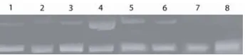

![Figure 8.DNA, lanes 1-5, final [DTT] = 5.7 mM, lanes 6-10, final [GSH] = 0.57 mM, assay run for 48h at designatedtype III linear DNA, bottom band is type I supercoiled DNA](https://thumb-us.123doks.com/thumbv2/123dok_us/1708274.1217023/113.612.101.532.270.355/figure-lanes-final-lanes-final-designatedtype-linear-supercoiled.webp)