A Portable Standalone Microscope by Attaching a Compact Module
to a Digital Camera
Youjun Li1, Xuan Zhu1, Jie Chen1, Fuhong Cai1, *, Jiong Zheng2, and Jianbo Guo3
Abstract—A portable microscope has been created through our work, so that we can observe and collect images in future scientific research whenever and wherever possible. The portable microscope is made up of a small LED chip, compact lens modules, a commonly used SLR camera and a USB driven power. The microscopic morphological features can be observed through our system. We also demonstrate that this standalone system can work well in moving state. Therefore, the portable microscope that has the potential for becoming a point-of-care setup in terms of health monitoring is appropriate for on-site micro-imaging.
1. INTRODUCTION
In scientific research, people utilize mature optic modules to perform microscopic imaging [1]. Recently, an infinite image distance achromatic microscope objective is used to amplify objects in micron scale [2]. In this way, a tube lens and a CMOS can be easily assembled to form a microscopic imaging system [3]. Nevertheless, the traditional microscope, which is large and heavy, is relatively strict in environmental requirements, and it is usually placed on a cumbersome optical table. In addition, professional training is required for the operation of traditional microscope. However, in severe areas, the condition is not allowed to utilize a traditional microscope. However, the micro-imaging is highly required in these areas. For example, in remote mountainous area, doctors want to perform red blood cell count to give an accurate diagnosis [3]. In our work, a portable standalone microscope has been invented for solving the dilemma when conventional optical microscopes cannot be used due to environmental restrictions. Nowadays, portable system is widely used in the microscopic field for point-of-care region, i.e., fluorescent detection [4, 5], blood oxygen saturation monitoring [6], spectral detection [7, 8], etc. Our portable microscope is mainly made up of a small LED chip, an infinite image distance achromatic objective, and a commonly used SLR camera. It is worth noting that this system can be driven by a traditional USB power. This portable microscope has many potential advantages due to its small size and ease of use. Compared to traditional microscopes, the portable microscope breaks the limitation of operation environment and is more convenient for real-time monitoring.
In this paper, we firstly describe the details of our portable microscope system. Next, we demonstrate the imaging results of this portable microscope for test target, as well as its performance while the microscope was in a bumpy car. Finally, we show the imaging results for bio-samples, i.e., the frog’s blood smear, onion skin, housefly’s legs, and cotton stalk cell.
Received 20 October 2017, Accepted 13 November 2017, Scheduled 24 November 2017
* Corresponding author: Fuhong Cai ([email protected]).
2. PORTABLE MICROSCOPE
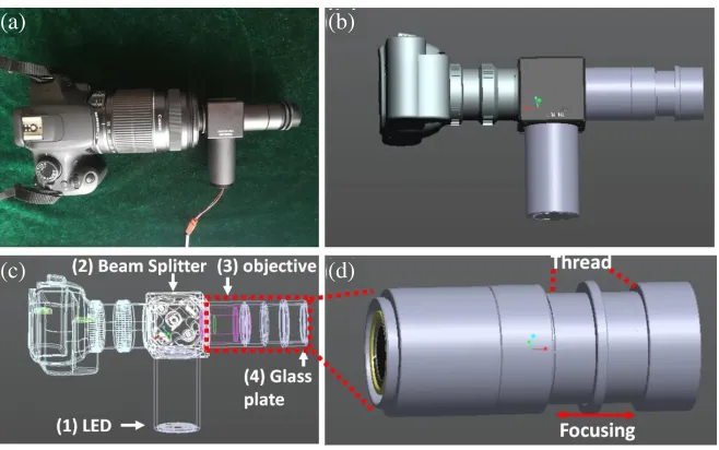
Figure 1 shows photos of the portable microscopes, whose size is moderate, and the microscope is easy to carry. As shown in Fig. 1, the portable microscope is mainly made up of a small white LED chip, infinite image distance achromatic microscope objective, a commonly used SLR camera and a beam splitter. The LED chip can provide a stable white light to illuminate the viewing area. Meanwhile, the LED is driven by a portable USB power. The infinite image distance achromatic microscope objective, which determines the resolution of the microscope, is an excellent module for sample illumination and micro-imaging. In our work, a 10X objective is selected to magnify the samples. The numerical aperture (NA) of objective is 0.17, and the resolution is 0.61λ/NA = 1.9µm, whereλ= 550 nm. The SLR camera consists of two parts: one is the imaging lens, and the other is the CMOS module. The built-in imaging lens of SLR camera can serve as the tube lens in microscope. The focal length of imaging lens installed in the smartphone is usually < 5 mm. However, in order to achieve ideal optical magnification, the focal length should > 50 mm. Hence, the lens of smartphone is not suitable for being tube lens in a microscope. Therefore, we select SLR as the sensor for microscope. The CMOS in an SLR camera is equivalent to the eyepiece of a conventional microscope and converts the imaging information to a digital image file [9]. The exposure time and ISO are set as 1/200 sec and 1600, respectively, and the aperture for the camera lens is set as F5.6 during the imaging. The beam splitter (50%R-50%T for visible light) is utilized to fold the illumination and imaging optical path. In this way, the system can become more compact. We can also observe images on the screen of SLR camera. In summary, the portable microscope utilizes an LED source and SLR camera as the illumination and detection modules. These two modules can be driven by battery power, hence our system can work without complex power systems. In addition, the focusing process can be achieved by rotating a threaded tube provided by Thorlabs company. As shown in Figs. 1(c) and (d), the glass plate (the sample was placed on this plate) is installed in one end of the tube, and the objective is installed in another tube. As we rotate the thread tube, the distance between objective and the sample can be changed. In this way, we can find the best focus distance for imaging.
(a) (b)
(c) (d)
3. STATIC AND DYNAMIC OBSERVATION OF PORTABLE MICROSCOPES
In order to demonstrate the advantage of real-time imaging of our portable microscope, we performed imaging experiments under static condition and dynamic conditions, respectively. Firstly in a static condition, we captured a micro-image, as shown in Fig. 2(a). The small grids (50µm interval) can be clearly observed in the micrometer. The size of the microscope was about 0.16 mm2. Secondly, the portable microscope was held by the fourth author to capture video as he was walking. In this case, the microscope suffered from the vibration. However, since our system was highly integrated, and each element suffered from the same vibration. In this way, the influence of vibration can be removed. Figs. 2(b), (c) and (d) show snapshots of the video when the time flag is 0 sec, 5 sec, and 10 sec. These images are as clear as Fig. 2(a), which was captured under a static condition. According to the result of these two experiments, this device is not only suitable for observation under a stationary condition, but also excellent under moving condition.
(a) (b) 0 s
(c) 5 s (d) 10 s
Figure 2. Microscopic photographs under stationary and moving states. (a) shows the observation of micrometer under stationary state. (b), (c), (d) are images of the micrometer under moving states, see text for details.
4. PHYSICAL OBSERVATION OF PORTABLE MICROSCOPES
Our device can capture an image of an insect. As the size of a housefly is very small, people cannot clearly see its structure by the naked eyes. The portable microscope can directly imaging the houseflies and is rarely restricted by the environmental condition. Placing the leg of the housefly on the glass plate, the microscopic image can be obtained immediately, as shown in Fig. 3(a). Similarly, we can perform experiments for other samples. Fig. 3(b) shows the cell morphology of an onion epidermis and cotton stalk. Because the cell shape is minimal, it can only be observed under the microscope. We can utilize this device to distinguish different types of cotton stalk fiber.
(a) (b)
(c) (d)
Figure 3. Microscopic images for (a) the leg of the housefly, (b) an onion epidermis, (c) cotton stalk fiber and (d) blood smear of a frog, (e) a sub-region of the blood smear, we can count the number of cells, which is 199 in this case.
5. CONCLUSION
In this work, we demonstrate a portable microscope, whose advantages are moderate weight, portability, and real time imaging. Some organisms and cells can be observed with the portable microscope. Furthermore, this device can capture a clear image in moving state. Recently, the development of rapid, simple, and accurate diagnostic tools with applicability at point of care and remote location is essential. We believe our device has the potential to be such a diagnostic tool for medical staff. Besides, we also demonstrate the optical setup of our portable microscope in detail. All elements of the portable microscope are available from optical component suppliers. Also, our device, which can be driven by a USB power, is standalone and easy to use. Based on the above discussion, our device is suitable for on-site rapid detection. Tseng et al. developed a lens free microscopy on a cellphone using hologram method to reconstruct the image [11]. Precise calculation is required in their method. Our portable microscope is relatively heavier, but we can acquire the microscopic image without computation.
ACKNOWLEDGMENT
REFERENCES
1. Dai, Y., M. W. Randy, and L. Li, “Confocal fluorescence microscopic imaging for investigating the analyte distribution in MALDI matrices,” Analytical Chemistry, Vol. 68, 2494–2500, 1996.
2. Keller, H. E., “Objective lenses for confocal microscopy,” Handbook of Biological Confocal Microscopy, 145–161, Springer, US, 2006.
3. Fahrbach, F. O., et al., “Rapid 3D light-sheet microscopy with a tunable lens,” Optics Express, Vol. 21, 21010–21026, 2013.
4. Shen, L., A. H. Joshua, and I. Papautsky, “Point-of-care colorimetric detection with a smartphone,”
Lab on a Chip, Vol. 12, 4240–4243, 2012.
5. Cai, F., M. Zhao, and D. Wang, “A compact perpendicular microscopy and imaging system for the detection of fluorescent solution flow,” Progress In Electromagnetics Research Letters, Vol. 67, 75–79, 2017.
6. Edwards, P., et al., “Smartphone based optical spectrometer for diffusive reflectance spectroscopic measurement of hemoglobin,”Scientific Reports, Vol. 7, 12224, 2017.
7. Das, A. J., et al., “Ultra-portable, wireless smartphone spectrometer for rapid, non-destructive testing of fruit ripeness,”Scientific Reports, Vol. 6, 32504, 2016.
8. Xie, J. and F. Cai, “A portable spectra detection system for ripeness detection and real-finger identification,”Progress In Electromagnetics Research Letters, Vol. 68, 73–77, 2017.
9. Shin, D., et al., “A fiber-optic fluorescence microscope using a consumer-grade digital camera for in vivo cellular imaging,” PLoS One, Vol. 5, e11218, 2010.
10. Briggs, C., “Quality counts: New parameters in blood cell counting,” International Journal of Laboratory Hematology, Vol. 31, 277–297, 2009.