DOI: http://dx.doi.org/10.26483/ijarcs.v9i5.6312
Volume 9, No. 5, September
International Journal of Advanced Research in Computer Science
Available Online at www.ijarcs.info
ROBUST AND AUTOMATED
IMAGE
Ph.D. Research Scholar, Dept.
Associate Professor, Dept.
Professor, Dept. CSE, Sreenidhi
Abstract: In the medical field among all the types in the beginning will recover the lifetime of images are very useful to find lung cancer enhancement and image segmentation. Watershed research paper aim is to find more precise
Keywords: Image processing, Watershed transformation,
I. INTRODUCTION
The main purpose of medical image analysis lung cancer using meaningful information disease diagnosis and therapy. Accuracy and quality factors of the image, image enhancement segmentation principles used in pre-processing feature extraction in the following steps: smoking cases of the male are 85% and the female [1]-[2]. 2. Gabor filter and watershed transform useful to remove the noise. 3. As shown in figure the theory of the various steps in the pre- processing 4. The patient life span can be increased at the of the cancer. Al Taraweeh et al [3] image enhancement methods strongly statistical operations and subjective observation variance and mean calculation. Sami et presented that lung disease scan images are used lung cancer. Ajil et al[5] described CT images sensitive in finding the tumor size and the lymph the lung.
Figure. 1:- Lung cancer detection system architecture
Volume 9, No. 5, September-October 2018
International Journal of Advanced Research in Computer Science
REVIEW ARTICLE
Available Online at www.ijarcs.info
ISSN No. 0976
AUTOMATED LUNG CANCER NODULE DETECTION
PROCESSING TECHNIQUES
M. Prem Chander
Dept. CSE, GITAM UNIVERSITY, Vishakhapatnam, India. Dr. M. Venkateshwara Rao
Dept. IT, GITAM UNIVERSITY, Vishakhapatnam, India. Dr. T. V. Rajini Kanth
Sreenidhi Institute of Science and Technology, Hyderabad, India.
types of cancers, lung cancer is more serious disease. Detection the patient. Using image processing techniques, Computed cancer nodule. The image pre-processing methods are feature Watershed transformation and based on Gabor filter to find lung
results using different segmentation and enhancement techniques.
transformation, Gabor filter, Lung cancer.
analysis is to detect information to support quality are two enhancement and processing to get 1. Cigarette female are 75% transform are more figure 1 explains processing stage. the early stage explained the depends on observation such as al [4] has used to find the images are more lymph nodes of
architecture diagram
I.I Image Enhancement
Chaudary et al.[7]-[8] explained with by the focus to improve the sensitivity, interpretability in an image pre-processing
The image enhancement methods are • Noise removal using a wiener filter • Median filtering
• Filtering with morphological operators • Histogram equalization
The quality of computed tomography improved by the transform coefficient enhancements benefits are as follows: • Frequency domain
• Computations of Complexity • Frequency coefficient of an image • Domain purpose improvement. Xiuhua et al.[6] explained early diagnosis can save a life, lung cancer is more disease in the world.
I.II Equalization of histogram
Histogram equalization is a enhancement, as explained a framework in histogram modification. The distributed obtained by cumulative distribution
image.
I.III Segmentation of image
Partition of a prototype into equivalent component part in which to each holding with as is attributes. These meaningful and useful information.
ISSN No. 0976-5697
DETECTION USING
India.
India.
Detection of lung cancer Computed tomography scan feature extraction, image lung cancer nodule. This
techniques.
with image enhancement sensitivity, quality, and processing stage.
given below. filter
operators
tomography images can be coefficient functions. The image
follows:
image manipulation
diagnosis and better treatment more critical and life taking
process of contrast framework technique is used distributed histogram is distribution function, in the input
image from low-level image processing into more other images such as objects and features, an accurate partitioning of an image in the image segmentation is a challenging problem [10]-[11].
Application areas of image segmentation are mentioned below.
• Pattern Recognition • Image Encryption • Image Processing
• Medical and Biomedical imaging • Computer Graphics
• Computer Vision I.IV Thresholding.
It is a process of segmenting the image into equal parts, by separating of light and dark pixels. If A(i, j) is a threshold version of B (i, j) at some global threshold T. A=1 if when B (i, j) ≥ T.
Huang et al. [13] explained about thresholding technique is based on the similarity in textural features.
I. V Feature Extraction
In image processing techniques feature extraction is a part of thresholding, resizing linearization, and
normalization. Feature extraction techniques used in image recognition and classification as character recognition.
II. PROPOSED METHODOLOGY
Rendon and gonzalez et al. [12] and [14] lung cancer detection is having four steps:
• Enhancement. • Feature extraction. • Segmentation and • Nodule detection.
The proposed methodology includes pre-processing of the images to extract the features of lung cancer in an automated process and subjected to the following steps. • The pre-processing stage that improves the nature of the
picture by evacuating the undesirable noise to feature the malignant information.
• The feature extraction acquires the discrete components of the image under processing.
• The classification stage identifies the objects of an image according to certain classes and helps in recognition of efficient data.
Figure. 2:-Different types of techniques for identifying lung cancer.
II .I Pre-processing
Smoothing is a process of image processing technique to remove noise and fluctuations in the image. To remove the image noise, reduce salt and pepper noise by using median filter. Ruchika et al.[9] explained about an image smoothing in the image pre-processing stage. The smoothing process is also used for detection of lung cancer nodule and in reduction of noise from the computed tomography images.
II.II Feature extraction
As shown in figure.2, a flowchart which explains various steps while detecting the malicious node, from various CT scan images.
A) Area of the nodule:
B) The array of edge that contains at least one value, among 0 to 255 values
created by transformation function. Perimeter of the nodule: P= (Pxy , I edge[P]=x, J edge [P]=y)
Where I and J are vectors, x & y are the pixels.
C) Eccentricity: Eccentricity is used to measure the region of tumor (I). If it is
circular I value is 1, otherwise < 1. (I) = major axis / minor axis
III. RESULTS AND DISCUSSION
Detection of cancer using image processing techniques, lung cancer stages are involved in the practical situation of cancer which transforms from the human body to
various parts [15]-[16]. The smallest lung cancer nodule sizes are all between 5 mm and 25 mm [17]-[18].
Wang J et al.[22] computed tomography(CT) and Computer-aided diagnosis(CAD) schemes for thoracic are widely used to detect lung abnormalities. Threshold value is verified after the edge of the lung boundary is matched [19]-[21] and [25].
For more details of lung cancer detection by using based on Gabor filter and watershed segmentation, see [23], [24], [26], [27] and [28]. And also knows that the presence or absence of cancer in lymph nodes.
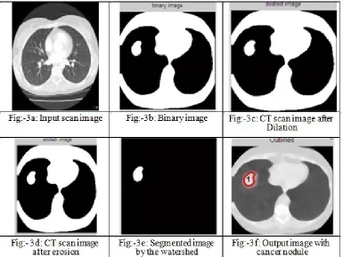
[image:3.612.111.496.278.567.2]As shown in Fig. 3, the lung cancer nodule image is shown in the above diagrams. For more details of image enhancement and feature extraction, see [29], [30], [31], [32], [33] and [34].
Figure 3
IV. CONCLUSION
The lung cancer detection system has been developed successfully. Lung cancer is the most critical disease which grows cancer cells in the abnormal way. The various image processing techniques are mostly useful in the medical areas. Computed tomography scan images are very useful for finding the lung tumor, based on the Gabor filter and various types of image segmentation techniques are used to detect the lung cancer. So this proposed method is highly suitable for identification of perimeter, eccentricity and area of the lung cancer tumor, according to this information diagnosis will be given by the physicians.
ACKNOWLEDGEMENTS
The research was supported by Dr. M. Venkateshwara Rao, and Dr. T. V. Rajini Kanth, For their valuable guidance for this paper.
REFERENCES
Advanced Engineering (ISSN 2250-2459, ISO 9001:2008 Certified Journal, Volume 7, Issue 7, July 2017).
[2] M. Premchander, Dr.T. V. Rajinikanth, and Dr. M . Venkateshwara Rao ,“Detection of Lung Cancer Using Digital Image Processing Techniques: A Comparative Study” International Journal of Medical Imaging, 2017; 5(5): 58-62,doi: 10.11648/j.ijmi.20170505.12, ISSN: 2330-8303 (Print); ISSN: 2330-832X (Online).
[3] Al Tarawneh MS. Lung cancer detection using image processing techniques. Leonardo Electronic Journal of Practices and Technologies. 2012.
[4] Gindi,A. M., Al Attiatalla, T. A., & Sami, M.M. “A Comparative Study for Comparing Two Feature Extraction Methods and Two Classifiers in Classification of Early stage Lung Cancer Diagnosis of chest x-ray images.” Journal of American Science, 2014.
[5] Ajil MV, Sreeram S. Lung cancer detection from CT images using various image processing techniques. International Journal of Advance Research in Computer Science and Management Studies.2015 May; 3(5), 249– 54.
[6] Xiuhua,G., Tao, S., &Zhigang, L, “Prediction Models for Malignant Pulmonary Nodules Based-on Texture Features of CT Image.” In Theory and Applications of CT Imaging and Analysis. 2011.
[7] Chaudhary A, Singh SS. Lung cancer detection on CT images by using image processing. International Conference on Computing Science, Phagwara. 2012. [8] Goswami A. For image enhancement and segmentation
by using evaluation of gabor filter parameters. International Journal of Advanced Technology and Engineering Research. 2012.
[9] Ruchika Kalra A. Detection of lung cancer in CT images using mean shift algorithm. International Journal of Advanced Research in Computer Science and Software Engineering. 2015.
[10]Roy, T., Sirohi, N., &Patle, A. (2015) “Classification of lung image and nodule detection using fuzzy inference system.” International Conference On Computing, Communication & Automation., 2015.
[11]Ignatious, S., & Joseph, R. (2015) “Computer aided lung cancer detection system.” 2015 Global Conference On Communication Technologies (GCCT).
[12]Rendon-Gonzalez, E., &Ponomaryov, V. (2016) “Automatic Lung nodule segmentation and classification in CT images based on SVM.” 2016 9Th International Kharkiv Symposium On Physics And Engineering Of Microwaves, Millimeter And Submillimeter Waves (MSMW).
[13]Ng HP, Huang S, Ong SH, Foong KWC, Goh PS, Nowinski WL. Medical image segmentation using watershedsegmentation with texture merging. IEEE Engineering in medicine and biology Society Conference, Canada. 2008.
[14]Patela SVK, Shrivastavab P. Implementation of medical image enhancement technique using gabor filter. International Journal of Current Engineering and Technology. 2012.
[15]Tanaka K, Sakuma T. Geographical difference of chromosome aberrations between Japanese and American small cell lung cancer cell lines. Indian Journal of Science and Technology. 2012.
[16]SPIE-AAPM-NCI Lung Nodule Classification
Challenge Dataset. The Cancer Imaging Archive; 2015.
[17]Hollings N., Shaw P. Diagnostic imaging of lung cancer. European Respiratory Journal. 2002.
[18]Li W., Nie S. D., Cheng J.Berlin, A fast automatic method of lung segmentation in CT images using mathematical morphology; pp. 2419–2422. (IFMBE Proceedings) Springer; 2007.
[19]Hu S., Hoffman E. A., Reinhardt J. M. Automatic lung segmentation for accurate quantitation of volumetric X-ray CT images. IEEE Transactions on Medical imaging, 2001.
[20]Shah S. Automatic cell images segmentation using a shape-classification model. IEICE Transactions on Information and Systems. 2007.
[21]Prasad M. N., Brown M. S., Ahmad S., et al. Automatic segmentation of lung parenchyma in the presence of diseases based on curvature of ribs. Academic Radiology. 2008.
[22]Wang J., Li F., Li Q. Automated segmentation of lungs with severe interstitial lung disease in CT. Medical Physics. 2009.
[23]Armato S., MacMahon H. Automated lung segmentation and computer-aided diagnosis for thoracic CT scans. Internationa l Congress Series. 2003.
[24]Sudha V, Jayashree P. Lung Nodule Detection in CT Images Using Thresholding and Morphological Operations. International Journal of Emerging Science and Engineering. 2012.
[25]Guo Y., Feng Y., Sun J., et al. Automatic lung tumor segmentation on PET/CT images using fuzzy markov random field model. Computational and Mathematical Methods in Medicine. 2014.
[26]Lam M., Disney T., Pham M., Raicu D., Furst J., Susomboon R. Content-based image retrieval for pulmonary computed tomography nodule images. Medical Imaging 2007: PACS and Imaging Informatics; March 2007.
[27]Dhara A. K., Chama C. K., Mukhopadhyay S., Khandelwal N. Content-based image retrieval system for differential diagnosis of lung cancer. Indian Journal of Medical Informatics. 2012.
[28]Nirmala J. B., Gowri S. A content based CT lung image retrieval by DCT matrix and feature vector technique. International Journal of Computer Science Issues. 2012.
[29]Huang Q, Gao W, Cai W. Thresholding technique with adaptive window selection for uneven lighting image. Pattern Recognition Letters. 2005 May. [30]Liu C.-T., Tai P.-L., Chen A. Y.-J., Peng C.-H., Lee
T., Wang J.-S. A content-based CT lung image retrieval system for assisting differential diagnosis images collection. Proceedings of the IEEE International Conference on Multimedia and Expo (ICME 2001), 2001.
[31]Nguyen worring HT, Van de Boomgaard R. Watersnakes: Energy-driven watershed segmentation. Pattern Analysis and Machine Intelligence. 2003. [32]Patil S. A., Kuchanur M. B. Lung cancer classification
[33]A. patil and M. B Kuchanur, ”Lung cancer classification using image processing“, International Journal of Engineering and Innovative Technology, vol. 2, no. 3, 2012.
[34]J. Kuruvilla and K. Gunavathi, “Lung cancer classification using neural networks for CT images“, Computer Methods and Programs in Biomedicine, vol. 113, no. 1, pp. 202–209, 2014.
AUTHOR’S BIOGRAPHY
M. Prem Chander has been pursuing Ph. D degree in Computer Science and Engineering from GITAM UNIVERSITY, Visakhapatnam, A.P India, as well as working as Asst. Professor of CSE Department in MNR C o l l e g e o f Engineering and Technology, Hyderabad. As well as obtained M. Tech. in Computer Science and Engineering from Jawaharlal Nehru Technological University(JNTU), Anantapurum, AP, INDIA in 2011. Prior to his professional career, he obtained Bachelor of Technology (B. Tech) in Computer Science and Information Technology from Sri Datta Institute of Engineering and Science, Hyderabad, Affiliated to JNTUH Hyderabad, Telangana, India in 2005. His current research interests include Image Processing and Data Warehousing & Mining for Medical Applications. His total teaching experience is 13 years. He guided around 30projects at B. Tech. and 10 projects at M. Tech. level. He has conducted two national Conferences namely CONVIVALITY 2K11, and CONVIVALITY 2K12 at MEC, Hyderabad as Convener .He is a Member in IEEE.
M. Venkateshwara rao has obtained his Ph. D degree in C.S.E branch. He is working as Associate Professor in the Department of Information Technology, in Gitam Institute of Technology, GITAM UNIVERSITY, Visakhapatnam, AP, and India. He obtained his M. Sc(Electronics), M. Phil (Opto Electronics), and M. Tech(CST). His teaching and Industrial experience is
25 years. His writings have appeared in numerous Professional conferences and Journals (International journals-5 National conferences-2.