The
Brain,
Explained:
A
Comprehensive
Theory
of
Brain
Function
Ari
Rappoport
The
Hebrew
University
of
Jerusalem,
Israel
[email protected]
www.cs.huji.ac.il/~arir
December
28,
2017
(first
version)
May
8,
2018
(minor
changes)
Abstract
Understandingbrainfunctionisoneofthemostimportantproblemsinhumanhistory. Atpresent,thereisnoconcretetheoryforhowthebrainworks.Here,atheoryis pre-sentedthatprovidesadetailedmechanisticbiologicalaccountofthebrain’scapacities, includingmotorcontrol,functionalstates,language,andthinking.
Brain function is managedby a well-definedre sponse(R )pr ocessth atis generally similar totheprocessunderlyingthe immunesystem. The Rprocessis strongly re-flectedinthebrain’sa natomy,p hysiology,andexternalin teractions.DifferentR pro-cess stagesaresupportedbydistinctexcitatorynetworkslocatedindifferentcortical layers,hippocampalfields,andbagalgangliapaths,bydistinctcoordinationnetworks comprisedofGABAergicinterneurons,andbydistinctmolecularagents.Therolesof norepinephrine, serotonin,dopamineandacetylcholineistopromotethealert, plan-ning, goal-setting and execution R process modes, respectively. Opioids and oxy-tocinpromoteterminationbysuccess,failure,fighto rr un,w hileg lucocorticoidsand cannabinoidssuppressacuteresponsestoprotectcells.
TheRprocesshastwoinstancesoccurringatdifferenttimescales. The millisecond-scaleQuaxprocessimplementstheexecutionofhierarchicalsequencesofmovements andthoughts, inwhich theselectionofthe nextactionisdeterminedvia interaction betweentop-downpredictionsandsensoryinputs. TheslowerNeedprocesscontrols thesatisfactionofinternalandexternalneeds.
The theorydiffers from theexisting standard accounts in manyof the majortopics (e.g., thebasalganglia, dopamine,language),andshowshowcognitionresultsfrom biologicalprocesses.
Keywords. Neuroscience, thalamus, basal ganglia, cortical layers, hippocampus, cerebellum,habenula,claustrum,amygdala,inhibition,predictions,automaticity, dopa-mine, serotonin, acetylcholine, opioids, oxytocin, CRH, glucocorticoids, cannabi-noids,orexin,melanin-concentratinghormone,mirrorneuron,placecells,gridcells, language,imagery,workingmemory,attention,consciousness,emotions.
Extended Abstract
• We present a comprehensive mechanistic biological theory of how the brain works.
• The brain’s role is to map sensory inputs (internal and external) to movement re-sponses. Responses can be innate or adaptive. Adaptive responses can be acute
(having a novel aspect) orlearned (automated).
• Brain operation is managed by a well-defined response (R) process having several stages called R modes. The R process determines the brain’s organization, in that different anatomical parts (e.g., auxiliary nuclei, cortical layers) and different molec-ular agents (e.g., dopamine, serotonin, GABA) are used by different R modes.
• There are two instances of the R process simultaneously executing at different time scales. The seconds to hoursneed (N) processaddresses needs and their satisfaction, while the millisecond scalequax (Q) processsupports execution.
• The R process proceeds as follows. The sensory input mode conveys bottom-up (BU) flow. If there is an innate response, it is executed. Otherwise, if there is a learned (automated) response, it is executed. Otherwise an acute response is executed. The acute response starts with thealertmode, which recruits resources and conveys widespread BU flow into adaptive areas. Alerts trigger thedecision making (DM)mode, which conveys top-down (TD) flow that activates response candidates. Candidates compete, and the winners join response execution. The R process has strong similarities with the immune system process, where inflammation is the acute response.
• The brain has two execution modes,predictionandresponse. The prediction mode enables learned response sequences, representing the parts of the winning response that do not yet hold in external reality, including specific prepared actions and sensory predictions. The response mode supports the present reality, including perceived objects and executing actions. The N process has two DM modes,competitionand
planning, for urgent and non-urgent decisions respectively. N process termination can be via protection, interaction, or specific innate actions.
• The R process determines the brain’s organization. Excitatory neurons belong to several content networks, each supporting a different R mode. In cortex, the dif-ferent networks occupy difdif-ferent layers. Sensory network: L4/6 (L3 in agranular areas). Alert network: L3b. DM network: L2/3. Prediction network: L5a (also L3 in agranular areas). Response network: L5b. In the hippocampus, the networks occupy different fields: dentate gyrus, CA3: alert/DM networks. CA1: prediction network. Subiculum: response network.
• Thethalamusprovides energy to cortex. Its core neurons, in both specific and non-specific nuclei, are part of the sensory network. Its matrix neurons convey TD flow that is essential for the formation and sustenance of adaptive responses.
• The R process determines the brain’s molecular physiology. Molecules called R agents (Rgens) control the R process, each Rgen promoting specific R modes and networks and suppressing competing ones. The major Rgens and their modes are as follows. Orexin, vasopressin, sex hormones, food agents: internal needs. Sub-stance P, histamine: external needs, fight. Norepinephrine: alert. Dopamine (DA): competition, prediction (goal setting). Acetylcholine (ACh): competition, response execution. Serotonin (SER): planning, mistake. CRH: extended alert. MCH: inter-action. Glucocorticoids, cannabinoids: energy, protection. DHEA: energy, continue. Opioids, oxytocin: termination by innates (oxytocin: satisfaction; beta-endorphin: consume; dynorphin: failure; nociceptin: fight; enkephalin: run). The hypothalamus controls the initiation and termination of the N process.
• Thehabenulasupports amistakeN mode by rapidly switching from the execution R mode (DA, ACh) to the DM planning mode (SER).
• Theamygdalais the innate-adaptive interface, allowing the triggering of innate re-sponses by non-innate inputs (conditioning) and the utilization of innate capacities by adaptive responses during planning and execution.
• The R process explains the role of GABAergic and glycinergic interneurons (IINs). IINs support coordination, which includes competition, sustained execution, and quax initiation. (Their role is not ‘inhibition’.) IINs can be assigned to several co-ordination networks, the main ones being the competition coordination network (CCN), the execution coordination network (ECN), and the response suppres-sion network (RSN). The networks utilize a join-or-stop (JOS) mechanism to select winners and synchronize activity.
The CCN includes SOM (Martinotti) IINs suppressing the apical dendrites of DM, prediction and response neurons, VIP IINs that suppress the SOMs to select the quax winners via disinhibition of these networks, and neurogliaform IINs that suppress quax losers. The ECN includes PV and CCK soma-targetting (basket) IINs that syn-chronize execution to sustain it. The RSN includes chandelier PV IINs that rapidly suppress responses.
• Cortex is arranged innodes, tightly connected microcolumns supporting vertical flow between the content networks. Nodes are the fundamental units of learned (adaptive) cortical representations.
• Execution involves the activation of a set of neurons called aquax. The Q process manages execution via quaxtransitionsin which neurons join and leave the execut-ing quax, thereby supportexecut-inghierarchical sequences. The executing quax contains action nodes active in response mode that drive movement, and object nodes active in prediction mode (i.e., their prediction network is active but their respone network is not) that represent the action’s sensorygoals. The executing actions excite (mobi-lize) all action and object nodes that have been useful in the past. When the active goals are attained, the resulting sensory inputs cause the nodes representing goals to switch to response mode, and select the next action via competition among the mobilized actions. In this way, hierarchical sequences emerge from sensory-action R process interactions without being centrally managed.
explainsmirror neurons. Transitions explain EEGoscillationsand the attentional blink. Goals explainplaceandgrid cellsandtheta phase precession.
• Learned (automated) responses are promoted each time responses are executed, starting from the initial acute response that trains a learned response while it exe-cutes. Learned responses are supported by the response network, by predictions in the prediction network and anticipations in the DM network, by the indirect BG path, and by the high-affinity Rgen receptors and autoreceptors (mainly D2R, alpha2-NER, and SER1/4/7).
• Dopaminepromotes the competition and prediction (goal-directed execution) R modes (together calleddecision), including immediate charging approach innate actions, via D1R, and learned transitions via D2R. (The role of DA is not ‘reward’.) Serotonin
promotes the planning R mode, by suppressing innate responses (‘emotions’) and executing responses via SER1R, exciting the DM network and the CCN via SER2R and SER3R, exciting memory/planning areas via SER4/6, and exciting motor circuits via SER7. (The role of SER is not ‘happiness’.)
• Language and other forms of communication are managed by the standard Q pro-cess. The only thing unique about them is that they use non-neuronal modalities (audio, movement, touch) in order to bridge the neuronal gap between organisms. Syntax is the interface between non-linguistic goal nodes and gap bridging actions, and its hierarchy and sequencing capacities are implemented as in all other actions.
• Imagery andthinkingoccur during the planning R mode and are implemented by the standard Q process. Motor imagery involves the activation of gamma motoneu-rons, which form the innate part of the prediction network.
• The state of R modes is accessible to adaptive actions via interoceptive sensory inputs reaching cortical nodes (mainly the insula and mPFC). Awareness (consciousness)
andemotions (feelings)result from Q processes that report events and R mode states.
Contents
List of Figures 6
1 Introduction 8
2 Overview: General Principles & Processes 9
2.1 Function . . . 9
2.2 General architecture. . . 9
2.3 The R process . . . 10
3 The Innate-Adaptive Axis 15 3.1 Ibrain, abrain, innates, valence . . . 15
3.2 The amygdala: the innate-adaptive interface . . . 16
4 The Process Axis: R Mode Networks 18 4.1 The innate sensory network . . . 18
4.2 The innate response network . . . 18
4.3 The adaptive sensory network . . . 20
4.4 The alert network . . . 22
4.5 The decision making (DM) network . . . 22
4.6 The adaptive response network . . . 22
4.7 The prediction (goals) network . . . 22
4.8 The thalamic TD network. . . 23
4.9 The basal ganglia (BG): gating execution . . . 23
4.10 The hippocampus . . . 24
4.11 Cognitive interpretation: predecision, reality, goals . . . 26
5 The Content Axis: Representations 27 5.1 Representations . . . 27
5.2 Nodes . . . 28
5.3 Object, action, valence areas . . . 29
5.4 Events, episodic memory . . . 30
6 The Q Process: Motor Control 31 6.1 Action goals, sequences & hierarchy . . . 32
6.2 Coordination: competition, synchrony, initiation . . . 35
6.3 Coordination networks . . . 37
6.4 ERPs, repetition suppression, predictions . . . 39
6.5 The cerebellum . . . 41
6.6 Attention: orienting, the claustrum . . . 42
6.7 Conditional wait. . . 43
6.8 Working memory . . . 44
6.9 Summary . . . 45
7 The N Process: Functional States 45 7.1 The hypothalamus: N mode hub . . . 46
7.2 Internal need: orexin (OX), arginine vasopressin (AVP), sex hormones . . . 46
7.3 Alert: norepinephrine (NE) . . . 48
7.5 Decision making, planning: serotonin (SER) . . . 51
7.6 Competition, prediction, goal-setting: dopamine (DA). . . 53
7.7 Response execution, attention: acetylcholine (ACh) . . . 56
7.8 Pain, aggression, mating: substance P (SP), histamine . . . 58
7.9 Mistake: the habenula . . . 60
7.10 Consume: beta-endorphin (BEND) . . . 61
7.11 Satisfaction: oxytocin (OT) . . . 64
7.12 Failure, withdrawal: dynorphin (DYN) . . . 65
7.13 Aggression: nociceptin (NOC, N/OFQ) . . . 66
7.14 Running: enkephalin (ENK) . . . 66
7.15 Interaction: melanin-concentrating hormone (MCH). . . 67
7.16 Energy, protection: glucocorticoids (GCs), endocannabinoids (ECBs), DHEA . . . 68
8 Language 70 8.1 The nature of language . . . 70
8.2 Representations & areas . . . 70
8.3 Processing . . . 72
8.4 Supporting evidence . . . 73
9 Internal Cognition 75 9.1 Awareness, consciousness . . . 75
9.2 Emotions . . . 77
9.3 Familiarity & recollection. . . 78
9.4 Imagery, thinking . . . 79
9.5 True & false recollection . . . 83
9.6 Summary . . . 84
10 Additional Evidence 84 10.1 Action observation, mirror neurons . . . 84
10.2 Place & grid cells, theta phase precession . . . 85
10.3 Conditioning, generalization & extinction . . . 86
10.4 EEG oscillations . . . 88
10.5 Attentional paradigms. . . 89
11 Discussion 90 12 Final Words 94 13 References 94 14 Supplementary Material 126 14.1 Abbreviations and new terms . . . 126
14.2 Method . . . 128
14.3 Generative linguistics, recursion, etc. . . 129
List of Figures
1 The R process (general version). . . 101
Introduction
How the brain works is one of the few truly major questions in human history. Follow-ing Newton, Einstein, Darwin and many others, we possess a good understandFollow-ing of many aspects of the physical world and of life. However, we do not understand how living or-ganisms act, think and feel. These capacities are known to be controlled by the brain, but currently there exists no brain theory or anything close to it, despite the enormous amount of research effort invested. Much progress has been made in our knowledge of cellular properties, but our understanding of higher brain levels (networks, systems, behaviors) is vague at best [Grillner, 2014], usually phrased in terms of abstract functions of large brain areas rather than through concrete mechanisms [Brown, 2014]. The brain is the most com-plex structure in nature; some researchers think that we may never understand how the human brain works [Adolphs, 2015], or that knowledge would only gradually accumulate, over many years [Koch and Marcus, 2014]. Large concerted data collection and measure-ment projects are widely viewed as essential for moving forward [Olds, 2016].
This situation is not surprising, given that a full theory of brain function faces the formidable task of explaining at least the following capacities: goal-directed movements and movement sequences; perception, including the identification of external objects and events; mental imagery (imagination, thinking, planning); attention; emotions; the nature of awareness (consciousness); motivated vs. automatic and habitual behavior; predictions and surprise; reward and aversion; innate responses (threat/fear, consummatory acts), their generalization and extinction; language; familiarity, novelty, and true and false recollection; episodic memory; learning; sleep.
In addition, a comprehensive theory must explain the function of the following compo-nents and phenomena: the different classes of excitatory and inhibitory neurons; cortical layers and columns; the major areas and structures, including the basal ganglia, thala-mus, cerebellum, amygdala, hypothalathala-mus, hippocampus, habenula, claustrum, and corti-cal areas such as the insula and Broca’s area; neuromodulators and neuropeptides such as dopamine, serotonin, acetylcholine, norepinephrine, opioids, cannabinoids, and oxytocin; synchrony and oscillations; ERP components; place cells and mirror neurons. The theory must also provide plausible accounts for disorders.
In this text I present a theory of brain1 function accounting for all of the items listed
above. It is a mechanistic biological theory, phrased in terms of concrete cellular processes. As such, the theory is refutable and provides a wealth of concrete predictions. The main idea is that brain function can be fully explained using a single response (R) process that has two brain instantiations, one driven by internal and external needs and the other managing response execution. The brain’s anatomy and molecular physiology are directly reflected by the R process, which is generally similar to the process guiding the immune system.
The methodology I used to develop the theory was to gather as many relevant facts as possible, and try to arrange them into a coherent picture. To find the facts, I examined a large number of papers and books2. I did not conduct experiments; when forming a
theory prediction, I searched for papers describing experiments that could refute or support it. The resulting theory turned out to be very different from existing accounts of specific phenomena in most important areas (e.g., the basal ganglia, GABA, dopamine, serotonin). I refer to the present (2017) version of the theory asR17, to distinguish it from inevitable future updates.
The present paper aims for a level of description that would be expected in textbooks.
Due to the scope of the subject, it is clearly impossible to go much deeper than that or to cite even a small part of the relevant literature here. The paper covers the brain aspects directly related to the main functional process, leaving topics having a different nature for other publications. These topics include brain learning and plasticity (including sleep, postnatal development, lateralization, and gender differences), and a mechanistic account of brain disorders. s.
The theory does not cover brain evolution, prenatal development, and species differ-ences, but it uses supporting data from these fields where appropriate.
2
Overview: General Principles & Processes
2.1
Function
The central nervous system (brain, for brevity) controls the organism’s interaction with the body-external environment. In particular, it drives and manages the external move-ments involved in satisfying survival-related needs, both internal needs (food, minerals, water, reproduction, waste disposal) and external ones (threats from other organisms and natural forces). Other aspects of such needs are managed by other body systems (e.g., the metabolism and reproduction systems). Another major need type, body-internal threats, is managed by the immune system. The brain and these systems cooperate in fulfilling their functions.
2.2
General architecture
The brain is a non-linearcomplex system, comprised of a large number of functional cells calledneurons(an order of1010in humans) that interact through many-to-many (101−105) connections. Neurons possess branching input and output processes called dendritesand
axons, respectively. Most connections (synapses) are point-to-point, with axons connect-ing to dendrites at precise locations (dendritic spines).
Brain function is based on electrically charged chemical agents (ions). The main positively charged agents are calcium, potassium, sodium and zinc ions, while the main negatively charged agents are chloride ions. Neurons are normally polarized (i.e., there is a voltage difference across their membrane). When a neuron is depolarized (hyperpolarized) it is said to be excited (suppressed, inhibited). Inter-neural communication is done via energyflow,the release of agents that act on membrane receptors to induce the opening of ion channels and other cell internal processes. When ion channels are open, the direction of ion movement is determined by concentration gradients across the cell’s plasma membrane. When a neuron is excited above a certain threshold, a strong response (action potential (AP)) of about one millisecond occurs that induces the axonal release of agents, which in turn affects the polarity of the axon’s targets. In general, depolarization by dozens of source neurons is needed to get a target neuron fire an AP.
The main excitatory agent is glutamate (glut), which mainly acts through two types of receptors, AMPA and NMDA. An important distinction is that between calcium-permeable (CP) and calcium-impermeable (CI) receptors, since CP receptors allow rapid excitation due to the substantially higher concentration of extracellular Ca2+. NMDARs and some AMPARs are CP. The main inhibitory agents are GABA and glycine.
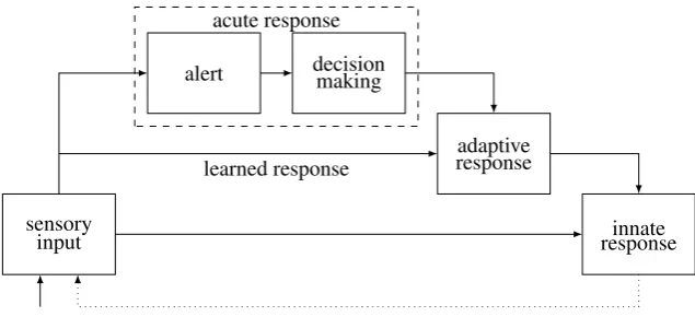
sensory input
adaptive response alert decisionmaking
acute response
innate response learned response
Figure 1: The general R process (without brain-unique aspects). Arrows indicate excitatory flow. The dotted line indicates that response execution affects incoming sensory inputs.
The brain is a learning system, continuously modifying its parts and connections to optimize its performance. We distinguish between innate brain capacities, genetically hard-coded by evolution and present in all healthy members of a species, and adaptive
capacities, different between organisms and acquired during a particular organism’s life-time. Adaptive capacities that have been optimized are called automated, and capacities that have a novel aspect are calledacuteor flexible.
2.3
The R process
The major contribution of R17 is in describing brain operation as following a general re-sponse (R) process, comprised of a sequence of stages calledR modes. The main R modes utilize different neuronal networks, and are promoted by different molecular agents (neu-romodulators). The R process provides a relatively simple conceptual framework through which virtually all brain phenomena can be understood.
There are five basic R modes (Figure 1): sensory input, innate response, alert, decision making (DM), and adaptive response. The process starts with sensory input. If there is an innate response to this input, it is executed. If there is no innate response, or if the input persists after executing it, flow reaches adaptive areas. If there is alearnedresponse, it is executed. Otherwise, an acute mode finds and executes a flexible response. The acute mode is comprised of two stages, an alert that directs resources to the event, and a decision making (DM) response that recruits response candidates (existing learned or innate responses that respond to some feature of the input) and lets them compete against each other. A focused adaptive response (decision) emerges from the recruited alternatives and is executed, using innate responses (usually low-level ones).
The R process has an obvious similarity to the processes underlying other biological systems. In the immune system, there is also a distinction between innate and adaptive immune responses, DM candidates are adaptive immune cells (T and B cells) that respond to some partial component of the pathogen (relatively short amino acid sequences displayed with major histocompatibility complex molecules by antigen presenting cells), the acute response is called inflammation, and the system learns the emerging focused response. In
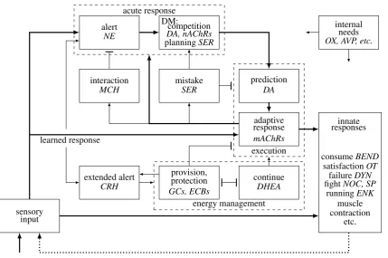
Brain operation is best described as having two instances of the R process unfolding at different time scales (intermediate and short term), addressing needs (N process) and response execution (Q process) respectively. The brain implementation of the R process has several brain-unique modes (Figure 2). Response execution is divided into two modes,
prediction and response, to support goal setting and response sequences. DM has two sub-modes, competition and planning, for urgent and non-urgent decisions. The N process has mistake, interaction, extended alert and energy management modes.
The different R modes use largely non-overlapping neuronal content networks. Innate areas have sensoryandresponsenetworks. Adaptive areas havesensory, alert, decision making (DM), predictionandresponse networks, which in cortex are located in different layers (L4/6, L3b, L2/3, L5a, and L5b respectively). The response network is the one directly inducing muscle contraction and movement.
Each R mode is promoted by one or more molecularR agents (Rgens). Rgens modify neuronal excitability via selective neuronal receptors, usually several receptor types per agent. Due to intracellular signaling, extracellular propagation, and distribution through the cerebrospinal fluid (in some cases), Rgen effect on brain execution can be spatially and temporally extended. In general, the Rgens promoting acute responses also promote automated responses, using lower amounts and different (high affinity) receptors. We say that Rgensmold the neuronal terrainto channel flow, thereby promoting their R modes.
The N process. The intermediate time scaleneed (N) process takes between seconds to hours, its stages (N modes) reflecting the state of internal and external needs (Table 1).
Internal needs are conveyed by specific agents sensed by neurons, thereby increasing the excitability of the neuronal paths involved in the satisfaction of specific needs. The alert N mode commonly follows surprises that trigger innate orienting of attention and innate sympathetic responses that recruit energy resources. If an alerting event is classified as unthreatening, theinteractionmode terminates the alert and by default stimulates interac-tion with the external object. If the situainterac-tion is potentially threatening but does not trigger specific innate or acute responses, theextended alertmode sustains a prolonged alert and triggers energy management.
There are two types of decision making. Planning(non-urgent DM) is promoted by SER.Competition(urgent DM), as well as execution, are promoted by DA and ACh. Both Rgens widely excite response candidates, push towards making a response, and sustain its execution. DA has a greater role in need-driven decisions (i.e., the N process) and prediction (goal setting), while ACh promotes attended execution (i.e., the Q process, see below). A mistakesignal can be communicated at any time to stop execution and switch back to non-urgent decision making mode (see habenula below).
Termination of the acute mode can be due to interaction, protection, or execution of innates3. Protectionis induced by the energy provision mode after excessive recruitment of energy resources. Such negative feedback is needed since a prolonged acute response may damage cells via oxidative stress and other processes. Protection is done by suppressing adaptive responses and by promoting innate responses, which are energetically efficient, without promoting any specific one. Protection competes with another energy management mode,continue, which optimizes energy utilization of acute responses.
Execution of specific innates is the common way in which acute execution is terminated, where each major innate is promoted by a different opioid agent or oxytocin. There are two success modes involving the satisfaction of internal needs, both comprising a brief boosting of responses followed by prolonged relaxation. Theconsumesuccess mode uses
sensory input
extended alert provision,protection continue
energy management
CRH
GCs, ECBs DHEA
adaptive response prediction
execution
mAChRs DA
mistake interaction
alert DM:competition
planningSER DA, nAChRs
acute response
SER MCH
NE
internal needs
innate responses
OX, AVP, etc.
consumeBEND
satisfactionOT
failureDYN
fightNOC, SP
runningENK
muscle contraction
etc. learned response
Figure 2: The brain’s R process. Arrows indicate excitatory flow, T junctions indicate sup-pression. Mode Rgens are shown in italics. The brain-unique aspects beyond the general R process are: (i) internal needs mold the neuronal terrain to affect response formation (the specific excitation of N process DA by OX is not shown); (ii) response execution includes two modes, prediction and response, with the former unidirectionally exciting the latter; (iii) decision making (DM) includes two sub-modes, competition (urgent DM) and plan-ning (non-urgent DM), which excite prediction; (iv) a mistake mode suppresses execution and promotes renewed DM; (v) an extended alert mode reciprocally excites alert and pro-motes energy management; (vi) energy management includes two competing sub-modes, provision (protection) and continue. Provision suppresses excessive execution (negative feedback), sustains the part of extended alert that sustains alerts, and uses negative FB on the part of extended alert that triggers energy management (not shown in the figure); (vii) an interaction mode suppresses alert in case the input is not threatening; (viii) in-nate responses include muscle contraction, success (consume, satisfaction), failure (freeze, withdraw), fight (aggression), flight (run), etc. The internal circuits of innate responses are not shown. The dotted line indicates that responses modify incoming sensory inputs. The connections serving both the N process and the Q process are shown in thick lines. Note the thick connection from the adaptive to the acute response, which enables the inner loop forming non-urgent DM (planning).
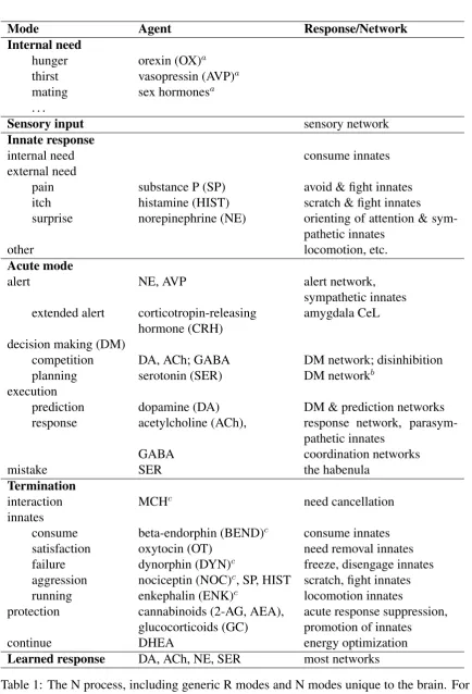
All modes except sensory inputs are promoted by specific Rgens.
Mode Agent Response/Network Internal need
hunger orexin (OX)a thirst vasopressin (AVP)a mating sex hormonesa
. . .
Sensory input sensory network
Innate response
internal need consume innates external need
pain substance P (SP) avoid & fight innates itch histamine (HIST) scratch & fight innates
surprise norepinephrine (NE) orienting of attention & sym-pathetic innates
other locomotion, etc.
Acute mode
alert NE, AVP alert network, sympathetic innates extended alert corticotropin-releasing amygdala CeL
hormone (CRH) decision making (DM)
competition DA, ACh; GABA DM network; disinhibition planning serotonin (SER) DM networkb
execution
prediction dopamine (DA) DM & prediction networks response acetylcholine (ACh), response network,
parasym-pathetic innates GABA coordination networks
mistake SER the habenula
Termination
interaction MCHc need cancellation
innates
consume beta-endorphin (BEND)c consume innates
satisfaction oxytocin (OT) need removal innates failure dynorphin (DYN)c freeze, disengage innates aggression nociceptin (NOC)c, SP, HIST scratch, fight innates running enkephalin (ENK)c locomotion innates
protection cannabinoids (2-AG, AEA), acute response suppression, glucocorticoids (GC) promotion of innates continue DHEA energy optimization
Learned response DA, ACh, NE, SER most networks
Table 1: The N process, including generic R modes and N modes unique to the brain. For each N mode, its Rgen(s), responses, and networks are indicated.
a: Important food, thirst and sex Rgens are not listed, see the need N mode section.
b: Planning also involves other networks, see the imagery section.
innates that utilize skeletal muscles, while thesatisfactionmode mostly uses innates that utilize smooth muscles and entail the secretion and intake of fluids. The failure mode utilizes disengage and freeze innates. The fight (aggression) mode utilizes fight innates to deal with the event, and therunningmode streamlines chase and flight via locomotion innates4.
In terms of cognitive experience, the N modes are an important component of the states underlying the meaning of the wordemotions(see internal cognition below).
The Q process. Post-competition N modes can involve a large number of lower level sensor-response (or stimulus-response) (SR) mappings. We use the termquax(plural qua-cia5) to denote the set of neurons participating in an active post-competition SR mapping. Quax formation and execution are managed by the second R process instance, the millisec-ond scale quax (Q) process. The space of the SR mappings made possible by a specific brain anatomy (i.e., neurons and their connectivity structure) is called theunderlying quax space (UQS).
All execution (innate, learned, non-urgent DM, acute) is implemented by a Q process. The Q process supports all aspects of cognitive execution, both external (movement, lan-guage) and internal (thinking, imagination). In the external Q process, action neurons ac-tivate actiongoalsrepresented by sensoriobject neurons active in prediction mode. When the goals are met, the sensoriobject representations switch to response mode and select the next action among competing ones. In this way, the prediction network facilitates the exe-cution ofhierarchical action sequences. In the internal Q process (thinking) some details may be different (see internal cognition below).
The Q process is assisted by GABAergic interneurons arranged incoordination net-works. Coordination is a term that includes competition, synchrony and initiation. There are three coordination networks: the competition coordination network (CCN) com-prises the non-PV-expressing IINs in the superficial networks, theexecution coordination network (ECN) comprises basket IINs (PV and CCK), and the response suppression networkcomprises PV IINs that target the axon initial segment. The DM stage of the Q process usescompetitionto determine which of the candidates activated during DM would be included in the precise UQS subset that the executing quax uses. GABAergic neurons mediate competition via a winner-takes-all mechanism calledjoin or stop (JOS). Compe-tition involvesdisinhibition, the inhibition of a default response suppression. GABAergic neurons also support quax initiation via brief excitation, and sustained quax execution throughsynchrony.
Generally, the N process determines what needs to be done, while the Q process man-ages how things are done. Since these two instances of the R process run simultaneously but at different time scales, they can be at different R modes at any particular time. Specif-ically, the N process can be in acute execution mode while employing learned Q process motor actions, and conversely, the automated N mode can involve Q process alerts trig-gered by low level surprises. Moreover, the Q and N instantiations of the same R mode can have very different natures. Notably, Q process DM involves pre-quax competition be-tween neurons, while N process DM can involve competition bebe-tween established quacia (thinking/planning, see below).
Learning. The brain continuously activates plasticity mechanisms to support behavioral
4From a predator’s point of view, the N process is not terminated by fight and chase but by subsequent
consume innates. However, if successful, fight and flight constitute termination from the prey’s perspective. In addition, like the other innate termination modes, they are promoted by opioids.
adaptivity. R17 includes a detailed account of this topic, R learning. Brain learning has one overarching principle, to facilitate the future activation of executed quacia. Each time a response is executed, learning mechanisms are invoked to improve its subsequent exe-cution, starting from the initial acute response that trains a learned one (e.g., see the BG paths).
Since execution is managed by the R process, learning tightly reflects the R process. In turn, learning strongly affects the R process, through short-term plasticity and long-term automaticity. Nonetheless, the learning process is not an instance of the R process as the Q and N processes are, since it generally does not involve neuronal firing that me-diate responses. There is a learning stage in which neurons fire, sleep, but is it driven by distributed intracellular processes, not by sensory inputs. For this reason we describe R learning elsewhere. Learning results in reduced acute responses and a quick suppression of quax neurons, discussed under automaticity in various parts of the paper.
Since learning agents such as calcium and zinc can damage cells (via apoptotic pro-cesses, oxidative stress, etc.), learning is intimately tied to neuroprotection and metabolism. Glia cells are important participants in these processes and are described elsewhere as well. Regarding learned content, innate responses are triggered by sensory features, but adaptive responses are commonly made according to feature combinations calledobjects. A major role of brain learning is to learn objects and the appropriate responses to them.
The brain’s anatomy reflects these R process notions and has three main axes (or dimen-sions). The innate-adaptive axis is the main flow axis connecting sensors to responses, and includes an innate-adaptive interface. Theprocess axisincludes the different flow net-works supporting the main R modes. The content axis supports a rich quax space via a multitude of specific representations.
3
The Innate-Adaptive Axis
3.1
Ibrain, abrain, innates, valence
The main flow path in the brain is along the axis connecting sensors to innate and then to adaptive responses. The axis connects posterior (caudal, tail) and anterior (rostral, nose) parts of the nervous system. The posterior and anterior parts implement innate and adap-tive responses and are called ibrainandabrain, respectively. The sensor-innate-adaptive direction is calledbottom-up (BU), and the opposite direction is calledtop-down (TD).
The ibrain consists of neuronal sensory and response paths extending throughout the body, spinal cord, and brainstem. It supports hard-wired actions (innates), triggered by specific sensory inputs in a hard-coded manner. Innates are species-specific and formed by evolution. Key innate structures are the periaqueductal gray (PAG), the trigeminal nu-clei, the parabrachial nucleus (PBN), the nucleus of the solitary tract (NTS), the pons, the medulla, the vagus nerve and the hypothalamus (HT)6.
The abrain supports adaptive capacities, acquired during the organism’s lifetime. Its main components are the cortex, hippocampus, extended amygdala, thalamus, basal ganglia (BG), cerebellum, claustrum, and habenula7. Adaptive actions can directly access muscles
and can utilize innate circuits.
Common innates include smell-triggeredapproachmovements,consumeactions (those involved in eating, drinking and mating),avoidactions (e.g., pain- and smell-triggered
re-6The HT is described in the N process section.
flexes, itch triggered scratching, flight, freeze, vomit), locomotion,orienting of attention, and need initiation and resolution. The latter two belong to a large family of innates used for communication with other bodily systems (e.g., the baroreceptor and immune reflexes) and are conveyed via the autonomic nervous system (ANS). Most brain-ANS communication is done via the HT. Locomotion (running) and aggression (fight) innates are used in both consume and avoid situations. Running innates are called by different names depending on the animal’s role, chase for predators and flight for prey.
We say that sensory inputs and learned objects that preferentially yield avoid responses have anegative valence, and those that yield consume and need termination responses have a positive valence. Locomotion, attention and aggression innates do not possess inherent valence, but they may carry positive or negative valence when associated with specific objects or situations.
3.2
The amygdala: the innate-adaptive interface
The amygdala is a set of nuclei in the deep temporal lobe. The amygdala serves as the interface between the abrain and ibrain, extending each system with the capacities of the other. It provides adaptivity to the innate system by allowing innates to be activated by non-innate sensory inputs, via a specific type of learning calledconditioning(below). We use innate+ to denote the resulting extended innate system. In the other direction, the amygdala allows the utilization of innates by adaptive responses, in two ways. First, it provides the abrain with access to object-innate mappings, letting adaptive responses be guided by object valence (e.g., during decision making). Second, it allows the abrain to select or disconnect specific innates for execution, as part of an adaptive response.
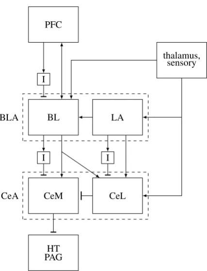
The amygdala has two main regions (Figure 3), the central (CeA) and basolateral (BLA) complexes, each having two main nuclei (centrolateral (CeL), centromedial (CeM), lateral (LA), basolateral (BL)), and two smaller medial and cortical olfactory nuclei. CeA CeL is innervated mostly by sensory inputs (direct interoceptive and somatosensory pain inputs, thalamic inputs via LA) and BLA, and projects to CeM. CeM projects to innate motor and autonomic output areas, including the PAG, the trigeminal nerve [Lazarov et al., 2011] and the hypothalamus. BLA receives inputs from the thalamus and cortex, projects unidirec-tionally to CeA and the BG, and is mutually connected with cortex and the hippocampus. Its projections to cortex are mostly into layers 1 and 2, and may be stronger than sensory inputs from the thalamus [Timbie and Barbas, 2014]. Thus, BLA strongly affects the re-sults of competitions. BLA and CeA projection neurons are glutamatergic and GABAergic, respectively.
CeA supports (and stores) executed SR mappings (quacia) by receiving their triggering sensory inputs (into CeL) and allowing their responses (via CeM->PAG). The ventrolat-eral (vl) PAG has a population of inhibitory interneurons (IINs) that are tonically active, suppressing excitatory vlPAG neurons that project to medulla neurons innervating spinal motor neurons. These IINs are suppressed by CeM neurons to disinhibit the excitatory re-sponse paths, allowing various types of competing innate rere-sponses [Tovote et al., 2016]8.
Thus, innate+ responses are supported by CeA, while BLA stores the associations between sensoriobject flow (from the thalamus into LA) and the executed innates (via BLA-CeM projections). Both CeA and BLA alert cortex when innates are executed, CeA via its HT projections (see the alert and extended alert N modes), and BLA directly. Through BLA’s bidirectional cortical connections and its BG projections, it lets adaptive responses use
in-8The amygdala is commonly viewed as being essentialfor executing innate actions. However, innates
HT PAG CeM BL PFC
CeL LA
thalamus, sensory
I I
I
CeA BLA
Figure 3: The amygdala. CeA controls the activation of innate responses (at the PAG, HT and other centers) via disinhibition. BLA serves as the interface between adaptive responses (mostly from PFC) and innate ones, via BLA-CeA projections. Only the main connections are shown. BNST, hippocampus, BG connections are not shown, as well as the cortical olfactory nuclei and circuits involving PKCD and non-PKCD neurons (see conditioning). ICMs are idealized. CeA: central complex. CeM: centromedial part of CeA. CeL: centrolateral part of CeA. BLA: basolateral complex. BL: basolateral part of BLA. LA: lateral part of BLA. I: intercalated masses (see BEND/consume section). HT: hypothalamus.
nates and be guided by object valence. The PAG is a BLA-CeM controlled switch that selects which innate response is activated, with specific selections corresponding to R ter-mination modes and mediated by terter-mination agents (opioids, OT etc.).
4
The Process Axis: R Mode Networks
Each location in the posterior-anterior flow axis contains separate neuronal networks sup-porting different R process modes. This is done differently in cortex, ibrain, the thalamus, the BG and the hippocampus. The cortical networks are located in different vertical layers (see Figures 4 and 5). The identification of networks associated with R modes is a major contribution of R17.
Flow network neurons are excitatory via glutamate9. Neurons in each cortical network
are bidirectionally interconnected to support quax stability through synchronous firing, but in all networks (except perhaps the response network) flow in one direction (BU or TD) is stronger than in the other [Markov et al., 2014]. The different networks show differ-ent patterns of Rgen receptor expression. These patterns provide strong support for our description of the networks, and are discussed in the N mode section.
4.1
The innate sensory network
Sensory input originates in sensors located throughout the body. The brain has three types of sensors, for exteroception (light, sound, taste, smell, and touch), proprioception (the state of the muscles and tendons) and interoception (the state of various internal body tissues). The input flows through the ibrain and splits into two streams, ibrain and abrain. The former innervates brainstem centers where it can activate innates and continue to the amygdala CeL, while the latter continues to cortex through the so-calledspecific thalamic nuclei (except olfaction, which reaches cortex directly). In most cases, each sensor type has its own path (‘labeled line’) until it reaches cortex (e.g., taste [Chen et al., 2011]). This yields topographic maps, i.e., cortical organization (receptive fields) that preserves in some way the relative structure of the input10. Because sensory flow reaches innate circuits before reaching adaptive areas, innate responses enjoy a latency advantage.
Proprioception is unique in that it has a special non-moving mechanism for stopping its inputs. Proprioceptive inputs arise from sensory neurons in the classical spinal cord motor unit. These neurons are excited by muscle spindles, which are sensitive to muscle length and contraction velocity, and by Golgi tendon organs sensitive to tension. The spinal cord in mammals has neurons calledgamma motoneurons (gMTNs), which innervate muscle spindles to set the threshold determining sensory neuron activation (in other species, this function is done by axon collaterals of the main motoneurons, aMTNs). Thus, gMTNs can adjust thresholds to prevent sensory neurons from stopping firing11or to suppress their firing. We view gMTNs as the innate part of the prediction network (see imagery below). If there exist gMTNs that are solely excited by ibrain neurons, these gTMNs constitute an innate prediction network.
4.2
The innate response network
Sensory inputs innervate ibrain motor centers where they can trigger innate actions. Proba-bly the simplest full quax is the motor unit, whose response network consists of alpha motor neurons (aMTNs) that innervate muscles to yield muscle contraction. In the stretch reflex,
9Disinhibition neurons, which are GABAergic, are naturally viewed as belonging to flow networks in
some areas (the BG and cerebellum), and to coordination networks in other areas (cortex, amygdala CeM).
10This specificity of sensory input can be contrasted with inputs that the brain receives via the circulation,
whose role is to affect R modes.
motor sensoryinput
DA ACh SER
BG GPi/SNr
GPe STN
STR
D1 CIN D2
thalamus
specific core matrix cmpf specific
non-core L5b
L5a L4 L3b L2/3
action node sensoriobject node
prediction nw DM nw
response nw sensory nw
alert nw
prediction nw DM nw
response nw sensory nw alert nw
Figure 4: Abrain architecture, including the BG, thalamus, two cortical nodes (cortical columns), in primary sensory cortex (right) and frontal cortex (left), and three Rgen nuclei. The cerebellum, hippocampus and amygdala are not shown. Arrows, T junctions and cir-cles indicate excitatory flow, GABAergic flow, and Rgen release respectively.
The BG output nuclei suppress the thalamic matrix, which projects diffusely to the inter-nal networks to sustain non-automated execution. The prediction and response networks project to the STR (to both the D1 and D2 paths), to recruit the BG and the thalamus to allow quax formation and execution. The response network also projects to the STN, thalamus, Rgen nuclei and motor centers. The STN sustains the BG response path (GPe, GPi/SNr). The thalamic cmpf nuclei excite the STR CINs when sensory input arrives. The CINs generally excite the D2 path to allow responses suppress the D1 path to allow deci-sion making.
To avoid clutter, some connections are not shown. These include the thalamic non-specific core projections to the action node, the intra-flow network connections, cortical ACh and SER projections, GPe projections to the STR and STN, DM network innervation of the alert network, BG projections to motor centers, cerebellum, and non-DA Rgen nuclei, the different projections of striosome and STR matrix neurons onto DA nuclei, and amygdala projections to the STR. The L6 part of the sensory network is not shown.
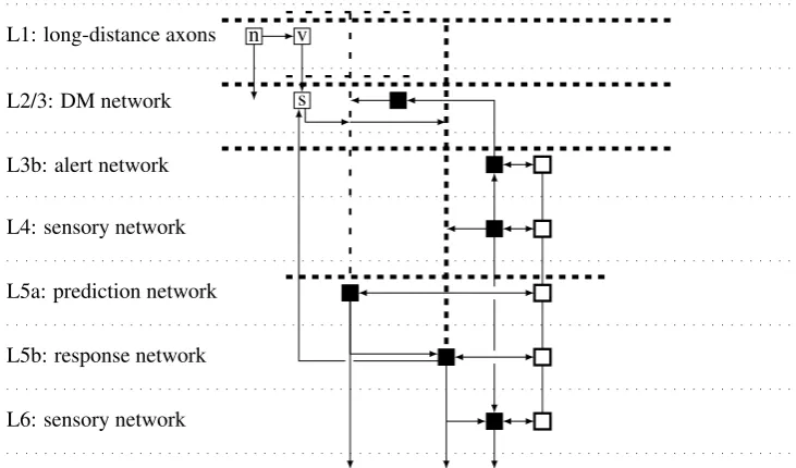
L6: sensory network L5b: response network L5a: prediction network L4: sensory network L3b: alert network L2/3: DM network L1: long-distance axons
s v n
Figure 5: Intra-node connections. Only the main patterns are shown. Thalamic inputs, inter-node connections and the RSN are not shown. The horizontal spacing between neu-rons is exaggerated. Filled squares: excitatory neuneu-rons. The thick (slender) tufted den-dritic tree of the response (prediction) neuron is shown in thick (thin) dashed lines. Empty squares: coordination IINs. The execution coordination network (ECN) is on the right in thick lines. ECN interconnections are shown without arrows to emphasize gap junc-tion coupling. The competijunc-tion coordinajunc-tion network (CCN) is on the left (s: SOMs, v: VIPs, n: NGFCs). NGFCs target all cell types, depicted by a single general arrow. The deep networks have subcortical projections, to motor centers, thalamus, and BG (response network), BG (prediction network), and thalamus (L6 sensory network).
forced limb movement yields muscle stretch, which activates sensory neurons. These ex-cite their agonistic motoneuron to yield movement in the opposite direction and stabilize posture. In normal movement, sensory neurons excite inhibitory interneurons that suppress the antagonistic muscle12. Higher level innate actions utilize more complex circuits, some of which known as ‘central pattern generators’ (e.g., for locomotion).
As the muscle contracts, the sensory neurons reporting its status reduce their firing rates. Normally, response neurons that directly generate movement require proprioceptive sensory inputs to be able to do so [Galán et al., 2015]. Without proprioceptive inputs, the abrain can learn to activate motor neurons adaptively via a different modality (e.g., vision), but this requires extensive training [Cole and Paillard, 1995]. As noted above, gMTNs can be used to ensure continued proprioceptive inputs to enable movement.
4.3
The adaptive sensory network
Most sensory input reaches the abrain via the thalamus, a large bilateral set of nuclei at the center of the brain (see Figure 4). Excitatory thalamic neurons can be classified into core
neurons, which project to middle cortical layers in a spatially focused manner and express parvalbumin (PV) in primates13, and matrix neurons, which are mostly present in
non-specific nuclei, project to layers 1-3 (L1-3) and L5 in a spatially diffuse manner, and express
12This is the simplest type of competition, see below.
calbindin (CB)14 [Clascá et al., 2012]. The thalamus also contains coordination networks. The adaptive sensory network consists of the thalamocortical core15, excitatory cortical L4 neurons, and corticothalamic L6 neurons. Core neurons project mainly to sensory network neurons in L416, but also to alert and response network neurons [Constantinople and Bruno, 2013]. The L4 sensory neurons project vertically to excitatory neurons in L6 (as well as to the alert network), and these project back to the thalamus17, closing an input flow loop that
ensures quax stability.
The thalamus contains two types of nuclei, specific and non-specific. The specific nu-clei are those conveying raw sensory flow to cortex. The non-specific nunu-clei are innervated by axon collaterals of cortical response network neurons and by the L6 sensory network neurons, and have core and matrix parts projecting to cortex. We view the non-specific thalamic core as part of the adaptive sensory network. Cortical response neurons, which are the main input of core non-specific neurons, can be activated by surprising BU sensory input (see bursts under the alert N mode). When this occurs, their activation is not part of a response, but part of still unanswered input. By innervating the non-specific thalamic core, response neurons convey such BU flow to higher level cortical areas.
Agranular & dysgranular areas. L4 conveys thalamic core flow to the alert network in L3b (see below). Given that the thalamic core also projects directly to L3b, it is not immediately clear why L4 is needed. Indeed, some cortical areas areagranular(lack L4) ordysgranular(have a smaller L4).
The main R17 observation in explaining this phenomenon is that in areas that must yield rapid responses to sensory input, L4 is redundant and only slows down the Q process. There are two types of such areas: those supporting low level motor control, and those providing immediate responses to alerts. Indeed, primary motor cortex (M1) is agranular, and premotor cortex, whose responses should be fast but not as fast as those of M1, is dysgranular. Similarly, entorhinal cortex (EC), which supports locomotion (see grid cells below), is agranular. In areas of this type, L3 is the sensory network (being the one that receives sensory input), and it also behaves like the prediction network. The role of the prediction network is to support action sequences by channelling responses to inputs to the next action (see below). When responses are very quick, predictions should arrive to the sensory network, because the additional synapse through the prediction network would slow down execution. Indeed, the hippocampus provides evidence that EC L3 behaves like a prediction network (see below).
The second type of agranular area in shown by medial prefrontal cortex (mPFC) and parts of the insula, both of which receive amygdalar alerts, as well as by the supplementary eye field, which supports attention orienting movements. Granular PFC areas support long term planning and execution and exist only in primates.
Thus, the role of L4 is to enable a longer DM process supporting longer response latencies. The separation of L4 from L3b allows L4 competition to be resolved while L3b competition (i.e., decision making) is still being held. This way, L4 allows sharp (focused) inputs to drive a coarse (disinhibited) DM process.
14CB is the fastest Ca2+ buffering protein, according with participation in acute responses.
15Thus, the thalamic core is thethalamic sensory(orBU)network. Generally, the networks that convey
BU flow are the sensory and alert networks, but the thalamus does not have an alert network.
16See also agranular/dysgranular areas below.
17L6 also has corticoclaustral and corticocortical populations. The claustrum attends to sensory input (see
4.4
The alert network
The alert network consists of pyramidal neurons in L3b (the lower part of L3). They are innervated by the sensory network, both vertically by L4 and by the thalamic core, and by the amygdala BLA. Alert neurons excite the L2/3 DM network vertically, and neighboring L4 neurons horizontally in all directions, mostly BU but also TD [Markov et al., 2014]. Thus, the network conveys unanswered BU flow that quickly extends to more frontal and ventral areas to drive decision making. We refer to the sensory and alert networks together as theexternalorBUnetworks.
4.5
The decision making (DM) network
The DM network consists of the pyramidal neurons in L2/3. They are vertically inner-vated by the alert and sensory networks, and can also be innerinner-vated by the prediction and response networks through long-distance connections [Rempel-Clower and Barbas, 2000]. They innervate other DM neurons horizontally through local and long-distance connec-tions, mostly in the TD direction, and through projections to the other hemisphere (i.e., they are commissural, callosal being the largest subgroup).
Since the alert and the DM networks are adjacent and are activated in the same R pro-cess stage (DM), their neurons are often intermingled such that all of L2/3 seems to be a single layer. Careful measurements identify two sub-networks with the lower/higher one conveying BU/TD flow [Markov et al., 2014], but the two networks are tightly related.
The DM network is where response alternatives are activated to participate in competi-tions. When a focused response emerges upon competition resolution, most of the activated DM neurons are suppressed, but the ones lying on the focused path actively participate in the executing focused response. Hence, DM neurons participate in non-DM (execution) modes as well. This may be why some DM neurons project to the BG (both the dorsal and ventral STR), at least in mPFC [Gabbott et al., 2005].
4.6
The adaptive response network
Cortical L5 contains two types of pyramidal neurons [Harris and Shepherd, 2015]. The cortical response network consists of the L5 pyramidal neurons that project ipsilaterally to the thalamus, the BG, the spinal cord, other motor centers, and Rgen nuclei. Viewing them as response neurons is natural, since they are the only cortical neurons that project to innate motor centers. Response neurons generally reside in L5b and are thick-tufted, with den-drites that reach L1 and exhibit hyperpolarization-activated cyclic nucleotide-gated (HCN, subunit HCH1) cation currents (Ih), which support repeated firing at slow rates. They re-ceive vertical input from the sensory network (both the thalamus and L4 neurons) and the prediction network, and provide vertical innervation to GABAergic (coordination) neurons, not to pyramidal neurons (see below). The response network is extensively internally con-nected across cortex (i.e., horizontally), but only ipsilaterally (see also nodes below). It sends horizontal long-distance connections through L1 to the DM network (see prediction below) and to apical dendrites of response neurons. The response network supports all adaptive responses, both learned and acute.
4.7
The prediction (goals) network
They are strongly inter-connected horizontally across cortex. They usually reside in L5a, some are tall and slender-tufted and some are short. Vertically, they are innervated by the DM network and innervate the response network (unidirectionally). We refer to them as the prediction network, because they represent the focused decision emerging from DM competition, but this decision is still not executed by the response network.
It may seem as if the prediction network is redundant, because the emerging decision could have been communicated directly from the DM network to the response network. However, this would immediately execute the response. The prediction network allows storing a competition winner without executing it, supporting intentions and predictions (see below). The DM and prediction networks are together called the internal or TD
networks, and the response and prediction networks are together called theexecution net-works.
The execution networks send long-distance connections to the DM network, even though decision making precedes execution in the R process. This way, identified objects can influence the formation of higher level responses, and executing tasks can affect the in-terpretation of sensory inputs. Many such connections are from non-granular PFC areas to granular ones [Rempel-Clower and Barbas, 2000], i.e., from lower level areas first respond-ing to alerts to higher level areas responsible for longer term responses. Such connections have a parallel function to that of the non-specific thalamic core, which conveys sensory flow to higher level areas.
4.8
The thalamic TD network
The thalamic core is the part of the BU sensory network that feeds inputs into cortex. The role of the more diffuse thalamic matrix is to sustain adaptive execution. Matrix neurons are innervated by the cortical response network (and possibly by L6 neurons as well) to convey TD flow. The matrix innervates the response and internal networks, especially the apical dendrites of L5 neurons, which extend to reach L1 (and the CCN, see below). When thalamic flow passes through the long narrow dendritic geometry above the cell’s soma it is greatly amplified, supporting sustained operation [LaBerge, 2002]18.
In addition to its sustaining effect on individual neurons, the matrix also provides quax stability by triangulating intra-cortical flow and by synchrony, mostly via coordination net-works (see below). Thus, the overall role of the thalamus is to provide energy drive to cortex, both BU and TD. Indeed, brain function critically depends on it [Ward, 2011].
4.9
The basal ganglia (BG): gating execution
Structure. The basal ganglia (BG) are a set of large bilateral abrain nuclei located at the center of the brain right above the thalamus [Graybiel and Mink, 2009] (see Figure 4). The flow between BG nuclei is largely directional, from input nuclei to output ones. There are three input nuclei: the dorsal striatum (dSTR), the ventral STR (or nucleus accumbens, NAc), and the subthalamic nucleus (STN). Output nuclei are the substantia nigra reticulata (SNr) and internal globus pallidus (GPi), with a smaller output from the ventral pallidum (VP). There are internal nuclei, the external globus pallidus (GPe) and its ventral analogue the VP. The out-going neurons from all nuclei are GABAergic, except the STN’s, which are glutamatergic. Those projecting from the STR are called medium spiny neurons (MSNs). BG neurons also project to Rgen (mostly DA and ACh) nuclei.
The prediction and response networks (and more weakly also the DM network) in all cortical areas project to the STR. Motor areas project to the dSTR, and higher level task areas and valence areas project to the vSTR. There is substantial convergence along the cortex-STR-GPi/SNr direction [Bar-Gad et al., 2003]. Neurons in the output nuclei are tonically active [Wilson, 2015], and project to matrix neurons [Kaneko, 2013] in the non-specific nuclei of the thalamus innervated by frontal areas, and to subcortical motor centers (e.g., the SC). Cortico-STR projections are topographic, and this is preserved in the thalamus and in its projections onto cortex, giving rise to the well-known cortico-BG-thalamocortical loops.
The BG nuclei are mutually and bidirectionally connected, but two main connectivity paths are evident: a STR-GPi/SNr path commonly called the direct path, and a STR-GPe/VP-STN-GPi/SNr path called the indirect path. The MSNs participating in the two paths are generally molecularly different: the direct MSNs express dopamine D1 receptors, dynorphin and substance P, while the indirect MSNs express D2 receptors and enkephalin. Both of the cortical execution networks project to both paths. There is neuronal overlap between the paths (e.g., one third of dSTR-GPe [Hegeman et al., 2016], and one half of NAc-VP projections, are from D1 neurons [Kupchik et al., 2015]).
Function. The tonic inhibition exerted by the BG output neurons on the non-specific tha-lamic nuclei prevents cortical quacia from recruiting thatha-lamic matrix neurons and thereby prevents adaptive responses. To allow cortical recruitment of the thalamus, the BG output neurons must be either neutralized or coordinated. An important novel thesis of R17 is that
the direct path neutralizes (disinhibits) the BG output neurons to support the competition R mode, while the indirect path coordinates output neurons during responses (acute and automated). In other words, the direct path supports DM and its resulting focused decision, while the indirect path is trained by the direct path to execute the focused decision. As a result, the direct path is active only in the acute mode (mostly in its initial stages), while the indirect path is active in both acute and automated modes. Note that this account is dramatically different from the common view, especially with respect to the indirect path. The precise mechanism is discussed in depth under the DA and ACh modes below.
The STN is innervated by cortex19 (mostly by motor areas) and thalamus ipsilaterally,
so we view it as part of the response network. Its main role is to provide excitatory drive to BG neurons to complement their inherent spiking. Indeed, suppression of the GPi and of the STN (e.g., by deep brain stimulation) is used to treat Parkinson’s disease, in which impaired DA signaling does not allow the BG to function correctly. The two treatments are similar in that they both neutralize the BG, allowing cortex to recruit the thalamus directly. The STN also projects to the superficial cortical networks and disynaptically to the cerebellum, promoting rapid transitions to support response sequences (see below).
An additional important BG topic, the striosomes, is discussed under the consume N mode below.
In summary, the role of the BG is to control the formation and sustained execution of adaptive quacia, by gating cortical access to the thalamus and motor centers and by recruiting Rgens.
4.10
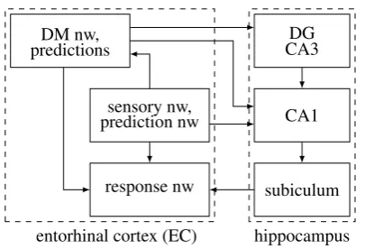
The hippocampus
Thehippocampus, adjacent to the amygdala at the medial temporal lobe, is an abrain struc-ture possessing a unique architecstruc-ture [Buzsaki, 2006]. It is a rolled sheet whose long axis is
hippocampus subiculum
CA1 DG CA3
entorhinal cortex (EC) DM nw,
predictions
response nw sensory nw, prediction nw
Figure 6: The hippocampus. The subiculum belongs to the response network. CA1 re-ceives input from the EC prediction network (L3, since it is an agranular area), and from an automated L2 population that also represents predictions. Hence, CA1 belongs to the prediction network. The dentate gyrus (DG) and CA3 receive input from an EC DM (L2) population conveying acute flow, and belong to the DM network. To focus on the content networks, connections with thalamic and hypothalamic nuclei, the amygdala, BNST, the BG, PFC and other areas are not shown. CA2 is not shown.
approximately dorsoposterior-ventroanterior and whose shorter axis is commonly divided into subfields. The main fields are the dentate gyrus (DG), CA3, CA2, and CA1, which are generally unidirectionally connected in this order. CA1 projects to an output structure, the subiculum. All fields except the DG have pyramidal content cells and coordination IINs as in cortex. Fields consist of three layers, the equivalents of L1, L5 and L6.
The hippocampus has attracted immense scientific and popular interest due to its role in memory [Moscovitch et al., 2016] and space [Buzsaki, 2006], but there is currently no coherent account for the roles of its subfields. We propose a novel account based on R process networks (Figure 6): (i) the DG and CA3 are part of the decision making network, (ii) CA1 is part of the prediction network, and (iii) the subiculum is part of the response network. CA2 is smaller and may be specific for social interaction, combining both DM and prediction.
There are six main arguments supporting this account. First, the subiculum is the output field of the hippocampus, projecting to the EC execution network (L5), the STR (mostly projections from the ventral subiculum to the vSTR), PFC, etc. Second, cortical inputs to the hippocampus arrive mostly from the entorhinal cortex (EC). An EC L2 population that conveys acute DM flow projects to DG and CA3 [Kitamura et al., 2015]. Adult neuro-genesis (NGNS) takes place in the DG [Christian et al., 2014], and in R17 the drive for NGNS is this DM network flow. Third, non-cortical inputs to the hippocampus are from the hypothalamic supramammillary nucleus (SUM) and the thalamic nucleus reuniens (NR) [Vertes, 2015]. SUM is connected with alert and innate areas (the septum, amygdala, PAG, VTA (DA), LC (NE)) and projects to the DG, CA3 and CA2, while NR conveys TD flow from mPFC to CA1 and the subiculum. Fourth, CA2 receives AVP inputs indicating acute needs, with emphasis on social needs and responses (see N process below). Fifth, the EC prediction network (L3, see agranular areas above) and an L2 population that participates in more automated actions (locomotion) project to CA1 [Kitamura et al., 2015]. Finally, learning (automaticity, and thus predictions) is reflected in CA1 more than in CA3 (it has many more place cells and they are more focused, see below).
subfields. The hippocampus is further discussed under event areas and place cells below.
4.11
Cognitive interpretation: predecision, reality, goals
A brain theory must explain how cognition arises from the biological level. Here we pro-vide a basic cognitive view of the roles of the flow networks, expanded under the Q process and internal cognition below.
During acute situations, the DM network is innervated by TD flow, its active neurons conveying many possible ways of perceiving the input and responding to it. In this situ-ation, the DM network can be said to provide rapid non-specific (coarse) feedback (FB), and the sensory, alert and DM networks can be said to conveypredecisionflow20.
Situations having a high degree ofuncertaintyinvolve prolonged DM network activity, since certainty would lead to decision and focus. For example, the DM network would be used when expecting a relatively unimportant event whose timing is not known. In this function, we say that the DM network conveys generalexpectationoranticipation.
The DM network contains the neuronal candidate pool from which adaptive quacia emerge. As such, a wider DM network yields a wider diversity of responses. Thus, the DM network can be said to support cognitive flexibilityandcreativity. Indeed, the superficial networks are evolutionarily more recent than the execution networks, and their spine den-sities have considerably increased in primates, especially in humans [Elston et al., 2006].
The prediction and response networks convey focusedpostdecisionflow resulting from competition resolution. Such flow is a prerequisite for perceiving an object or event, since perception (by definition) involves coherent entities and not an undefined flux or a simul-taneous multitude of entities induced by the same sensory input. The response network is excited by sensory flow and is essential for generating external movements. Combining these observations, we posit that we perceive something as occurring in the external en-vironment if and only if the response network in sensory cortices is activated21. For this reason, we refer to the response network as therealitynetwork.
The prediction network is activated in acute situations, it is driven by the decision mak-ing network, it conveys focused flow, and its activation immediately precedes that of the response network. These data have two complementary cognitive interpretations. First, fo-cused and effortful (acute driven) execution is what is usually meant by the termattention. Hence, we can refer to this network as the attention network. Second, when the focus network is activated but the response neurons it drives are still not activated, it represents responses activated before they occur in reality. Such responses are called predictions
when referring to sensory events that are not controlled by the animal (e.g., ‘this falling stone is going to hit the floor’), and goals, intentions or movement preparations when referring to sensory events controlled by the animal (e.g., ‘my fingers are prepared to grasp this cup’). For brevity, we usually use only one of these terms, referring to this network as the prediction network. Although prediction may sound like an advanced cognitive func-tion, the prediction network is a basic, evolutionarily ancient feature of the brain, and it is essential for executing actions and action sequences (see Q process below).
In summary of this section, brain anatomy strongly reflects the R process. Each R mode is associated with flow in a different neuronal network. Two prominent subcortical structures, the thalamus and basal ganglia, are involved in flow regulation and are essential for adaptive responses.
20Since the winning DM neurons also participate in focused execution, the DM network also exhibits
postdecision activity.