Western University
Scholarship@Western
Electronic Thesis and Dissertation Repository
September 2014
Characterizing the interaction between RanBPM
and c-Raf
Wesley Berube-Janzen The University of Western Ontario
Supervisor
Caroline Schild-Poulter
The University of Western Ontario
Graduate Program in Biochemistry
A thesis submitted in partial fulfillment of the requirements for the degree in Master of Science © Wesley Berube-Janzen 2014
Follow this and additional works at:https://ir.lib.uwo.ca/etd
Part of theCancer Biology Commons
This Dissertation/Thesis is brought to you for free and open access by Scholarship@Western. It has been accepted for inclusion in Electronic Thesis Recommended Citation
Berube-Janzen, Wesley, "Characterizing the interaction between RanBPM and c-Raf " (2014).Electronic Thesis and Dissertation Repository. 2290.
CHARACTERIZING THE INTERACTION BETWEEN RANBPM AND C-RAF
(Thesis format: Monograph)
by
Wesley Bérubé-Janzen
Graduate Program in Biochemistry
A thesis submitted in partial fulfillment of the requirements for the degree of
Master of Science
The School of Graduate and Postdoctoral Studies The University of Western Ontario
London, Ontario, Canada
Abstract
RanBPM/RanBP9 is a multi-domain nucleocytoplasmic protein which has been linked to numerous cellular processes including cell adhesion, migration, transcription and apoptosis. Although RanBPM is a member of the mammalian CTLH complex, the counterpart of a conserved yeast E3 ubiquitin ligase complex, its exact function remains unknown. Previous work in our laboratory has shown that RanBPM inhibits the ERK pathway by interacting with the kinase c-Raf and downregulating c-Raf levels. Here, we show that the N-terminus, LisH/CTLH and CRA domains of RanBPM are required for downregulation of c-Raf and that RanBPM interacts directly with c-Raf through its CRA domain. We also provide evidence that MAEA, another CTLH complex member, associates with c-Raf. Therefore, we propose a mechanism by which RanBPM downregulates c-Raf in a CTLH complex-dependent manner. This work contributes to our knowledge of the function of RanBPM and clarifies the relationship between RanBPM and c-Raf, two important proteins in oncogenesis.
Keywords
Co-Authorship Statement
This thesis was written by Wesley Bérubé-Janzen and edited by Caroline Schild-Poulter, Victoria Fell and Louisa Salemi. All experiments and procedures described in this thesis were performed by Wesley Bérubé-Janzen, with the exception of the following:
HeLa control and HeLa 2-7 stable cell lines were generated by Dawn Bryce.
ΔN2, ΔC4, pCMV-HA-RanBPM-ΔC1, pCMV-HA-RanBPM-Δ212, and pCMV-HA-RanBPM-Δ360, with mutations conferring resistance to shRNA, were generated by Caroline Schild-Poulter, Dawn Bryce, Patricia Christian and Tung Bai.
Acknowledgments
I would like to primarily thank my supervisor, Dr. Caroline Schild-Poulter, for giving me the opportunity to train in your lab. Your guidance, support and patience have allowed me to grow as a scientist and as a person and, without you, this degree would not have been possible. I would also like to thank my committee members, Dr. David Litchfield and Dr. Sean Cregan, for your helpful ideas and stimulating discussions during my committee meetings. I always left our meetings feeling refreshed, focused and motivated, and for that I am truly appreciative.
I sincerely thank all the past and current members of the Schild-Poulter lab that I have had the honour of working with and getting to know. Xu, your guidance and expertise has been invaluable over the course of my degree and there is no way I can thank you enough for all your help. Vicki, Louisa and Sarah, I not only thank you for your advice, knowledge and countless favours, but also for your friendship. I most enjoyed the blunt honesty we shared and our ruthless eagerness to make fun of each other, all in good fun. I can honestly say you guys made the lab an amazing place to work and I would have never imagined I would make such great friends in just two short years.
I must also thank my former supervisor, Dr. David Edgell, for your continuing support and direction. Your door has always been open to me and I sincerely appreciate everything you have done for me. I am also grateful it only comes at the small cost of being occasionally reminded that I left your lab to pursue the cure for cancer.
Table of Contents
Abstract and Keywords ...ii
Co-Authorship Statement ... iii
Acknowledgments ...iv
Table of Contents ... v
List of Tables ... vii
List of Figures ... viii
List of Abbreviations ...ix
Chapter 1 – Introduction ... 1
1.1. Cancer ... 1
1.2. RanBPM ... 2
1.2.1. Overview of RanBPM ... 2
1.2.2. Conserved domains ... 4
1.2.3. Cellular localization ... 4
1.2.4. Role in apoptosis ... 6
1.2.5. Functions in the reproductive and nervous systems ... 7
1.2.6. Regulation of transcriptional activity ... 9
1.2.7. Implications in cell morphology, adhesion and migration ... 10
1.2.8. RanBPM in cancer ... 11
1.2.9. CTLH complex ... 12
1.3. c-Raf ... 15
1.3.1. Overview of Raf family kinases ... 15
1.3.2. ERK signalling pathway ... 17
1.3.3. MEK1/2-independent signaling by c-Raf ... 19
1.3.4. c-Raf activation and deactivation ... 20
1.3.5. Regulation of c-Raf stability ... 22
1.4. Hypothesis and objectives ... 24
Chapter 2 – Materials and Methods ... 26
2.1. Chemicals and reagents ... 26
2.2. Antibodies ... 26
2.4. Stable shRNA cell lines and cell culture ... 30
2.5. In situ proximity ligation assay ... 30
2.6. Transfection assays ... 31
2.7. Preparation of mammalian cell extracts ... 31
2.8. Bacterial protein expression and preparation of E. coli extracts ... 31
2.9. Western blot analyses ... 32
2.10. GST pull-down assays ... 32
2.10.1. Using HeLa cell extracts ... 32
2.10.2. Using E. coli extracts ... 33
2.11. Statistical analyses ... 34
Chapter 3 – Results ... 35
3.1. Endogenous RanBPM and c-Raf are found in a complex ... 35
3.2. N-terminus, CRA and LisH/CTLH domains of RanBPM are required for c-Raf downregulation ... 38
3.3. The CRA domain of RanBPM is required for interaction with c-Raf ... 42
3.4. RanBPM interacts directly with c-Raf through its CRA domain ... 43
3.5. Endogenous c-Raf and MAEA are found in a complex ... 46
Chapter 4 – Discussion ... 50
4.1. Summary of findings ... 50
4.2. Model and rationale ... 50
4.3. Significance of the CRA domain as a binding-domain for c-Raf ... 57
4.4. Consequences of RanBPM-mediated regulation of c-Raf ... 58
4.5. Limitations of the study and future studies ... 59
4.6. Conclusion ... 61
References ... 62
Appendices ... 71
List of Tables
Table 1.1. Comprehensive list of proteins that have been shown to interact with RanBPM ... 3 Table 2.1. Concentrations of primary antibodies used for Western blot
analysis and in situ proximity ligation assay ... 28
Table 2.2. PCR primer sequences and descriptions ... 29 Table 4.1. Summary of results of the effects of RanBPM constructs on
List of Figures
Figure 1.1. Full-length RanBPM with conserved domains indicated ... 5
Figure 1.2. Mammalian orthologs of the members of the S. cerevisiae Gid complex ... 13
Figure 1.3. Model of the interactions between members of the S. cerevisiae Gid complex ... 14
Figure 1.4. Structure, conserved regions and regulatory phosphorylation sites of Raf family kinases ... 16
Figure 1.5. Summary of ERK pathway signaling ... 18
Figure 1.6. Summary of the activation cycle of c-Raf ... 21
Figure 3.1. Summary of the in situ PLA ... 36
Figure 3.2. Endogenous RanBPM and c-Raf are found in a complex ... 37
Figure 3.3. Full-length RanBPM and deletion mutants chosen for analysis ... 39
Figure 3.4. Δ360, ΔN2, ΔC4 and ΔC1 RanBPM deletion mutants do not effectively downregulate ΔN c-Raf compared to WT RanBPM ... 41
Figure 3.5. ΔC1 RanBPM is unable to interact effectively with ΔN c-Raf ... 44
Figure 3.6. Full-length RanBPM and individual domains chosen for analysis .... 45
Figure 3.7. WT RanBPM and C1 domain interact directly with ΔN c-Raf ... 47
Figure 3.8. Endogenous MAEA and c-Raf are foundin a complex ... 49
Figure 4.1. Model of the mechanism by which RanBPM downregulates c-Raf .. 52
List of Abbreviations
ACs adenylyl cyclases
AD Alzheimer’s Disease
ANOVA analysis of variance APP amyloid precursor protein
AR androgen receptor
ARMc8 armadillo repeat containing 8
ASK1 apoptosis signal-regulating kinase 1
ATP adenosine triphosphate
Aβ amyloid β
BACE1 β-secretase 1
Bax Bcl-2-associated X protein
Bcl-2 B cell lymphoma 2
Bcl-XL B cell lymphoma extra large
BioGRID Biological General Repository for Interaction Datasets BLT2 leukotriene B4 receptor 2
BRCA1 breast cancer type 1 susceptibility protein CDK11 cyclin-dependent kinase 11
c-Fos cellular FBJ murine osteosarcoma viral oncogene homolog CHIP C-terminus of Hsp70-interacting protein
CK2 casein kinase 2
CO2 carbon dioxide
CRA CT11-RanBPM
CREB cAMP response element-binding protein
CTLH C-terminal to LisH
DAPK death associated protein kinase
DD death domain
DMEM Dulbecco’s modified eagle medium
DTT dithiothreitol
ECL enhanced chemiluminescence
ERK extracellular signal-regulated kinase FBPase fructose-1,6-bisphosphatase
FBS fetal bovine serum
FMRP fragile X mental retardation protein
GDP guanosine diphosphate
GFP green fluorescent protein
Gid glucose induced degradation deficient
GR glucocorticoid receptor
Grb2 growth factor receptor-bound protein 2 GST glutathione-S-transferase
GTP guanosine triphosphate
GTPase guanosine triphosphatase
HA human influenza hemagglutinin
HDAC6 histone deacetylase 6
HEK human embryonic kidney
HEPES hydroxyethyl piperazineethanesulfonic acid HIPK2 homeodomain-interacting protein kinase 2
HMG high mobility group
HRS Hepatocyte growth factor-regulated tyrosine kinase substrate Hsp70 heat shock protein 70
Hsp90 heat shock protein 90 IAP inhibitor of apoptosis
IPTG isopropyl β-D-1-thiogalactopyranoside
IQGAP IQ motif containing GTPase-activating protein
IR ionizing radiation
JAK2 Janus kinase 2
KCl potassium chloride
KSR1 kinase suppressor of Ras 1
LB Luria Bertani
LisH lissencephaly type-1-like homology
MET mesenchymal epithelial transition factor
MSK1/2 mitogen- and stress-activated protein kinases 1/2 MST2 mammalian Ste20-like kinase 2
N/A not available
NaCl sodium chloride
NaF sodium fluoride
NaVO4 sodium orthovanadate
ND not determined
NES nuclear export signal
NFAT nuclear factor of activated T cells NLS nuclear localization signal
NP40 nonidet P-40
N-region negative-charge regulatory region OD600 optical density at 600nm
p75NTR p75 neurotrophin receptor PAK1 p21-activated kinase 1
PBS phosphate buffered saline
PCR polymerase chain reaction
PFA paraformaldehyde
PKA protein kinase A
PLA proximity ligation assay PMSF phenylmethylsulfonyl fluoride
PP1 protein phosphatase 1
PP2A protein phosphatase 2A
PP5 protein phosphatase 5
PVDF polyvinylidene fluoride
Raf rapidly accelerated fibrosarcoma Ran Ras-related nuclear protein RanBP10 Ran-binding protein 10
RanBPM Ran-binding protein in the microtubule-organizing center
Ras rat sarcoma
Rmnd5a required for meiotic nuclear division 5 homolog A Rok-α Rho-binding kinase α
SDS sodium dodecyl sulfate
SDS-PAGE sodium dodecyl sulfate polyacrylamide gel electrophoresis SEM standard error of the mean
SH3 Src homology 3
SNP single nucleotide polymorphism
SOS son of sevenless
SPRY dual-specificity kinase splA and ryanodine receptor
Src sarcoma
TAF4 transcription initiation factor TFIID TFIID transcription factor II D
TGF-β transforming growth factor β
TR thyroid receptor
TRAF6 tumor necrosis factor-receptor-associated factor 6 Tris Tris-hydroxymethyl amino methane
TrkA tropomyosin-related kinase A TrkB tropomyosin-related kinase B
Twa1 two hybrid-associated protein 1 with RanBPM
WCE whole cell extract
WT wild-type
Chapter 1 –
Introduction
1.1. Cancer
According to the Canadian Cancer Society, it is estimated that approximately 267,400 Canadians will be diagnosed with cancer in 2014, with the most common types being prostate, breast, lung, colorectal and non-melanoma skin cancer (1). Cancer is the leading cause of death in Canada, with an estimated 1 in 4 Canadians expected to die from the disease (1). Therefore, it is evident that cancer research focused on understanding the disease and developing new treatments to combat tumour growth are of utmost importance to preserve the health of our population.
Although the average one gram malignant tumour is estimated to contain 108–109 cancer cells, such tumour masses can start with the defiant behaviour of a single cell (2). Cells are programmed to grow, replicate and die when they have reached the end of their lifespan, however a cell can break free from these restraints in the event of genomic mutation and proliferate uncontrollably, resulting in cellular transformation and tumourigenesis. Cancer cells exhibit certain characteristics, termed the hallmarks of cancer, which are acquired during cellular transformation and are predominantly responsible for the progression of the disease (3). Namely, they sustain proliferative signaling, evade signaling from growth suppressors, resist cell death, replicate infinitely, induce angiogenesis and invade surrounding tissues (3).
1.2. RanBPM
1.2.1. Overview of RanBPM
RanBPM, also known as RanBP9, was initially discovered in a yeast two-hybrid assay as a 55kDa interacting partner for the small guanosine triphosphatase (GTPase), Ran (Ras-related nuclear protein) (4). However, subsequent studies soon uncovered that RanBPM is in fact a 90kDa protein, only weakly interacts with Ran and does not localize to the centrosome, as initially thought (4,5). Since then, RanBPM has generated significant interest and numerous studies have been conducted on the protein to attempt to characterize its function.
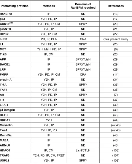
Table 1.1. Comprehensive list of proteins that have been shown to interact
with RanBPM. Methods used to demonstrate the interactions are indicated as yeast
two-hybrid (Y2H), mammalian two-hybrid (M2H), pull-down (PD), immunoprecipitation (IP), proximity ligation assay (PLA), confocal microscopy (CM) and/or fluorescence resonance energy transfer (FRET).
Interacting proteins Methods Domains of
RanBPM required References
RanBPM IP ND (13)
p73 Y2H, PD, IP ND (17)
CDK11p46 Y2H, PD, IP, CM SPRY (20)
p75NTR Y2H, IP ND (21)
HIPK2 Y2H, IP, CM ND (22)
c-Raf PD, IP, PLA CRA (24), present study
L1 Y2H, PD, IP SPRY (25)
MET Y2H, M2H, PD, IP SPRY (8)
TrkB IP, CM ND (26)
APP IP SPRY/LisH (29)
BACE1 IP SPRY/LisH (29)
LRP IP SPRY/LisH (29)
FMRP Y2H, PD, IP, CM CRA (14)
plexin-A Y2H, IP ND (34)
TrkA Y2H, PD, IP SPRY (35)
TAF4 Y2H, IP, CM ND (36)
AR Y2H, PD, IP SPRY (7)
TR Y2H, PD, IP ND (37)
LFA-1 Y2H, PD, IP ND (39)
β1 integrin Y2H, IP ND (39)
BLT-2 Y2H, PD, IP, CM ND (43)
BRCA1 Y2H ND (44)
Muskelin Y2H, IP ND (42,46)
Twa1 Y2H, IP, PD ND (42,46)
Rmnd5a IP ND (46)
MAEA IP ND (46)
ARMc8 IP ND (46)
HDAC6 IP, CM LisH/CTLH (103)
1.2.2. Conserved domains
Four conserved functional domains have been identified within the sequence of RanBPM: the SplA and Ryanodine receptor (SPRY) domain, the Lissencephaly type-1-like homology (LisH) domain, the C-terminal to LisH (CTLH) domain and the CT11-RanBPM (CRA) domain (Figure 1.1) (10). The SPRY domain is known to be involved in protein-protein interactions (11). The LisH domain is known to mediate protein dimerization, and is in fact predicted to moderate the dimerization and oligomerization of RanBPM (12,13). Additionally, the LisH domain, together with the CTLH domain, is thought to regulate microtubule dynamics and cell migration (12). The CRA domain, which is predicted to contain six α-helices and resembles a death domain (DD) superfamily domain, has also been shown to function as a protein interaction surface (14). In addition to these four conserved domains, RanBPM also contains a proline- and glutamine-rich N-terminus predicted to contain six Src homology 3 (SH3) binding domains (5,15).
1.2.3. Cellular localization
Although RanBPM was initially thought to be localized to the centrosome, further studies determined its localization to actually be predominantly nucleocytoplasmic (4,5). Recent studies in our laboratory have identified a primary nuclear localization signal (NLS) spanning amino acids 1–25, a secondary NLS spanning amino acids 635–649 and a nuclear export signal (NES) comprising amino acids 140–155, which together govern the subcellular localization of RanBPM (16). The SPRY and LisH/CLTH domains were also shown to be important for cytoplasmic retention of RanBPM, potentially through interactions with cytoplasmic proteins (16).
Figure 1.1. Full-length RanBPM with conserved domains indicated. RanBPM
the cytoplasm to the nucleus (17). Given that these two proteins physically interact and p73 is exclusively nuclear, it was hypothesized that an overabundance of p73 could sequester a high proportion of cellular RanBPM in the nucleus (17). Furthermore, under certain cellular conditions, RanBPM localization has been reported to be altered. For example, in response to ionizing radiation (IR), a DNA damage-inducing agent, RanBPM has been shown to shuttle from the nucleus to the cytoplasm (18). This change in localization could occur to allow RanBPM to interact with cytoplasmic apoptotic or DNA damage response proteins, although this speculation has yet to be confirmed (18). Altogether, such evidence suggests that RanBPM localization is important in dictating its function, as it allows RanBPM to interact with various specifically compartmentalized proteins and participate in different signaling pathways.
1.2.4. Role in apoptosis
(homeodomain-interacting protein kinase 2), which has been shown to activate and stabilize the tumour-suppressor protein p53 (21-23).
Important studies in our laboratory have demonstrated that RanBPM activates apoptosis in response to DNA damage caused by IR (18). Subsequent studies showed that RanBPM is in fact an inhibitor of the ERK (extracellular signal-regulated kinase) pathway and specifically downregulates the crucial ERK pathway kinase c-Raf at the protein level (24). This resulted in decreased downstream ERK pathway signaling, culminating in decreased levels of the anti-apoptotic protein Bcl-2 and its family member Bcl-XL (B cell lymphoma extra large) (24). This is consistent with previous findings that RanBPM regulates the intrinsic cell death pathway (19). In addition, it was found that downregulation of RanBPM leads to increased cell proliferation, an important hallmark of cellular transformation and cancer (24).
Other studies have also shown that RanBPM restricts ERK pathway signaling. For example, through its SPRY domain, it was observed that an N-terminal fragment of RanBPM interacts with the neural adhesion molecule L1 to inhibit downstream ERK signaling (25). Although it appears that RanBPM is a pro-apoptotic protein that inhibits the ERK pathway, there is some opposing evidence that RanBPM activates the ERK pathway through interactions with the receptor tyrosine kinases TrkB (tropomyosin-related kinase B) and MET (mesenchymal epithelial transition factor), although the latter was shown using a green fluorescent protein (GFP) tagged RanBPM construct (8,26). Therefore, some of the contradiction regarding RanBPM regulation of the ERK pathway could potentially be attributed to the different constructs used in each study and the unknown effects of large tags or truncations on the overall function of RanBPM.
1.2.5. Functions in the reproductive and nervous systems
characterization of RanBPM knockout mice. It is well documented that RanBPM knockout mice generally die neonatally, although a small number of newborn pups have been reported to survive into adulthood (27,28). The cause of this neonatal fatality remains unclear, although it has been suggested that these pups are unable to suckle milk, suggesting defects in brain function (28). RanBPM knockout mice suffer pronounced gonadal atrophy, severely compromised spermatogenesis and oogenesis as well as infertility, provided they reach adulthood (27). Furthermore, they display growth retardation and their brains are dramatically reduced in size, especially in the hippocampal and cortical regions, compared to wild-type (WT) mice (28).
A number of important studies have also implicated RanBPM in the development of the neurodegenerative disorder Alzheimer’s disease (AD). Interestingly, a truncated form of RanBPM has been shown to be expressed over six times higher in the brains of AD patients compared to those of healthy individuals (13). One of the defining pathological hallmarks of AD is the accumulation of Aβ (amyloid β) peptides in the brain and RanBPM has been shown to promote Aβ generation from its precursor APP (amyloid precursor protein) (29). RanBPM accelerates endocytosis of APP and acts as a scaffold for APP, BACE1 (β-secretase 1) and the endocytosis receptor LRP (low-density lipoprotein receptor-related protein) to facilitate BACE1 cleavage of APP into Aβ (29). Consistent with previously mentioned evidence regarding RanBPM involvement in apoptosis, these studies have shown that RanBPM overexpression causes apoptosis and also potentiates Aβ toxicity in the brain (30). In addition, RanBPM transgenic mice suffered neurodegeneration, spatial memory loss and a decreased number of neuronal synapses (31).
and sequesters the RNA-binding region of FMRP, rendering it unable to execute its RNA-binding function (14). RanBPM has also been reported to interact with the neural adhesion molecule L1, which can lead to various X-linked disorders if mutated (9). Inhibition of ERK signaling by an N-terminal fragment of RanBPM suppresses L1-meditated neurite outgrowth and branching in primary neurons (25).
Further functions for RanBPM in the nervous system include interaction with the plexin-A receptor to inhibit axonal outgrowth and induce neuronal contractility (34). RanBPM also been shown to interact with the receptor tyrosine kinases TrkB and TrkA, which both serve as neurotrophin receptors in the brain (26,35). Through its interaction with TrkB, RanBPM was shown to enhance neuronal morphogenesis, and through its interaction with TrkA, RanBPM was shown to reduce downstream expression of the transcription factor NFAT (nuclear factor of activated T cells), which is known to play a role in axon outgrowth and synaptic plasticity (26,35).
1.2.6. Regulation of transcriptional activity
Microarray analyses in our laboratory have shown that RanBPM influences transcriptional pathways primarily associated with cell, tissue and organ development as well as tumorigenesis and cancer (38). Upon RanBPM downregulation, global gene expression changes occurred and over-represented transcription factor binding sites were identified among the upregulated or downregulated genes (38). Among the most over-represented were binding sites for the Forkhead, homeodomain and HMG (high mobility group) transcription factors, providing further evidence that RanBPM regulates transcription by modulating transcription factor activity (38).
1.2.7. Implications in cell morphology, adhesion and migration
There is evidence of RanBPM involvement in cell morphology and polarity, based on its reported interactions with known regulators of these processes. It has been reported that RanBPM interacts with β1 integrin and the β2 integrin LFA-1 (lymphocyte function-associated antigen-1) (39,40). Integrins are transmembrane receptors that are well-known for mediating cell and cell-extracellular matrix interactions through focal adhesions, however, they also participate in many signaling pathways within the cell (39). RanBPM has been shown to accelerate endocytosis of β1 integrin to disrupt integrin-dependent cell adhesion, focal adhesion assembly and focal adhesion signaling (40). Some data also suggests that RanBPM acts in conjunction with Muskelin to regulate cell morphology and cell spreading, as they are found together in a complex and knockdown of either Muskelin or RanBPM in lung epithelial cells led to the same phenotype of increased cell perimeter and disrupted actin distribution (41,42).
1.2.8. RanBPM in cancer
Evasion of apoptosis, sustained proliferative signal and tissue invasion mark three of the six primary hallmarks of cancer demonstrated by malignant cells (3). Given its prominent roles in apoptosis as well as restricting cell growth and cell migration, it has been suggested that RanBPM might be playing a role in the prevention of tumour development and oncogenesis. As previously mentioned, RanBPM has been shown to interact with many pro-apoptotic tumour suppressors, inhibit proliferative cell pathways and directly induce apoptosis in response to DNA damage (17-25). It has also been shown to interact with proteins involved in cell motility and directly inhibit cell migration (24,43). Interestingly, RanBPM expression has been found to be altered in many human tumours, including lung, kidney and breast cancer samples (39). In most cases, expression was lost or greatly reduced, validating its characterization as a tumour suppressor protein (39).
1.2.9. CTLH complex
RanBPM is a known member of the mammalian CTLH complex, along with Muskelin, Twa1 (two hybrid-associated protein 1 with RanBPM), Rmnd5a (required for meiotic nuclear division 5 homolog A), MAEA (macrophage erythroblast attacher) and ARMc8 (armadillo repeat containing 8) (42,46). Each of these proteins, with the exception of Muskelin, have orthologs in
Saccharomyces cerevisiae which are part of the yeast Gid (glucose induced
Figure 1.2. Mammalian orthologs of the members of the S. cerevisiae Gid
complex. Proteins found within the Gid complex are represented on the right, along
Figure 1.3. Model of the interactions between members of the S. cerevisiae Gid
complex. Gid1, Gid2, Gid4, Gid5, Gid7, Gid8 and Gid9 interact to form an E3
conjugating enzymes and promote the ubiquitination of the prostatic tumour suppressor NKX3.1 (52).
1.3. c-Raf
1.3.1. Overview of Raf family kinases
The Raf family of serine/threonine kinases have been a hot topic of research since the discovery of the first raf gene, retroviral oncogene v-raf, in
1983 (53). Mammalian isoforms A-Raf, B-Raf and c-Raf soon generated interest due to their crucial role as signaling molecules in the ERK pathway, a pathway known to play an important role in many crucial cellular processes and whose loss of regulation can be devastating to an organism (54-56). Given that the ERK pathway is upregulated in approximately one-third of all human cancers (56), it has become clear that understanding Raf protein function is critical in understanding the ERK pathway as a whole and its role in cancer development.
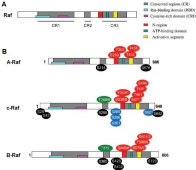
The structure of all three mammalian Raf kinases (Figure 1.4) can be divided into a regulatory N-terminal region and a catalytic C-terminal region. The N-terminus contains a primary Ras (rat sarcoma) binding site and a cysteine-rich secondary Ras binding site (54). The C-terminus contains a negative-charge regulatory region (N-region) and an activation segment, both containing multiple phosphorylation sites required for Raf activation (54,57), as well as an adenosine triphosphate (ATP) binding domain. Numerous regulatory phosphorylation sites, both activating and inhibitory, are also found throughout Raf (54).
Figure 1.4. Structure, conserved regions and regulatory phosphorylation sites
of Raf family kinases. (A) General structure of the Raf kinases with conserved
regions indicated. (B) Specific structures of A-Raf, c-Raf and B-Raf with activating
different set of complications (63,64). Although both types of mice demonstrate growth retardation, c-Raf knockout mice exhibit liver defects while B-Raf knockout mice exhibit vascular and neuronal deficiencies (63,64). Given the different phenotypes observed in these knockout mice and the apparent lack of compensation between Raf isoforms, and it is clear that A-Raf, B-Raf and c-Raf function differently despite their relatively conserved structure.
B-Raf, which is the strongest ERK pathway activator of the Raf family, has most recently generated interest due to the discovery of common oncogenic mutations in tumours, such as V600E (54). This mutation mimics phosphorylation of an activating site within the protein, resulting in a constitutively active form of B-Raf and persistent ERK pathway signaling (65). Prior to this discovery, however, c-Raf, the 70kDa isoform also known as Raf-1, was the primary isoform under investigation and thus still remains one of the best characterized Raf kinases (54).
1.3.2. ERK signaling pathway
Figure 1.5. Summary of ERK pathway signaling. Extracellular signals promote
ERK1/2 (54). Phosphorylated active ERK1/2 has over 150 reported targets in the cell, both nuclear and cytoplasmic (54,66).
For example, one of the outcomes of ERK1/2 phosphorylation is the activation of the transcription factor CREB (cAMP response element-binding protein) and the subsequent increase in transcription of certain anti-apoptotic factors, such as Bcl-2 and Bcl-XL (67). Other well-known targets of ERK1/2 include the transcription factors Elk1 (ETS domain-containing protein) and c-Fos (cellular FBJ murine osteosarcoma viral oncogene homolog), the kinases DAPK (death associated protein kinase) and MSK1/2 (mitogen- and stress-activated protein kinases 1/2) and the cytoskeletal element paxillin (66).
Furthermore, a number of scaffolding proteins have been shown to interact with components of the ERK pathway to facilitate signaling. The best-characterized scaffolds include KSR1 (kinase suppressor of Ras 1) and the IQGAP (IQ motif containing GTPase-activating protein) family of proteins, although there are many other scaffolds that have been reported to localize ERK pathway signaling to various compartments within the cell (55).
1.3.3. MEK1/2-independent signaling by c-Raf
Although c-Raf is a well-characterized kinase, it also affects signaling of some proteins in a kinase-independent manner. For instance, the pro-apoptotic proteins ASK1 (apoptosis signal-regulating kinase 1) and MST2 (mammalian Ste20-like kinase 2) are negatively regulated through direct binding with c-Raf (72,73). Rok-α (Rho-binding kinase α) is also inhibited solely by c-Raf binding, a phenomenon that regulates cell motility and protects against apoptosis (74,75).
1.3.4. c-Raf activation and deactivation
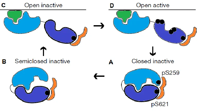
Figure 1.6. Summary of the activation cycle of c-Raf. (A) The N-terminal
regulatory region of c-Raf (light blue) sequesters the C-terminal catalytic region (dark blue) in a closed inactive conformation stabilized by 14–3–3 (orange). The interaction between c-Raf and 14–3–3 is stabilized by the phosphorylated residues (black) S259 and S621 on c-Raf. (B) S259 is dephosphorylated and 14–3–3 is
released from the c-Raf N-terminus, creating a semiclosed inactive conformation.
(C) c-Raf adapts an open inactive conformation, where the N-terminus unmasks the
C-terminus and binds membrane-bound Ras (green). (D) The C-terminus of c-Raf is
been proposed to increase kinase activity compared to monomeric or homodimeric versions of either protein (85).
During c-Raf deactivation, the phosphorylated N-region serves as a binding site for RKIP (Raf kinase inhibitor protein) (86), which dissociates MEK1/2 from c-Raf (87). PP5 (protein phosphatase 5) binds c-Raf and promotes the dephosphorylation of S338 (88) while PP2A dephosphorylates other activating sites (89). PKA (protein kinase A) has also been reported to contribute to c-Raf deactivation, phosphorylating S43 and S233, which interfere with Ras binding, as well as S259, which interferes with Ras binding and contributes to 14–3–3 binding (90,91). Altogether, these events return c-Raf to its inactive state, stabilized in a closed conformation by 14–3–3.
1.3.5. Regulation of c-Raf stability
In addition to the abundance of phosphorylation and dephosphorylation events that regulate c-Raf activity, there are also some systems known to regulate c-Raf stability and overall c-Raf levels within the cell. One well-known regulator of c-Raf stability is the chaperone protein Hsp90 (heat shock protein 90). Hsp90 is a highly conserved molecular chaperone that mediates the folding of newly synthesized or misfolded client proteins, assembles and disassembles molecular complexes and prevents protein aggregation (92). Hsp90 does not perform these tasks alone, however, as it has been shown to form complexes with over 20 co-chaperones (92).
degradation of c-Raf (97). There has been evidence that CHIP is able to ubiquitinate c-Raf, suggesting that CHIP is an E3 ubiquitin ligase responsible for proteasomal degradation of the kinase (98,99). XIAP (X-linked inhibitor of apoptosis), a member of the IAP (inhibitor of apoptosis) family of proteins, has been shown to be a modulator of CHIP-mediated c-Raf degradation. Although XIAP itself is an E3 ubiquitin ligase, evidence suggests that XIAP interferes with c-Raf stability and promotes recruitment of CHIP to Hsp90 and c-Raf, independently of its ubiquitin ligase activity (99).
There has also been evidence of c-Raf degradation by mechanisms that do not rely on CHIP. It has been reported that autophosphorylation of S621 is necessary for c-Raf stabilization, as kinase-dead mutants were ubiquitinated and targeted to the proteasome (100). This occurred even when CHIP levels were knocked-down by siRNA, suggesting that other E3 ubiquitin ligases may also play a role in c-Raf downregulation (100). c-Raf has also been shown to be ubiquitinated and degraded by the proteasome in response to disruption of cell adhesion and treatment with the oxidative glucose metabolite methylglyoxal, however the mechanisms by which these events occurred were not determined (101,102).
Hsp90, providing insight on a potential mechanism by which c-Raf could be de-stabilized by RanBPM (24). However, further studies exploring this concept have yet to be conducted and other mechanisms could also contribute to c-Raf downregulation by RanBPM.
1.4. Hypothesis and objectives
Work in our laboratory has shown that RanBPM and c-Raf are found together in a complex and that RanBPM downregulates c-Raf at the protein level (24). However, how the two proteins interact and the mechanism by which RanBPM downregulates c-Raf remains unknown. Therefore, it is hypothesized that specific domains of RanBPM are required for direct interaction with c-Raf and regulation of c-Raf stability by a mechanism that could involve the CTLH complex. The work presented in this thesis aims to specifically address the following objectives:
(1) Determine which domain(s) of RanBPM are required for regulation of c-Raf stability.
(2) Identify which domain(s) of RanBPM are required for interaction with c-Raf.
(3) Investigate the possibility of CTLH complex involvement in c-Raf downregulation.
Chapter 2 – Materials and Methods
2.1. Chemicals and reagents
All enzymes and buffers used for cloning were obtained from either New England Biolabs Inc. (Ipswich, MA, USA) or Fermentas Thermo Fisher Scientific Inc. (Waltham, MA, USA) and were used according to the manufacturer’s protocol. Hydrochloric acid (HCl) was also acquired from Thermo Fisher Scientific Inc. Fetal bovine serum (FBS), trypsin, L-glutamine, sodium pyruvate, paraformaldehyde (PFA), sodium chloride (NaCl), ethylenediaminetetraacetic acid (EDTA), hydroxyethyl piperazineethanesulfonic acid (HEPES), sodium dodecyl sulfate (SDS), Tris-hydroxymethyl amino methane (Tris) and potassium chloride (KCl) were purchased from Wisent Inc. (St. Bruno, QC, Canada), while Dulbecco’s modified eagle medium (DMEM) and phosphate buffered saline (PBS) were purchased from both Gibco by Life Technologies Inc. (Burlington, ON, Canada) and Wisent Inc. G418 sulphate, Triton X-100, Nonidet P-40 (NP40), aprotinin, leupeptin, pepstatin, dithiothreitol (DTT), sodium fluoride (NaF), sodium orthovanadate (NaVO4), pheylmethylsulfonyl fluoride (PMSF) and isopropyl β-D-1-thiogalactopyranoside (IPTG) were purchased from BioShop Inc. (Burlington, ON, Canada) while glycerol was acquired from Caledon Laboratory Chemicals Ltd. (Georgetown, ON, Canada).
2.2. Antibodies
sc-Cambridge, MA, USA). For each application, primary antibodies were used in the concentrations indicated in Table 2.1.
Secondary antibodies used for Western blot analyses were Peroxidase-conjugated AffiniPure Goat anti-Mouse IgG (H+L) (Jackson ImmunoResearch Laboratories Inc., West Grove, PA, USA) and Blotting Grade Goat anti-Rabbit IgG (H+L) (Human IgG Adsorbed) Horseradish Peroxidase Conjugate (Bio-Rad Laboratories Inc., Mississauga, ON, Canada). Both were used at a concentration of 1:5000.
2.3. Plasmid constructs
pCMV-HA-RanBPM was a gift from Dr. Mark Nelson (University of Arizona, Tucson, AZ, USA) and was rendered resistant to shRNA degradation via the introduction of two silent point mutations as described in (18). RanBPM deletion mutants RanBPM-ΔN2, RanBPM-ΔC4, pCMV-HA-RanBPM-ΔC1, pCMV-HA-RanBPM-Δ212, and pCMV-HA-RanBPM-Δ360 were generated as described in (18,103). pEBG-GST-ΔN-c-Raf was a gift from Dr. Zhijun Luo (Boston University, Boston, MA, USA).
Table 2.1. Concentrations of primary antibodies used for Western blot and in
situ proximity ligation assay.
Antibody Species Western blot
concentration
In situ proximity ligation assay concentration
RanBPM (K-12) goat N/A 1:400
RanBPM (F-1) mouse N/A 1:50
c-Raf (E-10) mouse 1:500 1:50
c-Raf (C-12) rabbit 1:500 N/A
β-actin (I-19) rabbit 1:2000 N/A
HA (HA-7) mouse 1:1000 N/A
GST (B-14) mouse 1:500 N/A
Hsp90 α/β (H-114) rabbit N/A 1:100
Table 2.2. PCR primer sequences and descriptions.
Primer Tm Sequence (5’ to 3’) Description
RBPMfwdBamHI 70.1°C GCTAGGATCCATGTCCGGGCAGCCGCCG
forward PCR primer to amplify
WT RanBPM and N2 domain
RBPMrevSalI 65.6°C CGCGGTACGTCGACTAATGTAGGTAGTCTTCC
reverse PCR primer to amplify
WT RanBPM and C1 domain
N2domrevSalI 68.8°C GTATGTCGACTACCCGCTGGCGGGGGC
reverse PCR primer to amplify
N2 domain
C1domfwdBamHI 64.3°C CGATGGATCCAAGGATGCATTCAGTCTACTAGC
forward PCR primer to amplify
2.4. Stable shRNA cell lines and cell culture
HeLa cells were obtained from the American Type Culture Collection (Manassus, VA, USA). HeLa cell lines stably expressing either RanBPM shRNA (clone 2-7) or control shRNA were generated as described in (18). HeLa 2-7 cells and HeLa control cells were cultured in DMEM supplemented with 8% FBS, 1% sodium pyruvate, 1% L-glutamine, 4.5g/L glucose and 0.35g/L G418 sulphate at 37°C in 5% CO2. Cells were washed with PBS and detached with trypsin upon passaging.
2.5. In situ proximity ligation assay
To prepare for the Duolink II in situ proximity ligation assay (PLA)
(Sigma-Aldrich Inc.), cover slips were pre-treated by outlining with the hydrophobic ImmEdge pen (Vector Laboratories Inc., Burlingame, CA, USA). HeLa cells were seeded on these cover slips at approximately 50,000 cells per cover slip, fixed with 4% PFA for 13 minutes at 4°C, permeabilized with 0.5% Triton X-100 for 10 minutes at room temperature, and blocked for 1 hour with 5% FBS in PBS at room temperature. Cover slips were incubated in the appropriate primary antibodies at the concentrations indicated in Table 2.1 overnight at 4°C. Manufacturer’s instructions were followed for the in situ PLA. Cover slips were
2.6. Transfection assays
ExGen 500 in vitro Transfection Reagent (Fermentas Thermo Fisher
Scientific Inc.), TurboFect Transfection Reagent (Thermo Fisher Scientific Inc.) and JetPRIME Transfection Reagent (PolyPlus Transfection, Illkirch, France) have all been used according to the manufacturer’s instructions for transfection of HeLa cells. For each pCMV-HA RanBPM deletion mutant, the amount of construct transfected was adjusted to account for variations in stability between the expressed proteins. In all cases, the amount of DNA used was brought up to the manufacturer’s recommendations with the vector pBS-SK (Agilent Technologies, Santa Clara, CA, USA). Transfected cells were incubated 24–48 hours at 37°C in 5% carbon dioxide (CO2).
2.7. Preparation of mammalian cell extracts
HeLa cells were scraped in cold PBS, centrifuged at 8000rpm for 3 minutes, lysed for 40 minutes on ice in whole cell extract (WCE) buffer (150mM NaCl, 1mM EDTA, 50mM HEPES pH 7.4 and 10% glycerol) and supplemented with 0.5% NP40, 0.5% Triton X-100, 10μg/mL aprotinin, 2μg/mL leupeptin, 2.5μg/mL pepstatin, 1mM DTT, 2mM NaF, 2mM NaVO4, and 0.1mM PMSF. The lysate was centrifuged at 13,000rpm for 20 minutes at 4°C and the resulting supernatant was collected.
2.8. Bacterial protein expression and preparation of Escherichia coli
extracts
For each bacterial expression construct, plasmids were transformed into
E. coli strain BL21DE3. Single transformants were selected and grown in Luria
overnight at 16°C. Bacteria was centrifuged at 4000rpm for 20 minutes at 4°C and subsequently resuspended in lysis buffer (25mM HEPES pH 7.4, 10mM KCl, 2mM EDTA and 20% glycerol) supplemented with 0.1% NP40, 10μg/mL aprotinin, 2μg/mL leupeptin, 2.5μg/mL pepstatin, 1mM DTT, 2mM NaF, 2mM NaVO4, and 0.1mM PMSF. The cell suspension was sonicated three times for 10 seconds on ice using the Sonic Dismembrator Model 100 (Thermo Fisher Scientific Inc.), centrifuged at 13,000rpm for 10 minutes at 4°C and the resulting supernatant was collected.
2.9. Western blot analyses
Samples were resolved by sodium dodecyl sulfate polyacrylamide gel electrophoresis (SDS-PAGE) on either 8% or 10% acrylamide gels and subsequently transferred for either for 1 hour at 100V or overnight at 25V onto a polyvinylidene fluoride (PVDF) membrane. Blots were blocked in 5% non-fat dry milk for at least 1 hour at room temperature, then incubated in primary antibody diluted in 5% non-fat dry milk as indicated in Table 2.1 overnight at 4°C or for 1 hour at room temperature. Blots were incubated in secondary antibody diluted in 5% non-fat dry milk for 1 hour at room temperature and developed using either Western Lightning Enhanced Chemiluminescence (ECL) Substrate (Perkin Elmer Inc., Waltham, MA, USA) or Clarity Western ECL Substrate (Bio-Rad Laboratories Inc.). Images were captured using either Kodak X-OMAR LS film (Carestream Health Inc., Rochester, NY, USA) or the ChemiDoc MP (Bio-Rad Laboratories Inc.) and Image Lab software (Bio-Rad Laboratories Inc.).
2.10. GST pull-down assays
2.10.1. Using HeLa cell extracts
concentration of 0.4% NP40, 0.4% Triton X-100, 20μg/mL aprotinin, 4μg/mL leupeptin, 5μg/mL pepstatin, 2mM DTT, 4mM NaF, 4mM NaVO4, and 0.2mM PMSF. Glutathione-Agarose beads (Sigma-Aldrich Inc.) suspended in PBS were added to each sample to a final concentration of 5μL beads/100μg total protein and pull-down samples were incubated overnight at 4°C. Beads were subsequently washed three times in WCE buffer supplemented with 0.4% NP40, 0.4% Triton X-100, 1mM DTT and 0.1mM PMSF. Beads were resuspended in SDS loading dye (0.105g/mL SDS, 0.093g/mL DTT, 0.35M Tris HCl pH 6.8 and 30% glycerol), boiled for 5 minutes and centrifuged at 10,000rpm for 10 seconds. The resulting supernatant was collected and analyzed by Western blot.
2.10.2. Using E. coli extracts
2.11. Statistical analyses
Chapter 3 – Results
3.1. Endogenous RanBPM and c-Raf are found in a complex
Previous co-immunoprecipitation and pull-down experiments in our laboratory have demonstrated that RanBPM and c-Raf exist together in a complex (24). Since this was shown using ectopically expressed protein constructs, we sought to confirm that the complex occurs with the endogenous proteins in cells. The in situ PLA, an assay that allows visualization of
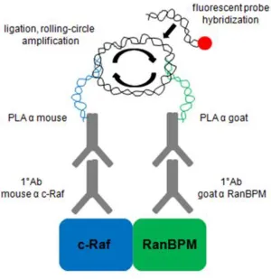
protein-protein interactions in cells using fluorescence microscopy (Figure 3.1) (104), was thus used to visualize the interaction between endogenous RanBPM and c-Raf in HeLa cells.
To summarize the PLA, fixed and permeabilized cells are incubated with primary antibodies against two proteins of interest. Two types of specialized secondary antibodies fused to short DNA strands, called PLA probes, are then incubated with the cells to bind their respective primary antibodies. When two different PLA probes come within 40nm of each other, the DNA strands are ligated together and amplified by a polymerase in a process termed rolling-circle amplification. The amplified DNA product is then hybridized with a fluorescently labeled complementary oligonucleotide probe, which is then visualized by fluorescence microscopy. Each fluorescent dot seen represents a protein-protein interaction between the two proteins of interest.
Figure 3.1. Summary of the in situ PLA. Primary antibodies against two proteins of
Figure 3.2. Endogenous RanBPM and c-Raf are found in a complex. An in situ
PLA was performed in (A) HeLa control cells, without the addition of primary
antibodies (negative control); (B) HeLa 2-7 cells, using primary antibodies against
c-Raf and RanBPM (negative control); (C) HeLa control cells, using primary antibodies
against Hsp90 and RanBPM (positive control); (D) HeLa control cells, primary
Given that c-Raf is known to interact with Hsp90 (96), a positive control was included where HeLa control cells were incubated with antibodies against c-Raf and Hsp90. This did expectedly produce fluorescent dots representing interactions (Figure 3.2C). In HeLa control cells in which antibodies against c-Raf and RanBPM were included for the assay, fluorescent dots were observed, confirming that the two endogenously expressed proteins are found together in a complex(Figure 3.2D).
3.2. The N-terminus, CRA and LisH/CTLH domains of RanBPM are required
for c-Raf downregulation
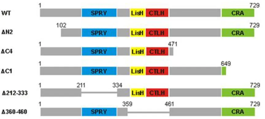
Figure 3.3. Full-length RanBPM and deletion mutants chosen for analysis. WT,
As previously reported (24), WT RanBPM was able to significantly downregulate ΔN c-Raf compared to the levels of ΔN c-Raf seen in the pCMV-HA control, resulting in a 2.08 ± 0.44 fold decrease of ΔN c-Raf expression (Figures 3.4A-D). Furthermore, Δ212 RanBPM was also able to downregulate ΔN c-Raf compared to the levels of ΔN c-Raf seen in the pCMV-HA control, demonstrating a significant 1.58 ± 0.45 fold decrease of ΔN c-Raf expression (Figures 3.4A and 3.4C). Although it would appear from the representative image that Δ212 RanBPM was able to downregulate ΔN c-Raf better than WT RanBPM, Δ212 RanBPM actually demonstrated better expression in this experiment and therefore more protein was likely available to exert its effect on ΔN c-Raf (Figure 3.4A). Overall, the effects that Δ212 RanBPM and WT RanBPM demonstrated on ΔN c-Raf were not significantly different from one another (Figure 3.4C), indicating that the SPRY domain is not required for c-Raf destabilization.
ΔN2 RanBPM, however, was unable to effectively downregulate ΔN c-Raf and resulted in a significant 3.14 ± 0.60 fold increase when compared to the levels of ΔN c-Raf seen in response to WT RanBPM (Figures 3.4A and 3.4C). ΔN c-Raf expression in response to this mutant was not significantly different than that observed in the pCMV-HA control (Figure 3.4A and 3.4C). The expression of ΔN2 RanBPM is consistently much lower than that of the other RanBPM deletion mutants, despite identical transfection conditions, and thus our laboratory has hypothesized that the protein is very unstable (16,18). Despite this phenomenon, levels of ΔN2 RanBPM near those of WT RanBPM were achieved in this experiment (Figure 3.4A) and therefore it is reasonable to conclude that ΔN2 RanBPM has lost its ability to downregulate ΔN c-Raf. This implies that the N-terminus of RanBPM is required for its effect on c-Raf.
Figure 3.4. Δ360, ΔN2, ΔC4 and ΔC1 RanBPM deletion mutants do not
effectively downregulate ΔN c-Raf compared to WT RanBPM. HeLa 2-7 cells
were transfected with pEBG-GST-ΔN-c-Raf and either pCMV-HA, pCMV-HA-RanBPM or pCMV-HA pCMV-HA-RanBPM deletion mutant constructs. Extracts were analyzed by Western blot. (A,B) Representative images are shown. (C,D) Multiple
These results suggest that the LisH/CTLH domains and the C-terminus of RanBPM play a role in c-Raf downregulation. However, considering that the ΔC4 deletion removes a very large portion of RanBPM, we decided to repeat the experiment using a construct harboring only a deletion of the CRA domain, namely the ΔC1 RanBPM construct. ΔC1 RanBPM behaved nearly identically to ΔC4 RanBPM in its inability to downregulate ΔN c-Raf and resulted in a 3.76 ± 1.15 fold increase when compared to the levels of ΔN c-Raf seen in response to WT RanBPM, (Figures 3.4B and 3.4D). This implies that within the C-terminus of RanBPM, it is specifically the CRA domain that is needed for c-Raf downregulation.
Altogether, these results demonstrate that the N-terminus, LisH/CTLH and CRA domains are required for c-Raf destabilization, since loss of any of these regions render RanBPM unable to effectively downregulate c-Raf.
3.3. The CRA domain of RanBPM is required for interaction with c-Raf
To continue to characterize the interaction between RanBPM and c-Raf, we aimed to determine the domain or domains of RanBPM required for the interaction. To accomplish this, we used the same system outlined in section 3.2 and used the extracts to perform GST pull-down assays to test which RanBPM deletion mutants have retained their ability to interact with ΔN c-Raf.
Specifically, HeLa 2-7 cells were transfected with either pEBG-GST and pCMV-HA-RanBPM, or pEBG-GST-ΔN-c-Raf and either pCMV-HA-RanBPM or a pCMV-HA RanBPM deletion mutant. Due to its poor stability, ΔN2 RanBPM was not among the mutants tested as we were unable to obtain sufficient levels of the protein to detect it in this type of assay. Cell extracts were prepared and Glutathione-Agarose beads were incubated with the resulting extracts to pull-down GST or GST-ΔN-c-Raf, as well as any RanBPM deletion mutant associated with it. Pull-down samples were analyzed by Western blot.
the negative control, GST alone only pulled-down background levels of WT RanBPM (Figures 3.5A-C). Δ212 RanBPM and Δ360 RanBPM both retained their abilities to interact with ΔN c-Raf, as levels of pulled-down RanBPM were not significantly different than those of the positive control but were significantly higher than those of the negative control (Figures 3.5A-C). This indicates that the SPRY and LisH/CTLH domains are not required for the interaction between RanBPM and c-Raf, since deletion of these regions does not nullify the interaction.
However, ΔC1 RanBPM was not able to effectively interact with ΔN c-Raf, as the amount of ΔC1 RanBPM associating with ΔN c-Raf resulted in a significant 2.12 ± 0.19 fold decrease compared to the amount of WT RanBPM associating with ΔN c-Raf, but was not significantly different than the level of interaction seen in the negative control (Figures 3.5A and 3.5C). Altogether, this data suggests that the CRA domain is the only domain tested that appears to be required for the interaction between RanBPM and c-Raf, since deletion of this region abolishes the interaction.
3.4. RanBPM interacts directly with c-Raf through the CRA domain
Further tests were needed to confirm the interaction between the CRA domain of RanBPM and c-Raf. Also, the nature of the interaction, whether it be direct or mediated by another factor, remained to be determined. Therefore, to address this matter, we aimed to repeat pull-down experiments using bacterial extracts. By expressing our mammalian RanBPM and c-Raf constructs in E. coli,
we ensured that no other mammalian proteins were present to mediate the interaction between our two proteins of interest. Thus, if the interaction was to persist in this system, it was assumed to be direct.
Figure 3.5. ΔC1 RanBPM is unable to interact effectively with ΔN c-Raf. HeLa
2-7 cells were transfected with either GST and pCMV-HA-RanBPM or pEBG-GST-ΔN-c-Raf and either pCMV-HA-RanBPM or a pCMV-HA RanBPM deletion mutant. A GST down assay was performed on the resulting extracts and pull-downs were analyzed by Western blot. (A,B) Representative images are shown. (C)
Figure 3.6. Full-length RanBPM and individual domains chosen for analysis.
domain, which remained unexamined due to the poor level of expression of the ΔN2 RanBPM deletion mutant in previously conducted mammalian cell-based GST pull-down assays (Figure 3.5).
Each GST-tagged construct, as well as GST alone, was expressed separately in E. coli and purified using Glutathione-Agarose beads. These
constructs were each subsequently incubated with a crude cell lysate from E. coli
expressing ΔN c-Raf. The GST-tagged constructs were pulled-down and analyzed by Western blot to detect levels of associated ΔN c-Raf.
Both GST-WT-RanBPM and GST-C1 were able to pull-down ΔN c-Raf significantly above background levels pulled down by GST alone, demonstrating 5.02 ± 0.98 and 2.71 ± 0.36 fold increases, respectively (Figures 3.7A and 3.7B). Although it appears that WT RanBPM associates with ΔN c-Raf better than the C1 domain does, the variability within the levels of ΔN c-Raf pulled-down with WT RanBPM was relatively high and in fact the amount of ΔN c-Raf pulled-down with WT RanBPM is not significantly different than that pulled-down with the C1 domain (Figure 3.7B). Therefore, this result confirms that the CRA domain of RanBPM is able to interact with c-Raf and the interaction between RanBPM and c-Raf is direct.
GST-N2, unlike GST-WT-RanBPM and GST-C1, was unable to pull-down ΔN c-Raf significantly better than GST alone (Figures 3.7A and 3.7B). This suggests that the N-terminus of RanBPM is unable to directly interact with c-Raf, although an indirect interaction cannot be ruled out based on these results.
3.5. Endogenous c-Raf and MAEA are found in a complex
Figure 3.7. WT RanBPM and C1 domain interact directly with ΔN c-Raf. GST
pull-down assays were performed using GST, GST-WT-RanBPM, GST-N2-domain and GST-C1-domain E. coli extracts as well as ΔN c-Raf E. coli extracts. Pull-downs
were analyzed by Western blot. (A) A representative image is shown. (B) Multiple
investigate whether MAEA, a CTLH complex protein, is able to form a complex with c-Raf.
Figure 3.8. Endogenous MAEA and c-Raf are found in a complex. An in situ PLA
was performed in HeLa cells (A) without the addition of primary antibodies (negative
control), (B) using primary antibodies against MAEA and RanBPM (positive control)
and (C) using primary antibodies against MAEA and c-Raf. DAPI staining was used
Chapter 4 – Discussion
4.1. Summary of findings
The aim of this study was to characterize the interaction between RanBPM and c-Raf. We hypothesized that specific domains of RanBPM are required for direct interaction with c-Raf and regulation of c-Raf stability by a mechanism involving the CTLH complex. Specifically, we sought to determine
which domain(s) of RanBPM are required for regulation of c-Raf stability, identify which domain(s) of RanBPM are required for interaction with c-Raf and investigate the mechanism by which RanBPM downregulates c-Raf. In summary, we found that RanBPM and c-Raf in fact do form a complex in cells. The N-terminus, CRA domain and LisH/CTLH domains of RanBPM are required for downregulation of c-Raf but only the CRA domain is required for complex formation with c-Raf (Table 4.1). RanBPM interacts directly with c-Raf and the CRA domain is sufficient for this direct interaction to occur. Finally, the CTLH complex member MAEA and c-Raf are also found together in a complex, suggesting that c-Raf could associate not only with RanBPM and MAEA, but with the entire CTLH complex.
4.2. Model and rationale
Table 4.1. Summary of results of the effects of RanBPM constructs on downregulation of ΔN c-Raf and interaction with ΔN c-Raf.
RanBPM construct Downregulates ΔN c-Raf Interacts with ΔN c-Raf
WT RanBPM YES YES
Δ212 RanBPM (ΔSPRY) YES YES
ΔC4 RanBPM (ΔC-terminus) NO NO
ΔC1 RanBPM (ΔCRA) NO NO
Δ360 RanBPM (ΔLisH/CTLH) NO YES
ΔN2 RanBPM (ΔN-terminus) NO ND
C1 domain (CRA) ND YES
Figure 4.1. Model of the mechanism by which RanBPM downregulates c-Raf.
Therefore, it is hypothesized that some other protein or protein complex is interacting with RanBPM through its LisH/CTLH domains to downregulate c-Raf. Deletion of the N-terminus of RanBPM also resulted in a loss of c-Raf downregulation and this region was also shown to be unable to interact directly with c-Raf. Therefore, the N-terminus might aid the LisH/CTLH domains in mediating the interaction between RanBPM and the unidentified complex potentially responsible for downregulating c-Raf. This is plausible since the N-terminus is a potentially flexible proline-rich region of RanBPM which could fold over to stabilize the protein or, in this case, stabilize interactions with other proteins (16). Given that deletion of the SPRY domain did not perturb the ability of RanBPM to downregulate or interact with c-Raf, we propose that this domain does not participate in regulation of c-Raf.
We have presented a plausible mechanism which suggests the involvement of an additional protein or protein complex in the downregulation of c-Raf. We propose that c-Raf might be targeted for degradation by the CTLH complex in a RanBPM-dependent manner, with c-Raf being tethered to the complex by RanBPM through its CRA domain, and that the CTLH complex interacts with RanBPM primarily through its LisH/CTLH domains. RanBPM is a known member of the CTLH complex and our results show that c-Raf can associate with both RanBPM and MAEA, another member of the CTLH complex. This mammalian complex is comprised of six proteins in total, all of which have known orthologs in S. cerevisiae that form the yeast Gid complex (6). Given that
with other members of the complex mainly through its LisH and CTLH domains (47).
Although the major piece of evidence in our study tying the CTLH complex to c-Raf downregulation is the fact that c-Raf and MAEA associate in cells, subtleties in our data are also consistent with the idea that the CTLH complex may be involved in c-Raf downregulation. The number of interactions seen between c-Raf and MAEA were noticeably fewer than the number of interactions seen in the positive control between RanBPM and MAEA. This is compatible with the idea that RanBPM and MAEA are fixed members of the CLTH complex, whereas c-Raf may only associate with the complex temporarily to target it for degradation. Also, previous studies in our laboratory have shown that RanBPM has a greater effect on the active form of c-Raf, which represents only a fraction of the total pool of endogenous c-Raf in the cell (24). This further supports the observation that Raf associates with MAEA infrequently, as only activated c-Raf may be targeted for degradation by RanBPM, MAEA and the rest of the CTLH complex.
A large number of studies have described proteins that interact with RanBPM, albeit they seldom provide functional significance for the interactions. A number of these interacting proteins have been described in Chapter 1, but a broader record has been retrieved from the BioGRID (Biological General Repository for Interaction Datasets) version 3.2.114 (Figure 4.2) (105). In addition to the evidence discussed in Chapter 1 of ARMc8, Rmnd5a and MAEA being involved in ubiquitination, the collection of proteins retrieved from the BioGRID that interact with RanBPM involved in ubiquitination, deubiquitination, ubiquitin-like modification or management of ubiquitinated proteins give further weight to the argument that RanBPM and the CTLH complex may play a role in these processes.
Figure 4.2. RanBPM interactome retrieved from the BioGRID version 3.2.114
showing 71 proteins that interact with human RanBPM. Interactions between
misfolded or damaged proteins in conditions where the proteasome and the UPS are overwhelmed (106). This crucial role in aggresome formation implies that RanBPM has the ability to handle ubiquitinated proteins targeted for degradation. In addition, RanBPM has been found to interact with the E3 ubiquitin ligase TRAF6 (tumor necrosis factor-receptor-associated factor 6) (Figure 4.2) and to reduce the TGF-β (transforming growth factor β) dependent auto-ubiquitination of TRAF6 (107). Some high-throughput screens studying the ubiquitinome have even found RanBPM among a pool enriched for ubiquitinated proteins (Figure 4.2) (108). This may simply be evidence of RanBPM itself being targeted for ubiquitination, given that RanBPM has been shown to be ubiquitinated as well as deubiquitinated specifically through interaction with USP11 (ubiquitin-specific protease 11) (Figure 4.2) (109). However, there is still substantial reason to suspect that RanBPM, in concert with the CTLH complex, could be playing a role in protein ubiquitination and subsequent degradation by the proteasome.
c-ubiquitinate c-Raf and send it for proteasomal degradation, it has also been suggested that c-Raf may be ubiquitinated and degraded by CHIP-independent mechanisms (98-100).
4.3. Significance of the CRA domain as a binding-domain for c-Raf
Many studies have identified binding partners for RanBPM and suggested roles for the protein in various cellular processes, but a clear function for RanBPM has yet to be elucidated. Our study further contributes to our growing knowledge of the protein and demonstrates the importance of the CRA domain of RanBPM for its interaction with c-Raf. Some studies have already identified certain regions of RanBPM to be required for interactions with specific proteins. For example, the SPRY domain of RanBPM has been shown to be required for interaction with CDK11p46, L1, MET, TrkA, AR and USP11 (7,8,20,25,35,109). The SPRY domain, along with the LisH domain, has been shown to be sufficient for interaction with BACE1, LRP and APP to increase Aβ generation (29). The LisH and CTLH domains have both been shown to be required for RanBPM interaction with HDAC6 and only FMRP has previously been shown to interact with the CRA domain of RanBPM (14,103). Our study defines c-Raf as only the second protein to be found to interact with RanBPM through the CRA domain and further confirms this domain to be a protein interaction surface.