Scholarship@Western
Scholarship@Western
Electronic Thesis and Dissertation Repository
5-9-2019 10:00 AM
Investigating Grip Range of Motion and Force Exerted by
Investigating Grip Range of Motion and Force Exerted by
Individuals with and without Hand Arthritis during Functional
Individuals with and without Hand Arthritis during Functional
Tasks and while Swinging a Golf Club
Tasks and while Swinging a Golf Club
Sara Frances Holland
The University of Western Ontario Supervisor
Lalone, Emily
University of Western Ontario; Roth McFarlane Hand and Upper Limb Centre (HULC), St. Joseph's Healthcare Centre Co-Supervisor
Ferreira, Louis
University of Western Ontario; Roth McFarlane Hand and Upper Limb Centre (HULC), St. Joseph's Healthcare Centre
Graduate Program in Mechanical and Materials Engineering
A thesis submitted in partial fulfillment of the requirements for the degree in Master of Engineering Science
© Sara Frances Holland 2019
Follow this and additional works at: https://ir.lib.uwo.ca/etd
Part of the Biomechanical Engineering Commons, and the Sports Studies Commons
Recommended Citation Recommended Citation
Holland, Sara Frances, "Investigating Grip Range of Motion and Force Exerted by Individuals with and without Hand Arthritis during Functional Tasks and while Swinging a Golf Club" (2019). Electronic Thesis and Dissertation Repository. 6214.
https://ir.lib.uwo.ca/etd/6214
This Dissertation/Thesis is brought to you for free and open access by Scholarship@Western. It has been accepted for inclusion in Electronic Thesis and Dissertation Repository by an authorized administrator of
i
Hand arthritis is the leading cause of disability in individuals over the age of 50;
resulting in dysfunction and pain, making activities of daily living and recreational activities
such as golf difficult. Few studies have been conducted on the biomechanical response of
individuals with hand arthritis when performing functional activities. This research quantified
hand grip movements and strength differences seen in individuals with hand arthritis. Using a
video-based motion capture system (Dartfish), a grip limitation of 17.2% (maximum flexion),
and 12.7% (maximum extension) was discovered. A wireless finger force measurement system
(FingerTPS), was used to show that larger diameter, softer firmness golf grips assisted in
reducing the grip force in individuals with and without hand arthritis during a golf swing. This
research will benefit the sport biomechanics and clinical fields, providing quantitative results
to develop more sophisticated joint protection devices and gain a better understanding of hand
arthritis mechanics.
Keywords
Activities of daily living (ADL), Dartfish, FingerTPS, golf grips, hand arthritis, golf
ii
Co-Authorship Statement
Chapter 1: Sara Holland – wrote manuscript
Emily Lalone – reviewed manuscript
Chapter 2: Sara Holland – data collection, statistical analysis, wrote manuscript
Emily Lalone – study design, statistical analysis, reviewed manuscript
Joy MacDermid – statistical analysis review, reviewed manuscript
Kathryn Sinden – assisted in the study design
Louis Ferreira – reviewed manuscript
Jim Dickey – reviewed manuscript
Timothy Burkhart – assisted in the statistical analysis
Ahmed Tanashi – assisted in data collection
Lauren Straatman – assisted in data collection
Chapter 3: Sara Holland – study design, data collection, statistical analysis, wrote
manuscript
Emily Lalone – study design, statistical analysis, reviewed manuscript
Joy MacDermid – statistical analysis review, reviewed manuscript
Timothy Burkhart – assisted in the statistical analysis
Baraa Daher – assisted in the data analysis and collection
Amelia Carver – assisted in the data analysis and collection
Ahmed Tanashi – assisted in data collection
Chapter 4: Sara Holland – wrote manuscript
iii
Acknowledgments
I would like to acknowledge and thank those who assisted me throughout this project.
Firstly, a tremendous thanks to Dr. Jim Dickey for allowing me to use your Biomechanics
Laboratory for my testing and for always finding time to assist me with various components
of my project. I would also like to thank Dr. Joy MacDermid for funding the purchase of the
FingerTPS sensor system. I am very grateful for your guidance on the statistical approaches
needed for my study, and for your insight into the clinical side of the biomechanics field. A
huge thanks to Paul Sheller and Stephen Mallinson of the Western Engineering Finance and
Stores (EFS), for dealing with all of my warranty returns, travel issues, and for participating in
my study. Thank you to my co-supervisor Dr. Louis Ferreira, for expanding my academic
knowledge throughout my time at Western. I appreciate the time that you spent advising me
on troubleshooting techniques and on the structure of my study protocol.
To Marc Elliott and Marc Webster – you two were extremely helpful throughout this
entire process, running trial tests, being there to bounce ideas off of, recruiting participants,
and participating in my study yourselves. I also want to thank you for connecting me with
Martin Neil, the Ping representative at Sunningdale Golf and Country Club, who supplied me
with the golf clubs used in my project.
Most importantly I would like to thank my supervisor Dr. Emily Lalone. I cannot begin
to express the gratitude and respect that I have for you, and the mentorship that you provided
me over the course of this project. You gave me the opportunity to combine my passion for
golf with my engineering background and I cannot thank you enough!
Thank you to the members of the Human Biomechanics Laboratory: Ahmed, Madeline,
Michael, Andrea, and Puneet. Special thanks to Ahmed Tanashi for helping me during testing,
conducting study statistics, and for being such a great person to work alongside. Thank you to
all the students who helped keep this project moving smoothly: Lauren, Sydney, Baraa, Amelia
and Kevin. I would also like to thank all of my friends, specifically my best friend Michaela
Khan of the Wolf Orthopedic Biomechanics Laboratory at Western, for keeping me sane and
supporting me in so many ways throughout the last few years.
Lastly, I’d like to thank my parents, Scott and Shirley Holland, who always support
and encourage me to do my best and to never stop trying, even when things get tough or do
iv
Table of Contents
Abstract ... i
Co-Authorship Statement... ii
Acknowledgments... iii
Table of Contents ... iv
List of Tables ... viii
List of Figures ... x
Symbols and Acronyms ... xiv
Chapter 1 – Introduction ... 1
1 Overview ... 1
1.1 Anatomy ... 1
1.1.1 Distal Upper Extremity ... 1
1.1.2 Biomechanics of the Hand and Wrist ... 7
1.1.3 Grip Range of Motion ... 8
1.1.4 Grip Strength ... 9
1.2 Hand Arthritis ... 10
1.2.1 Hand Arthritis in Relation to Grip Strength and Range of Motion... 11
1.2.2 Joint Protection Programs ... 11
1.3 Joint Motion Analysis ... 13
1.4 Grip Force Measurement Systems ... 14
1.5 Golf Background ... 16
1.5.1 Golf Terminology ... 16
1.5.2 Golf Biomechanics... 17
1.5.3 Golf Grips and Gloves ... 18
v
1.7 Objectives and Hypothesis ... 20
1.8 Thesis Overview ... 21
Chapter 2 – The Development and Validation of a Novel Grip Motion Analysis Technique to Evaluate Hand Motion During Activities of Daily Living... 22
2 Overview ... 22
2.1 Introduction ... 22
2.2 Materials and Methods ... 25
2.2.1 Participants ... 25
2.2.2 Data Collection ... 26
2.2.3 Data Processing ... 31
2.2.4 Grip Configuration ... 32
2.2.5 Statistical Analysis ... 33
2.2.6 Validation ... 34
2.2.7 Assumptions ... 36
2.3 Results ... 36
2.3.1 Finger Sleeves Analysis ... 41
2.3.2 Reliability Analysis ... 41
2.4 Discussion ... 45
2.5 Conclusion ... 47
Chapter 3 – Investigating the Grip Forces Exerted by Individuals with and without Hand Arthritis while Swinging a Golf Club with the Use of a New Wearable Sensor Technology ... 49
3 Overview ... 49
3.1 Introduction ... 49
3.2 Study Protocol ... 52
vi
3.2.3 Data Processing ... 61
3.2.4 Statistical Analysis ... 62
3.2.5 Assumptions ... 63
3.3 Results ... 63
3.3.1 Golf Grip Configuration Results ... 63
3.3.2 Golf Grip Force Results ... 65
3.3.3 Reliability Analysis ... 71
3.4 Discussion ... 73
3.4.1 Strengths and Limitations ... 77
3.5 Conclusion ... 80
Chapter 4 – Conclusion ... 82
4 Overview ... 82
4.1 Summary ... 82
4.2 Strengths and Limitations ... 85
4.3 Future Direction ... 87
4.3.1 Future Golf Grip Design and Grip Measurement System ... 88
4.4 Significance... 89
Bibliography ... 92
Appendices ... 99
Appendix A – Synovial Joint Structure (Chapter 1) ... 99
Appendix B – Gripping Style Variations (Chapter 2) ... 100
Appendix C – Specifications of the Golf Grips (Chapter 3) ... 102
Appendix D – Study Letter of Information and Consent Form (Chapter 3) ... 106
Appendix E – Participant Survey and Data Results (Chapter 3) ... 109
vii
Appendix H – Ethics Approval (Chapter 3) ... 129
Appendix I – Golf Grip Force Study Supplementary Information (Chapter 3) ... 130
viii
List of Tables
Table 1: Demographic information of participants. ... 26
Table 2: Average maximum extension and average maximum flexion angles (in degrees) for
the individuals with and without hand arthritis (n=20 healthy participants: n=20 participants
with hand arthritis). ... 32
Table 3: The average grip configuration values of the two test groups for the five activities of
daily living including the positive standard deviation (SD) and upper and lower 95%
confidence interval (CI) values (n=20 healthy participants: n=20 participants with hand
arthritis). ... 38
Table 4: The average maximum flexion and extension angles for the index PIP and MCP, and
thumb MCP and CMC joints of the healthy participants and participants with hand arthritis.
The positive standard deviation (SD) values are also presented (n=20 healthy participants:
n=20 participants with hand arthritis). ... 39
Table 5: Demographic of Chapter 3 golf grip force population. ... 53
Table 6: The 12 standard and ‘arthritic’ designed golf grips used for testing, with material,
and geometry (diameter) size categorization. The grip order presented (top to bottom) pairs
with the grip order from left to right in Figure 22 [54], [77], [80], [81]. ... 55
Table 7: The grip configuration percentage values for the healthy participants and
participants with hand arthritis. The upper and lower 95% confidence intervals (CI) are
presented for each heath status, the positive standard deviations (SD), and the overall
percentage difference between the two groups (n=10 healthy participants: n=8 participants
with hand arthritis). ... 65
Table 8: The average grip forces for each of the 12 grips and the golf grip strength values for
the healthy participants, organized from highest to lowest force output. The upper and lower
95% confidence intervals (CI), and the positive standard deviations (SD) are also presented
ix
the individuals with hand arthritis, organized from highest to lowest force output. The upper
and lower 95% confidence interval (CI), and the positive standard deviations (SD) are also
x
List of Figures
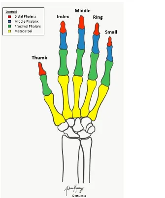
Figure 1: Exhibiting the 19 hand bone locations of the distal phalanx, middle phalanx,
proximal phalanx, and metacarpal bones. ... 3
Figure 2: Joint locations of the hand with the distal interphalangeal (DIP), proximal
interphalangeal (PIP), metacarpophalangeal (MCP), and carpometacarpal (CMC) joints of
the finger segments and the interphalangeal (IP), MCP, and basal CMC of the thumb
identified. ... 4
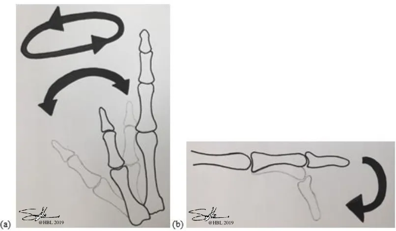
Figure 3: Long finger segments range of motion capabilities of (a) the MCP joint for radial
and ulnar deviation, and (b) the DIP, PIP, and MCP joints for flexion/extension (lateral
view). ... 5
Figure 4: The thumb segment range of motion capabilities of (a) the basal CMC joint for
flexion/extension, radial/ulnar deviation, and circumduction, and (b) the IP and MCP joints
for flexion/extension (lateral view). ... 5
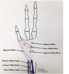
Figure 5: Muscles of the hand which move the thumb through its respective ranges of
motion. ... 6
Figure 6: Range of motion (ROM) capabilities of the hand demonstrating (a) maximum
extension, and (b) maximum flexion. ... 9
Figure 7: Joint protection tools and techniques to avoid positions that foster deformity. The
images on the left represent the positions to avoid where the images on the right incorporate
joint protection adaptions. ... 13
Figure 8: Progression of the golf swing broken down into three phases being the backswing,
downswing, and follow through. ... 17
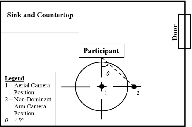
Figure 9: Testing room camera positions set up, shown as a schematic sketch of the room
with camera positions for a right hand dominant participant (not scaled to the measurements
xi
MCP and PIP joints for the healthy participants (n=20) during maximum extension (a) with
the finger sleeves, and (b) without the finger sleeves. ... 30
Figure 11: Electromagnetic (EM) tracking system in vivo sensor placement on the thumb
distal phalanx, proximal phalanx, and the metacarpal as well as the index finger distal,
middle, and proximal phalanges. ... 35
Figure 12: Validation measurement test comparing the Dartfish Movement Analysis
software, the EM tracking system, and a manual goniometer. ... 35
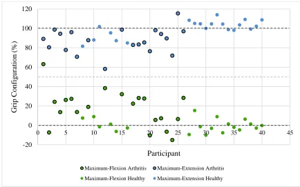
Figure 13: Maximum flexion and maximum extension grip configuration values. The
horizontal dashed lines represent the healthy baseline threshold values at 0% and 100%. A
mid-range is also defined at 50% with points above being in extension and below in flexion
(n=20 healthy participants: n=20 participants with hand arthritis). ... 37
Figure 14: The average grip configuration values comparing the individuals with and without
hand arthritis to demonstrate the difference between the five activities of daily living
evaluated. Positive standard deviation (SD) bars and significance indications (*) are also
included (n=20 healthy participants: n=20 participants with hand arthritis). ... 38
Figure 15: The average joint flexion angles comparing the individuals with and without hand
arthritis to demonstrate the differences between each of the joints evaluated. Positive
standard deviation (SD) bars and significance indications (*) are also included (n=20 healthy
participants: n=20 participants with hand arthritis). ... 40
Figure 16: The average joint extension angles comparing the individuals with and without
hand arthritis, to demonstrate the differences between each of the joints evaluated. Positive
standard deviation (SD) bars and significance indications (*) are also included (n=20 healthy
participants: n=20 participants with hand arthritis). ... 40
Figure 17: Bland-Altman Graph for the inter-rater reliability analysis for maximum flexion
activity. The upper and lower 95% are represented by the horizontal red lines, calculated
from the mean flexion angle values between each rater for the five trials and the total mean
xii
activity. The upper and lower 95% are represented by the horizontal red lines, calculated
from the mean flexion angle values between each rater for the five trials and the total mean
within both raters. The total mean value is represented by the horizontal blue line... 43
Figure 19: Bland-Altman Graph for the intra-rater reliability analysis for maximum flexion
activity. The upper and lower 95% are represented by the horizontal red lines, calculated
from the mean flexion angle values between each rater for the five trials and the total mean
within both raters. The total mean value is represented by the horizontal blue line... 43
Figure 20: Bland-Altman Graph for the intra-rater reliability analysis for maximum extension
activity. The upper and lower 95% are represented by the horizontal red lines, calculated
from the mean flexion angle values between each rater for the five trials and the total mean
within both raters. The total mean value is represented by the horizontal blue line... 44
Figure 21: Golf study test set-up in the Thames Hall Biomechanics Laboratory at Western
University. ... 54
Figure 22: The 12 golf grips tested in this study from left to right: CP2 Pro Undersized, CP2
Pro Standard, CP2 Pro Mid-Sized, CP2 Pro Jumbo, MCC PLUS4 Undersized, MCC PLUS4
Standard, MCC PLUS4 Mid-Sized, Z-Grip Standard, Z-Grip Mid-Sized, Tacki-Mac Arthritis
Serrated Standard Grip, Winn Excel Soft Oversized Grip and Lamkin Arthritis Grip. ... 55
Figure 23: FingerTPS sensors on the bottom gripping hand of a golfer’s thumb, index, middle, and ring finger. The signal conditioning wrist assembly is secured to the golfer’s
wrist by a Velcro strap. ... 58
Figure 24: The rechargeable electronic interface module (D710 interface) attaches to a belt or
the waistline of the participant’s pants and is responsible for transmitting the data collected
by the finger sensors to the computer through Bluetooth. ... 59
Figure 25: The Chameleon Visualization Software displays a graph showing the force
xiii
designed golf grips for both the healthy participants and participants with hand arthritis
(n=10 healthy participants: n=8 participants with hand arthritis). Positive standard deviation
(SD) bars are also included. ... 64
Figure 27: The average golf grip forces for regular and maximum golf grip strength are
displayed for the healthy participants and participants with hand arthritis. Positive standard
deviation (SD) bars and significance indications (*) are also included (n=17 healthy
participants: n=10 participants with hand arthritis). ... 66
Figure 28: The healthy participant’s golf grip force values exerted by the thumb, index,
middle, and ring fingers when swinging with each of the 12 golf grips tested. The positive
standard deviation (SD) bars are presented (thumb n=16, index n=15, middle n=17, ring
n=14). ... 67
Figure 29: The participant’s with hand arthritis golf grip force values exerted by the thumb,
index, middle, and ring fingers when swinging with each of the 12 golf grips tested. The
positive standard deviation (SD) bars are presented (thumb n=9, index n=10, middle n=9,
ring n=8). ... 68
Figure 30: The average grip force for each of the 12 grips, normalized to their maximum golf
grip strength for both the healthy participants and participants with hand arthritis (n=17
healthy participants: n=10 participants with hand arthritis). ... 69
Figure 31: Bland-Altman analysis for the golf grip configuration inter-rater reliability
evaluation with the upper and lower 95% confidence intervals represented by the horizontal
xiv
Symbols and Acronyms
◦ Degree(s)
𝑋𝐸 Baseline maximum extension (grip configuration equation)
𝑋𝐹 Baseline maximum flexion (grip configuration equation)
𝛾 Conversion factor (grip configuration equation)
% Percentage
3D Three-dimensional
2D Two-dimensional
ADL Activities of daily living
ANOVA Analysis of variance
CI Confidence interval
CMC Carpometacarpal
DIP Distal interphalangeal
DOF Degrees of freedom
EM Electromagnetic tracking
IP Interphalangeal
JPP Joint protection program
MCP Metacarpophalangeal
mm Millimeter(s)
MRI Medical response imaging
N Newtons
OA Osteoarthritis
P Significance accepted when P<0.05
PIP Proximal interphalangeal
RA Rheumatoid arthritis
ROM Range of Motion
s Seconds
Chapter 1 – Introduction
1
Overview
One can argue that the hands execute the most important kinematic functions of
the body. They mold, shape, and grasp various objects with either stability and
strength, or delicate precision. The hands are responsible for performing many
of the activities that are executed daily. This results in the joints being constantly
under stress which can lead to injuries and diseases. The most common joint
disease is arthritis. One of the most prevalent locations for arthritis to occur is
in the hands. Hand arthritis, specifically osteoarthritis (OA), causes difficulties
in range of motion (ROM) and grip strength, which greatly limits one’s ability
to perform various activities of daily living (ADL). Hand arthritis also makes it
challenging to live an active lifestyle through the participation in recreational
activities such as golf. Through understanding the biomechanics of the hand, an
appreciation of the importance of one’s grip can be gained. This chapter
provides an overview of hand anatomy, ROM, grip strength, and hand arthritis.
Key background information on the game of golf, the equipment (specifically the
grips), and gripping styles will be highlighted. Finally, various measurement
techniques and tools for assessing joint motion and grip strength forces will be
presented.
1.1
Anatomy
1.1.1
Distal Upper Extremity
1.1.1.1
Distal Upper Extremity Bone and Joint Structures
The upper limb is an extension of the torso, linked through a kinematic chain comprised of
the shoulder, elbow, wrist, and hand. The wrist consists of eight carpal bones having four
degrees of freedom (DOF) being flexion/extension, supination/pronation, radial/ulnar
deviation, and circumduction [1]. The wrist complex connects the distal aspects of the
radius and ulna of the forearm to the hand. Controlled by muscles in the forearm, the hand
of 19 bones, 15 articulations, and 19 muscles [2]. Of these 19 bones, there are five
metacarpals, five proximal phalanges, four intermediate phalanges, and five distal
phalanges (Figure 1) [1]. Each of the long finger segments consist of three joint meeting
points being condyloid structures: metacarpophalangeal (MCP), proximal interphalangeal
(PIP), and distal interphalangeal (DIP) (Figure 2) [3]. The long finger segments each have
four DOF. The DIP and PIP joints each have one DOF being flexion/extension, where the
MCP joints have two DOF of radial/ulnar deviation (Figure 3a), and flexion/extension
(Figure 3b) [4]. The thumb is unique as it has two joints being the interphalangeal (IP) and
MCP joint which are condyloid structures having one DOF (Figure 4a) [3, 5]. The joint
types that connect the proximal aspect of the metacarpals of the finger segments, and thumb
to the wrist complex are the carpometacarpal (CMC), and basal CMC joint, respectively.
For the long finger segments, the CMC joints have only one DOF. The basal CMC of the
thumb is of convex structure with three DOF being flexion/extension, radial/ulnar
deviation, and circumduction (Figure 4b).
Each of these joint types, though different in physical structure, have a similar
encapsulated features in that they are synovial joints (Appendix A). Synovial joints join
the meeting ends of two bones with a fibrous joint capsule creating a highly vascular and
active cavity [5]. Each end of the bones have a layer of cartilage acting as cushions between
the meeting points of the bones [5]. With these components and the joint cavity containing
a lubricant known as synovial fluid, it allows the bones to easy slide into their respective
Figure 2: Joint locations of the hand with the distal interphalangeal (DIP), proximal interphalangeal (PIP), metacarpophalangeal (MCP), and carpometacarpal (CMC) joints of the finger segments and the
Figure 3: Long finger segments range of motion capabilities of (a) the MCP joint for radial and ulnar deviation, and (b) the DIP, PIP, and MCP joints for flexion/extension (lateral view).
Figure 4: The thumb segment range of motion capabilities of (a) the basal CMC joint for flexion/extension, radial/ulnar deviation, and circumduction, and (b) the IP and MCP joints for flexion/extension (lateral
view).
@HBL 2019 @HBL 2019
1.1.1.2
Distal Upper Extremity Muscles and Tendons
The intricate movements of the hand require the use of multiple muscles to maneuver each
joint of the hand through its respective ranges of motion. One of the major muscles
responsible for these movements is the flexor digitorum profundus. This is a deep forearm
muscle which extends out into four tendons that connect into the tips of the index, middle,
ring, and small finger [3]. The flexor digitorum profundus gives the fingers their sequential
strength capabilities, particularly during grasping tasks involving long circular objects such
as a hammer, tennis racquet, baseball bat, or golf club handle. Unlike the long finger
segments, the thumb uses multiple individual muscles to perform various grasping and
pinching actions. The extensor pollicis brevis, and flexor pollicis longus are located on the
dorsal side of the forearm and extend into the base of the proximal phalanx [3]. These
muscles assist in moving the IP and MCP joints through flexion and extension [3]. The
adductor pollicis provides power for pinching tasks, and the abductor pollicis longus
abducts the thumb away from the long finger segments [3]. These muscles move the basal
CMC joint of the thumb through radial/ulnar deviation and circumduction (Figure 5).
1.1.2
Biomechanics of the Hand and Wrist
As discussed above, multiple joint structures are present within the hand and wrist to allow
for various movements. With tendons in the hand being extensions of muscles in the
forearm, there is a unity between the fingers and wrist. This gives explanation to the
restricted movements of either the wrist or finger segments when particular actions are
performed. These biomechanical constraints predominantly relate to the connective tissues
and muscle fiber lengths in the hand, wrist, and forearm. A muscle is made up of multiple,
long cylindrical fiber chains called filaments, consisting of smaller subunits called
sarcomeres [6]. Sarcomeres are arranged end to end and contain two types of contractile
proteins, actin, and myosin. Actin protein myofilaments (thinner of the two) slide along the
myosin protein myofilaments (thicker of the two) to extend and contract the respective
muscle [6]. The muscle fiber length dominates the full movement potential in which a joint
can maneuver through. The muscles and tendons in the forearm that cross over the wrist to
connect into the hand, do not have the physical length to allow for full ROM in multiple
joints simultaneously. Oatis et al. identified these occurrences by demonstrating that when
the fingers move into full flexion (a fist), the wrist naturally moves into a slightly extended
position [7]. This reaction gives extra length in the tendons to allow the finger segments to
fully flex. This occurrence is not only seen in the hand and wrist network, but in other
locations of the body where tendons cross over multiple joints. Textbooks by Hamill and
Knutzen et al., [8], and Nordin and Frankel et al., [9], have investigated this topic.
However, they fail to inspect the ROM capabilities of a single joint when another is in a
certain position, such as the individual finger joints ROM when the wrist is in flexion or
extension. These concepts have however been investigated in terms of the hands’ grip
strength capabilities when the wrist is in various positions. Plewa et al. demonstrated that
ulnar deviation torques were highest in a neutral wrist position [10]. For other wrist
positions (combinations of flexion/extension, radial/ulnar deviation, and
pronation/supination) the torques were highest when the wrist was not in a neutral position.
Understanding these basic kinematic capabilities of the hand and wrist, provides insight
into the operation of the hand’s multiple gripping patterns.
Throughout the course of a day, a person can go through over 4000 different
function in conducting everyday activities. The two common grip categorizations are the
precision and power grip. Variations of these grips are used for activities conducted on a
daily basis. The precision, or pinch grip, refers to the small, controllable gripping patterns,
using the thumb, and distal palmar section of the long fingers. The precision grip can be
broken down into different styles being the tip-to-tip, pulp-to-pulp, lateral pinch, and the
chuck pinch [12]. The power grip typically uses the fingers and palm, placing the hand in
a fist position, where the fingers are typically in flexion [12]. Recruiting multiple flexor
muscles to generate a large grip force is commonly used for grasping large, heavy objects
such as a hammer, or carrying a grocery bag. The choice between a precision or power
gripping style is dominantly influenced by the shape and size of the object, rather than the
task being performed [12]. However, the current literature only provides a cursory
understanding into the complexity of the kinematic motion of the fingers and has only
evaluated a limited number of activities of daily living (ADL). With the two main
indispensable functions of the hand being ROM and grip strength, these components need
to be fully understood in order to gauge how different ADL should be performed or
adapted.
1.1.3
Grip Range of Motion
The ROM capabilities of the hand allow it to mold and shape objects in order to perform
different tasks. A joint motion and functional assessment book written by Clarkson et al.,
presented the maximum ROM of the joints in the long finger segments (MCP: 0-90°, PIP:
0-100°, DIP: 0-90°) and thumb (CMC: 0-15°, MCP: 0-50° and IP: 0-80°) [13]. These
multiple ROM capabilities are performed within a small space. This makes evaluating the
dynamic movements of the hand difficult. In previous research, gripping ROM evaluations
have been limited to using only standard cylindrical objects. Through affixing
pressure-sensitive sheets to a cylindrical tube, measurements of both grip ROM and strength have
been evaluated [14], [15]. Instrumented gloves have also been used to measure finger joint
ROM and grip force when grasping cylindrical objects [16]. Results demonstrated that grip
strength was more affected by the MCP joint flexion angles than PIP joint flexion angles.
However, with these systems being bulky, ridged, and having low accuracy, it undermines
been used such as optoelectronic motion analysis system, and digital photographic
imagery. A three-dimensional (3D) quantitative and objective method based measurement
technique [15], and reconstructed 3D locations of markers drawn on the skin [17], have
been established and utilized. Though more sophisticated, implementing these large
complex systems to measure the small joints of the hand, still pose issues of accuracy and
the ability to appropriately capture natural movements. The full flexion and extension
capabilities of the hand can be seen below in Figure 6.
Figure 6: Range of motion (ROM) capabilities of the hand demonstrating (a) maximum extension, and (b) maximum flexion.
1.1.4
Grip Strength
A person’s grip strength capability, varies depending on numerous factors: age, sex, range
of motion, and muscle size and length [18]. Mathiowetz et al. evaluated 628 volunteers
(310 men, 318 women) aged 20 to 94 years of age, conducting four hand strength measures:
tip pinch, key pinch, palmar pinch, and grip strength [19]. It was found that the mean grip
strength for both male, and female participants is 439.0 Newtons (N) and 259.5 N,
respectively [19]. It was also discovered that grip force varied between the different
gripping patterns of pinch and power grip. The pinch grip was found to only exert roughly
one quarter of the full power grip force in both sex groups. This difference in grip force
would be due to each grip relying on different muscle groups. The angles in which the
which can be generated. The distribution of force across each finger segment is also
influenced by the gripping position, and the shape and size of an object. This was
demonstrated by Takano et al., who discovered that when grasping cylindrical objects
smaller than 30mm in diameter, the thumb generated smaller forces than other fingers,
whereas cylinders greater than 50mm, the thumb produced larger forces [20]. As different
shaped objects place the hand in distinct orientations, these findings demonstrate the
influence that the shape of an object has on the finger forces generated.
The complexity in the ROM and grip strength functions of the hand, shows their
contributions to the ability of the hand to perform various precision and power operations.
Abnormalities and diseases of the hand can greatly influence these functions. Hand
arthritis, specifically osteoarthritis, is a disease which causes difficulties in both ROM and
grip strength causing dysfunction and pain.
1.2
Hand Arthritis
Arthritis is the number one cause of disability in Canada, affecting 1 in 5 individuals over
the age of 15 [21]. It is an incurable disease that causes dysfunction and pain making daily
activities difficult. The most common forms of arthritis are osteoarthritis (OA), rheumatoid
arthritis (RA), and psoriatic arthritis [22]. All of these forms of arthritis cause inflammation
and joint instability. Differences arise in their initial triggers, and stages of disease
progression. Rheumatoid arthritis (RA) is an autoimmune disease where the immune
system attacks the joint structure. Psoriatic arthritis initially begins as a skin condition and
progresses to joint inflammation causing pain, stiffness, and swelling [22]. Osteoarthritis
is the most prevalent form of arthritis, more frequently occurring in women, and is
predominantly induced by overuse and aging. Osteoarthritis affects synovial joints
promoting inflammation, swelling, cartilage break down, nerve damage, and in severe
cases bone-on-bone contact [23]. In the early stages of OA, inflammation causes
enlargement of the synovial joint cavity. Over time this stretches and weakens the
surrounding ligaments, prompting malalignment and improper movement of the joint [24].
Locations in the body most commonly affected by OA are of the hands, and weight bearing
joints such as the hips and knees. Of the hand, the DIP, PIP, and the base of the thumb
Primarily, osteoarthritis is clinically diagnosed by a physician through physical
examination. Key characteristics identified are swelling in three or more joints, pain, and
morning stiffness [23]. Imaging techniques and lab tests allow physicians to verify, or
contradict their physical assessments, as they provide a visual representation of the
individual’s internal joint structure. Devices such as x-rays and magnetic response imaging
(MRI) are used to observe joint space narrowing, or the specific cartilage structure [26].
Joint fluid analysis tests can also be used to assess if there is an increase of the cell count
in the joint cavity which indicates inflammation [26]. Tehranzadeh et al. compared MR
enhanced, and non-enhanced imaging techniques to evaluate tenosynovitis of the hand and
wrist on patients with inflammatory arthritis and tenosynovitis [27]. It was found that
contrast-enhanced images allowed for early detection of tenosynovitis in acute and
subacute stages [27]. This technique allows for early treatment and possible prevention of
the disease developing into different forms of arthritis [27].
1.2.1
Hand Arthritis in Relation to Grip Strength and Range of
Motion
As stated above, the hands’ grip strength and ROM capabilities, give the hand its unique
versatility. Hand arthritis, specifically OA, causes frailty of these functions due to the
breakdown of the synovial joints. Zhang et al. demonstrated that OA leads to a 10%
reduction in maximal grip strength [25]. However minute this difference may seem to an
individual with healthy hand function, it can have a large impact on one’s quality of life.
This level of impairment limits one’s natural ability to tightly grasp objects such as when
carrying bundles, hand writing, and handling small objects [25]. Individuals with hand
arthritis also exhibit difficulties in situations such as pushing one’s body mass upward
against gravity when standing from a chair [25]. Assistive devices and joint protection
programs (JPP) have been put into place to assist individuals with hand arthritis to continue
to perform daily activities with as little pain as possible.
1.2.2
Joint Protection Programs
Joint protection programs (JPP) are self-management strategies to protect the joints and
adjustments to how daily activities are performed. Depending on the cause of arthritis, JPP
can help individuals preserve the function of their joints, maintain joint alignment, and
slow the progression of arthritis [24]. These programs can also be used by individuals with
healthy hand function to minimize the damaging stresses placed on the joints when
conducting repeated daily tasks. Kleinert et al. wrote a joint protection handbook which
illustrates 10 joint protection principles, and offers information about arthritis, exercises to
improve quality of life, strategies to minimize flare-ups, and reducing further deformities
[24]. Key recommendations for individuals with hand arthritis are to avoid tight pinching
and gripping activities (Figure 7), positions that place excessive or constant pressure on
joints, and prolonged static positions [24]. If continued strain is applied to already damaged
joints, it will cause further deformities and discomfort. It is also recommended that
individuals with arthritis perform a variety of daily exercises to increase muscle strength
and ROM, to promote healthy synovial joint function [24]. As the synovial lining is
permeable, performing daily exercises assists in bringing new cells into the joint capsule
and filters out the old cells to maintain proper joint health and articulation. Strengthening
activities can include a variation of simple hand flexion and extension movements, lifting
small weights, or participating in recreational activities such as golf. This joint protection
hand book, though informative, is not evidence based. Having also been published in 1988,
it consists of outdated activities of daily living (ADL), such as a rotary phone. This
demonstrates the need to provide updated, evidence based strategies, to assist the growing
Figure 7: Joint protection tools and techniques to avoid positions that foster deformity. The images on the left represent the positions to avoid where the images on the right incorporate joint protection adaptions.
1.3
Joint Motion Analysis
Joint motion tracking has been used to study human body kinematics during different ADL.
It has also been used to evaluate the progression of different joint diseases. In 1965, the
American Academy of Orthopaedic Surgeons were the first to publish a text for measuring
joint ROM [28]. The main point stated was that visual estimations were just as reliable as
goniometer measures [28]. This was later disproved by van de Pol et al. [29]. From this
discovery, various techniques and tools for measuring ROM have made great strides.
However, manual and digital goniometers are still the most commonly used method by
both researchers and clinicians [30]. Concerns of human error in reading measurements,
repeatability in identifying consistent joint center locations, and only having the ability to
measure static positions, greatly limits their use [31]. To address these concerns, more
advanced tracking systems have been developed.
Electromagnetic (EM) tracking uses a magnetic field to digitally track the position
and orientation of joints in 3D space [32]. Research has concluded on the effectiveness of
EM tracking in cadaveric specimens for elbow joint kinematics [33]. However, in-human
trials are lacking. Reasons for this are due to the system being bulky and restrictive,
consisting of multiple bundles of wires, and issues concerning skin movement [33]. A
technique which addresses these limitations is optical tracking.
Optical tracking tools such as Microsoft Kinect V2 Sensor, Vicon, and Optitrack
[34]–[36] utilizes less invasive techniques, to easily capture dynamic human movements.
This technique uses video cameras to track the movement of anatomical landmarks for both
two-dimensional (2D), and more advanced 3D analyses. Markers allow for easy and
consistent identification of the joints being evaluated. The two types of markers used are
passive and active. Passive markers reflect infrared light, where active markers emit
infrared light [37]. Both types of markers can be either small reflective stickers, or large
retroreflective markers [37]. Constant line-of-sight between the markers and camera is
necessary to obtain the appropriate measures. Optical tracking has the advantage of being
minimally invasive, having high accuracy and resolution, while remaining small, and of
low cost [38]. Current literature has demonstrated the accurate tactics of EM and optical
tracking of large joint motions (knee, hip, shoulder, and elbow) in 3D space during both
recreational and ADL.
As addressed above, the hand consists of multiple small components, operating
within a small space. This makes it challenging to track the kinematic movements. Van De
Noort et al. compared a new 3D PowerGlove containing multiple miniature inertial
sensors, with an opto-electronic marker system, during specific finger tasks in three healthy
subjects [39]. Angle differences were the largest during fast, and circular pointing tasks
[39]. Large amplitude difference of 15.8mm were also shown during a fast finger tapping
task [39]. These variabilities between a new 3D measurement system and a more
commonly used opto-electronic tracking system, demonstrate the challenges of measuring
the kinematics of the hand.
1.4
Grip Force Measurement Systems
Common measurement devices such as load cells, strain gauges, and capacitive sensors,
have been used to measure grip force. In the current literature, pressure-sensitive sheets
fixed to a cylindrical tube, and instrumented gloves, have been designed and tested [14]–
[16]. Each of these techniques have produced similar results. However, each of these
recognized the limitations in 3D load measurement tools and manufactured a new
six-degree-of-freedom force transducer to measure individual finger loads [40]. The device
consisted of a strain-gauged tubular section and a cantilever load cell to measure both shear
and bending moments, to a high degree of sensitivity and accuracy [40]. Fowler and Nicol
et al. incorporated this six-degree-of-freedom force transducer into several ‘opening’ and
‘closing’ household objects such as a jar, a tap, a key in a lock, and a jug kettle [41]. Twelve
healthy volunteers exhibited large external forces of up to 25N and moments up to 1.8 Nm,
applied to their index PIP and DIP joints [41]. This technique enabled for more accurate
measures than what had been previously presented, and provided input for a full 3D model
of the PIP joint [41]. This information could potentially improve surgical techniques for
patients with hand arthritis, by establishing a more realistic representation of the loading
properties that occur at the interphalangeal joints of the hand [41]. It could also benefit
occupational therapists in their understanding of how household tools are used from a
detailed biomechanics standpoint.
Advancements in software and hardware systems have allowed for the development
of new technologies to measure a variety of gripping tasks. Wearable multifunctional
sensors have been developed by Yao and Zhu et al. being stretchable conductors made of
screen printed silver nanowires [42]. They can detect strain, pressure up to approximately
1.2 MPa, and finger touch having a fast response time of approximately 40 ms [42]. These
printed wearable silver nanowire sensors have been used for monitoring thumb movement,
motions such as walking, running and jumping, as well as sensing the strain of the knee
joint in patellar reflex [42]. The Kato Tech HapLog sensor is another wearable senor which
detects the deformation of the finger pad through measuring the horizontal displacement
forces experienced at the fingertip when a linear force is applied [43]. This technique does
not come in contact with the fingertip pad therefore limiting the interference between the
hand/grip interface. Each of these newly developed sensor systems are compact and
wireless, allowing for a wide range of activities to be easily analysed. In analysing various
1.5
Golf Background
The game of golf was first established in Scotland back in 1457 [44]. A driver, woods,
hybrids, irons, wedges, and a putter, make up the 14 club allowance in a player’s bag. On
a standard championship course with par of 72 (the number of strokes taken for 18 holes
played), on average, a 0 handicap player (a professional player) will hit roughly 14 tee
shots with a driver, 22 iron or wedge shots, and 36 putts. This means that roughly 31% of
the shots taken during a round of golf are with irons. Unlike the driver, woods, or hybrids,
irons are designed with shorter, steel alloy shafts and smaller heads made of solid iron or
steel [45]. In the early years, golf clubs were designed from tough woods, such as beech,
holly, persimmon, pear, apple, and hickory [46]. In using these materials, club breakage
was extremely common, resulting in players having to constantly replace them [46]. This
led to the game being associated with the upper class due to the cost of replacing clubs. To
better accommodate for a larger percentage of the population, stronger, more durable
materials were implemented. Today, clubs are designed using high strength, light weight
metals, including titanium, stainless steel, aluminum, carbon graphite, and carbon steel
[47]. These advancements in materials and manufacturing techniques, paired with the
strides made in the biomechanical analysis of the golf swing, have shaped equipment
designs to positively impact each player’s performance. This section will summarize the
basics of the game of golf, with common terminology defined. Golf grips and gripping
styles will be evaluated, with the biomechanics of the swing highlighted.
1.5.1
Golf Terminology
In 2014, the National Golf Foundation (NGF) reported that nearly 25 million people in the
world played the game of golf [48]. Individuals over 50 years of age make up 35% of this
population [48]. Golf is played on an 18 hole course, with the basic goal of completing
each hole in as few strokes/shots as possible. A handicap or average scoring range is used
to gauge player’s skill level. According to Golf Canada, a Handicap Factor is a service
mark, that indicates the measurement of a player’s potential ability on a course of standard
playing difficulty [49]. In the United States, the average handicap of male and female
1.5.2
Golf Biomechanics
The golf swing is one of the most complex biomechanical movements in sport. It generates
large amounts of power, while maintaining stability [51]. The golf swing can be broken
down into three main phases: the backswing, downswing, and follow through (Figure 8)
[52]. The start of the swing begins with the player in an athletic “ready position”, with the
club face at address (behind the golf ball). When the player begins to pull the club back,
several movements happen simultaneously. The shoulders and hips rotate, body weight is
transferred to the back leg, and the wrists hinge, all to create a large energy storage [52].
Once the golf club reaches the top of the backswing with the shaft of the club relatively
parallel to the ground, the downswing phase begins. The downswing harnesses the built up
energy generated from the backswing and transfers it to the club. This is done through the
rotation of the shoulders and hips back to their original starting position. The player’s body
weight is transferred to the front foot which assists in accelerating the club head. The final
phase of the golf swing is the follow through. The player rotates their torso to an upright
position facing their target with the club wrapping behind their body.
Figure 8: Progression of the golf swing broken down into three phases being the backswing, downswing, and follow through.
1.5.3
Golf Grips and Gloves
Each club is fitted with a grip residing at the end of the club opposite to the head of the
club. The two main design components of a golf grip are the material composition and
geometry (diameter size). It is unclear when the first golf grips were introduced. However,
it is known that they were made of pieces of leather or suede, wrapped around the end of
the club [53]. Today, grips are made of synthetic, rubber, or hybrid materials, manufactured
into a single piece and slipped over the end of the shaft [53]. For marketing purposes,
materials are commonly categorized into soft, medium, and hard firmness [54]. The main
goal of these material choices are to provide comfort, sensory feedback, and a high friction
coefficient between the hands and grip. In terms of geometry (diameter size), grips are
categorized into four sizes: undersized, standard, mid-sized, and jumbo [54]. These
categorizations are based on an individual’s hand length measures, from the crease of the
wrist to the tip of the middle finger.
Often, players feel that the grip alone does not provide enough traction. The
common solution to this lack of traction felt is to wear a glove on their top gripping hand
(closer to the body). Golf gloves are made from synthetic or leather materials, offering a
second skin feel [55]. Since golf is an outdoor sport, players are faced with many different
weather conditions. A golf glove provides a better grip on the club in humid or wet
conditions, and when a player’s hands sweat [55]. FootJoy, who is a notable golf glove
manufacturer, estimated that 85% of amateur golfers and 95% of professional tour players,
wear a golf glove [56]. This distribution was confirmed by our online survey conducted
showing nearly 87% of a sample size of 54 wearing a glove on one or both hands (online
survey questions and data shown in Appendix F). The limitations of a glove are that they
can restrict the flexion capabilities of a golfer’s fingers and thumb, creating excess material
bulk. A golf glove’s life span is roughly 5 rounds of golf, meaning that they constantly
need to be replaced therefore increasing overall cost [55]. They also desensitise the player’s
grip, not providing the same ‘feel’ aspect as having direct contact with the grips. However,
with golf gloves giving players both comfort and security, they provide a simple solution
without having to replace the grips. This club security is especially important for golfers
with a weakened grip strength, as a golf glove would provide a higher friction coefficient
Golf companies are beginning to develop custom grips and assistive devices for
players with injuries and disabilities. Specially designed gloves and braces are among the
tools that have been developed. Also, ‘arthritic’ designed golf grips have been marketed,
with designs consisting of a serrated material surface, as well as a soft material oversized
design. These tools are designed with the goal of providing more security when swinging
the club, while limiting the pain experienced when gripping the club. However, these tools
and techniques are not evidence based to quantitatively prove whether their claims are true.
1.5.4
Gripping Styles
When gripping a golf club, the entire palmar side of the hand is utilized with the fingers
wrapping around the grip. The top hand (closer to the body) creates a fist position,
demonstrating a power grip to provide support when swinging. The precision gripping style
is shown by the thumb, index, and middle finger positions of the bottom gripping hand (the
hand closer to the head of the club). A palmar pinch orientation allows for the bottom hand
to have a lighter grip force to allow for the wrist to easily extend through the back swing.
These components are the basis for the three types of golf gripping styles used by players:
(1) the interlocking grip, which involves linking the bottom hand small finger with the top
hand index finger, (2) the overlapping grip sees the bottom hand small finger lay over top
of the top hand index finger, and (3) the baseball grip (ten finger grip). The overlapping
gripping style was first popularized by the famous British golfer Harry Vardon in the late
19th/early 20th century [44]. Modifications to this gripping style were made to create the
baseball (10 finger) and interlocking gripping style.
1.6
Study Rationale
Hand arthritis, specifically OA, is a common, degenerative disease with no known cure. It
affects an individual’s grip ROM and strength capabilities, making it difficult and painful
to perform daily tasks. Recreational activities such as golf, where the hands are the only
contact point between the play and the club, present further obstacles for individuals with
hand arthritis. Research in biomechanics, injury prevention, and safety, have become a
focus for many sporting equipment manufacturers. However, the current literature
techniques previously used involve large interferences such as wires, bulky gloves and
materials, between the hand/golf grip interface. This interference changes the natural
environment of a player’s grip. These outdated studies coupled with the advancements in
technology and materials, begs the question of which golf grip design is better for
individuals with and without hand arthritis in reducing the grip ROM and force when
gripping a golf club?
1.7
Objectives and Hypothesis
The overall objective of this thesis was to determine if grip material and geometry can
reduce the grip ROM and forces required to appropriately grip a golf club, and to determine
whether or not specifically designed golf grips for individuals with hand arthritis can
further reduce ROM and finger forces. The specific objectives were:
1. To develop and validate a simple, video-based motion analysis measurement
technique, to quantify the hands’ grip range of motion (Grip Configuration Model).
The Grip Configuration Model will be used to:
a. Evaluate kinematic changes of the hand
b. Quantify the grip range of motion differences in individuals with and without
hand arthritis during:
i. Maximum flexion and maximum extension
ii. Functional tasks involving the hand
iii. Gripping different golf grip designs
2. To employ a commercially available force measurement system to evaluate the
applied individual finger forces, and obtain a total grip force in golfers with and
without hand arthritis when swinging a golf club.
3. To detect differences in golfers’ total grip force when swinging golf clubs with a
It was hypothesized that:
1. Functional limitations in range of motion in individuals with hand arthritis will be
translated to activities of daily living.
2. Larger diameter golf grips will result in a larger golf grip configuration (less flexion
in the fingers), where the smaller diameter golf grips will result in smaller (more
flexed) grip configurations, in golfers with and without hand arthritis.
3. The larger diameter, softer firmness golf grips will reduce the overall finger forces
in both individuals with and without hand arthritis.
1.8
Thesis Overview
In the game of golf, there are three fundamental aspects that work together to create a sound
golf grip being grip position, ROM, and grip force [57]. Two of these three fundamental
aspects will be explored and tested in this thesis. Chapter 2 will describe a new
measurement system known as the Grip Configuration Model. This model evaluates
individual grip ROM (grip configuration) when conducting five ADL (Chapter 2), and
when gripping various standard and ‘arthritic’ golf grip designs (Chapter 3). The grip force
values produced by individuals with and without hand arthritis when swinging each of the
different golf grip designs, will be presented in Chapter 3. Finally, Chapter 4 contains an
overall conclusion on the results discussed in Chapters 2 and 3, with a summary of future
Chapter 2 – The Development and Validation of a Novel Grip
Motion Analysis Technique to Evaluate Hand Motion During
Activities of Daily Living
2
Overview
Obtaining quantitative joint angle measures of the hand is difficult. Motion of
the hand involves the complex coordination of numerous rigid bodies with 27
degrees of freedom (DOF). Without having a complete understanding of the
kinematics of the hand, the assessment of joint diseases such as arthritis when
performing various activities of daily living (ADL) and recreational activities, is
not fully understood. This chapter explores currently presented literature on grip
ROM in terms of technologies used and their limitations. A cohort of 40
participants (20 healthy participants: 20 participants with hand arthritis,
specifically OA) performed maximum flexion/extension of the hand along with
five ADL. Using a video-based motion capture system, (Dartfish Movement
Analysis Software), the thumb MCP and CMC, and index MCP and PIP joint
angles, was measured for each activity. A Grip Configuration Model was
established to provide a single percentage value to describe an individual’s grip
ROM capabilities. This allowed for simple comparisons of the activities
performed between the two test groups. Results were compared against the
clinically used manual goniometer, and a more advanced electromagnetic (EM)
motion tracking system. This chapter presents the results of this study, and
provides a discussion and conclusion on the findings.
2.1
Introduction
Arthritis can be costly in both direct illness costs and indirect costs such as those related to
work disability as it affects two-thirds of the working population (18-64 years) [58]. One
of the most common locations in the body for arthritis to occur is the hands, with
osteoarthritis (OA) being the most common form having no known cure [21]. It often leads
to substantial pain and disability. In the hand, arthritis is most prevalent in the DIP, PIP,
and base of the thumb (CMC) joints, and is more common in women than men [25].
care, hygiene, and domestic) rely on proper hand function. Dollar et al. reported that
individuals execute 500-625 grasp changes per hour while conducting domestic work in
the home [11]. Optimizing the ability to perform these tasks is an important goal for
clinicians when treating individuals with musculoskeletal injuries and diseases such as
arthritis.
As stated in Chapter 1, individuals with hand arthritis have impairments in their
grip strength and ROM. This leads to difficulties when carrying heavy bundles,
handwriting, handling small objects, and pushing oneself upwards against gravity from a
chair [25]. These challenges can have great impacts on the quality of a person’s life. Joint
protection programs (JPP) provide adaptive strategies and tools for individuals with
reduced ROM and grip strength. They enable individuals to maintain their independence,
through the ability of continuing to perform daily tasks [24]. However, many of the tools
and techniques are outdated and include strategies that are no longer relevant in today’s
modern society [24].
Through the use of new motion analysis technologies, a better understanding of the
mechanics of how ADL are performed can be quantified. This is central to designing
updated and fact driven JPP to optimize hand function. Conducting kinematic analyses of
the hand is complex. The hand consists of 19 finger bones, totaling 27 DOF, all working
within a small space [59]. Measurement techniques such as goniometry, optical, and EM
tracking, have been used to evaluate specific joint motions [32], [60]. As discussed in
Chapter 1, these techniques have several advantages, but are not without limitations.
Goniometry is the most commonly used but is restricted to static positions, with human
error being of major concern [61]. Electromagnetic tracking can measure both position and
orientation in three-dimensional (3D) space, however, this technique is restrictive and
difficult to mount onto anatomical landmarks and can interact with metal in the testing
environment [32]. Conducting in situ assessments of the hand, requires the functional tasks
to be performed in a relatively small testing space. Chapter 1 also explored several other
motion tracking techniques to specifically quantify the kinematic joint motions of the hand.
Techniques included optoelectronic motion analysis [15], digital photographic imagery
demonstrating the correlation between grasp force and finger joint angles when gripping a
cylindrical object.
Video or optical tracking analysis systems, are an alternative option for tracking the
small joint motions of the hand. This technique is less invasive and can readily be used for
numerous functional tasks. One such video tracking system is the Microsoft Kinect V2
Sensor. The validity and reliability of this sensor system was compared against a 3D motion
capture system (Vicon, Oxford Metrics Ltd., Oxford, UK) using a sample of 10 healthy
male university students performing several upper limb motions such as zipping up a
jacket, brushing their hair, and lifting a mug [34]. The Kinect V2 system is a vision device
used to recognize contour and special positions, as well as the shape and dimensions of
different objects [62]. High-level agreements were observed for shoulder and elbow
kinetics for the Kinect V2 system which demonstrated good accuracy in measuring
shoulder and elbow flexion/extension angles [34]. Another system is the Dartfish
Movement Analysis Software. A previous study evaluated the inter-rater reliability of
video gait assessments in children with cerebral palsy [63]. The Dartfish software was
found to be a more user-friendly tool than 3D gait analysis systems for measuring
movement patterns [63]. Being a high-speed video analysis software, it can be used to
monitor and evaluate individuals with arthritis treatment progress in a clinical setting or
during a performance based task (golfing etc.). However, it does not substitute for more
sophisticated 3D tools or offer the same joint kinematic information [63]. Large joints of
the body have been frequently evaluated using Dartfish, as they provide a greater surface
area and an unobstructed line-of-site. Previously, Dartfish has been used to describe the
functional posture of the shoulder during common ADL. Results demonstrated the clinical
relevance in both the Dartfish software itself, and the importance of collecting data in this
field [64]. These components demonstrate the usability of video tracking analysis and more
specifically the Dartfish Movement Analysis Software for evaluating the small dynamic
movements of the hand.
In the current literature, little focus has been given to assessing the kinematic
movements of the hand during basic ADL and recreational activities such as golf where
the hands are heavily relied upon. Without these evaluations, it has limited the opportunity
for more sophisticated tools and techniques to be developed and implemented. This will
enable individuals with hand arthritis to be able to more easily perform these activities with
minimal pain, and potentially protect healthy individuals’ joints from developing arthritis.
Therefore, the purpose of this Chapter is to develop and validate a simple, video-based
motion analysis measurement technique, to quantify the hands’ grip range of motion (Grip
Configuration Model), to evaluate kinematic changes of the hand (Objective 1a.) in
individuals with and without hand arthritis, during maximum flexion and maximum
extension (Objective 1b.i.), and five functional tasks (Objectives 1b.ii.). This will enable
the variability between the healthy individuals and individuals with hand arthritis to be
easily detected.
2.2
Materials and Methods
2.2.1
Participants
Forty (40) participants (20 healthy participants: 20 participants with hand arthritis,
specifically OA) were evaluated for this study (study demographic shown in Table 1).
Participants with hand arthritis were previously diagnosed with OA by a clinician, prior to
study participation. This was not a criteria for participating in the study, but was a
commonality between the participants with hand arthritis. Western University’s Research
Ethics Board approved the study protocol (Appendix G). Each participant provided
informed, written consent prior to study participation. The Patient Rated Wrist/Hand
Evaluation was used to rate the participants pain and disability in their wrist and hand, as
well as functional difficulties when performing various ADL [65]. In this study, a total
score below 20 classified participants as having none or minimal pain/disability, a score
between 20 and 80 was considered to have a moderate level of pain/disability, and a score
above 80 was identified as extreme pain/disability. This allowed for the degree of
wrist/hand related musculoskeletal disabilities to be quantified within the two test groups
[66]. Participants were excluded if they were under the age of 18, had any prior disabling
hand conditions or wrist injuries within the last year, or had any other form of hand arthritis