Polysaccharide Serotype 14 Does Not
Reduce the Induction of Cross-Reactive
Antibodies
Míriam A. da Silva,
a*
Thiago R. Converso,
a,bViviane M. Gonçalves,
aLuciana C. C. Leite,
aMartha M. Tanizaki,
aGiovana C. Barazzone
aLaboratório Especial de Desenvolvimento de Vacinas, Instituto Butantan, São Paulo, Brazila; Programa de
Pós-Graduação Interunidades em Biotecnologia USP-IPT-IB, São Paulo, Brazilb
ABSTRACT
Current pneumococcal vaccines are composed of bacterial
polysaccha-rides as antigens, plain or conjugated to carrier proteins. While efficacious against
vaccine serotypes, epidemiologic data show an increasing incidence of infections
caused by nonvaccine serotypes of
Streptococcus pneumoniae
. The use of
pneumo-coccal surface protein A (PspA) as a carrier protein in a conjugate vaccine could help
prevent serotype replacement by increasing vaccine coverage and reducing selective
pressure of
S. pneumoniae
serotypes. PspA is present in all pneumococcal strains, is
highly immunogenic, and is known to induce protective antibodies. Based on its
se-quence, PspA has been classified into three families and six clades. A PspA fragment
derived from family 2, clade 4 (PspA4Pro), was shown to generate antibodies with a
broad range of cross-reactivity, across clades and families. Here, PspA4Pro was
modi-fied and conjugated to capsular polysaccharide serotype 14 (PS14). We investigated
the impact of conjugation on the immune response induced to PspA4Pro and PS14.
Mice immunized with the PS14-mPspA4Pro conjugate produced higher titers of
anti-PS14 antibodies than the animals that received coadministered antigens. The
conjugate induced antibodies with opsonophagocytic activity against PS14-carrying
strains, as well as against a panel of strains bearing PspAs from five clades
(encom-passing families 1 and 2) bearing a non-PS14 serotype. Furthermore, mice
immu-nized with PS14-mPspA4Pro were protected against nasal colonization with a
nonre-lated
S. pneumoniae
strain bearing PspA from clade 1, serotype 6B. These results
demonstrate that the cross-reactivity mediated by PspA4Pro is retained following
conjugation, supporting the use of PspA4 as a carrier protein in order to enhance
pneumococcal vaccine coverage and encourage its further investigation as a
candi-date in future vaccine designs.
KEYWORDS
conjugate vaccine,
Pneumococcus
, PspA, cross-reactivity, pneumococcus
S
treptococcus pneumoniae
is an important worldwide pathogen that can cause
several diseases, such as pneumonia, meningitis, acute otitis media, and bacteremia
(1). The most efficient prevention of pneumococcal diseases is achieved by vaccination.
So far, all commercialized pneumococcal vaccines use the capsular polysaccharide
(CPS) as the antigen, plain or conjugated to a carrier protein. Plain CPS induces
T-cell-independent immune responses and therefore fails to elicit immunological
mem-ory and to confer protection in children under 2 years old (2, 3). When CPS is covalently
linked to a protein, it can engage T helper cells, resulting in long-lived immunity even
in children under 2 years of age (4, 5).
Received13 April 2017 Returned for modification3 June 2017 Accepted15 June 2017
Accepted manuscript posted online21 June 2017
Citationda Silva MA, Converso TR, Gonçalves VM, Leite LCC, Tanizaki MM, Barazzone GC. 2017. Conjugation of PspA4Pro with capsular
Streptococcus pneumoniaepolysaccharide serotype 14 does not reduce the induction of cross-reactive antibodies. Clin Vaccine Immunol 24:e00118-17.https://doi.org/10 .1128/CVI.00118-17.
EditorMarcela F. Pasetti, University of Maryland School of Medicine
Copyright© 2017 American Society for Microbiology.All Rights Reserved.
Address correspondence to Giovana C. Barazzone, [email protected].
*Present address: Míriam A. da Silva, Laboratório de Bacteriologia, Instituto Butantan, São Paulo, Brazil.
on August 17, 2020 by guest
http://cvi.asm.org/
Although antibodies to CPS confer protection against disease, this protection is
serotype specific and therefore limited to those included in the vaccine. There are more
than 90 serotypes described in
S. pneumoniae
(6, 7), and each CPS has a unique
chemical structure. Polyvalent polysaccharide conjugate vaccines (PCVs) have been
shown to effectively reduce invasive pneumococcal disease (IPD) due to vaccine
serotypes (8). However, the introduction of PCVs has been followed by an increase in
disease rates caused by serotypes not included in their formulation, also known as
serotype replacement (9, 10). In order to circumvent this limitation, we have been
evaluating pneumococcal surface protein A (PspA) as a carrier protein in a
pneumo-coccal conjugate in order to broaden the vaccine coverage and possibly reduce
serotype replacement (11–13).
PspA is a surface protein that is highly immunogenic and expressed by all clinically
important capsular serotypes of
S. pneumoniae
(14). It is associated with pneumococcal
virulence, and its ability to inhibit complement deposition on the bacterial surface is
one of its well-studied functions (15–19). PspA and other pneumococcal proteins, such
as pneumococcal surface antigen A (PsaA), pneumococcal choline-binding protein A
(PcpA), a polyamine binding protein, PotD, and pneumolysin (Ply) and its detoxified
forms (20), have been studied as protein vaccine candidates.
Based on the sequence variations of its N-terminal region, PspA can be grouped into
three families that are divided in six clades, distributed as follows: clades 1 and 2 belong
to family 1, clades 3, 4, and 5 belong to family 2, and clade 6 belongs to family 3 (21).
More than 90% of clinical isolates are from families 1 and 2 (22, 23).
Furthermore, a recombinant PspA fragment from family 1 (clade 2), rPspA2, has
been tested in humans in a phase I clinical trial and shown to be safe and immunogenic
(24). Antibodies induced by immunization with rPspA2 in this trial were demonstrated
to passively protect mice against invasive pneumococcal challenge with strains of
several PspA clades, highlighting the potential of PspA to induce cross-protection (25).
The extension of immunological cross-reactivity induced by different PspA
mole-cules within and between clades, however, remains controversial. It has been reported
that the cross-reactivity of antibodies is greater between PspA molecules from
homol-ogous families (24, 26). Goulart et al. (27) expressed different PspA molecules from
family 1, five from clade 1 and five from clade 2, and only two from each clade showed
high cross-reactivity with bacteria bearing heterologous PspAs. Recombinant PspA
hybrid proteins comprising fragments from families 1 and 2 (PspA clades 2 and 4, PspA
clades 2 and 5, and PspA clades 3 and 2) were tested for cross-reactivity, and the
broader protective effect was obtained by immunization with PspA clades 3 and 2 (28).
Furthermore, there seems to be greater cross-reactivity between PspAs from family 1
than between those from family 2 (26, 27). These apparently discordant results can be
attributed to the specific PspA sequence of each molecule analyzed.
On the other hand, Moreno et al. (29) showed that immunization with PspA4 and
PspA5 (both from family 2) elicited functional antibodies with broad cross-reactivity
and were able to induce protection against intranasal challenge with strains bearing
PspA clades 2 and 5. They also analyzed the contribution of the proline and nonproline
(nonPro) regions to the cross-reactivity of PspA4. Using a PspA4Pro fragment
contain-ing the N-terminal
␣
-helical region plus the first block of prolines, but lacking the
nonPro region and the second block of prolines, they were able to demonstrate that
the exclusion of the nonPro region of PspA4 induced antibodies with a higher capacity
to bind to PspA4 in strains that lack this region.
Here we investigated the conjugation of capsular polysaccharide serotype 14 to
PspA4Pro to verify whether it would affect the previously described cross-reactivity. To
evaluate this, sera of mice immunized with the PS14-mPspA4Pro conjugate or with the
free components were tested regarding the levels of antibodies produced, and their
protective activity was measured by opsonophagocytic assay and nasal colonization
challenge.
on August 17, 2020 by guest
http://cvi.asm.org/
RESULTS
Characterization of the PS14-mPspA4Pro conjugate.
The PS14-mPspA4Pro conjugate
was synthetized as described previously (12). The polysaccharide hydrolysis with HCl
resulted in PS14 molecules of approximately 25 kDa. Reaction between hydrolyzed
PS14 and NaIO
4generated 7 aldehydes per PS14 molecule. These aldehyde groups
were reacted with adipic acid dihydrazide (ADH). Previously, rPspA4Pro was treated
with formaldehyde in order to avoid intramolecular reactions. The final product,
PS14-mPspA4Pro conjugate, was purified by gel filtration chromatography (Superose
12 prep grade). The protein/PS ratio in the purified conjugate was 2:1 (wt/wt).
Immunogenicity of PS14-mPspA4Pro conjugate.
The anti-PS14 and anti-PspA4
IgG titers induced by the conjugate and coadministered groups were measured by
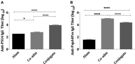
enzyme-linked immunosorbent assay (ELISA) and are shown in Fig. 1. As expected, the
conjugation of PS14 to mPspA4Pro resulted in a higher induction of anti-PS14 IgG than
that provided by the free PS14 (Fig. 1A). Although conjugation to the PS14 seemed to
slightly reduce the levels of anti-PspA4Pro IgG antibodies by approximately 0.5 log
compared to those induced by free mPspA4Pro (Fig. 1B), both conjugated and free
mPspA4Pro (Co-adm) were shown to induce high titers of anti-PspA4Pro IgG antibodies
in mice (Fig. 1B).
Opsonophagocytic assay.
The functionality of anti-PspA4 and anti-PS14 antibodies
was measured by their opsonophagocytic activity. First we investigated whether sera
from mice immunized with conjugate PS14-mPspA4Pro or with mPspA4Pro
coadmin-istered with PS14 differ in their opsonophagocytic activities against strains bearing
PspA4 and a heterologous PS (non-PS14) (Table 1). Sera from mice immunized with the
conjugate reduced the survival of the three strains of pneumococci tested, while sera
from mice immunized with coadministered mPspA4Pro and PS14 significantly
dimin-ished the number of CFU recovered in two of the three strains tested (Fig. 2).
Interestingly, the level of opsonophagocytic activity differed between the conjugated
and coadministered groups according to the strain. In one case, the number of CFU
recovered was significantly reduced in the conjugated group in comparison with the
group immunized with mPspA4Pro coadministered with PS14 (strain P166, serotype
6A), and in the two cases involving serotype 19F strains, the numbers of CFU were
either comparable (strain P101) or the sera from mice immunized with free mPspA4Pro
coadministered with PS14 was shown to reduce the survival of strain P40 more
efficiently (Fig. 2).
We next sought to characterize the opsonophagocytic activity against heterologous
strains from the same family 2: PspA clades 3 and 5. Sera from mice immunized either
with the conjugate or with the coadministered components were able to significantly
FIG 1IgG titers induced by immunization of mice with conjugate PS14-mPspA4Pro. IgG titers of individual serum samples from mice (n⫽10) immunized s.c. with the PS14-mPspA4Pro conjugate, PS14 (2.5g) and mPspA4Pro (5.0g) (Co-adm), or 50g Al(OH)3in PBS (Alum) were analyzed by ELISA and
compared by one-way ANOVA with Tukey’s multiple-comparison test. Alum was used as an adjuvant in all groups. (A) Anti-PS14 IgG titer; (B) anti-PspA4Pro IgG titer. The bars represent the means⫾standard errors of the means (SEM), and asterisks indicate statistically significant differences (*,Pⱕ0. 05;****,Pⱕ 0.0001).
on August 17, 2020 by guest
http://cvi.asm.org/
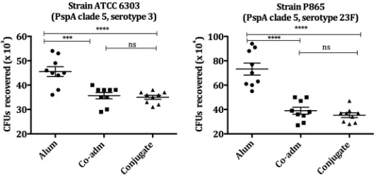
reduce the numbers of CFU recovered in the four strains tested (Fig. 3 and 4).
Conjugation maintained opsonophagocytic activity against strains Tigr4 (PspA3,
sero-type 4), ATCC 6303 (PspA5, serosero-type 3), and P865 (PspA5, serosero-type 23 F) and,
surpris-ingly, increased activity against P1148 (PspA3, serotype 6B), in comparison to sera from
mice immunized with free mPspA4Pro coadministered with PS14 (Fig. 3 and 4).
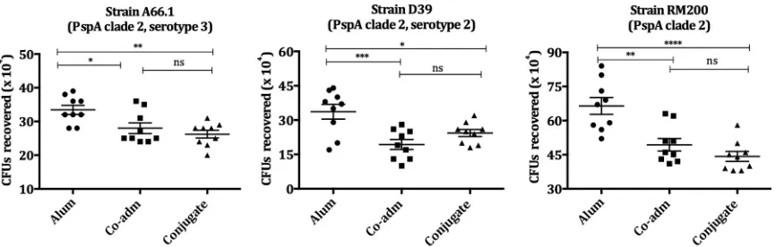
Opsonophagocytosis activity was also evaluated in strains from PspA family 1, PspA
clades 1 and 2. The sera from conjugate-immunized mice showed significant
op-sonophagocytic activity in all strains tested bearing PspA clades 1 and 2 (Fig. 5 and 6).
The reduction in survival was comparable to that obtained by sera from mice
immu-nized with free mPspA4Pro coadministered with PS14 for all strains. Strains tested were
from serotypes 6A, 1, 2, and 3; strain RM200 is a nonencapsulated strain (Fig. 5 and 6).
Finally, we determined the opsonophagocytosis activity against homologous PS
strains (PS14 strains) bearing a heterologous PspA (PspA1, strain 245/00) or a
homol-ogous PspA (PspA4, strain P1140). The sera from conjugate-immunized mice showed
significant opsonophagocytic activity in both strains tested, demonstrating the
func-tionality of anti-PS14 antibodies after the conjugation process (Fig. 7). Additionally, it is
noteworthy that when using a strain bearing homologous PS and PspA in the
op-FIG 2Opsonophagocytic activity of sera from mice immunized with conjugate PS14-mPspA4Pro on pneumococcal strains bearing PspA clade 4. Pneumococcal strains P101, P40, and P166, bearing PspA from clade 4, were incubated with the pooled sera of mice (10 animals/group) immunized with the PS14-mPspA4Pro conjugate, mPspA4Pro coadministered with PS14 (Co-adm), or Al(OH)3in
PBS (Alum) plus a complement source (NMS). Al(OH)3was used as an adjuvant in all groups. The opsonized pneumococci were
incubated with peritoneal cells and plated on blood agar plates. The numbers of CFU recovered after 18 h were compared by one-way ANOVA with Tukey’s multiple-comparison test. The results are representative of an experiment performed in triplicate. The lines represent the means⫾SEM, and asterisks indicate statistically significant differences (*,Pⱕ0.05;**,Pⱕ0.01;***,Pⱕ0.001;****,Pⱕ 0.0001). ns, nonsignificant differences.
TABLE 1Pneumococcal strains used in the opsonophagocytosis assay
Strain Serotype PspA clade Sourcea
A66.1 3 2 UAB
ATCC 6303 3 5 IAL
D39 2 2 UAB
P40 19F 4 UFG
P101 19F 4 UFG
P105 6A 1 UFG
P166 6A 4 UFG
P865 23F 5 UFG
P1079 1 1 UFG
P1148 6B 3 UFG
RM200 —b 2 BCHHMS
Tigr4 4 3 UAB
0603 6B 1 BCHHMS
245/00 14 1 IAL
P1140 14 4 UFG
aUAB, University of Alabama at Birmingham, Birmingham, AL, USA; UFG, Universidade Federal de Goiás,
Goiânia, Brazil; IAL, Instituto Adolpho Lutz, São Paulo, Brazil; BCHHMS, Boston Children’s Hospital, Harvard Medical School, Boston, MA, USA.
bNonencapsulated.
on August 17, 2020 by guest
http://cvi.asm.org/
sonophagocytic assay (strain P1140), conjugation led to a significantly lower number of
CFU recovered compared to that of the coadministered group (free mPspA4 and PS14)
and conjugate groups (Fig. 7).
Colonization challenge.
To determine the protective effect of the PS14-mPspA4Pro
conjugate against a nonrelated pneumococcal strain (non-PspA4, non-PS14), we
im-munized mice subcutaneously (s.c.) with the conjugate or coadministered antigens, in
a three-dose scheme, and challenged them intranasally using pneumococcus type 6B
strain 0603 bearing PspA from clade 1 (10
7CFU). The number of CFU recovered was
determined in the mouse nasal washes after 7 days of challenge. As shown in Fig. 8,
immunization with the PS14-mPspA4Pro conjugate significantly protected the animals
against pneumococcal colonization, with protection at levels similar to those observed
for the animals that received the coadministered antigens PS14 and mPspA4Pro.
DISCUSSION
The use of pneumococcal surface proteins as an alternative to heterologous
pro-teins, such as CRM
197or tetanus toxoid as a carrier for CPS, would be a way to improve
FIG 3Opsonophagocytic activity of sera from mice immunized with conjugate PS14-mPspA4Pro on pneumococcal strains bearing PspA clade 3. Pneumococcal strains P1148 and Tigr4, bearing PspA from clade 3, were incubated with the pooled sera of mice (10 animals/group) immunized with mPspA4Pro coadministered with PS14 (Co-adm), the PS14-mPspA4Pro conjugate, or Al(OH)3in PBS (Alum) plus a
complement source (NMS). Al(OH)3was used as an adjuvant in all groups. The opsonized pneumococci
were incubated with peritoneal cells and plated on blood agar plates. The numbers of CFU recovered after 18 h were compared by one-way ANOVA with Tukey’s multiple-comparison test. The results are representative of an experiment performed in triplicate. The lines represent the means⫾SEM, and asterisks indicate statistically significant differences (**,Pⱕ0.01;***,Pⱕ0.001;****,Pⱕ0.0001). ns, nonsignificant differences.
FIG 4Opsonophagocytic activity of sera from mice immunized with conjugate PS14-mPspA4Pro on pneumococcal strains bearing PspA clade 5. Pneumococcal strains ATCC 6303 and P865, bearing PspA from clade 5, were incubated with the pooled sera of mice (10 animals/group) immunized with mPspA4Pro coadministered with PS14 (Co-adm), the PS14-mPspA4Pro conjugate, or Al(OH)3in PBS
(Alum) plus a complement source (NMS). Al(OH)3was used as an adjuvant in all groups. The opsonized
pneumococci were incubated with peritoneal cells and plated on blood agar plates. The numbers of CFU recovered after 18 h were compared by one-way ANOVA with Tukey’s multiple-comparison test. The results are representative of an experiment performed in triplicate. The lines represent the means⫾SEM, and asterisks indicate statistically significant differences (***,Pⱕ0.001;****,Pⱕ0.0001). ns, nonsignif-icant differences.
on August 17, 2020 by guest
http://cvi.asm.org/
pneumococcal vaccine coverage. PspA has been shown to be an efficient carrier for
capsular polysaccharide, as well as an antigen able to induce protective antibodies
when conjugated (11–13, 30). PspA is one of the pneumococcal surface proteins
described as a protein vaccine candidate, and the main reason is its capacity to induce
protective antibodies (25, 28, 31). PspA is a mosaic protein with a variable domain in its
N-terminal sequence that induces antibodies cross-reacting within proteins from the
same family (21) but to a lesser extent across families; this is a restriction for a vaccine
candidate. Different approaches have been used in order to circumvent this limitation,
such as the combination of PspAs from different clades and families (28, 32).
On the other hand, the antiserum induced against rPspA clade 4 has been described
as broadly cross-reactive, recognizing pneumococcal strains containing PspAs of all
clades from families 1 and 2 (33). The functionality of the anti-rPspA4 antibodies was
analyzed as to the capacity to bind to and mediate complement deposition on intact
bacteria
in vitro
, and mice immunized with rPspA4 and rPspA5 were protected against
intranasal lethal challenge with pneumococcal strains (A66.1 and ATCC 6303) bearing
PspAs from families 1 and 2 (29). We here evaluated the functionality and
immuno-FIG 6Opsonophagocytic activity of sera from mice immunized with conjugate PS14-mPspA4Pro on pneumococcal strains bearing PspA clade 2. Pneumococcal strains A66.1, D39, and RM200, bearing PspA from clade 2, were incubated with the pooled sera of mice (10 animals/group) immunized with mPspA4Pro coadministered with PS14 (Co-adm), the PS14-mPspA4Pro conjugate, or Al(OH)3in PBS
(Alum) plus a complement source (NMS). Al(OH)3was used as an adjuvant in all groups. The opsonized pneumococci were incubated with
peritoneal cells and plated on blood agar plates. The numbers of CFU recovered after 18 h were compared by one-way ANOVA with Tukey’s multiple-comparison test. The results are representative of an experiment performed in triplicate. The lines represent the means⫾ SEM, and asterisks indicate statistically significant differences (*,Pⱕ0.05;**,Pⱕ0.01;***,Pⱕ0.001;****,Pⱕ0.0001). ns, nonsignificant differences.
FIG 5Opsonophagocytic activity of sera from mice immunized with conjugate PS14-mPspA4Pro on pneumococcal strains bearing PspA clade 1. Pneumococcal strains P105 and P1079, bearing PspA from clade 1, were incubated with the pooled sera of mice (10 animals/group) immunized with mPspA4Pro coadministered with PS14 (Co-adm), the PS14-mPspA4Pro conjugate, or Al(OH)3in PBS (Alum) plus a
complement source (NMS). Al(OH)3was used as an adjuvant in all groups. The opsonized pneumococci
were incubated with peritoneal cells and plated on blood agar plates. The numbers of CFU recovered after 18 h were compared by one-way ANOVA with Tukey’s multiple-comparison test. The results are representative of an experiment performed in triplicate. The lines represent the means⫾SEM, and asterisks indicate statistically significant differences (*,Pⱕ0.05;****,Pⱕ0.0001). ns, nonsignificant differences.
on August 17, 2020 by guest
http://cvi.asm.org/
logical cross-reactivity of the antibodies induced by the s.c. immunization of BALB/c
mice with mPspA4Pro conjugated to PS14.
Polysaccharide as a thymus-independent antigen induces low-avidity antibodies,
mainly of the IgM type, and does not induce immunological memory. When covalently
linked to a protein, PS becomes a T-cell-dependent antigen inducing long-term
immu-nity in young children (4). Therefore, the increased anti-PS14 IgG after conjugation with
mPspA4Pro indicates that the reaction was successful. Our results showed that after
conjugation, the titer of anti-rPspA4 was lower than that induced by the free
mPspA4Pro coadministered with PS14. This indicates that the conjugation may have
changed the protein epitope profile. Considering these results, it is essential to evaluate
whether the decrease in the anti-rPspA antibody level could affect the functionality of
these antibodies.
Opsonophagocytic activity is a means of evaluating the functional activity of the
antibodies
in vitro
and is dependent on the antibody binding and complement
deposition that lead to killing and clearance. The opsonophagocytic activities exhibited
by anti-PspA4Pro antibodies in the sera of mice immunized with the PS14-mPspA4Pro
FIG 7Opsonophagocytic activity of sera from mice immunized with conjugate PS14-mPspA4Pro on pneumococcal strains of serotype 14. Pneumococcal strains 245/00 and P1140 (serotype 14), bear-ing PspA from clade 1 and clade 4, were incubated with the pooled sera of mice (10 animals/group) immunized with mPspA4Pro coadministered with PS14 (Co-adm), the PS14-mPspA4Pro conjugate, or Al(OH)3in PBS (Alum) plus a complement source (NMS). Al(OH)3was used as an adjuvant in all groups.
The opsonized pneumococci were incubated with peritoneal cells and plated on blood agar plates. The numbers of CFU recovered after 18 h were compared by one-way ANOVA with Tukey’s multiple-comparison test. The results are representative of an experiment performed in triplicate. The lines represent the means⫾SEM, and asterisks indicate statistically significant differences (*,Pⱕ0.05;**,Pⱕ 0.01;***,Pⱕ0.001;****,Pⱕ0.0001). ns, nonsignificant differences.
FIG 8Colonization challenge. Groups of 10 BALB/c mice immunized subcutaneously with the PS14-mPspA4Pro conjugate, PS14 and PS14-mPspA4Pro (Co-adm), or Al(OH)3 in PBS (Alum) were challenged
intranasally 7 days after the third immunization with pneumococcal type 6B strain 0603 (1⫻107CFU),
bearing PspA from clade 1. Al(OH)3was used as an adjuvant in all groups. The nasal wash was collected
after 7 days, diluted, and plated on blood agar plate. Each dot represents the CFU recovered from one mouse. A horizontal line denotes the median for each group. Data were compared by one-way ANOVA with Tukey’s multiple-comparison test. Asterisks indicate statistically significant differences (**,Pⱕ0.01; ***,Pⱕ0.001). ns, nonsignificant differences.
on August 17, 2020 by guest
http://cvi.asm.org/
conjugate were evaluated using 14 pneumococcal strains carrying PspAs from clade 1
to clade 5 and a variety of serotypes. Despite the lower antibody levels induced by the
conjugate, these antibodies were shown to be functional against all strains tested
comprising PspAs from both families, including clades 1 to 5 and serotypes 1, 2, 3, 4,
6A, 6B, 19F, and 23F. The reduction in the number of CFU recovered obtained with the
sera from the conjugate was either comparable to or greater than that obtained with
mPspA4Pro coadministered with PS14. The only exception was found in strain P40
(PspA clade 4, serotype 19F).
It is also very important to observe that the anti-PS14 antibodies generated by
immunization with the conjugate are functional, as can be seen in the
opsonophago-cytosis assay in the PS14 strains tested (245/1000 and P1140). PS14 antibodies from
mice immunized with conjugated PS14-mPspA4Pro showed opsonophagocytic activity
that was significantly higher than that from nonconjugated PS. In the case of testing a
strain bearing a homologous PspA4 (P1140), it was possible to see the effect of PspA4,
as the reduction in the number of CFU recovered was greater than that in strain 245/00,
which expresses PspA1.
Pneumococcal disease is generally initiated by bacterial colonization of the
naso-pharynx mucosa. Protection against nasal carriage is crucial for herd immunity to
pneumococcal disease and would have a direct effect on the reduction of invasive
disease. In our study, subcutaneous immunization with free or conjugated mPspA4Pro
was able to reduce the bacterial load in colonized mice. It has been described that
cellular rather than humoral immunity promotes the clearance of pneumococcal nasal
colonization requiring CD4
⫹cells, where monocytes seem directly related to the
bacterial clearance and interleukin 17A (IL-17A) is critical to the recruitment of
phago-cytic cells (34, 35). Therefore, we could speculate that the induction of cellular immune
responses would be similar for the free and conjugated mPspA4Pro, since mice
immunized with either vaccine were equally protected from nasal challenge.
On a whole, our results show that the conjugation of mPspA4Pro with PS14 provides
the expected IgG increase against the polysaccharide moiety, indicating that the
conjugation was effective in modifying it into a T-cell-dependent antigen. In addition,
the anti-PS14 antibodies are functional and reduced the number of CFU in the
opsonophagocytic killing assay. Moreover, the PS14-mPspA4Pro conjugate retains
the antigenic properties of the mPspA4Pro molecule, as the antisera generated against
the conjugate has opsonophagocytic properties comparable to those of the free
protein in a large panel of strains and shows comparable protection against
coloniza-tion. These results reinforce the potential of using highly immunogenic pneumococcal
proteins as carriers for capsular polysaccharide in order to broaden their immunogenic
cross-reactivity.
MATERIALS AND METHODS
The nontagged recombinant fragment of PspA from clade 4 (PspA4Pro) and capsular polysaccharide fromS. pneumoniaeserotype 14 were produced and purified at Departamento de Engenharia Química (UFSCar) and Centro de Biotecnologia (Instituto Butantan) as described previously (36, 37). Adipic acid dihydrazide (ADH) and 4-(4,6-dimethoxy-1,3,5-triazin-2-yl)-4-methylmorpholinium chloride (DMT-MM) were from Sigma-Aldrich. All other reagents were analytical grade.
PS14-mPspA4Pro conjugation.PS14-mPspA4Pro conjugation was performed as described earlier (12). Briefly, PS14 (5 mg ml⫺1) was hydrolyzed by 0.5 M HCl at 80°C for 30 min in a reflux system, followed
by the addition of NaOH (pH 7.5). The hydrolyzed PS14 was oxidized with NaIO4(10 mM) in
phosphate-buffered saline (PBS), pH 7.5, for 30 min in the dark at room temperature. The reaction was stopped by glycerol (10 eq) and diafiltered using a 5-kDa cutoff membrane (Pellicon XL; Millipore). Oxidized PS14 was reacted with ADH at a molar ratio of 1:50 (aldehyde:ADH) and NaBH3CN (5 M) for 24 h in PBS, pH 7.5. The
reaction was stopped with NaBH4(5 M) at a molar ratio of 10:1 (NaBH4:PS). The reaction mixture was
diafiltered using a 5-kDa cutoff membrane (Pellicon XL; Millipore). Modified PS14 (10 mg ml⫺1) was
incubated with mPspA4Pro (20 mg ml⫺1) previously reacted with formaldehyde and DMT-MM (0.05 M)
in PBS buffer, pH 7.5, for 24 h and quenched by ammonium acetate 0.1 M. The product was purified by Superose 12 prep grade gel filtration chromatography in an HR 16/50 column (GE Healthcare) using PBS, pH 7.5, as the mobile phase (0.5 ml min⫺1).
Analytical methods.PS14 was quantified by the phenol sulfuric acid method (38). PS14 from the ATCC was used as the standard. The extension of oxidation was estimated by the colorimetric method using bicinchoninic acid (BCA) (39). The extension of the reaction of ADH with lysine-aminogroups was
on August 17, 2020 by guest
http://cvi.asm.org/
ELISA.Anti-PS14 and anti-mPspA4Pro antibodies were determined by conventional ELISA as follows. ELISA 96-well microtiter plates (Nunc MaxiSorp) were coated with 50g/well of PS14 or 1g/well of rPspA4Pro in PBS (pH 7.2) overnight at 4°C. The plates were washed three times with PBS containing 0.05% Tween 20 (PBS-T) and blocked with PBS and 10% skim milk for 1 h at 37°C. The plates were washed with PBS-T and incubated with serial dilutions of serum from individual mice in PBS and 1% bovine serum albumin for 2 h at 37°C for anti-PS14 or 1 h at 37°C for anti-PspA, and the plates were washed with PBS-T. Peroxidase-conjugated polyclonal goat anti-mouse IgG (1:5,000) was then added, and the plates were incubated at 37°C for 2 h (PS14) or 1 h (PspA). The plates were washed three times with PBS-T, and the substrate o-phenylenediamine dihydrochloride in citrate buffer (pH 5.0) with 0.5 l ml⫺1 of 30%
hydrogen peroxide was added for 15 min in the dark. The enzyme reaction was quenched by adding 4 M H2SO4. The plates were read at 492 nm on a Multiskan EX ELISA reader (Labsystems Uniscience). Titers
were calculated by using the dilution resulting in an absorbance value of 0.1 at 492 nm.
Opsonophagocytic assay.The modified opsonophagocytic assays were performed as described by Goulart et al. (27). The S. pneumoniaestrains shown in Table 1 were grown in Todd-Hewitt broth supplemented with yeast extract (THY) to an optical density at 600 nm (OD600 nm) of 0.4 to 0.5, which
corresponds to approximately 1⫻108CFU/ml, and centrifuged at 4,000⫻gfor 3 min. The pellets were
washed once with PBS and resuspended in Hanks’ buffer (Invitrogen) containing 0.1% gelatin. Aliquots containing 2.5⫻106CFU were incubated with heat-inactivated pooled sera in the test at a final dilution
of 1:100, in triplicate, for 30 min at 37°C. Sera from mice that received saline plus Al(OH)3were used as
a control for all the assays. The samples were incubated with 10% normal mouse serum (NMS) diluted in Hanks’ buffer containing 0.1% gelatin (opsono-buffer) at 37°C for 30 min. After that, the samples were washed once with PBS and incubated with 4⫻105peritoneal cells diluted in opsono-buffer at 37°C for
30 min with shaking (200 rpm). The reaction was stopped by incubation on ice for 5 min. Tenfold dilutions of the samples were plated on blood agar plates in triplicate. The plates were incubated at 37°C, in 5% CO2, and the pneumococcal CFU recovered were counted after 18 h.
In order to obtain the peritoneal cells, BALB/c mice were injected intraperitoneally with 10g of concanavalin A from Canavalia ensiformis (ConA) (Sigma), and 2 days later, after the mice were anesthetized, they were sacrificed and their peritoneal cavities were washed with 5 ml of cold PBS.
Colonization challenge.The colonization assay was based on the protocol described by Malley et al. (41). Pneumococcal strain 0603 (serotype 6B, PspA clade 1) was inoculated (1⫻107CFU/animal in 10
l of cold PBS) into the nostrils of previously immunized mice. On day 7 after the colonization challenge, the animals were euthanized (with 1.25g of xylazine and 3g of ketamine mixture/dose) and upper respiratory tract samples were collected from the transected trachea. To determine the colonization density in each sample, aliquots were plated on blood agar plates with 2.5g ml⫺1gentamicin. The
number of CFU was determined after 18 h of incubation at 37°C.
Statistical analysis. Tukey’s multiple-comparison test was performed for all the experiments, followed by the one-way analysis of variance (ANOVA). For all comparisons, aPvalue of⬍0.05 was considered to represent statistical significance.
ACKNOWLEDGMENTS
This study was supported by Fundação de Amparo à Pesquisa do Estado de São
Paulo (FAPESP) grants numbers 2008/05207-4 and 2015/07225-3.
We thank Douglas B. Figueiredo and Rafaela T. Zanardo for providing PspA4Pro and
PS14, respectively. We also thank professor Martin Wesley for his revision of English and
Catia T. Perciani for her contribution to the manuscript.
REFERENCES
1. Musher DM. 1992. Infections caused byStreptococcus pneumoniae: clin-ical spectrum, pathogenesis, immunity and treatment. Clin Infect Dis 14:801– 807.https://doi.org/10.1093/clinids/14.4.801.
2. Peltola H, Käythy H, Sivonen A, Mä kelä PH. 1977.Haemophilus influenzae type b capsular polysaccharide vaccine in children: a double-blind field study of 100,000 vaccinees 3 months to 5 years of age in Finland. Pediatrics 60:730 –737.
3. Parke JJC, Schneerson R, Robbins JB, Schlesselman JJ. 1977. Interim report
of a controlled field trial of immunization with capsular polysaccharides of Haemophilus influenzaetype b and group CNeisseria meningitidisin Meck-lenburg County, North Carolina (March 1974 –March 1976). J Infect Dis 136:S51–S56.https://doi.org/10.1093/infdis/136.Supplement.S51. 4. Schneerson R, Barrera O, Sutton A, Robbins JB. 1980. Preparation,
char-acterization, and immunogenicity of Haemophilus influenzae type b polysaccharide-protein conjugates. J Exp Med 152:361–376.https://doi .org/10.1084/jem.152.2.361.
on August 17, 2020 by guest
http://cvi.asm.org/
5. Stein KE. 1992. Thymus-independent and thymus-dependent responses to polysaccharide antigens. J Infect Dis 165:S49 –S52.https://doi.org/10 .1093/infdis/165-Supplement_1-S49.
6. Henrichsen J. 1995. Six newly recognized types ofStreptococcus pneu-moniae. J Clin Microbiol 33:2759 –2762.
7. Park IH, Geno KA, Yu J, Oliver MB, Kim K-H, Nahm MH. 2015. Genetic, biochemical, and serological characterization of a new pneumococcal serotype, 6H, and generation of a pneumococcal strain producing three different capsular repeat units. Clin Vaccine Immunol 22:313–318. https://doi.org/10.1128/CVI.00647-14.
8. Yildirim I, Shea KM, Pelton SI. 2015. Pneumococcal disease in the era of pneumococcal conjugate vaccine. Infect Dis Clin North Am 29:679 – 697. https://doi.org/10.1016/j.idc.2015.07.009.
9. Huang SS, Hinrichsen VL, Stevenson AE, Rifas-Shiman SL, Kleinman K, Pelton SI, Lipsitch M, Hanage WP, Lee GM, Finkelstein JA. 2009. Continued impact of pneumococcal conjugate vaccine on carriage in young children. Pediatrics 124:e1–e11.https://doi.org/10.1542/peds .2008-3099.
10. Weinberger DM, Malley R, Lipsitch M. 2011. Serotype replacement in disease after pneumococcal vaccination. Lancet 378:1962–1973.https:// doi.org/10.1016/S0140-6736(10)62225-8.
11. Csordas FCL, Perciani CT, Darrieux M, Gonçalves VM, Cabrera-Crespo J, Takagi M, Sbrogio-Almeida ME, Leite LCC, Tanizaki MM. 2008. Protection induced by pneumococcal surface protein A (PspA) is enhanced by conjugation to a Streptococcus pneumoniae capsular polysaccharide. Vaccine 26:2925–2929.https://doi.org/10.1016/j.vaccine.2008.03.038. 12. Santamaria R, Goulart C, Perciani CT, Barazzone GC, Carvalho R, Jr,
Gonçalves VM, Leite LC, Tanizaki MM. 2011. Humoral immune response of a pneumococcal conjugate vaccine: capsular polysaccharide serotype 14-lysine modified PspA. Vaccine 29:8689 – 8695. https://doi.org/10 .1016/j.vaccine.2011.08.109.
13. Perciani CT, Barazzone GC, Goulart C, Carvalho E, Cabrera-Crespo J, Gon-calves VM, Leite LCC, Tanizaki MM. 2013. Conjugation of polysaccharide 6B fromStreptococcus pneumoniaewith pneumococcal surface protein A: PspA conformation and its effect on the immune response. Clin Vaccine Immunol 20:858 – 866.https://doi.org/10.1128/CVI.00754-12.
14. Crain MJ, Waltman WD, II, Turner JS, Yother J, Talkington DF, McDaniel LS, Gray BM, Briles DE. 1990. Pneumococcal surface protein A (PspA) is serologically highly variable and is expressed by all clinically important capsular serotypes of Streptococcus pneumoniae. Infect Immun 58: 3293–3299.
15. Briles DE, King JD, Gray MA, McDaniel LS, Swiatlo E, Benton KA. 1996. PspA, a protection-eliciting pneumococcal protein: immunogenicity of isolated native PspA in mice. Vaccine 14:858 – 867.https://doi.org/10 .1016/0264-410X(96)82948-3.
16. Tu A-HT, Fulgham RL, McCrory MA, Briles DE, Szalai AJ. 1999. Pneumo-coccal surface protein A inhibits complement activation byStreptococcus pneumoniae. Infect Immun 67:4720 – 4724.
17. Mukerji R, Mirza S, Roche AM, Widener RW, Croney CM, Rhee D-K, Weiser JN, Szalai AJ, Briles DE. 2012. Pneumococcal surface protein A inhibits complement deposition on the pneumococcal surface by competing with the binding of C-reactive protein to cell-surface phosphocholine. J Immunol 189:5327–5335.https://doi.org/10.4049/jimmunol.1201967. 18. Ren B, Szalai AJ, Hollingshead SK, Briles DE. 2004. Effects of PspA and
antibodies to PspA on activation and deposition of complement on the pneumococcal surface. Infect Immun 72:114 –122. https://doi.org/10 .1128/IAI.72.1.114-122.2004.
19. Ren B, Li J, Genschmer K, Hollingshead SK, Briles DE. 2012. The absence of PspA or presence of antibody to PspA facilitates the complement-dependent phagocytosis of pneumococci in vitro. Clin Vaccine Immunol 19:1574 –1582.https://doi.org/10.1128/CVI.00393-12.
20. Darrieux M, Goulart C, Briles D, Leite LCC. 2015. Current status and perspectives on protein-based pneumococcal vaccines. Crit Rev Micro-biol 41:190 –200.https://doi.org/10.3109/1040841X.2013.813902. 21. Hollingshead SK, Becker R, Briles DE. 2000. Diversity of PspA: mosaic
genes and evidence for past recombination in Streptococcus pneu-moniae. Infect Immun 68:5889 –5900.https://doi.org/10.1128/IAI.68.10 .5889-5900.2000.
22. Brandileone MC, Andrade AL, Teles EM, Zanella RC, Yara TI, Di Fabio JL, Hollingshead SK. 2004. Typing of pneumococcal surface protein A (PspA) inStreptococcus pneumoniae isolated during epidemiological surveil-lance in Brazil: towards novel pneumococcal protein vaccines. Vaccine 22:3890 –3896.https://doi.org/10.1016/j.vaccine.2004.04.009.
23. Hollingshead SK, Baril L, Ferro S, King J, Coan P, Briles DE. 2006.
Pneu-mococcal surface protein A (PspA) family distribution among clinical isolates from adults over 50 years of age collected in seven countries. J Med Microbiol 55:215–221.https://doi.org/10.1099/jmm.0.46268-0. 24. Nabors GS, Braun PA, Herrmann DJ, Heise ML, Pyle DJ, Gravenstein S,
Schilling M, Ferguson LM, Hollingshead SK, Briles DE, Becker RS. 2000. Immunization of healthy adults with a single recombinant pneumococ-cal surface protein A (PspA) variant stimulates broadly cross-reactive antibodies to heterologous PspA molecules. Vaccine 18:1743–1754. https://doi.org/10.1016/S0264-410X(99)00530-7.
25. Briles DE, Hollingshead SK, King J, Swift A, Braun PA, Park MK, Ferguson LM, Nahm MH, Nabors GS. 2000. Immunization of humans with recom-binant pneumococcal surface protein A (rPspA) elicits antibodies that passively protect mice from fatal infection withStreptococcus pneu-moniaebearing heterologous PspA. J Infect Dis 182:1694 –1701.https:// doi.org/10.1086/317602.
26. Miyaji EN, Ferreira DM, Lopes APY, Brandileone MCC, Dias WO, Leite LCC. 2002. Analysis of serum cross-reactivity and cross-protection elicited by immunization with DNA vaccines against Streptococcus pneumoniae expressing PspA fragments from different clades. Infect Immun 70: 5086 –5090.https://doi.org/10.1128/IAI.70.9.5086-5090.2002.
27. Goulart C, Darrieux M, Rodriguez D, Pimenta FC, Brandileone MCC, de Andrade ALSS, Leite LCC. 2011. Selection of family 1 PspA molecules capable of inducing broad-ranging cross-reactivity by complement de-position and opsonophagocytosis by murine peritoneal cells. Vaccine 29:1634 –1642.https://doi.org/10.1016/j.vaccine.2010.12.074.
28. Piao Z, Akeda Y, Takeuchi D, Ishii KJ, Ubukata K, Briles DE, Tomono K, Oishi K. 2014. Protective properties of a fusion pneumococcal surface protein A (PspA) vaccine against pneumococcal challenge by five dif-ferent PspA clades in mice. Vaccine 32:5607–5613.https://doi.org/10 .1016/j.vaccine.2014.07.108.
29. Moreno AT, Oliveira MLS, Ferreira DM, Ho PL, Darrieux M, Leite LCC, Ferreira JMC, Jr, Pimenta FC, Andrade AL, Miyaji EN. 2010. Immunization of mice with single PspA fragments induces antibodies capable of mediating complement deposition on different pneumococcal strains and cross-protection. Clin Vaccine Immunol 17:439 – 446.https://doi.org/ 10.1128/CVI.00430-09.
30. Lin H, Peng Y, Lin Z, Zhang S, Guo Y. 2015. Development of a conjugate vaccine against invasive pneumococcal disease based on capsular poly-saccharides coupled with PspA/family 1 protein ofStreptococcus pneu-moniae. Microb Pathog 83– 84:35– 40.https://doi.org/10.1016/j.micpath .2015.04.006.
31. Talkington DF, Crimmins DL, Voellinger DC, Yother J, Briles DE. 1991. A 43-kilodalton pneumococcal surface protein, PspA: isolation, protective abilities, and structural analysis of the amino-terminal sequence. Infect Immun 59:1285–1289.
32. Darrieux M, Miyaji EN, Ferreira DM, Lopes LM, Lopes APY, Ren B, Briles DE, Hollingshead SK, Leite LCC. 2007. Fusion proteins containing family 1 and family 2 PspA fragments elicit protection againstStreptococcus pneumoniaethat correlates with antibody-mediated enhancement of complement deposition. Infect Immun 75:5930 –5938.https://doi.org/10 .1128/IAI.00940-07.
33. Darrieux M, Moreno AT, Ferreira DM, Pimenta FC, de Andrade ALSS, Lopes APY, Leite LCC, Miyaji EN. 2008. Recognition of pneumococcal isolates by antisera raised against PspA fragments from different clades. J Med Microbiol 57:273–278.https://doi.org/10.1099/jmm.0.47661-0. 34. Zhang Z, Clarke TB, Weiser JN. 2009. Cellular effectors mediating
Th17-dependent clearance of pneumococcal colonization in mice. J Clin Invest 119:1899 –1909.https://doi.org/10.1172/JCI36731.
35. Trzcinski K, Thompson CM, Srivastava A, Basset A, Malley R, Lipsitch M. 2008. Protection against nasopharyngeal colonization byStreptococcus pneumoniaeis mediated by antigen-specific CD4⫹T cells. Infect Immun 76:2678 –2684.https://doi.org/10.1128/IAI.00141-08.
36. Figueiredo DB, Carvalho E, Santos MP, Kraschowetz S, Zanardo RT, Campani G, Silva GG, Sargo CR, Horta ACL, Giordano RdC, Miyaji EN, Zangirolami TC, Cabrera-Crespo J, Gonçalves VM. 2016. Production and purification of an untagged recombinant pneumococcal surface protein A (PspA4Pro) with high-purity and low endotoxin content. Appl Micro-biol Biotechnolhttps://doi.org/10.1007/s00253-016-7983-9.
37. Zanardo RT, Ferri ALS, Figueiredo DB, Kraschowetz S, Cabrera-Crespo J, Gonçalves VM. 2016. Development of a new process for purification of capsular polysaccharide from Streptococcus pneumoniaeserotype 14. Braz J Chem Eng 33:435– 443. https://doi.org/10.1590/0104-6632 .20160333s20150140.
38. Cuesta G, Suarez N, Bessio MI, Ferreira F, Massaldi H. 2003. Quantitative