O R I G I N A L R E S E A R C H
Virulence Constitution of Multi-Drug-Resistant
Pseudomonas aeruginosa
in Upper Egypt
This article was published in the following Dove Press journal: Infection and Drug Resistance
Noha A Hassuna 1
Sahar A Mandour 2
Ebtisam Samir Mohamed 1
1Medical Microbiology and Immunology,
Faculty of Medicine, Minia University, Minia, Egypt;2Microbiology and
Immunology Department, Faculty of Pharmacy, Deraya University, Minia, Egypt
Purpose:Ventilator-associated pneumonia caused byPseudomonas aeruginosa(P. aeruginosa) is a major health-care problem. In this study, we explored the epidemiology of virulence determinants among multi-drug-resistant (MDR) clinicalP. aeruginosaisolates from hospitalized patients with ventilator-associated pneumonia in intensive care units in Upper Egypt.
Patients and Methods: MDRP. aeruginosa isolates were screened for the presence of eight virulence factors and typed by ERIC-PCR.
Results:A total of 39 clinical MDR isolates were selected out of 173 isolatedP. aeruginosa
showing a combination of adhesion and cytotoxicity virulence patterns, with the detection of
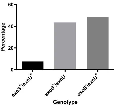
aprA,exoU,exoS,lasB,algD,toxA in 74.3%, 58.9%, 46.1%, 41.2%, 30.7%, 20.5% of the isolates, respectively. The MDR isolates were grouped into 13 different virulence profiles according to the pattern of virulence gene distribution.exoU genotype was more predomi-nant among theP. aeruginosaisolates with more than 48% of the isolates harboring this gene alone, 7% harboring bothexoU andexoS and 43.5% harboringexoS gene. An intermediate degree of diversity was detected by ERIC-PCR typing where the isolates were clustered in 7 major groups, indicating possible cross-infection within the hospital.
Conclusion:Our results highlight the increased frequency of virulentP. aeruginosaisolates with a shift to the more virulent cytotoxicexoU genotype. Further hospital infection-control measures are mandatory to control the hospital cross-transmission of these highly virulent isolates. This study could vastly be a help to develop efficient treatment policies against
P. aeruginosainduced ventilator-associated pneumonia.
Keywords:P. aeruginosa, virulence,exoU, MDR
Introduction
Pseudomonas aeruginosa (P. aeruginosa) is one of the most common causes of healthcare-associated infections being responsible for urinary tract, respiratory and surgical site infections.1,2 This opportunistic pathogen is considered as a major health hazard, especially in immunodeficient patients.3 Furthermore, infections caused by multi-drug-resistant (MDR) P. aeruginosa isolates are associated with longer duration of hospitalization, increased costs, as well as increased morbidity and mortality rates.4
P. aeruginosahas a repertoire of virulence factors that markedly contribute to its pathogenicity5 e.g., lipopolysaccharides, adhesion factor (pili type IV), flagella, exo-proteases (alkaline protease “AprA”, elastase, staphylolysin, protease IV), phospholipase C, exotoxins “Exo A”, exoenzymes S, T, and U, as well as sialidase.5–9 In addition, P. aeruginosa has the exquisite ability to form biofilms, which render it more resistant to antimicrobials.10
Correspondence: Noha A Hassuna Medical Microbiology and Immunology, Faculty of Medicine, Minia University, Minia 61111, Egypt
Tel +20 862342813 Email [email protected]
Infection and Drug Resistance
Dove
press
open access to scientific and medical research
Open Access Full Text Article
Infection and Drug Resistance downloaded from https://www.dovepress.com/ by 118.70.13.36 on 24-Aug-2020
Exoenzymes S, Y, T, and U are encoded by the genes exoS,exoYexoT, andexoU, respectively, withexoS being the most predominant.11While former studies have shown that the existence ofexoS is related to increased virulence in burn wounds and lung infections,12 ExoU is 100 times more cytotoxic than ExoS.5,13LasB elastase, a zinc metal-loprotease encoded by the lasB gene, attacks eukaryotic proteins such as elastin and collagen.10,14In addition, two phospholipases C encoded by plcH and plcN genes can hydrolyze phospholipids. The high frequency of virulence factor phospholipase C gene (plcH) in MDRP. aeruginosa clinical isolates probably plays an important role in the pathogenesis of this bacterium. The genealgD encodes the GDP-mannose dehydrogenase enzyme, which is the first element in the alginate biosynthetic cluster essential for alginate biosynthesis. Alginate is a linear exopolysacchar-ide, which protects the bacterium from antibiotics and the host’s immune response.10
The increased invasiveness and pathogenicity as a result of harboring these virulence factors upsurge the morbidity despite the use of antibiotics.15 However, the arsenal of virulence mechanisms harbored by P. aeruginosa varies according to the settings and the environment of infection.16 In previous studies done in this region, a high inci-dence of MDRP. aeruginosawas observed among isolates from infected burn and surgical wounds.17,18The identifi -cation of virulence genes’profile is crucial for developing efficient policies againstP. aeruginosainfections; the aim of this study was to evaluate the distribution of different virulence genes among MDRP. aeruginosaisolated from different Egyptian ICUs.
Materials and Methods
In this cross-sectional study, samples were collected from respiratory tract infections over a period of 15 months between 2017 and 2019 from patients admitted to the differ-ent ICUs in Minia University Hospital (a tertiary care hospi-tal). A written informed consent was obtained from every patient or his caregiver. The study was carried out as per the Helsinki declarations and was approved by the Ethical com-mittee of the Faculty of Medicine, Minia University. A total of 173P. aeruginosaisolates were phenotypically identified by routine cultural and biochemical methods. MDR isolates were selected for further characterization by antibiotic sensi-tivity testing using the disc-diffusion method according to CLSI 2017, usingP. aeruginosaATCC 27853 as a reference strain. MDR is identified as resistance to three or more groups of antimicrobials.
DNA Extraction, Identi
fi
cation and
Virulence Gene Detection
Genomic DNA was extracted using a boiling method as described previously; one loopful of fresh bacteria (grown overnight on Brain-Heart Infusion agar plates) was picked up and suspended in 200 µL of sterile DNase/RNase-free water and boiled for 10 min. The bacterial suspension was then centrifuged at 12,000 rpm at 4°C for 10 min, and the supernatant was collected and kept at−20°C.19
Detection of the following virulence genes was carried out by conventional PCR: GDP-mannose dehydrogenase enzyme for alginate (algD), alkaline protease (aprA), elastase (lasB), exoenzyme S and U (exoS andexoU) and exotoxin A (toxA), hemolytic phospholipase C (plcH) and non-hemolytic phos-pholipase C (plcN), and the used primers are listed inTable 1. The amplification was carried in a 25μL volume, containing 12.5μL PCR Master mix (DreamTaq Green PCR master mix, Thermo scientific), 1μL of each primer (forward and reverse), 1μL of template DNA and nuclease-free water.
The PCR conditions forlasB andaprA comprised dena-turation at 94°C (4 mins), followed by 25 cycles of: 94°C (1 min), 46°C (40 s), 72°C (1 min) and afinal extension at 72°C (2 mins). TheexoS andexoU amplification conditions included denaturation at 95°C (5 mins), followed by 35 cycles of 95°C (1 min), 55°C (1 min), 72°C (1 min) followed byfinal extension at 72°C (10 mins).11,20For the rest of the genes, the conditions were as follows: preliminary denaturation at 94°C (5 min), 35 cycles of denaturation at 94°C (60 s), annealing at 48°C (60 s) and extension at 72°C (90 s), with a last extension cycle at 72°C for 10 min.21PCR products were run on 1.5% agarose gel and were afterward visualized under UV lamb.
Enterobacterial Repetitive Intergenic
Consensus-PCR (ERIC-PCR Typing)
ERIC PCR was performed to produce the repetitive sequence between the two primers.22The PCR started with an initial denaturation at 94°C for 7 min, followed by 30 cycles of denaturation at 94°C for 1 min, annealing at 53°C for 1 min and extension at 72°C for 2 min and afinal extension cycle at 72°C for 15 min. Amplicons were loaded in 1.5% agarose gel, which was run at 80 V for 3 hrs. Dendrogram was generated by using GelJ software using Dice Similarity method; UPGMA linkage.23
Statistical Analysis
Statistical analysis was performed using Graph-pad Prism 6.
Infection and Drug Resistance downloaded from https://www.dovepress.com/ by 118.70.13.36 on 24-Aug-2020
Results
Identi
fi
cation and Virulence-Pro
fi
ling
This study included 39 (22.5%) MDR isolates from a total of 173 P. aeruginosa isolates (the antimicrobial-susceptibility profile of the MDR isolates to different anti-pseudomonal drugs is illustrated inSupplementary Figure 1). The preva-lence of eight virupreva-lence genes among MDR P. aeruginosa nosocomial isolates was investigated, and the frequencies of toxA, plcN;plcH;algD;lasB;aprA;exoU andexoS genes among the MDR isolates were 9 (23%); 0 (0%); 5 (12.8%); 12 (30.7%); 17 (43.5%); 27 (67.2%); 22 (50.4%) and 20 (51.3%), respectively (Figure 1). None of the isolates har-bored plc N gene. Only three isolates (7.6%) showed a simultaneous presence ofexoU andexoS genes (Table 2).
The majority of the isolates (23 isolates) had between 1 and 4 virulence genes, and only two isolates showed 6 virulence genes altogether, while no isolates possessed more than 6 virulence genes. There were 13 different virulence profiles of MDR P. aeruginosa iso-lates with types 1, 2 and 3 being the most common,
while the rarest were types 10–13 (Table 3).
Furthermore, due to the danger of exoU-harboring iso-lates, the isolates were classified into three categories
according to the presence or absence of exoU and
exoS genes (Figure 2), where exoS−/exoU+ genotype was the commonest genotype (48.7%).
Genotyping by ERIC-PCR
This study revealed intermediate diversity of ERIC fi n-gerprints, with 2 major clusters (1 and 2) enclosing 22 Table 1Primers Used in This Study
Target Gene Sequence (5ʹ–3ʹ) Amplicon Size (bp) Annealing Temperature
toxA F: 5ʹGGTAACCAGCTCAGCCACAT 3ʹ 352 48°C
R: 5ʹTGATGTCCAGGTCATGCTTC 3’
plcN F: 5ʹGTTATCGCAACCAGCCCTAC 3ʹ 466 48°C
R: 5ʹAGGTCGAACACCTGGAACAC
plcH F: 5ʹGAAGCCATGGGCTACTTCAA 3ʹ 307 48°C
R: 5ʹAGAGTGACGAGGAGCGGTAG 3’
algD F:5ʹATGCGAATCAGCATCTTTGGT 3ʹ 1311 48°C
R:5ʹCTACCAGCAGATGCCCTCGGC 3ʹ
lasB F:5ʹCCAGCCCGCTGACCCACAAGCTGTA 3ʹ 650 46°C
R: 5ʹCATTCCTTCCTGGAGTGCYRGCCG 3’
aprA F:5ʹCCTGATCKGGCCGATAACTGCAAT 3ʹ 1580 46°C
R:5ʹGGAAGACASCTATCAATTCGAACAG 3’
exoU F: 5ʹGGCACATATCTCCGGTTCCTTC 3ʹ 761 55°C
R: 5ʹTCAACTCAGCTGCCAACCATGC 3’
exoS F: 5ʹATGGCGTGTTCCGAGTCA 3ʹ 1587 55°C
R: 5ʹAGGTGTCGGTTCGTGACGTCT 3’ ERIC primers ERIC-1R, 5VCACTTAGGGGTCCTCGAATGTA-3V
ERIC-2, 5V-AAGTAAGTGACTGGGGTGAGCG-3V
Abbreviations:toxA, exotoxin A;plcH, hemolytic phospholipase C; plcN, non-hemolytic phospholipase C;algD, GDP-mannose dehydrogenase enzyme for alginate;lasB, elastase;aprA, alkaline protease;exoU, exoenzyme U;exoS, exoenzyme S; ERIC, Enterobacterial Repetitive Intergenic Consensus-PCR.
toxA plcN plcH algD lasB aprA exoU exoS
0 5 10 15 20 25 30 35 40
Virulence genes
Nu
m
b
er
o
f isolat
es
Figure 1Frequency of virulence genes in MDRP. aeruginosa.
Infection and Drug Resistance downloaded from https://www.dovepress.com/ by 118.70.13.36 on 24-Aug-2020
isolates, three groups with 3 isolates and two groups with 2 closely related isolates. Four isolates showed a unique pattern (9, 10, 15, and 25), while groups (3 and 7) were composed of 2 isolates (Figure 3). The difference in the ERIC regions is not strictly related to the occurrence of virulence genes. Furthermore, isolates with shared ERIC types displayed different virulence factor arrays.
Discussion
One of the very important pathogens causing opportunis-tic health-care-acquired infections is P. aeruginosa, because of its capacity to invade tissues using a myriad of enzymes and toxins.24It is contemplated as a threat to health particularly in patients with deficient immune response: as those in intensive care units (ICU) and Table 2Characteristics of MDRP.aeruginosaIsolates from Different ICUs in Egypt
Isolate No. toxA plcN plcH algD lasB aprA exoU exoS Number of
Virulence Genes
PA-M1 - - - + + - 2
PA-M2 - - - + + - 2
PA-M3 - - - - + + - + 3
PA-M4 - - - + 1
PA-M5 - - - - + + + - 3
PA-M6 - - - + + 2
PA-M7 - - - + - 1
PA-M8 - - - - + + - + 3
PA-M9 - - - + 1
PA-M10 - - + + - - + - 3
PA-M11 - - - + + + + - 4
PA-M12 - - - - + + - + 3
PA-M13 - - - - + + + - 3
PA-M14 - - - - + + + - 3
PA-M15 + - + + + + - + 6 PA-M16 - - - + + - 2
PA-M17 + - - + - + - + 4
PA-M18 + - - + - + + - 4
PA-M19 + - - + - + - + 4
PA-M20 - - - + + - 2
PA-M21 - - - + + - 2
PA-M22 - - - - + + - + 3
PA-M23 - - - + 1
PA-M24 - - - + 1
PA-M25 - - - + + 2
PA-M26 - - - + - 1
PA-M27 + - + - + - + - 4 PA-M28 - - - - + + + - 3
PA-M29 - - + + - - + - 3
PA-M30 - - - + - + + + 4
PA-M31 - - - - + + - + 3
PA-M32 + - - + + - - + 4
PA-M33 - - - - + + + - 3
PA-M34 + - + + + + - + 6 PA-M35 - - - + + - 2
PA-M36 - - - - + + + - 3
PA-M37 - - - - + + - + 3
PA-M38 + - - + - + - + 4
PA-M39 + - - + - + - + 4
Abbreviations:PA,P. aeruginosa; toxA, exotoxin A;plcH, hemolytic phospholipase C;plcN, non-hemolytic phospholipase C;algD, GDP-mannose dehydrogenase enzyme for alginate;lasB, elastase;aprA, alkaline protease;exoU, exoenzyme U;exoS, exoenzyme S; ERIC, Enterobacterial Repetitive Intergenic Consensus-PCR.
Infection and Drug Resistance downloaded from https://www.dovepress.com/ by 118.70.13.36 on 24-Aug-2020
burn units.3In fact,P. aeruginosais the most commonly isolated pathogen from patients with ventilator-associated pneumonia (VAP) in ICUs.25Epidemiological evaluation of P. aeruginosa on the molecular level is extremely important so as to comprehend the potential risk elements related to the infection process, assist infinding epidemic strain(s) in a particular population of patients, and recog-nize virulent strain(s).26
In the current study, MDR P.aeruginosa showed
a frequency of 22.5%, which is far less than previous results found in the same region.18These could be due to
differences in the nature of infection with different asso-ciated risk factors.27In addition to the fact that the
epide-miology of MDR microorganism infections fluctuates
from year to year between hospitals, wards, departments in addition to the geographical zone.28 Furthermore, a better implementation of antimicrobial stewardship in different ICUs is another reason for this decrease in MDR rate.
Identification of the P. aeruginosa armor of virulence factors is crucial to understand the pathogenesis of this oppor-tunistic pathogen and is important in exploring new antimi-crobial objectives especially in MDR strains. In this study, most of the isolates (23 isolates) had at least one virulence gene with a maximum of 6 virulence genes detected in 2 isolates only. The virulence phenotype was a mixture of adhe-sion and cytotoxicity, whereaprA (encoding alkaline metallo-proteinase) detected in 74.3% of the isolates;exoU (encoding exoenzyme U) in 58.9%;exoS (encoding exoenzyme S) in
46.1%; lasB (encoding elastase LasB) in 41.2%;
algD (encoding GDP-mannose 6-dehydrogenase) in 30.7%; toxA (encoding exotoxin A) in 20.5% of the isolates and plcH (encoding hemolytic phospholipase C precursor) in 10.2% of the isolates.
The high incidence of aprA among our isolates is consistent with other studies,21,29as this enzyme is essen-tial for bacterial survival in tissues by diminishing the activation of Toll-like receptor 5 (TLR-5) and degrading host proteins as cytokines and complement.30
We found a high frequency of exoU in our isolates indicating that more than half of the isolates display the cytotoxic phenotype,31 which is associated with more
pulmonary damage than invasive phenotypes.32 This
finding is consistent with Rodulfo et al,33 who found
a high frequency of exoU in MDR P. aeruginosa
iso-lates. On the other hand, exoS was detected in
a relatively similar frequency asexoU; with 17 isolates (43%) harboring exoS gene alone and 3 isolates posses-sing both genes (7.6%). These results are different from those obtained earlier by our team, whereexoS genotype was much more frequent thanexoU.34 This could be due to the difference in sample source as in the earlier study samples were from infected surgical wounds.
ElastaselasB, an important virulence attribute detected in a relatively high frequency in our isolates, is known to evade the immune response and particularly target alveolar macrophages.20,32Our results are concurring with several studies showing a high frequency oflasB in isolates from different sources.35–37
Table 3Virulence Profiles of MDRP. aeruginosaIsolates
Profile Virulence Gens Number/%
(Profile 1) lasB-aprA-exoS 6 (15%) (Profile 2) lasB-aprA-exoU 6 (15%) (Profile 3) aprA-exoU 6 (15%) (Profile 4) toxA-algD-aprA-exoS 5(12.8%)
(Profile 5) exoS 4(10%)
(Profile 6) exoU 2 (5%)
(Profile 7) exoU/exoS 2(5%) (Profile 8) toxA-plcH-algD-lasB-aprA-exoS 2(5%) (Profile 9) plcH-algD-exoU 2(5%) (Profile 10) toxA-algD-aprA-exoU 1(2%) (Profile 11) toxA-plcH-lasB-exoU 1(2%) (Profile 12) algD-lasB-aprA-exoU 1(2%) (Profile 13) algD-aprA-exoU-exoS 1(2%)
Abbreviations:toxA, exotoxin A;plcH, hemolytic phospholipase C; plcN, non-hemolytic phospholipase C;algD, GDP-mannose dehydrogenase enzyme for algi-nate;lasB, elastase;aprA, alkaline protease;exoU, exoenzyme U;exoS, exoenzyme S; ERIC, Enterobacterial Repetitive Intergenic Consensus-PCR.
exo S+/e
xoU +
exo S+/e
xoU
-exo S-/exo
U+ 0
20 40 60
Genotype
Pe
rc
en
ta
g
e
Figure 2Distribution ofexoU,exoS genotypes among MDRP. aeruginosaisolates.
Infection and Drug Resistance downloaded from https://www.dovepress.com/ by 118.70.13.36 on 24-Aug-2020
GDP-mannose dehydrogenase enzyme encoded by algD (which contributes to bacterial adhesion) was not very frequent in our isolates. These findings are not in agreement with Al Dawodeyah et al who found a very
high frequency of algD among their respiratory
isolates.37 As this polysaccharide is a biofilm-related one, this might indicate the absence or lack of biofilm
formation by our isolates, which needs further confi rma-tion in future work.
Exotoxin A (a very potent and is the most toxic virulence determinant of P. aeruginosa) was detected in a smaller number of our isolates. Thisfinding is not concurring with a study carried by Nikbin et al or Khattab et al, who found that almost all of their pulmonary isolates harboredtoxA.38,39 Figure 3Dendrogram illustrating the genetic relationship among MDRP. aeruginosaisolates.
Infection and Drug Resistance downloaded from https://www.dovepress.com/ by 118.70.13.36 on 24-Aug-2020
Two phospholipases C determined by plcH and plcN genes are known to hydrolyze the phospholipids contained in pulmonary surfactants. We did not find any plcN in our isolates; however, plcH was detected in
about 10% of MDR P. aeruginosa. This adds more
threat, as plcH increases the virulence (causing organ injury and increased mortality) compared to plcN, which has no pathogenic effect.21 Our results are comparable to those done by Fadhil et al,24 while higher incidence of both genes was reported in different studies involving P. aeruginosa from various hospital sources.6,21
Bacterial phenotypic typing methods as phage typing and serotyping are now replaced by molecular methods as pulse-field gel electrophoresis (PFGE), ribotyping, and different PCR-based methods such as ERIC-PCR. ERIC-PCR is a technique that uses the difference in the position and number of ERIC sequences as an indicator of bacterial diversity.40 Bacterial genotyping using ERIC-PCR is of tremendous
importance in epidemiological studies carried on
P. aeruginosa to evaluate genetic relations, particularly in health-care-acquired infections. In the current study, we found intermediate diversity of ERIC patterns, with no close relation to the presence of different virulence genes. In addi-tion, we found that sharing the same ERIC group did not imply having the same virulence determinant pattern. Great genetic heterogeneity inP. aeruginosaisolates was reported by several studies using ERIC-PCR, RAPD, and PFGE.3,36,41We used ERIC-PCR as it is cheap, reliable and has a good discrimina-tory power forP. aeruginosagenotyping.42Our results suggest that nosocomial transmission of the isolates in this study and its cross-infection is relatively limited due to the relative degree of diversity among the ERIC-patterns apart from iso-lates in the first cluster, where cross-acquisition may have occurred.P. aeruginosacross-infection is reported in several studies done in ICUs with variable rates between different hospitals.43–45
Conclusion
In conclusion, pulmonary isolates obtained from ICUs in Upper Egypt possess a wide array of virulence determi-nants in addition to its MDR attributes. Furthermore, we report here a high incidence of the virulentexoU genotype, the presence of which in MDRP. aeruginosafound in our ICUs is of tremendous significance due to the harmful blend of increased cytotoxicity and limited treatment options for these patients.
Ethics Approval and Consent to
Participate
A written consent was obtained from each patient or his/ her caregiver and the study was ethically approved by the Ethical Committee of the Faculty of Medicine, Minia University.
Funding
This research did not receive any specific grant from funding agencies in the public, commercial, or not-for-profit sectors.
Disclosure
The authors declare no conflicts of interest in this work.
References
1. Fazeli H, Nasr Esfahani B, Sattarzadeh M, Mohammadi BH. Antibiotyping and genotyping of Pseudomonas aeruginosa strains isolated from Mottahari Hospital in Tehran, Iran by ERIC-PCR.
Infect Epidemiol Microbiol.2017;3(2):41–45.
2. Rossolini G, Mantengoli E. Treatment and control of severe infec-tions caused by multiresistant Pseudomonas aeruginosa. Clin Microbiol Infect. 2005;11:17–32. doi:10.1111/j.1469-0691.2005. 01161.x
3. Nanvazadeh F, Khosravi AD, Zolfaghari MR, Parhizgari N. Genotyping of Pseudomonas aeruginosa strains isolated from burn patients by RAPD-PCR.Burns.2013;39(7):1409–1413. doi:10.1016/ j.burns.2013.03.008
4. Gales AC, Castanheira M, Jones RN, Sader HS. Antimicrobial resis-tance among gram-negative bacilli isolated from Latin America: results from SENTRY antimicrobial surveillance program (Latin America, 2008–2010). Diag Microbiol Infect Dis. 2012;73 (4):354–360. doi:10.1016/j.diagmicrobio.2012.04.007
5. Benie CKD, Dadié A, Guessennd N, et al. Characterization of viru-lence potential of Pseudomonas aeruginosa isolated from bovine meat, fresh fish, and smoked fish. Euro J Microbiol Immunol. 2017;7(1):55–64. doi:10.1556/1886.2016.00039
6. Fazeli N, Momtaz H. Virulence gene profiles of multidrug-resistant Pseudomonas aeruginosa isolated from Iranian hospital infections.
Iran Red Cresc Med J.2014;16:10.
7. Krall R, Schmidt G, Aktories K, Barbieri JT. Pseudomonas aerugi-nosa ExoT is a Rho GTPase-activating protein. Infect Immun. 2000;68(10):6066–6068. doi:10.1128/IAI.68.10.6066-6068.2000 8. Shaver CM, Hauser AR. Relative contributions of Pseudomonas
aeruginosa ExoU, ExoS, and ExoT to virulence in the lung.Infect Immun. 2004;72(12):6969–6977. doi:10.1128/IAI.72.12.6969-6977. 2004
9. Bricha S, Ounine K, Oulkheir S, Haloui N, Attarassi B. Virulence factors and epidemiology related to Pseudomonas aeruginosa.Tunis J Infect Dis.2009;2:7–14.
10. Wolska K, Szweda P. Genetic features of clinical Pseudomonas aeruginosa strains.Pol J Microbiol.2009;58(3):255–260.
11. Agnello M, Wong-Beringer A, Cascales E. Differentiation in quino-lone resistance by virulence genotype in Pseudomonas aeruginosa.
PLoS One.2012;7(8):e42973. doi:10.1371/journal.pone.0042973 12. de Almeida K, Calomino MA, Deutsch G, et al. Molecular
character-ization of multidrug-resistant (MDR) Pseudomonas aeruginosa iso-lated in a burn center.Burns. 2017;43(1):137–143. doi:10.1016/j. burns.2016.07.002
Infection and Drug Resistance downloaded from https://www.dovepress.com/ by 118.70.13.36 on 24-Aug-2020
13. Sawa T, Shimizu M, Moriyama K, Wiener-Kronish JP. Association between Pseudomonas aeruginosa type III secretion, antibiotic resis-tance, and clinical outcome: a review. Crit Care.2014;18(6):668. doi:10.1186/s13054-014-0668-9
14. Toder D, Ferrell S, Nezezon J, Rust L, Iglewski B. lasA and lasB genes of Pseudomonas aeruginosa: analysis of transcription and gene product activity.Infect Immun.1994;62(4):1320–1327. doi:10.1128/ IAI.62.4.1320-1327.1994
15. Makedou KG, Tsiakiri EP, Bisiklis AG, et al. Changes in antibiotic resistance of the most common gram-negative bacteria isolated in intensive care units.J Hosp Infect.2005;60(3):245–248. doi:10.1016/ j.jhin.2005.01.013
16. Dubern JF, Cigana C, De Simone M, et al. Integrated whole-genome screening for P seudomonas aeruginosa virulence genes using multiple disease models reveals that pathogenicity is host specific. Environ Microbiol. 2015;17(11):4379–4393. doi:10.1111/ 1462-2920.12863
17. Hassuna NA, Mohamed AHI, Abo-Eleuoon SM, Rizk HA. High prevalence of multidrug resistant Pseudomonas aeru. Arch Clin Microbiol.2015;6:4.
18. Raouf MR, Sayed M, Rizk HA, Hassuna NA. High incidence of MBL-mediated imipenem resistance among Pseudomonas aeruginosa from surgical site infections in Egypt.J Infect Dev Countr.2018;12 (07):520–525. doi:10.3855/jidc.9936
19. Gholami A, Majidpour A, Talebi-Taher M, Boustanshenas M, Adabi M. PCR-based assay for the rapid and precise distinction of Pseudomonas aeruginosa from other Pseudomonas species recovered from burns patients.J Prev Med Hyg.2016;57(2):E81.
20. Andrejko M, Zdybicka-Barabas A, Janczarek M, Cytryńska MJABP. Three Pseudomonas aeruginosa strains with different protease profiles.Acta Bioch Polo.2013;60(1):83–90.
21. Badamchi A, Masoumi H, Javadinia S, Asgarian R, Tabatabaee A. Molecular detection of six virulence genes in Pseudomonas aeruginosa isolates detected in children with urinary tract infection.Microb Pathog. 2017;107:44–47. doi:10.1016/j.micpath.2017.03.009
22. Syrmis MW, O’carroll MR, Sloots TP, et al. Rapid genotyping of Pseudomonas aeruginosa isolates harboured by adult and paediatric patients with cystic fibrosis using repetitive-element-based PCR assays.J Med Microbiol.2004;53(11):1089–1096. doi:10.1099/jmm. 0.45611-0
23. Heras J, Domínguez C, Mata E, et al. GelJ–a tool for analyzing DNA
fingerprint gel images. BMC Bioinform.2015;16:270. doi:10.1186/ s12859-015-0703-0
24. Fadhil L, Al-Marzoqi AH, Al Taee ZM, Shalan AA. Molecular and phenotypic study of virulence genes in a pathogenic strain of Pseudomonas aeruginosa isolated from various clinical origins by PCR: profiles of genes and toxins.Res J Pharm Biolog Chem Sci. 2016;7(1):590–598.
25. Barbier F, Andremont A, Wolff M, Bouadma L. Hospital-acquired pneumonia and ventilator-associated pneumonia: recent advances in epidemiology and management. Cur Opin Pulm Med. 2013;19 (3):216–228. doi:10.1097/MCP.0b013e32835f27be
26. Tazumi A, Maeda Y, Buckley T, et al. Molecular epidemiology of clinical isolates of Pseudomonas aeruginosa isolated from horses in Ireland.Irish Vet J.2009;62(7):456. doi:10.1186/2046-0481-62-7-456 27. Bassetti M, Vena A, Croxatto A, Righi E, Guery B. How to manage Pseudomonas aeruginosa infections.Drugs in Context.2018;7:1–18. doi:10.7573/17404398
28. Horcajada JP, Montero M, Oliver A, et al. Epidemiology and treat-ment of multidrug-resistant and extensively drug-resistant Pseudomonas aeruginosa infections. Clin Microbiol Rev. 2019;32 (4):e00031–e000119.
29. Ra’oof WA. Distribution of algD, lasB, pilB and nan1 genes among MDR clinical isolates of Pseudomonas aeruginosa in respect to site of infection.Tikrit Med J.2011;17(2):148–160.
30. Bardoel BW, van Kessel KPM, van Strijp JAG, Milder FJ. Inhibition of Pseudomonas aeruginosa virulence: characterization of the AprA– AprI interface and species selectivity. J Mol Biol. 2012;415 (3):573–583. doi:10.1016/j.jmb.2011.11.039
31. Roy-Burman A, Savel RH, Racine S, et al. Type III protein secretion is associated with death in lower respiratory and systemic Pseudomonas aeruginosa infections. J Infect Dis. 2001;183 (12):1767–1774. doi:10.1086/320737
32. Fleiszig S, Wiener-Kronish JP, Miyazaki H, et al. Pseudomonas aeruginosa-mediated cytotoxicity and invasion correlate with distinct genotypes at the loci encoding exoenzyme S.Infect Immun.1997;65 (2):579–586. doi:10.1128/IAI.65.2.579-586.1997
33. Rodulfo H, Arcia A, Hernández A, et al. Virulence Factors and Integrons are Associated with MDR and XDR Phenotypes in Nosocomial Strains of Pseudomonas Aeruginosa in a Venezuelan University Hospital. Vol. 61. Revista do Instituto de Medicina Tropical de São Paulo;2019.
34. Hassuna NA. Molecular Detection of the virulent ExoU genotype of Pseudomonas aeruginosa isolated from infected surgical incisions.Surg Infect (Larchmt).2016;17(5):610–614. doi:10.1089/sur.2016.065 35. Karatuna O, Yagci A. Analysis of quorum sensing-dependent virulence
factor production and its relationship with antimicrobial susceptibility in Pseudomonas aeruginosa respiratory isolates. Clin Microbiol Infect. 2010;16(12):1770–1775. doi:10.1111/j.1469-0691.2010.03177.x 36. Asadpour L. Antimicrobial resistance, biofilm-forming ability and
virulence potential of Pseudomonas aeruginosa isolated from burn patients in northern Iran.J Glob Antimicrob Resist.2018;13:214–220. doi:10.1016/j.jgar.2018.01.018
37. Al Dawodeyah HY, Obeidat N, Abu-Qatouseh LF, Shehabi AA. Antimicrobial resistance and putative virulence genes of Pseudomonas aeruginosa isolates from patients with respiratory tract infection.Germs. 2018;8(1):31–40. doi:10.18683/germs.2018.1130
38. Nikbin V, Aslani MM, SharafiZ, et al. Molecular identification and detection of virulence genes among Pseudomonas aeruginosa isolated from different infectious origins.Iran J Microbiol.2012;4(3):118. 39. Khattab M, Nour M, ElSheshtawy NJJMBT. Genetic identification of
Pseudomonas aeruginosa virulence genes among different isolates.
Microb Biochem Technol.2015;7(5):274–277.
40. Van Belkum A, Tassios P, Dijkshoorn L, et al. Guidelines for the validation and application of typing methods for use in bacterial epidemiology. Clin Microbiol Infect. 2007;13:1–46. doi:10.1111/ j.1469-0691.2007.01786.x
41. Zarei O, Shokoohizadeh L, Hossainpour H, Alikhani MY. Molecular analysis of Pseudomonas aeruginosa isolated from clinical, environ-mental and cockroach sources by ERIC-PCR. BMC Res Notes. 2018;11(1):668. doi:10.1186/s13104-018-3765-z
42. Wilson LA, Sharp PM. Enterobacterial repetitive intergenic consen-sus (ERIC) sequences in Escherichia coli: evolution and implications for ERIC-PCR.Mol Biol Evol.2006;23(6):1156–1168. doi:10.1093/ molbev/msj125
43. Ruimy R, Genauzeau E, Barnabe C, et al. Genetic diversity of Pseudomonas aeruginosa strains isolated from ventilated patients with nosocomial pneumonia, cancer patients with bacteremia, and environmental water.Infect Immun. 2001;69(1):584–588. doi:10.11 28/IAI.69.1.584-588.2001
44. Bertrand X, Thouverez M, Talon D, et al. Endemicity, molecular diversity and colonisation routes of Pseudomonas aeruginosa in intensive care units. Int Care Med.2001;27(8):1263–1268. doi:10. 1007/s001340100979
45. Di Martino P, Gagnière H, Berry H, Bret L. Antibiotic resistance and virulence properties of Pseudomonas aeruginosa strains from mechanically ventilated patients with pneumonia in intensive care units: comparison with imipenem-resistant extra-respiratory tract iso-lates from uninfected patients. Microb Infect. 2002;4(6):613–620. doi:10.1016/S1286-4579(02)01579-4
Infection and Drug Resistance downloaded from https://www.dovepress.com/ by 118.70.13.36 on 24-Aug-2020
Infection and Drug Resistance
Dove
press
Publish your work in this journal
Infection and Drug Resistance is an international, peer-reviewed open-access journal that focuses on the optimal treatment of infection (bacterial, fungal and viral) and the development and institution of preventive strategies to minimize the development and spread of resis-tance. The journal is specifically concerned with the epidemiology of
antibiotic resistance and the mechanisms of resistance development and diffusion in both hospitals and the community. The manuscript manage-ment system is completely online and includes a very quick and fair peer-review system, which is all easy to use. Visit http://www.dovepress.com/ testimonials.php to read real quotes from published authors.
Submit your manuscript here:https://www.dovepress.com/infection-and-drug-resistance-journal
Infection and Drug Resistance downloaded from https://www.dovepress.com/ by 118.70.13.36 on 24-Aug-2020