A Review on Efficient Brain Tumor Detection Using
Various Methods
Ku. Mayuri R. Khode
1, Prof. S. R. Salwe
2, Prof. A.P. Bagade
3, Dr. R.D. Raut
4 M.Techstudent1,Assistant Professor2, 3,Associate Professor,Department of Electronics andTelecommunication1,2,3,Department of Electronics4 B.D.College of Engineering,Sewagram,Wardha,Maharashtra,India.
E-mail: [email protected], [email protected], [email protected], [email protected]
Abstract-The brain is the anterior most part of the central nervous system. The location of tumors in the brain is one of the factors that determine how a brain tumor effects an individual's functioning and what symptoms the tumor causes. Brain tumor is an abnormal growth caused by cells reproducing themselves in an uncontrolled manner. During past few years, brain tumor diagnosis using magnetic resonance imaging (MRI) has become an emergent research area in the field of medical imaging system. Accurate detection of size and location of brain tumor plays a vital role in the diagnosis of tumor. An efficient algorithm is proposed for tumor detection using wavelet transform.
Index Terms- Brain tumor, MRI, wavelet transform, enhancement, segmentation, feature extraction.
1. INTRODUCTION:
The body is made up of many cells which have their own special function. Most of the cells in the body grow and divide to form a new cell of the same kind as they are needed for the proper functioning of the human body. When these cells lose control and grow in an uncontrollable way. It gives rise to a mass of unwanted tissue forming a tumor.
Brain tumor is a mass of tissue which cells grow and multiply uncontrollably. These brain tumors may be embedded in the regions of the brain that makes the sensitive functioning of the body to be disabled. Its location and vigorous spreading capacity makes its treatment very complex and risky.
The symptoms of a brain tumor depend on tumor size, type and location. Symptoms may be caused when a tumor presses on a nerve or harms a part of a brain. Also they may be caused when a tumor blocks the fluid that flows through and around the or when the brain swells because build up of fluid. Headaches, nausea and vomiting, Changes in speech, vision or hearing, problem balancing or walking, changes in mood, personality or ability to concentrate, problems with memory, muscle jerking or itching, numbness or tingling in the arms or legs.
Accurate detection of the type of brain
abnormality is highly essential for treatment planning which can minimize the fatal results. Manual detection of brain tumor is a tedious job and takes a lot of time and not accurate, varies from one doctor to another. Accurate results can be obtained only through computer aided automated systems. Besides being accurate, these techniques must coverage quickly in order to apply them for real time applications. Brain tumor can be diagnosed by using magnetic resonance imaging (MRI), ultrasonic, CT images and X-rays. Magnetic Resonance. Imaging is an important tool used in many fields of medicine and is capable of generating a detailed image of any part of the human body.
Classification of the brain tumor is also a important task for treatment planning. There are two types of tumor which are- benign (non-cancerous) and malignant (cancerous) tumors. Conventional methods involve invasive techniques such as biopsy, lumbar puncture and signal tap method, to detect and classify brain tumor into benign and malignant which are very painful and time consuming.
the images at multiple levels, the method is able to extract finer details from them and in turn improves the quality of the image. In addition, wavelet analysis is capable of compressing or de-noising a signal without appreciable degradation. Wavelet analysis is of at most importance in case of delicate information, such as in case of medical imaging.
It is an important tool used in many fields of medicine and is capable of generating a detailed image of any part of the human body.
A MRI scanner uses powerful magnets to polarise and excite hydrogen nuclei (single proton) in human tissue, which produces a signal that can be detected and it is encoded spatially, resulting in images of the body. The MRI machine emits radio frequency (RF) pulse that specifically binds only to hydrogen. The system sends the pulse to that specific area of the body that needs to be examined. Due to the RF pulse, protons in that area absorb the energy needed to make them spin in a different direction. This is meant by the resonance of MRI. The RF pulse makes the protons spin at the larmour frequency, in a specific direction. This frequency is found based on the particular tissue being imaged and the strength of the main magnetic field.
2. LITERATURE REVIEW
2.1 Image enhancement methods:
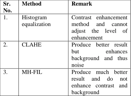
Histogram equalization is a pre-processing technique to enhance contrast in all type of images. Equalization implies mapping from given intensity distribution so the intensity values are spread over the whole range. The main drawback of histogram equalization is that it does not provide any mechanism by which we can adjust the level of enhancement. HE uses a level transformation function to enhance the image contrast, this transformation function also redistributes the input image histogram such that the resultant histogram is closed to uniform distribution. Another new method
of histogram modification for contrast
enhancement. The proposed method has the ability to control the level of contrast enhancement in the output image.
Using HE we can enhance the contrast in a given mammogram image. The limitation of HE is that the mean gray level of output image changes drastically and we do not have any control over it. CLAHE produces better results but the drawback of CLAHE is that this method enhances the background and foreground at equal level, this leads
the enhancement of noise in the background area and hence some artifacts appears in background of enhanced image, similar problem occurs with US .
MH-FIL produces much better results as compared to results of other given methods; as this method neither over enhances the contrast nor does it enhance background and foreground at equal grey level. MH-FIL produces visually better results than other methods [8].
[image:2.595.299.515.352.511.2]Recently, multiscale techniques have sparked the interest of researchers for contrast enhancement of images especially wavelet transform. For contrast enhancement based on mathematical morphology theory there are methods that deals with the contrast of the original image to enhance the segmentation process [14].
Table 1: Overview of enhancement techniques.
Sr. No.
Method Remark
1. Histogram
equalization
Contrast enhancement
method and cannot
adjust the level of enhancement
2. CLAHE Produce better result
but enhances
background and thus noise
3. MH-FIL Produce much better
result and do not enhance contrast and background
2.2 Image segmentation methods:
Segment the threshold image by watershed segmentation because it is the best method to segment an image to separate a tumor but it suffers from over and under segmentation. It not give the better result after that some morphological operations are applied on the image after converting it into binary form [1].
In this paper various clustering methods that have been used for segmentation in MRI are reviewed.
Thresholding- In this process of separating pixels in different classes depending on their pixel gray levels. Pixels are grouped based on threshold values. Disadvantage is that only two classes are generated and it cannot be applied to multichannel images.
Region growing-In this method the seed point is selected. Pixels in the region accordance with the homogeneity criteria and continues until all pixels belongs to some region. Disadvantage is that manual selection of seed point that is requires user interface and time consuming and is not used alone as do not sufficient to segment accurately.
Mean shift- It used for cluster analysis in computer vision and image processing. Mean shift algorithm used for clusters an n- dimensional data set.
Clustering techniques- Clusters the process of collection of objects which are similar between them and are dissimilar objects belonging to other cluster.
• K-mean is the unsupervised algorithms
that solve clustering problem and segment the image using basic knowledge of cluster value [6]. The K-means clustering technique is a pixel-based method. K-means clustering is suitable for biomedical image segmentation as the number of clusters is usually known for images of particular regions of the human anatomy [10].
• Fuzzy c- mean clustering is popular
method for Segmentation but it only considers image intensity thereby producing noisy images so used with k-mean [6].
The methods include k-means clustering with watershed segmentation algorithm, optimized k-means clustering with genetic algorithm and optimized c- means clustering with genetic algorithm.
1. K-means clustering method helps to segment the brain tumor image and the second method improves the primary results of segmentation of tumor.
2. Over segmentation and sensitivity to false edges are difficulties in ordinary k-means method. GAs with the modification of mutation operations improves the speed of convergence and computing time is reduced also.
3. The c-means clustering method has been implemented and its performance can be improved by using optimization with the use of genetic algorithm.
4. The implementation of genetic algorithm begins with an initial population of chromosomes which are randomly selected Particulars traits determine the hereditary [11].
In ACO, a set of software agents called artificial ants search for good solutions to a given optimization problem. In Block based technique, both the given reference MR brain image and the normal image has been divided into several blocks [12].
The method consists of automatic segmentation of tumors from 2D MRI slices using morphological methods and these segmented tumor slices are exported to a 3D tool kit in MATLAB. For verification of rendered 3D tumor volume, we used 3D-DOCTOR software package that is usually using by radiologists. In this work, T2 weighted and T1 FLAIR image was used for extracting tumor from 2D slices [13].
Two major classes of segmentation techniques: edge based segmentation approach and region based segmentation approach. Edge approach looks for limits between regions with different characteristics. Its aims at finding object boundaries and segmenting regions enclosed by the contours. They prove to be computationally fast and don’t require prior information about the image content. However a drawback of the edge approach is that the edges do not enclose the object completely. In region-based techniques, segmentation is applied by identifying all pixels that belong to the object based on the intensity of pixels. Their aim is the regions satisfying a given homogeneity criterion [14].
Table 2: overview of segmentation techniques.
Sr. No.
Method Remark
1 Watershed segmentation
Best method to segment but suffers from over and under segmentation.
2 Thresholding Based on threshold values but cannot be applied to multichannel images. 3 Region
growing
Based on seed points but requires user interface. 4 Mean- shift Used for cluster analysis. 5 Clustering
techniques
K-mean is a pixel-based method and Fuzzy c-mean considers image intensity.
2.3 Feature extraction methods:
Probabilistic Neural Network (PNN) is a Radial Basis Neural Network, which provides a general solution to pattern classification problems by following an approach developed in statistics, called Bayesian classifiers. It is employed to implement an automatic MR image classification of brain tumors into normal, benign and malignant [2].
The automated recognition of tumor cell in given MRI image a neuro fuzzy classifier is realized. The classifier module implements a hybrid algorithm integrating neural network and fuzzy system. Fuzzy neural approach found to have more accurate decision making as compare to their counterparts. The obtained features are processed using fuzzy classification layer before passing it to neural network. Texture features or more Precisely, Gray Level Co- occurrence Matrix (GLCM) features are used to distinguish between normal and abnormal brain tumors. The different features like entropy, Angular second moment (ASM), Contrast, Inverse Difference Moment (Homogeneity), Dissimilarity. The tumor region is extracted and the severity of the tumor is found out [9].
In a PNN, the operations are organized into a multi-layered feed forward network with four layers: Input layer, Hidden layer, Pattern layer/Summation layer and Output layer. When an input is present, the first layer computes the distance from the input vector to the training input vectors. The second layer sums the contribution for each class of inputs and produces its net output as a vector of probabilities. Finally, a complete transfer function on the output of the second layer picks the maximum of these probabilities, and produces a 1
(positive identification) for that class and 0 (negative identification) for non-targeted classes [10].
The feature extraction is carried out by using an overlapping and sliding window of 7*7 pixels on each slice of a specific plane. In the feature extraction process, window size plays an important role since smaller windows are not able to capture the second order feature that is texture information. The first order features we extract from the image are intensity, mean and variance. The intensity is referred to the gray level of the centre pixel on the window. The mean and variance are calculated taking into account the gray level present on the window. On the other hand, we additionally use second order features such as textural features [15].
Table 3: overview of feature extraction techniques.
Sr. No.
Method Remark
1. Probabilistic Neural Network
Radial Basis Neural
Network and classify brain tumor into normal, benign and malignant. 2. Neuro fuzzy
classifier
Implement a hybrid
algorithm and distinguish
between normal and
abnormal portion of brain.
3. CONCLUSION:
In this paper enhancement methods, segmentation methods, classification of brain tumor and its feature extraction is discussed. Many algorithms have been proposed in the literature for each processing stage. So the methods gives the better result are histogram equilization for enhancement, probabilistic neural network for feature extraction.
REFERENCES
[1] 1.Sudipta Roy, Samir K. Bandyopadhyay “Detection and Quantification of Brain Tumor from MRI of Brain and it’s Symmetric Analysis” International Journal of Information and Communication Technology Research Volume 2 No. 6, June 2012.
[3] Dina AboulDahab, Samy S. A. Ghoniemy, Gamal M. Selim “Automated Brain Tumor Detection and Identification Using Image Processing and Probabilistic Neural Network Techniques” International Journal of Image Processing and Visual Communication Volume 1 , Issue 2 , October 2012.
[4] Prof.B.K.Saptalakar, Miss. Rajeshwari.H “Segmentation Based Detection Of Brain Tumor” et al International Journal of Computer and Electronics Research [Volume 2, Issue 1, February 2013]
[5] VivekAngoth, CYN Dwith, Amarjot Singh “A Novel Wavelet Based Image Fusion for Brain Tumor Detection” International Journal of Computer Vision and Signal Processing, 2(1), 1-7(2013).
[6] Jay Patel and KaushalDoshi “A Study of Segmentation Methods for Detection of Tumor in Brain MRI” Advance in Electronic and Electric Engineering Volume 4, Number 3 (2014).
[7] Ed-EdilyMohd. Azhari, Muhd.
MudzakkirMohd. Hatta, ZawZawHtike and Shoon Lei Win “Brain Tumor Detection And
Localization In Magnetic Resonance
Imaging”International Journal of Information
Technology Convergence and Services
(IJITCS) Vol.4, No.1, February 2014.
[8] Tarun Kumar Agarwal, Mayank Tiwari, Subir Singh Lamba “Modified Histogram Based Contrast Enhancement using Homomorphic Filtering for Medical Images” 978-1-4799-2572-8/14/$31.00 c 2014 IEEE
[9] Anisha M. Lal, M. Balaji, D. Aju “Multi-Level Fusion of CT and MRI Brain Images for Classifying Tumor” International Journal of Enhanced Research in Management & Computer Applications, Vol. 3 Issue 8, August 2014
[10]Miss. PriyankaKatti, Mr. V. R. Marathe “Implementation of Classification System for Brain Tumor using Probabilistic Neural Network” International Journal of Advanced Research inComputer and Communication Engineering Vol. 4, Issue 10, October 2015. [11]Kailash Sinha1, G.R.Sinha “Efficient
Segmentation Methods for Tumor Detection in MRI Images” IJCSI International Journal of Computer Science Issues, Vol. 9, Issue 2, No 1, March 2012.
[12]Dr.M.Karnan, K.Selvanayaki “Improved
Implementation of Brain MR Image
Segmentation Using Meta Heuristic
Algorithms” 2: Research Scholar 978-1-4244-5967-4/10/$26.00 ©2010 IEEE.
[13]S. AnandaResmi, Tessamma Thomas “A semi- automatic method for segmentation and 3D modelling of gliomatumors from brain MRI” J. Biomedical Science and Engineering, 2012.
[14]Ahmed KHARRAT, Mohamed Ben
MESSAOUD,Nacéra BENAMRANE,
Mohamed ABID “Detection of Brain Tumor in Medical Images” InternationalConference on Signals, Circuits and Systems 978-1-4244-4398-7/09/$25.00 2009 IEEE.
[15]A. Ortiz, J. M. Gorriz, J. Ramirez, D. Salas- Gonzalez “Unsupervised Neural techniques Applied to Brain Images Segmentation” Hindavi publication corporation Advance in Artificial Neural Systems volume 2012.
[16]Sivasundari .S, Dr.R. Siva Kumar,
Dr.M.Karnan “Review of MRI Image
Classification Techniques” International Journal of Research Studies in Computer Science and Engineering (IJRSCSE) Volume 1, Issue 1, May 2014, PP 21-28.
[17]KimmiVerma, AruMehrotra, Vijayeta Pandey,
Shardendu Singh “Image Processing
Techniques For The Enhancement Of Brain Tumor Patterns” International Journal of Advanced Research in Electrical, Electronics and Instrumentation Engineering Vol. 2, Issue 4, April 2013.
[18]Mohammed SabbihHamoud Al-Tamimi,
GhazaliSulong “Tumor Brain Detection Through Mr Images: A Review Of Literature ”Journal of Theoretical and Applied Information Technology 20th April 2014. Vol. 62 No.2.
[19]RoopaliR.Laddha, S.A.Ladhake “A Review on Brain Tumor Detection Using Segmentation
And Threshold Operations” (IJCSIT)
International Journal of Computer Science and Information Technologies, Vol. 5 (1), 2014, 607-611.