R E S E A R C H A R T I C L E
Open Access
Short-term changes of choroidal vascular
structures after phacoemulsification surgery
Haisong Chen
1, Zheming Wu
1, Yun Chen
1, Manshan He
1and Jiawei Wang
2*Abstract
Background:To evaluate the changes of choroidal vascular structures in patients after phacoemulsification surgery. Methods:A self-control study was conducted on 36 eyes of 36 patients who had uneventful phacoemulsification. Choroidal images were acquired preoperatively, 7 days (D7), 1 month (M1), and 3 months (M3) after surgery from enhanced depth imaging (EDI) optical coherence tomography (OCT) scans. Choroidal vascularity index (CVI) was used to assess vascular status of the choroid using image binarization by the Niblack method. The postoperative values of mean CVI were compared with baseline by paired t-test. Univariate and multiple linear regression analyses were performed to determine the associations between CVI and other factors.
Results:The mean age of the recruited patients was 63.1 ± 6.9 years. The mean CVI at baseline was 60.1 ± 5.5%. After surgery, the CVI significantly increased to 61.7 ± 5.3% at D7, 63.6 ± 4.4% at M1 and 64.8 ± 4.0% at M3 (p= 0.035, 0.0006, < 0.0001, respectively). Univariate and multiple regression analysis revealed a positive association between CVI and subfoveal choroidal thickness (SFCT) at pre-operation and no significant association with age, axial length (AL), intraocular pressure (IOP) and gender at all timepoints.
Conclusions:Phacoemulsification induced increased CVI in patients diagnosed with cataract. Evaluation of the long-term change of CVI following surgery may provide valuable information for studying the relationship between phacoemulsification and disorders of the choroid.
Keywords:Choroid, Vascular structures, Phacoemulsification
Background
Cataract with phacoemulsification surgery is the most extensively performed eye surgery. There are more than 1300 cases per million people undergoing phacoemulsifi-cation surgery per year in China and greater than 5000 cases per million people per year in Europe, America and India [1]. Phacoemulsification surgery is safe and generally associated with successful visual outcomes.
Choroid, the highest blood circulation in the human body, is composed of blood vessels, connective tissues, nerves, melanocytes and extracellular fluid. A great deal of analysis and research indicates that even uncomplicated phacoemulsification induces disorders of the choroid, especially an increase in the choroid thickness [2–4]. However, only the choroidal thickness does not supply convincing evidence on what structures change, especially
about the blood volume within the choroid in patients after Phacoemulsification surgery.
Further morphological and vascular analyses of the choroid may certify the change of choroidal blood vol-ume in patients after Phacoemulsification surgery. With the advent of enhanced depth imaging (EDI) optical co-herence tomography (OCT), it is possible to assess the choroidal stromal and vascular structures. Recently, ap-plication of image binarization of choroid structures has further provided a novel measure index for vascular sta-tus of the choroid [5]. To the best of our knowledge, there is no report about the change of choroidal vascular structures after Phacoemulsification surgery.
In the current study, we aimed to determine the influ-ence of phacoemulsification on the proportion of chor-oidal vascular structures in patients after surgery. The choroidal vascularity index (CVI) from EDI-OCT scans will be used and we speculated that CVI may provide more additional information about the morphology and * Correspondence:[email protected]
2Eye Center of Shandong University, The Second Hospital of Shandong
University, Shandong University, Jinan 250000, China
Full list of author information is available at the end of the article
physiology of the choroid and may be useful to interpret the disorders of the choroid after cataract surgery.
Methods
Thirty-six healthy patients undergoing uncomplicated Phacoemulsification surgery were recruited for this self-controlled case series study. All the patients were re-cruited consecutively (from October 2016 to December 2016) from the cataract department of Guangzhou Aier Eye Hospital and signed the consent form after a fullest explanation of the purpose and procedures of the study. The study was adhered to the provisions of the Declar-ation of Helsinki for research involving human subjects and was approved by the Ethical Review Committee of Guangzhou Aier Eye Hospital.
All the study participants were healthy individuals with no history of ocular disease or visual symptoms; aged at least 40 years; intraocular pressure (IOP) < 21 mmHg; nor-mal appearance of optic nerve head; nornor-mal anterior cham-ber angles; Patients were excluded if they had glaucoma, high myopia or hyperopia (magnitude exceeding ±6 diop-ters (D) of spherical equivalent refraction), AMD, or other retinal diseases that could interfere the choroidal thickness. The diagnosis of glaucoma was based on the findings from gonioscopy, optic disc characteristics, and visual fields re-sults. Patients with severe systemic diseases, such as dia-betes mellitus, rheumatism, or malignant tumors, serious opacity of refractive media or unstable fixation that could prevent EDI-OCT measurement were also excluded.
All the patients underwent a comprehensive ophthal-mologic examination, including IOP measurement using Goldmann applanation tonometry, autorefraction examin-ation, measurement of visual acuity and a best-corrected visual acuity (BCVA), axial length using ocular biometry (IOL Master, Zeiss, Germany), fundus examination and EDI-OCT measurement (Spectralis, Heidelberg Engineer-ing, Heidelberg, Germany) before surgery and postopera-tively at 7 days (D7), month 1 (M1) and months 3 (M3).
All patients received standard phacoemulsification sur-gery through clear corneal incisions under superficial anesthesia (0.5% Proparacaine hydrochloride Eye Drops, Alcon, Fort Worth, TX). All phacoemulsification surger-ies was performed by the same experienced surgeon (HSC) using the Infiniti system® (Alcon Labs Inc). In all cases, after removal of the lens cortex, a foldable intraoc-ular lens was implanted uneventfully in the capsintraoc-ular bag. Within 1 month after surgery, Tobradex (0.3% tobra-mycin and 1% dexamethasone, Alcon, Fort Worth, TX) eye drops were applied four times a day, a non-steroidal anti-inflammatory eye drops (Pranoprofen Eye Drops, Senju Pharmaceutical Co.,Ltd. Osaka, Japan) were ap-plied four times a day, and TobraDex eye ointment (Alcon, Fort Worth, TX) was applied once every evening before bed.
Image acquisition
EDI-OCT scans of the macular were performed for the operated eye using the EDI mode of SD-OCT (Spectralis, Heidelberg Engineering, Heidelberg, Germany). Horizon-tal 6-mm line scans centred on the fovea were acquired. Due to the diurnal variation of choroidal thickness, all the measurements were performed at the same time of the day (08:00 AM~ 12:00 AM) and accomplished in triplicate by two independent examiners. The sections going dir-ectly through the center of the fovea were selected for fur-ther analysis. The subfoveal choroidal thickness (SFCT) was measured using the in-built calipers tool. SFCT was defined as the vertical distance between the outer surface of the retinal pigment epithelium and the choroidal–sclera interface [6].
Procedures of image binarization
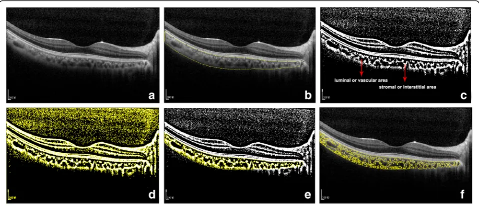
Image binarization of the subfoveal choroidal area was performed by one public domain software, Image J (ver-sion 1.47, provided in the public domain by the National Institutes of Health, Bethesda, MD, USA; http://imagej. nih.gov/ij/) [7, 8]. In brief, the images with one central scan passing through the fovea were chosen. The region of interest was manually selected using the polygon tool and added to ROI manager. After measuring the bright-ness of the selected luminal areas of the original OCT, the average brightness was set at the minimum value to minimize the noise in the OCT image. Then the original images were converted to 8 bits and adjusted by the Niblack Auto Local Threshold. The binarized image was converted to RGB (red, green, blue) image again, and the luminal area was determined using the Threshold Tool. After the image binarization, the total circumscribed area (TCA) and area of dark pixels were calculated. The dark pixels represent the luminal or vascular area (LA) and stromal or interstitial area (SA) was defined as the area of light pixels (Fig. 1). CVI was defined as the pro-portion of LA to TCA.
Inter-rater and intra-rater agreement
all the image binarization was performed by single author (JWW).
Statistical analysis
All statistical analyses were performed using SPSS software version 20.0 (IBM-SPSS, Chicago, Illinois, USA). Normally distributed data were expressed as mean ± standard devi-ation (SD). Each postoperative value was compared with baseline by paired t-test. Univariate and multiple linear re-gression analyses were performed to determine the associa-tions between CVI and other factors. Values of p< 0.05 were considered to be statistically significant.
Results
A total of 36 patients (20 male and 16 female) were fi-nally recruited for the current study and the demo-graphic characteristics of the patients are shown in Table1. Mean age of the volunteers was 63.1 ± 6.9 years (range, 49~ 78).
The inter-rater agreement for CVI was 0.932 (95% CI: 0.866–0.965) and the inter-rater reliability was 0.959 (95% CI: 0.920–0.979), which indicates excellentagree-ment for image binarization and CVI calculation. Bland Altman plot analysis was constructed to display the high agreement (Fig.2).
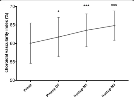
The baseline CVI in patients was 60.1 ± 5.5%. After sur-gery, the CVI significantly increased to 61.7 ± 5.3% at D7, 63.6 ± 4.4% at M1 and 64.8 ± 4.0% at M3 (p= 0.035, 0. 0006, < 0.0001 for D7, M1 and M3 when compared with the preoperative values). The greatest progression of CVI was observed between D7 and M1 after surgery (Fig.3).
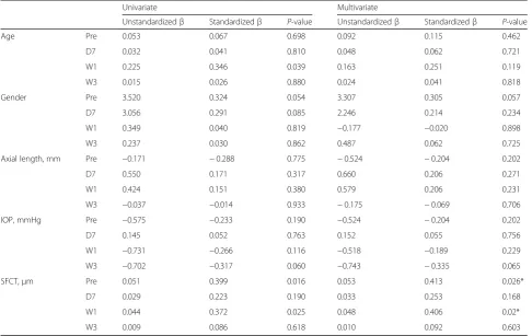
Univariate linear regression analysis revealed a positive association between CVI and SFCT at baseline and M1 postoperative follow-up. Age was found to be related with CVI at W1 after surgery. However, in the multiple regression model, only SFCT was significantly associated with CVI. Univariate and multivariate linear regression analyses revealed no significant association of CVI with AL, IOP and gender (Table2).
Fig. 1Image binarization for choroid with Niblack auto local thresholding technique.aOriginal EDI-OCT scan image.bManual segmentation of the choroidal area with one central scan passing through the fovea.cConversion of the image with the Auto Local Threshold tool.dClear segmentation of black and white areas on the choroid with Niblack autolocal threshold.ebinarized image was reconverted back to RGB image.fbinarized image over the original EDI-OCT scan
Table 1Demographics characteristics of the recruited subjects
Characteristics Mean ± SD
Baseline D7 W1 W3
Axial length, mm 23.55 ± 1.58 23.37 ± 1.65 23.30 ± 1.58 23.11 ± 1.56
Intraocular pressure, mmHg 14.19 ± 2.13 13.18 ± 1.91 12.54 ± 1.62 11.93 ± 1.80
Subfoveal choroidal thickness (SFCT),um 234.8 ± 42.49 239.7 ± 40.61 266.6 ± 37.71 276.3 ± 36.20
Age, yrs 63.1 ± 6.9
Gender, male (%) 20 (55.6%)
Discussion
A number of publications have reported the possible in-fluence of cataract surgery on the choroid [2, 3, 9, 10]. Aslan BS et al. [9] have investigated the effects of unevent-ful phacoemulsification surgery on choroidal thickness using spectral domain optical coherence tomography (SD-OCT). They found that phacoemulsification may cause significant increase in choroid at all regions evaluated at 1 month postoperative follow-up. Yılmaz T et al. [10] have reported a long-term change in SFCT after cataract sur-gery. They have measured the SCT at baseline and postop-eratively at week 1 and months 1, 3, 6 and 12 and the results indicated that uncomplicated phacoemulsification induced insignificant increases in SFCT and this did not return to baseline during follow-up. In our study, we found that the mean SFCT, as tested by EDI-OCT, signifi-cantly increased after surgery, which was consistent with the published literatures (Fig. 4). As well known, the choroid is predominantly composed of blood vessels surrounded by stromal tissue. Although many studies, including our study, have described the increased SCT after phacoemulsification, nobody really knows which
structures within the choroid increased. To answer this question, we used the binarization of EDI-OCT images and firstly attempted to assess the changes of choroidal vascular and stromal structures following cataract surgery. With the advent of EDI-OCT, it is possible to analyze the structural changes allowing for quantitative measure-ments of choroidal vasculature in patients. Recently, an OCT based metric termed CVI has been used to assess the choroidal vascularity, using the image binarization tech-nique for EDI-OCT scans [5]. CVI is more stable and less interferences from physiologic factors as opposed to the thickness of choroid [5,7,11]. In our previous study, we have found that SFCT is affected by many physiological factors, like AL and gender [12]. When comparing the fac-tors influencing CVI, we found no significant association of CVI with AL, IOP and gender and there were positive associations between CVI and SFCT at pre-operation and 1 month postoperative follow-up. CVI was affected by few variables and demonstrated greater stability than SFCT. Measuring of the CVI would provide deeper understanding of the vascular structural changes in the process of choroid diseases, and therefore may be more informative compared to SFCT measurements alone.
By calculating the CVI, it is able to determine if there was an increase or decrease in vascularity and provide us more information on the proportion of vascularity in the choroid. Agrawal R and collaborators [13] found that eyes with acute central serous chorioretinopathy (CSC) had significantly higher CVI compared with their fellow eyes and age-matched healthy subjects. They demon-strated that CVI might be useful for the early diagnosis of CSC and to be a therapeutic index for the treatment response after laser photocoagulation or photodynamic therapy. In patients diagnosed with exudative age-related macular degeneration (AMD), there was a significantly lower CVI and CVI was probably a potential noninvasive tool for studying structural changes in choroid and ex-udative AMD development monitored [14]. Therefore, CVI is considered to be a relatively stable index to moni-tor the progression of choroidal diseases [15–17].
Fig. 2Bland-Altman plot analysis of the intra- and inter-rater agreement.aandbshows the high reliability of the inter-rater and intra-rater agreement for the image binarization and CVI calculation
Phacoemulsification is the most frequently performed surgical intervention worldwide and considered to be safe and effective. However, in the past, concern has been raised about the association between cataract sur-gery with the incidence or progression of AMD. Disor-ders of the choroid after Phacoemulsification may cause
the onset of many choroid diseases, including AMD. Some researchers have raised concerns that phacoemulsi-fication may constitute a risk factor for the development of exudative AMD [18–20]. In contrast, recent evidence does not find the surgery to cause or worsen the progres-sion of AMD [21–24]. In our study, the CVI was 60.1 ± 5. 5% in the baseline and significantly increased D7, M1 and M3 after the surgery. The greatest progression of CVI was observed between D7 and M1 after surgery. Therefore, our data suggested that phacoemulsification seemed to be able to induce the expansion of choroidal vascular struc-tures within 3 months after surgery. As a more stable par-ameter for disease monitoring, it will be very useful to evaluate CVI of patients after phacoemulsification at fur-ther follow-up over a longer period. Evaluation of CVI may provide further evidence about the relationship be-tween phacoemulsification and AMD.
We found that phacoemulsification induced progressive increases in CVI. On the other hand, the surgery increased the proportion of vascular structures in the choroid. We suspect that the increased CVI may depend crucially on the choroidal inflammation induced by surgical trauma [25]. With the disruption to the blood-aqueous barrier, the inflammatory mediators in the aqueous humor pass Table 2Univariate and multivariate linear regression analyses of age, gender and ocular factors associated with choroidal vascularity index
Univariate Multivariate
Unstandardizedβ Standardizedβ P-value Unstandardizedβ Standardizedβ P-value
Age Pre 0.053 0.067 0.698 0.092 0.115 0.462
D7 0.032 0.041 0.810 0.048 0.062 0.721
W1 0.225 0.346 0.039 0.163 0.251 0.119
W3 0.015 0.026 0.880 0.024 0.041 0.818
Gender Pre 3.520 0.324 0.054 3.307 0.305 0.057
D7 3.056 0.291 0.085 2.246 0.214 0.234
W1 0.349 0.040 0.819 −0.177 −0.020 0.898
W3 0.237 0.030 0.862 0.487 0.062 0.725
Axial length, mm Pre −0.171 −0.288 0.775 −0.524 −0.204 0.202
D7 0.550 0.171 0.317 0.660 0.206 0.271
W1 0.424 0.151 0.380 0.579 0.206 0.231
W3 −0.037 −0.014 0.933 −0.175 −0.069 0.706
IOP, mmHg Pre −0.575 −0.233 0.190 −0.524 −0.204 0.202
D7 0.145 0.052 0.763 0.152 0.055 0.756
W1 −0.731 −0.266 0.116 −0.518 −0.189 0.229
W3 −0.702 −0.317 0.060 −0.743 −0.335 0.065
SFCT,μm Pre 0.051 0.399 0.016 0.053 0.413 0.026*
D7 0.029 0.223 0.190 0.033 0.253 0.168
W1 0.044 0.372 0.025 0.048 0.406 0.02*
W3 0.009 0.086 0.618 0.010 0.092 0.603
*Adjusted for variables with ap-value< 0.05 in the univariate analysis.β, regression coefcient
through the vitreous to the retina and choroid, subse-quently leading to the change of choroidal vascular struc-tures. Another possibility is the IOP decrease after cataract surgery. The increased ocular perfusion pressure caused by reduced IOP may induce the increased CVI in the early period after phacoemulsification. How-ever, the concrete mechanism has yet to be fully ex-plained and further study is needed.
There are some limitations in the current study. Firstly, although this binarization technique is valid and widely used, there is no concrete evidence that the dark areas represented the vascular areas. Secondly, the post-operative anti-inflammatory eye drops may affect the CVI evaluation; Thirdly, the average ultrasonic emulsifi-cation time (UST) and the cumulative dissipated energy (CDE) were not recorded, which may also influence the CVI analysis. Lastly, the cohort of patients was not large enough and we only assessed the short-term changes of CVI after surgery. The future bigger sample research and longer follow-up periods are needed, especially to clarify the relationship between phacoemulsification and the frequency of disorders of the choroid such as AMD.
Conclusions
In the follow-up study, we firstly used CVI to assess the change of choroidal vascular structures in patients under-going phacoemulsification surgery. Our results showed that the proportion of vascularity in the choroid, termed as CVI, significantly increased within 3 months following surgery. Evaluation of CVI may provide valuable informa-tion for studying the relainforma-tionship between phacoemulsifi-cation and disorders of the choroid such as AMD.
Abbreviations
AL:Axial length; AMD: Age-related macular degeneration; CSC: Central serous chorioretinopathy; CVI: Choroidal vascularity index; EDI: Enhanced depth imaging; IOP: Intraocular pressure; LA: Luminal or vascular area; OCT: Optical coherence tomography; SA: Stromal or interstitial area; SFCT: Subfoveal choroidal thickness; TCA: Total circumscribed area
Acknowledgements Not applicable.
Funding
This study was supported by the National Natural Science Foundation of China (81700831), the Natural Science Foundation of Shandong province, China (ZR2017BH049) and the Medicine and Technology Program of Guangzhou, China (20161A011099).
Availability of data and materials
The datasets used and/or analysed during the current study are available from the corresponding author on reasonable request.
Authors’contributions
All authors conceived of and designed the experimental protocol. HSC, YC, MSH and JWW collected the data and involved in the analysis and interpretation of the data. HSC wrote the first draft of the manuscript. JWW and ZMW reviewed and revised the manuscript and produced the final version. All authors read and approved the final manuscript.
Ethics approval and consent to participate
The study protocol adhered to the tenets of the Declaration of Helsinki and and was approved by the Ethical Review Committee of Guangzhou Aier Eye Hospital. All the patients signed the consent form after a fullest explanation of the purpose and procedures of the study.
Consent for publication
The consents for publication from all the patients were obtained after a detailed explanation of the purpose and procedures of the project.
Competing interests
The authors declare that they have no competing interests.
Publisher’s Note
Springer Nature remains neutral with regard to jurisdictional claims in published maps and institutional affiliations.
Author details
1Guangzhou Aier Eye Hospital, Guangzhou, China.2Eye Center of Shandong
University, The Second Hospital of Shandong University, Shandong University, Jinan 250000, China.
Received: 25 October 2017 Accepted: 16 March 2018
References
1. Tan X, Lin H, Li Y, et al. Cataract screening in a rural area of southern China: a retrospective cohort study. Lancet. 2016;388(Suppl 1):S53.
2. Cheong KX, Tan CS. Long-term increase in subfoveal choroidal thickness after surgery for senile cataracts. Am J Ophthalmol. 2015;159(3):608–9. 3. Ohsugi H, Ikuno Y, Ohara Z, et al. Changes in choroidal thickness after
cataract surgery. J Cataract Refract Surg. 2014;40(2):184–91.
4. Jiang H, Li Z, Sun R, Liu D, Liu N. Subfoveal choroidal and macular thickness changes after phacoemulsification using enhanced depth imaging optical coherence tomography. Ophthalmic Res. 2017;https://doi.org/10.1159/ 000480240.
5. Agrawal R, Salman M, Tan KA, et al. Choroidal vascularity index (CVI)–a novel optical coherence tomography parameter for monitoring patients with Panuveitis. PLoS One. 2016;11(1):e0146344.
6. Huang W, Wang W, Gao X, et al. Choroidal thickness in the subtypes of angle closure: an EDI-OCT study. Invest Ophthalmol Vis Sci. 2013;54(13):7849–53. 7. Agrawal R, Gupta P, Tan KA, Cheung CM, Wong TY, Cheng CY. Choroidal
vascularity index as a measure of vascular status of the choroid: measurements in healthy eyes from a population-based study. Sci Rep. 2016;6:21090. 8. Agrawal R, Li LK, Nakhate V, Khandelwal N, Mahendradas P. Choroidal
vascularity index in Vogt-Koyanagi-Harada disease: an EDI-OCT derived tool for monitoring disease progression. Transl Vis Sci Technol. 2016;5(4):7. 9. Aslan BS, Bayhan HA, Muhafiz E, Kırboğa K, Gürdal C. Evaluation of choroidal
thickness changes after phacoemulsification surgery. Clin Ophthalmol. 2016; 10:961–7.
10. Yılmaz T, Karci AA, Yilmazİ, Yılmaz A, Yıldırım Y, Sakalar YB. Long-term changes in subfoveal choroidal thickness after cataract surgery. Med Sci Monit. 2016;22:1566–70.
11. Agrawal R, Wei X, Goud A, Vupparaboina KK, Jana S, Chhablani J. Influence of scanning area on choroidal vascularity index measurement using optical coherence tomography. Acta Ophthalmol. 2017;95(8):e770–5.
12. Wang J, Gao X, Huang W, et al. Swept-source optical coherence tomography imaging of macular retinal and choroidal structures in healthy eyes. BMC Ophthalmol. 2015;15:122.
13. Agrawal R, Chhablani J, Tan KA, Shah S, Sarvaiya C, Banker A. Choroidal vascularity index in central serous CHORIORETINOPATHY. Retina. 2016;36(9): 1646–51.
14. Wei X, DSW T, Ng WY, Khandelwal N, Agrawal R, CMG C. CHOROIDAL VASCULARITY INDEX: A Novel Optical Coherence Tomography Based Parameter in Patients With Exudative Age-Related Macular Degeneration. Retina. 2017;37(6):1120–5.
16. Tan KA, Laude A, Yip V, Loo E, Wong EP, Agrawal R. Choroidal vascularity index - a novel optical coherence tomography parameter for disease monitoring in diabetes mellitus. Acta Ophthalmol. 2016;94(7):e612–6. 17. Ng WY, Ting DS, Agrawal R, et al. Choroidal structural changes in myopic
choroidal neovascularization after treatment with Antivascular endothelial growth factor over 1 year. Invest Ophthalmol Vis Sci. 2016;57(11):4933–9. 18. Freeman EE, Munoz B, West SK, Tielsch JM, Schein OD. Is there an association
between cataract surgery and age-related macular degeneration? Data from three population-based studies. Am J Ophthalmol. 2003;135(6):849–56. 19. Cugati S, Mitchell P, Rochtchina E, Tan AG, Smith W, Wang JJ. Cataract
surgery and the 10-year incidence of age-related maculopathy: the Blue Mountains eye study. Ophthalmology. 2006;113(11):2020–5.
20. Wang JJ, Klein R, Smith W, Klein BE, Tomany S, Mitchell P. Cataract surgery and the 5-year incidence of late-stage age-related maculopathy: pooled findings from the beaver dam and Blue Mountains eye studies. Ophthalmology. 2003;110(10):1960–7.
21. Chew EY, Sperduto RD, Milton RC, et al. Risk of advanced age-related macular degeneration after cataract surgery in the age-related eye disease study: AREDS report 25. Ophthalmology. 2009;116(2):297–303.
22. Ehmann DS, Ho AC. Cataract surgery and age-related macular degeneration. Curr Opin Ophthalmol. 2017;28(1):58–62.
23. Park SJ, Lee JH, Ahn S, Park KH. Cataract surgery and age-related macular degeneration in the 2008-2012 Korea National Health and nutrition examination survey. JAMA Ophthalmol. 2016;134(6):621–6.
24. Bockelbrink A, Rasch A, Roll S, Willich SN, Greiner W. What effects has the cataract surgery on the development and progression of Age-Related Macular Degeneration (AMD). GMS Health Technol Assess. 2006;2:Doc21. 25. Xu H, Chen M, Forrester JV, Lois N. Cataract surgery induces retinal
pro-inflammatory gene expression and protein secretion. Invest Ophthalmol Vis Sci. 2011;52(1):249–55.
• We accept pre-submission inquiries
• Our selector tool helps you to find the most relevant journal
• We provide round the clock customer support
• Convenient online submission
• Thorough peer review
• Inclusion in PubMed and all major indexing services
• Maximum visibility for your research
Submit your manuscript at www.biomedcentral.com/submit