R E S E A R C H
Open Access
Identifying more reliable parameters for the
detection of change during the follow-up
of mild to moderate keratoconus patients
Ivo Guber
1, Colm McAlinden
2,3, François Majo
4and Ciara Bergin
4*Abstract
Background:Reaching a consensus on which parameters are most reliable at detecting progressive keratoconus
patients with serial topography imaging is not evident. The aim of the study was to isolate the parameters best positioned to detect keratoconus progression using the Pentacam HR® measures based on the respective limits of repeatability and range of measurement.
Method:Using the Pentacam HR®, a tolerance index was calculated on anterior segment parameters in healthy and keratoconic eyes. The tolerance index provides a scale from least to most affected parameters in terms of
measurement noise relative to that observed in healthy eyes. Then, based on the“number of increments”from no disease to advanced disease, a relative utility (RU) score was also calculated. RU values close to 1 indicate parameters best positioned to detect a change in keratoconic eyes.
Results:The tolerance index values indicated that 36% of ocular parameters for keratoconic eyes had repeatability limits which were wider than normative limits (worse), but 28% of the ocular parameters were narrower than
normative limits (better). Considering only those parameters with a RU greater than 0.95, a small number of parameters were within this range, such as corneal curvature and asphericity indices.
Conclusions:This study demonstrates that measurement error in keratoconic eyes is significantly greater than healthy eyes. Indices implemented here provide guidance on the levels of expected precision in keratoconic eyes relative to healthy eyes to aid clinicians in distinguishing real change from noise. Importantly maximal keratometry (Kmax), central corneal thickness (CCT) and thinnest corneal thickness (TCT) were highlighted as problematic indices for the follow-up of keratoconus in terms of repeatability.
Keywords:Pentacam, Keratoconus, Progression, Repeatability, Precision, Corneal crosslinking
Background
The clinician who follows keratoconus patients with serial topography imaging desires to know which parameters are most reliable at detecting progression. This is important as the detection of progression will often determine treatment choice e.g., collagen cross-linking (CXL). However, the abil-ity of a parameter to detect progression is decreased with increased measurement noise (signal to noise ratio). Previ-ously, in healthy eyes the repeatability limit, of the maximal corneal curvature Kmax (with the Pentacam HR®) were
reported to be 0.8 Dioptres (D), however, we found repeat-ability limits in keratoconic eyes to be 1.97 D [1, 2]. This re-sult means that the current main criteria for progression detection and CXL is inadequate (i.e. a change of 1 D in Kmax after one year of follow-up) [1, 3].
To date, comparison of repeatability between subgroups has been limited to a comparison of the repeatability limits or the correlation of variation values within a given parameter [4–16]. Noting the important changes observed in repeatability limits with keratoconus, we aimed to de-termine which parameters were least affected. To isolate these parameters, we employed the tolerance and relative utility (RU) indices [17]. The tolerance index creates a scale of least to most affected parameters and the RU * Correspondence:[email protected]
4Jules-Gonin Eye Hospital, University of Lausanne, Fondation Asile des Aveugles, Lausanne, Switzerland
Full list of author information is available at the end of the article
index highlights which parameters will theoretically de-scribe the most number of stages of severity of disease and hence progression.
In this article, we aim to highlight the topographic pa-rameters obtained with the Pentacam HR® (V 1.20r02) that are more reliable in detecting keratoconus progres-sion. We aim to achieve this by providing a table of the associated tolerance and RU indices and demonstrating their use.
Methods
This study was approved by the local cantonal ethics committee and adhered to the tenets of Declaration of Helsinki for research on human subjects. Informed con-sent was obtained from all participants.
Patients
Healthy group
Ethical approval was granted by the Flinders clinical re-search ethics committee. Data from a previous study by McAlinden et al. were used as the healthy control group [2]. This study reported the repeatability limits of Penta-cam HR parameters for 100 healthy eyes. These reported repeatability limits were used to calculate the tolerance index and RU index. The study by McAlinden et al. in-volved the use of one randomly selected eye. For the re-peatability assessment, each eye was scanned twice with the Pentacam HR in the 25 pictures per second mode using automatic release by one observer. Participants remained positioned during all repeated measurements. Only scans that had an examination quality specification graded as “OK” were saved. Fifty-three left eyes of 100 subjects (68 female) with a mean age of 33.7 years (range 19–68) were included. A sample size of 100 eyes will give 99% confidence limits around estimates that are within 13% of the true value. McAlinden et al. reported an estimate of 95% limit of repeatability in K-max to be 0.8 D in normal subjects, therefore the 99% confidence interval (CI) around the estimate of the 95% limit is 0.7 D and 0.9 D.
Keratoconus (KCN) group
Ethical approval was granted by the ethical commission of the canton de Vaud, Switzerland under protocol num-ber 375/11. Thirty-three eyes of 20 patients with mild to moderate KCN were recruited from a specialized anter-ior segment unit at the Jules-Gonin eye hospital in Lausanne, Switzerland. Tomography measurements were obtained using the Pentacam HR® (V 1.20r02). Three re-peated measurements by two independent observers were taken with the Pentacam HR in the 25 pictures per second scanning automatic release mode by two inde-pendent observers. Only measurements with a quality factor (Q) “OK” or when over 95% of the data was
validated by the system were used for analysis. Images from 32 eyes (16 right, 16 left) of 20 patients (6 females, 14 males) were taken. The mean age of patients was 31 years (range 18–47). Baseline mean and standard de-viation (SD) for thinnest corneal thickness (TCT), max-imal corneal curvature (Kmax), mean corneal curvature anterior (Km ant), astigmatism, anterior chamber (AC) depth and corneal volume (CV) at 7 mm were 482.1 ± 36.8μm, 52.3 ± 3.7 D, 46.0 ± 2.2 D, −3.25 ± 1.6 D, 3.3 ± 0.3 mm, and 23.5 ± 1.6 mm3, respectively. A sample size of 32 will give 99% confidence limits that are within 23% of the true value; here we have reported that K max has a repeatability of 1.97 D, therefore the 99% CI of this estimate is 1.5 D and 2.4 D.
Tolerance index
Repeatability (Sr) and reproducibility (SR) were assessed based on the recommendations from the British Stan-dards Institute and the International Organization for Standardization [18]. Repeatability and reproducibility limits from the normal population are denoted as rNand
RN [2]. Repeatability and reproducibility limits derived
from our KCN population are denoted as rK and RK [1].
These were used to calculate the tolerance index, de-noted as Tr and TR for repeatability and reproducibility limits, respectively [17].
Tri¼Logn rKi
rNi ; TRi¼
Logn RKi
RNi
ð1Þ
Where i represents the ith parameter e.g., Kmax, K1 etc. A tolerance index value of 0 represents perfect agreement with normal limits; the larger the difference from 0 the greater divergence from normative limits. Negative numbers indicate narrower (better) CI limits in the pathological group relative to normal subjects and positive numbers indicate wider (worse) CI limits.
Sample size
Based on the estimates of repeatability of each parameter (e.g., K-max) in both populations, healthy (n= 100) and keratoconic (n= 32), the respective CI around each esti-mate can be calculated and CI overlap can be assessed. In this way, any significant changes in repeatability can be detected and highlighted. The tolerance index allows us to summarize this information systematically. Based on the central limit theorem, with a sample size of 32 and 100, a│tolerance value│of >0.24 indicates that the confidence limits do not overlap and there is a statisti-cally significant difference at the 5% level.
Relative utility index
analysis of variance (ANOVA)(Sri), the between observer standard deviation that is derived by ANOVA (SRi), and the between patient standard deviation (SPi) were calcu-lated using the data in keratoconus eyes (Eq. 2).
RUi¼
ffiffiffiffiffiffiffiffiffiffiffiffiffiffiffiffiffiffiffiffiffiffiffiffiffiffiffiffiffiffiffiffiffi SP2
i SP2i þSR2i þSr2i s
ð2Þ
The RU scale is from 0 to 1, with poor latent ability nearer 0 and good latent ability nearer to 1. Analysis was performed with R software version 2.15.1 [19].
Results
Repeatability and tolerance index
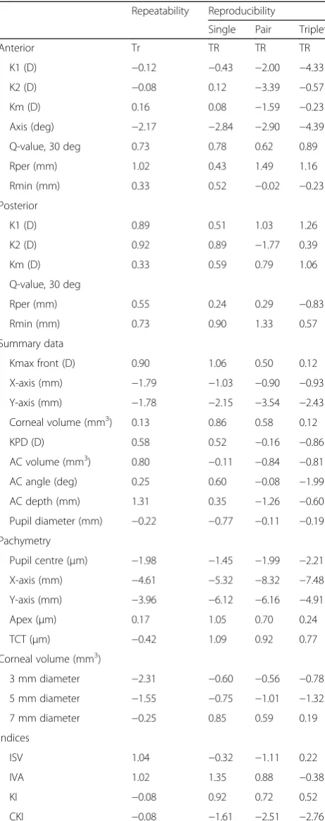
The tolerance index values reported for anterior and posterior curvatures were on average greater than +0.35, in particular, Kmax had a Tr of 0.90 indicating a much wider repeatability limit in keratoconus eyes compared to normal eyes (Table 1). On the other hand, anterior and posterior axis values were found to demonstrate bet-ter repeatability limits (r = 11°; 23° respectively), with better (high negative) Tr values (Tr < −1.7). Summary data in terms of keratometric power deviation (KPD), AC depth, AC volume and AC angle estimates were all greater than normative values (Table 1; Tr > 0). Front surface elevation maps at TCT were more repeatable than back surface elevation maps at TCT. Pachymetry estimates had good repeatability limits for pupil centre, corneal apex, and TCT, with most measures inside nor-mal limits (Table 1). Corneal volume measurements at all diameters were repeatable and had similar or better than normative limits of repeatability (Tr < 0.2). The topometric Q-values were repeatable, however, anterior
Q-value repeatability limits were outside normal limits. Centre keratoconus index (CKI) and index of height decentration (IHD) were repeatable with tighter limits of repeatability (Tr < −1.1) but index of surface variance (ISV), index of vertical asymmetry (IVA) and particularly index of height asymmetry (IHA) were markedly less re-peatable and significantly outside normative limits (Tr> 1.0).
Reproducibility and tolerance index
With a single image, Kmax had reproducibility limits well outside normal with aTR value of 1.06, but when the average of three images was used instead, reproduci-bility was similar to normal limits (TR = 0.12). Of the pachymetry estimates, apex measures were the least re-producible followed by those at the TCT. The measures at pupil centre had the best R-value (R-values, Table 1). R-values of corneal volume increased with increasing diameter, however central corneal volume R-limits were greater than any of the peripheral estimates. AnteriorQ -values had worse reproducibility than normal limits and
did not markedly improve when estimates from pairs or triplets of images were used. IHD and CKI had tight re-producibility limits, remaining within normative limits, suggesting these are among the most reproducible pa-rameters in KCN patients.
Relative utility index
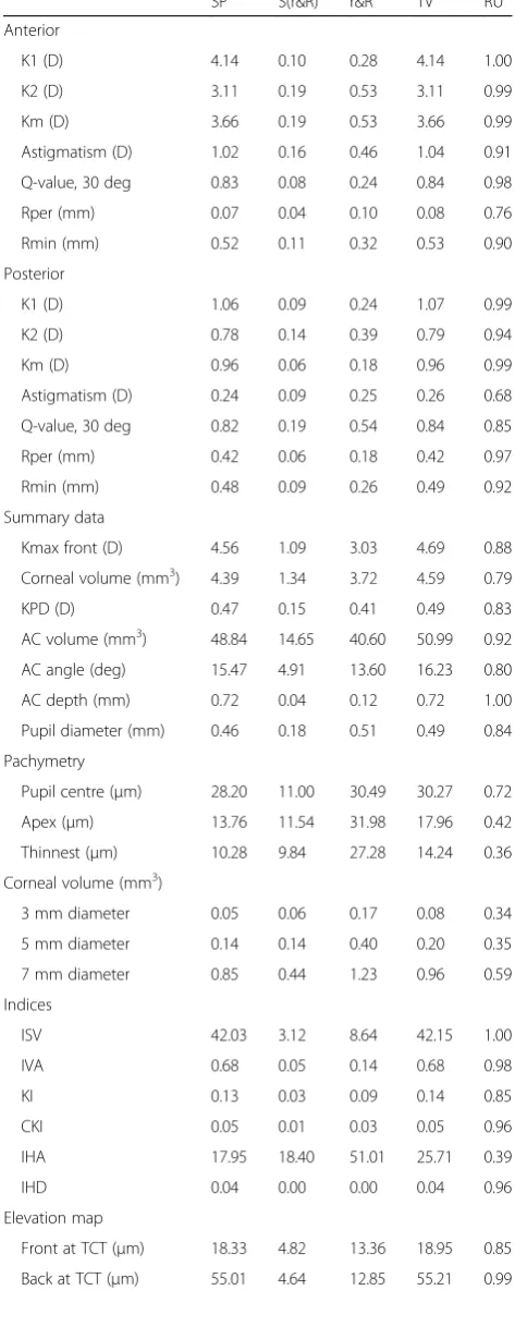
RU was used to indicate which parameters are less vari-able relative to the respective dynamic range of that par-ameter in our cohort (Table 2). Pachymetry at the corneal apex, for example, is unlikely to be useful clinic-ally, as this parameter has an RU of 0.42, suggesting that 58% of the differences in CT apex between any two kera-toconic eyes from the study cohort can be attributed to measurement variability (Table 2). On the other hand, corneal curvature estimates all have RU values above 0.94, except for Kmax that has an RU of 0.88 (Table 2). Considering only those parameters with a RU value greater than 0.95, a small number of parameters within the acceptable range were identified, namely: K1, K2 and Km;Q-value (anterior), R-peripheral posterior, CKI, ISV, IVA, IHD, AC depth, the back-elevation map at TCT and ectasia map indices D and Db (Table 2).
Discussion
Clinically, it is difficult to choose which parameter to use to determine whether disease progression has oc-curred, a consensus on the accepted parameters is emer-ging but there is still significant divergence between authors [1–18, 20–26]. This article provides an overview of the reliability of these parameters, removing the clin-ical interpretation component. We have summarized the differences in measurement noise between healthy and keratoconus patients across all topographic parameters from the Pentacam HR device using thetolerance index. Comparing “r” and “R” reported by McAlinden et al. in healthy eyes to our data in keratoconic eyes, 36%/44% (n = 13/36; 16/36) of parameters were significantly worse (Tr/TR > 0.45), and 28%/36% (n = 11/36; 13/36) were significantly better (Tr/TR < −0.45) (e.g., axis is more repeatable in KCN patients) [1, 2].
Using this information, the RU index isolated the group of parameters theoretically best positioned to de-tect progression. Summarizing the RU values: 37% (n= 15/41) of parameters had an RU greater than 0.95, indicating good ability to detect progression, 29% (n = 12/41) of parameters had an RU <0.80 indicating poor ability to detect progression. It may seem counter-intuitive, but it is possible that a parameter has poor TI but still a good RU. This is because some parameters have large differences between mild and moderate KCN or in other words has a large dynamic range, and it is the balance between the limits of repeatability and the size dynamic range that determines the RU.
Clinically, there are three primary motivations for col-lecting serial topography images in keratoconus patients: to help distinguish healthy from early keratoconus, to detect progression of keratoconus, or to determine the effectiveness of treatments for keratoconus. Regardless of the motivation, when comparing the RU values re-ported in this article with the area under the curve (AUC) values reported in the literature, we observe that there is notable agreement [4, 5, 7–15, 24].
In studies which attempt to distinguish between healthy and keratoconic eyes, the pachymetry values, posterior elevations maps, keratometry asymmetry and decentration indices have been mainly reported [4, 5, 7– 10, 13, 14]. Pachymetry at centre and thinnest location have good sensitivity and specificity, however, the AUC is lower than that reported with the asymmetry indices [7, 8, 25]. Comparing the parameters with >0.90 AUC values reported by Correia et al. to those parameters with >0.95 RU values reported here, there is good agree-ment [7]. Likewise, comparing the poorest AUC results (<0.85) reported by Uçakhan et al. to the poorest RU values (<0.8) reported here, there is good agreement in majority of parameters [8].
Table 1The tolerance indices (Tr, TR)
Repeatability Reproducibility
Single Pair Triplet
Anterior Tr TR TR TR
K1 (D) −0.12 −0.43 −2.00 −4.33
K2 (D) −0.08 0.12 −3.39 −0.57
Km (D) 0.16 0.08 −1.59 −0.23
Axis (deg) −2.17 −2.84 −2.90 −4.39
Q-value, 30 deg 0.73 0.78 0.62 0.89
Rper (mm) 1.02 0.43 1.49 1.16
Rmin (mm) 0.33 0.52 −0.02 −0.23
Posterior
K1 (D) 0.89 0.51 1.03 1.26
K2 (D) 0.92 0.89 −1.77 0.39
Km (D) 0.33 0.59 0.79 1.06
Q-value, 30 deg
Rper (mm) 0.55 0.24 0.29 −0.83
Rmin (mm) 0.73 0.90 1.33 0.57
Summary data
Kmax front (D) 0.90 1.06 0.50 0.12
X-axis (mm) −1.79 −1.03 −0.90 −0.93
Y-axis (mm) −1.78 −2.15 −3.54 −2.43
Corneal volume (mm3) 0.13 0.86 0.58 0.12
KPD (D) 0.58 0.52 −0.16 −0.86
AC volume (mm3) 0.80 −0.11 −0.84 −0.81
AC angle (deg) 0.25 0.60 −0.08 −1.99
AC depth (mm) 1.31 0.35 −1.26 −0.60
Pupil diameter (mm) −0.22 −0.77 −0.11 −0.19
Pachymetry
Pupil centre (μm) −1.98 −1.45 −1.99 −2.21
X-axis (mm) −4.61 −5.32 −8.32 −7.48
Y-axis (mm) −3.96 −6.12 −6.16 −4.91
Apex (μm) 0.17 1.05 0.70 0.24
TCT (μm) −0.42 1.09 0.92 0.77
Corneal volume (mm3)
3 mm diameter −2.31 −0.60 −0.56 −0.78
5 mm diameter −1.55 −0.75 −1.01 −1.32
7 mm diameter −0.25 0.85 0.59 0.19
Indices
ISV 1.04 −0.32 −1.11 0.22
IVA 1.02 1.35 0.88 −0.38
KI −0.08 0.92 0.72 0.52
CKI −0.08 −1.61 −2.51 −2.76
Table 1The tolerance indices (Tr, TR)(Continued)
Repeatability Reproducibility
Single Pair Triplet
IHA 2.09 2.41 2.49 2.43
IHD −1.10 −1.66 −0.57 −0.73
K1, K2= Keratometry readings 1,2;Km= Mean keratometry reading; Rper= Mean radius of curvature in the 7-9 mm area of the cornea;Rmin= Mini-mum radius of curvature;KPD= Keratometric power deviation;AC= Anterior chamber;ISV= Index of surface variance;IVA= Index of vertical asymmetry; KI= Keratoconus index;CKI= Centre keratoconus index;IHA= Index of height asymmetry;IHD= Index of height decentration;Sr= Within-subject standard deviation;r= The limits of repeatability;Tr= The log of the ratio between the limits of repeatability of keratoconus patients and normal subjects;−0.45 < Tr/ TR < 0.45 is within normative limits
There are several articles examining keratoconus pro-gression [5, 11, 12, 15]. The corneal curvature parame-ters perform well in distinguishing between different stages of the disease [5], furthermore progressing eyes have significantly different change rates in these parame-ters than in non-progressing eyes [15], which corre-sponds well with RU values recorded here for K1, K2 and Km. Despite central corneal thickness (CCT) and TCT being well established clinically and both demon-strating significant difference in mean values for differ-ent stages of the disease [11], the annual change rates are not significantly different between progressing and stable eyes for these parameters [15], which corresponds to the poor RU values for pachymetry reported in this study (RU < 0.75).
There are a small number of studies that have exam-ined topographic parameters following CXL: those pa-rameters with positive outcomes in these studies correspond well with the better RU values reported in this study [22–24]. In our study, the large change in re-peatability in eyes with keratoconus versus healthy eyes indicates that repeatability in eyes following CXL should be critically examined, as there are many possible add-itional confounders. A change in repeatability in eyes following CXL could be important, as currently there is more than 70 clinical trials listed on the National Insti-tute for Health Research (NIHR) clinical trial registry Table 2Summary of variation between patients and ratio of
variability attributable to instrument and observers
SP S(r&R) r&R TV RU
Anterior
K1 (D) 4.14 0.10 0.28 4.14 1.00
K2 (D) 3.11 0.19 0.53 3.11 0.99
Km (D) 3.66 0.19 0.53 3.66 0.99
Astigmatism (D) 1.02 0.16 0.46 1.04 0.91
Q-value, 30 deg 0.83 0.08 0.24 0.84 0.98
Rper (mm) 0.07 0.04 0.10 0.08 0.76
Rmin (mm) 0.52 0.11 0.32 0.53 0.90
Posterior
K1 (D) 1.06 0.09 0.24 1.07 0.99
K2 (D) 0.78 0.14 0.39 0.79 0.94
Km (D) 0.96 0.06 0.18 0.96 0.99
Astigmatism (D) 0.24 0.09 0.25 0.26 0.68
Q-value, 30 deg 0.82 0.19 0.54 0.84 0.85
Rper (mm) 0.42 0.06 0.18 0.42 0.97
Rmin (mm) 0.48 0.09 0.26 0.49 0.92
Summary data
Kmax front (D) 4.56 1.09 3.03 4.69 0.88
Corneal volume (mm3) 4.39 1.34 3.72 4.59 0.79
KPD (D) 0.47 0.15 0.41 0.49 0.83
AC volume (mm3) 48.84 14.65 40.60 50.99 0.92
AC angle (deg) 15.47 4.91 13.60 16.23 0.80
AC depth (mm) 0.72 0.04 0.12 0.72 1.00
Pupil diameter (mm) 0.46 0.18 0.51 0.49 0.84
Pachymetry
Pupil centre (μm) 28.20 11.00 30.49 30.27 0.72
Apex (μm) 13.76 11.54 31.98 17.96 0.42
Thinnest (μm) 10.28 9.84 27.28 14.24 0.36
Corneal volume (mm3)
3 mm diameter 0.05 0.06 0.17 0.08 0.34
5 mm diameter 0.14 0.14 0.40 0.20 0.35
7 mm diameter 0.85 0.44 1.23 0.96 0.59
Indices
ISV 42.03 3.12 8.64 42.15 1.00
IVA 0.68 0.05 0.14 0.68 0.98
KI 0.13 0.03 0.09 0.14 0.85
CKI 0.05 0.01 0.03 0.05 0.96
IHA 17.95 18.40 51.01 25.71 0.39
IHD 0.04 0.00 0.00 0.04 0.96
Elevation map
Front at TCT (μm) 18.33 4.82 13.36 18.95 0.85
Back at TCT (μm) 55.01 4.64 12.85 55.21 0.99
Table 2Summary of variation between patients and ratio of variability attributable to instrument and observers(Continued)
SP S(r&R) r&R TV RU
Ectasia map
Df 6.41 2.30 6.38 6.81 0.75
Db 5.57 0.77 2.14 5.62 0.99
Dp 2.94 1.45 4.01 3.28 0.60
Dt 0.12 0.13 0.37 0.18 0.39
Da 0.10 0.18 0.51 0.21 0.21
D 4.13 0.46 1.28 4.16 0.99
examining the effectiveness of CXL, where the primary or secondary outcome is a change in corneal curvature. Therefore, the parameters used to validate keratoconus progression in these clinical trials may require updating.
This agreement between RU and AUC values is of sig-nificance as the data required to calculate RU values is collected at one sole visit, while the AUC data requires data from several years of clinical observation. RU values are not a replacement for AUC values, but they can be used to help optimize clinical trials, by helping to pro-vide guidelines on the parameters of interest, the opti-mal number of scans and the frequency of consultation.
Some of the differences in precision noted between keratoconic and normal eyes are likely to be related to the fitting algorithm used by the Pentacam HR device. Alignment algorithms rely on alignment markers such as pupil centre, thinnest corneal location, and corneal apex. Some alignment markers will be less evident in normal eyes than keratoconic eyes. For example, due to the conical shape of the cornea in keratoconic eyes, the location of Kmax is clear in most images, therefore the same x, y coordinates will be calculated between images. Furthermore, the fitting algorithm uses a model of the smooth spherical cornea in the form of a “best fit sphere”more akin to the normal cornea than the conical cornea observed in keratoconic eyes. With this tech-nique, the presence of the cone is unexpected and likely distorts estimates of many of the topographic parameters [6]. Lastly, in eyes with a steep cone, the eye movements associated with the loss of fixation have the potential to cause much larger errors in the estimation of parameters such as Kmax and TCT. This may be exacerbated by multifocality associated with these “steep cones”, thus greater higher intraocular straylight [16, 21], and poorer fixation. Lastly, this study examined only early to mo-derate KCN, those parameters identified as useful in this group may differ from those used in more advanced disease [25–27].
Conclusion
The indices implemented in this article were designed to provide an “at a glance” guideline on the levels of ex-pected precision in keratoconic eyes relative to healthy eyes to aid clinicians in distinguishing real change from variability [18]. Furthermore, the RU index isolates topo-graphic parameters with a large dynamic range in com-parison to measurement noise. This index gives an indication of those parameters with the potential for de-tecting a change when no longitudinal data are available e.g. when a new device/software is released. Our hypoth-esis is that parameters with high RU are best positioned to detect change, whether it is disease progression or assessing the efficacy of a therapeutic intervention. For example, the Kmax and CCT parameters, which are
currently the standard measures used for the monitoring of keratoconus have been shown to have poor RU in our study, indicating that these parameters are not best posi-tioned to detect change. Further investigation is required to verify these results and develop this methodology for clinical practice.
Abbreviations
AC:Anterior chamber; AUC: Area under the curve; CKI: Centre keratoconus index; CXL: Collagen cross-linking; D: Belin/Ambrosio ectasia total deviation value; D: Dioptre; Da: Deviation of the apex thickness; Db: Deviation of the back-elevation map; Df: Deviation of the front elevation map; Dp: Deviation of average pachymetric progression; Dt: Deviation of the minimum thickness; IHA: Index of height asymmetry; IHD: Index of height decentration; ISV: Index of surface variance; IVA: Index of vertical asymmetry; K1, K2: Keratometry readings 1 and 2; KCN: Keratoconus; KI: Keratoconus index; Km: Mean central keratometry; KPD: Keratometric power deviation; r: Limits of repeatability; R: Limits of reproducibility; Rmin: Minimum radius of curvature; Rper: Mean radius of curvature in the 7-9 mm area of the cornea; RU: Relative utility; Sr: Repeatability; SR: Reproducibility; TR: Tolerance index (the log of the ratio between the limits of reproducibility of keratoconus patients and normal subjects)
Acknowledgements
None.
Availability of data and materials
The datasets used and/or analysed during the current study are available from the corresponding author on reasonable request.
Authors’contributions
IG and CM collected the data, CB and IG analysed and interpreted the patient data. IG and CB were the major contributors in writing the manuscript. All authors read and approved the final manuscript.
Funding
None.
Ethics approval and consent to participate
This study was approved by the local cantonal ethics committee and adhered to tenets of Declaration of Helsinki for research on human subjects. Healthy group: Ethical approval was granted by the Flinders clinical research ethics committee.
Keratoconus (KCN) group: Ethical approval was granted by the ethical commission of the canton de Vaud, Switzerland under protocol number 375/11.
Consent for publication
All patients gave informant consent.
Competing interests
The authors declare that they have no competing interests.
Author details
1
Department of Ophthalmology, University of Geneva, Geneva, Switzerland. 2Department of Ophthalmology, Glangwili Hospital, Hywel Dda University Health Board, Carmarthen, UK.3School of Ophthalmology and Optometry, Wenzhou Medical University, Wenzhou, Zhejiang, China.4Jules-Gonin Eye Hospital, University of Lausanne, Fondation Asile des Aveugles, Lausanne, Switzerland.
Received: 31 May 2017 Accepted: 10 October 2017
References
2. McAlinden C, Khadka J, Pesudovs K. A comprehensive evaluation of the precision (repeatability and reproducibility) of the Oculus Pentacam HR. Invest Ophthalmol Vis Sci. 2011;52:7731–7.
3. Hafezi F, Randleman B. Corneal collagen cross-linking. Slack Inc: Thorofare, NJ; 2013.
4. Schlegel Z, Hoang-Xuan T, Gatinel D. Comparison of and correlation between anterior and posterior corneal elevation maps in normal eyes and keratoconus-suspect eyes. J Cataract Refract Surg. 2008;34:789–95. 5. Piñero DP, Alió JL, Alesón A, Escaf Vergara M, Miranda M. Corneal volume,
pachymetry, and correlation of anterior and posterior corneal shape in subclinical and different stages of clinical keratoconus. J Cataract Refract Surg. 2010;36:814–25.
6. Belin MW, Khachikian SS, Ambrósio R Jr, Salomão M. Keratoconus/Ectasia Detection with the Oculus Pentacam: Belin/Ambrósio Enhanced Ectasia Display. Highlights Ophthalmol. 2011;35:5–12.
7. Faria-Correia F, Ramos I, Lopes B, Salomão QM, Luz A, Correa RO, et al. Topometric and tomographic indices for the diagnosis of Keratoconus. Int J Keratoconus Ectatic Corneal Dis. 2012;1:92–9.
8. Uçakhan ÖÖ, Çetinkor V, Özkan M, Kanpolat A. Evaluation of Scheimpflug imaging parameters in subclinical keratoconus, keratoconus and normal eyes. J Cataract Refract Surg. 2011;37:1116–24.
9. Ruiseñor Vázquez PR, Galletti JD, Minguez N, Delrivo M, Fuentes Bonthoux F, Pförtner T, et al. Pentacam Scheimpflug tomography findings in topographically normal patients and subclinical keratoconus cases. Am J Ophthalmol. 2014;158(1):32–40.
10. de Sanctis U, Loiacono C, Richiardi L, Turco D, Mutani B, Grignolo FM. Sensitivity and specificity of posterior corneal elevation measured by Pentacam in discriminating keratoconus/subclinical keratoconus. Ophthalmology. 2008;115:1534–9.
11. Ishii R, Kamiya K, Igarashi A, Shimizu K, Utsumi Y, Kumanomido T. Correlation of corneal elevation with severity of keratoconus by means of anterior and posterior topographic analysis. Cornea. 2012;31(3):253–8. 12. Mannion LS, Tromans C, O’Donnell C. Reduction in corneal volume with
severity of keratoconus. Curr Eye Res. 2011;36(6):522–7.
13. Ahmadi Hosseini SM, Abolbashari F, Niyazmand H, Sedaghat MR. Efficacy of corneal tomography parameters and biomechanical characteristic in keratoconus detection. Cont Lens Anterior Eye. 2014;37(1):26–30. 14. Fam HB, Lim KL. Corneal elevation indices in normal and keratoconic eyes. J
Cataract Refract Surg. 2006;32:1281–7.
15. Choi JA, Kim MS. Progression of keratoconus by longitudinal assessment with corneal topography. Invest Ophthamol Vis Sci. 2012;53(2):927–35. 16. Jinabhai A, O’Donnell C, Radhakrishnan H, Nourrit V. Forward light scatter
and contrast sensitivity in keratoconic patients. Cont Lens Anterior Eye. 2012;35(1):22–7.
17. Bergin C, Guber I, Hashemi K, Majo F. Tolerance and Relative Utility:Two Proposed Indices for Comparing Change in Clinical Measurement Noise Between Different Populations (Repeatability) or Measurement Methods (Agreement). Invest Ophthalmol Vis Sci. 2015;56:5543–7.
18. McAlinden C, Khadka J, Pesudovs K. Precision (repeatability and reproducibility) studies and sample-size calculation. J Cataract Refract Surg. 2015;41(12):2598–604.
19. R Core Team. A language and environment for statistical computing. Vienna, Austria: Foundation for Statistical Computing; 2012.
20. Epstein RL, Chiu YL, Epstein GL. Pentacam HR criteria for curvature change in keratoconus and postoperative LASIK ectasia. J Refract Surg. 2012;28(12): 890–4.
21. Guber I, Guber J, Kaufmann C, Bachmann LM, Thiel MA. Visual recovery after corneal crosslinking for keratoconus: a 1-year follow-up study. Graefes Arch Clin Exp Ophthalmol. 2013;251(3):803–7.
22. Vinciguerra P, Albè E, Trazza S, Rosetta P, Vinciguerra R, Seiler T, et al. Refractive, topographic, tomographic, and aberrometric analysis of keratoconic eyes undergoing corneal crosslinking. Ophthalmology. 2009; 116:369–78.
23. Greenstein SA, Fry KL, Hersh PS. Corneal topography indices after corneal collagen crosslinking for keratoconus and corneal estasia: one-year results. J Cataract Refrac Surg. 2011;37:1282–90.
24. Asri D, Touboul D, Fournié P, Malet F, Garra C, Gallois A, et al. Corneal collagen crosslinking in progressive keratoconus: Multicenter results from the French National Reference Center for Keratoconus. J Cataract Refrac Surg. 2011;37:2137–43.
25. Kanellopoulos AJ, Asimellis G. Revisiting keratoconus diagnosis and progression classification based on evaluation of corneal asymmetry indices, derived from Scheimpflug imaging in keratoconic and suspect cases. Clin Ophthalmol. 2013;7:1539–48.
26. Hashemi H, Yekta A, Khabazkhoob M. Effect of keratoconus grades on repeatability of keratometry readings: comparison of 5 devices. J Cataract Refract Surg. 2015;41(5):1065–72.
27. Flynn TH, Sharma DP, Bunce C, Wilkins MR. Differential precision of corneal Pentacam HR measurements in early and advanced keratoconus. Br J Ophthalmol. 2016;100(9):1183–7.
• We accept pre-submission inquiries
• Our selector tool helps you to find the most relevant journal
• We provide round the clock customer support
• Convenient online submission
• Thorough peer review
• Inclusion in PubMed and all major indexing services • Maximum visibility for your research
Submit your manuscript at www.biomedcentral.com/submit