S T U D Y P R O T O C O L
Open Access
Long term cerebral and vascular complications
after irradiation of the neck in head and neck
cancer patients: a prospective cohort study: study
rationale and protocol
Joyce Wilbers
1*, Arnoud C Kappelle
1, Roy PC Kessels
4, Stefan CA Steens
3, Frederick JA Meijer
3, Johannes H Kaanders
2,
Roy AM Haast
10, Laura E Versteeg
4, Anil M Tuladhar
1, Chris L de Korte
10, Hendrik HG Hansen
10, Frank J Hoebers
8,
Willem Boogerd
5, Erik D van Werkhoven
7, Marlies E Nowee
6, Guus Hart
7, Harry Bartelink
6, Lucille D Dorresteijn
9and Ewoud J van Dijk
1Abstract
Background:Successful treatment options for cancer result in more young long-term survivors prone for long-term complications. Carotid artery vasculopathy is a potential long-term complication after radiotherapy of the neck, resulting in cerebrovascular events and probably deficits in cognitive and motor functioning. Better insight into the underlying pathofysiology of radiotherapy induced carotid artery vasculopathy is needed for prognostic purposes and to develop preventive strategies.
Methods/Design:The current study is a prospective cohort study on the long-term cerebral and vascular complications after radiotherapy of the neck, in 103 patients treated for head and neck cancer, included in our study database between 2002 and 2008. Baseline protocol (before radiotherapy) included screening for cerebrovascular risk factors and intima media thickness measurement of carotid arteries by ultrasonography. Follow-up assessment more than 5 years after radiotherapy included screening of cerebrovascular risk factors, cerebrovascular events, neurological examination with gait and balance tests, extensive neuropsychological examination, self-report questionnaires, ultrasonography of the carotid arteries with measurement of intima media thickness and elastography, magnetic resonance imaging of the brain and magnetic resonance angiography of the carotid arteries.
Discussion:The current study adds to the understanding of the causes and consequences of long-term cerebral and vascular changes after radiotherapy of the neck. These data will be helpful to develop a protocol for diagnostic and preventive strategies for long-term neurological complications in future head and neck cancer patients with anticipated radiotherapy treatment.
Keywords:Head and neck cancer, Cerebrovascular disease, Carotid artery, Radiation vasculopathy
* Correspondence:[email protected]
1Department of Neurology, Radboud University Nijmegen Medical Center, Donders Institute for Brain, Cognition and Behaviour Centre for Neuroscience, PO box 9101, 6500 HB Nijmegen, The Netherlands Full list of author information is available at the end of the article
Background
Successful treatment options for cancer have resulted in more long-term survivors, prone to long-term com-plications of chemotherapy and radiotherapy (RT). A well-known long-term complication of RT of the neck is carotid artery vasculopathy, which ultimately can lead to stroke [1,2].
Apart from acute neurological complications, like a cerebral infarction, evidence from daily clinical practice shows more subtle, slowly progressive deficits in cognitive and motor functioning. These long-term cerebral and vascular complications in young survivors of cancer are a serious potential problem that hamper reintegrating in social functioning and work. The longer survival stresses the need for a long-term follow-up study that assesses the long-term complications of RT induced carotid artery vasculopathy. To date, no such studies exist to the best of our knowledge.
We hypothesize that RT of the neck accelerates carotid atherosclerosis with secondary development of cerebral vascular lesions. These vascular cerebral changes are subsequently related to cognitive and motor functioning with a major impact on activities of daily social functioning. A better understanding of the underlying pathophysiology of this RT induced vasculopathy is needed for prognostic purposes and can be used to develop strategies that prevent further progression and thereby reducing the risk of future stroke and neurological impairments.
The current study aims to investigate four questions. The first question focuses on the relation between RT and long-term carotid artery-wall changes. We hypothesize that irradiation induces premature atherosclerosis which is different from more common atherosclerosis due to classic cerebrovascular risk factors. We therefore study the location and composition of post-irradiation vessel-wall changes with advanced imaging techniques; plaque and Intima Media Thickness (IMT) analysis on ultrasound with innovative vessel-wall elastography and magnetic resonance imaging/angiography (MRI)/(MRA) [3-5]. The second question examines the relation between RT and long-term cerebral vascular changes. In the general population the prevalence of silent brain infarcts (SBI) is up to fivefold higher than that of acute stroke [6]. SBI are related to impaired cognitive functioning, dementia, stroke and gait and balance disorders [7]. Carotid artery pathology increases the risk of SBI [8]. To assess this, all study patients underwent MRI of the brain to analyze silent brain infarcts and related pathology like white-matter lesions and cerebral atrophy.
The third question addresses the relation between RT and clinical outcome measures (motor and cognitive functioning, as well as quality of life; QoL). Silent brain infarcts are associated with gait disturbances, decline in cognitive function and increased risk of developing
dementia [9-11]. We performed quantitative gait/balance analysis and an extensive neuropsychological assessment in all patients. Finally, we want to study the relation between patient and treatment baseline characteristics and the development of vasculopathy, in order to find high risk patients that can benefit from preventive strategies.
Methods
The current study is a prospective cohort study that aims to investigate the frequency, pathophysiology and long-term outcome of radiotherapy induced vasculopathy. The Medical Ethics Review Committee region of Arnhem-Nijmegen approved the study (NL 41008.091.12).
Patients
A prospective cohort study of head and neck cancer pa-tients in two centers of the Netherlands (the Netherlands Cancer Institute/Antoni van Leeuwenhoek Hospital (NCI/ AvL), Amsterdam and the Radboud University Medical Center Nijmegen) was initiated.
At baseline, patients were eligible for inclusion if they received RT of the neck because of a T1/T2 (N0M0) laryngeal carcinoma, T1/T2 (N0M0) parotid carcinoma/ pleomorphic adenoma, T1/2 (N1/2 M0) naso/oro/hypo-pharynxcarcinoma or non-Hodgkin/Hodgkin lymphoma. Originally, the study was designed as an open-label multi-centre Prospective Randomized Open Blinded Endpoint (PROBE) study to assess the effect of an HMG-coA reduc-tase inhibitor (atorvastatine) on carotid IMT in the first 2 years after irradiation of the neck. Because of dwindling accrual the study was redesigned to a prospective cohort study. The initial exclusion criteria were: a history of cere-brovascular disease, pregnancy or breast-feeding, ongoing treatment with an HMG-coA reductase or cytochroom P450 inhibitor, active liver disease or > 3 times the upper limit of serum transaminases, 5 times the normal level of creatine phosphokinase, serum cholesterol > 7 mmol/L and a life expectancy < 2 years. All patients gave written informed consent.
Controls
Baseline
Patients were assessed at baseline (before RT) for height, weight and blood pressure, underwent a neurological and laboratory (C-reactive protein) examination and the following cerebrovascular risk factors were assessed:
1. Cigarette smoking. 2. Hypertension, defined as using antihypertensive medication or blood pressure > 130/ 80 mmHg. 3. Diabetes mellitus, defined as using anti-diabetic medication or a non-fasting serum glucose >11.1 mmol/L. Medication prescription was checked with the pharmacy database. 4. Hypercholesterolemia, defined as serum total cholesterol > 6.5 mmol/L. 5. Obesity, defined as Body Mass Index (BMI) > 30 kg/m2.
Carotid ultrasound
In patients with laryngeal or hypopharyngeal carcinoma IMT was measured on the Common Carotid Artery (CCA), according to the RT-field. In patients with other tumors IMT was measured in both the CCA and the Internal Carotid Artery (ICA), also according to the RT- field.
IMT was measured with a linear array transducer (iU22 Philips NZE172 probe: L17-5, Hewlett Packard, Sonos 2000, probe: 7.5/5.5, ALOKA 5000 and G.E Logiq E9). IMT pictures were digitally stored on the ultrasound machine or printed in case digital storage was not available. Printed images were then scanned at high resolution and interpolated (using Matlab 2010a) to obtain the same resolution as the digitally stored im-ages. IMT was automatically measured by one blinded investigator (JW), using QLAB (Version 4.2.1, Philips). An edge-detection algorithm identified the lumen-intima and the media-adventia interface within a region of inter-est (selected by the operator) over a maximum of 10 mm long segment and calculated the average thickness.
Follow-up > 5 years after radiotherapy
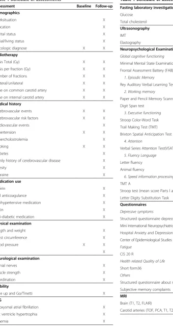
At follow-up all patients underwent a structured program (Table 1), consisting of assessment of demographics and life style, medical history and medication use, fasting laboratory investigation, electrocardiogram (ECG), physical and neuro-logical examination, extensive neuropsychoneuro-logical assess-ment battery (NPA), carotid artery ultrasound (Intima Media Thickness measurement and elastography), MRI/ MRA of the brain and carotid arteries, gait and balance analysis and self-report questionnaires.
Demographics and life style
Standardized questions on demographics (age at radiother-apy, age at follow-up, sex) were administered. Education was classified using seven categories; one being less than primary school and seven reflecting an academic degree. Type of work, working situation, marital status and living conditions were asked using a structured interview.
Medical history and medication use
Standard questionnaires about medication use (carbaspirin calcium, oral anticoagulance, antihypertensive medication, statin, antidiabetic medication), cerebrovascular risk factors (hypertension, hypercholesterolemia, smoking, diabetes, family history, obesity, migraine), vascular diseases in the past (stroke, cerebral bleeding, acute myocard infarc-tion, atrial fibrillainfarc-tion, angina pectoris, peripheral arterial disease) were asked by one of the investigators (JW). If patients had had an ischemic stroke (IS) or Transient Ischaemic Attack (TIA), detailed medical information was collected from their neurologist and reassessed by one neurologist (EvD).
Fasting laboratory investigation
Fasting blood samples were taken. Analysis included glucose and total cholesterol.
Electrocardiogram (ECG)
An ECG was performed and evaluated with a standard-ized assessment by an experienced cardiologist. Out-come measures were (paroxysmal) atrial fibrillation, left ventrical hypertrophia and signs of ischemia (pathologic Q’s, ST depression or elevation).
Physical and neurological examination
Height and weight were measured and body mass index (BMI) was calculated. The maximal weight circumference was measured in standing position, between the lowest rib and the iliac crest, at the end of normal expiration [14]. Blood pressure was measured on the right and left side. All patients underwent a standardized neurologic examination by an experienced neurologist (JW), consist-ing of assessment of functionconsist-ing of cranial nerves, muscle strength and cerebellar functions.
Assessment of gait and balance
We used a widely used version of the Tinnetti with 17 items: 9 items for body balance (score 0-16) and 8 for gait (score 0-12), with a maximum score of 28 [15]. It grades balance while sitting, standing with eyes open and closed, nudging and turning, gait initiation, stride length and width and symmetry. Functional mobility was classified by using the widely-used Timed Up-and-Go (TUG) test which is a timed test during which the participant is asked to rise from a standard armchair, walk 3 m, turn, walk back and sit down again [16]. Each participant performed the test three times.
Neuropsychological assessment
Table 1 Schedule of assessments
Assessment Baseline Follow-up
Demographics
Worksituation X
Education X
Marital status X
Social/living status X
Oncologic diagnose X X
Radiotherapy
Dosis Total (Gy) X X
Dosis per fraction (Gy) X X
Number of fractions X X
Bilateral/unilateral X X
Dose on common carotid artery X X
Dose on internal carotid artery X X
Medical history
Cerebrovascular events X X
Cerebrovascular risk factors X X
Cardiovascular events X
Hypertension X X
Hypercholostrolemia X
Smoking X X
Diabetes X
Family history of cerebrovascular disease X
Obesity X
Migraine X
Medication use
Aspirin X
Oral anticoagulance X
Antihypertensive medication X
Statin X
Anti-diabetic medication X
Physical examination
Length and weight X
Waist circumference X
Blood pressure X X
BMI X
Neurological examination
Cranial nerves X
Muscle strength X
Coordination X
Mobility
Time up and Go/Tinetti X
ECG
Paroxysmal atrial fibrillation X
Left ventricle hypertrophia X
Ischemia X
Table 1 Schedule of assessments(Continued)
Fasting laboratory investigation
Glucose X
Total cholesterol X
Ultrasonography
IMT X X
Elastography X
Neuropsychological Examination
Global cognitive functioning
Minimal Mental State Examination (MMSE) X
Frontal Assessment Battery (FAB) X
1. Episodic Memory
Rey Auditory Verbal Learning Test (RAVLT) X
2. Working memory
Paper and Pencil Memory Scanning Tasks X
Digit Span test X
3. Executive functioning
Stroop Color-Word Task X
Trail Making Test (TMT) X
Brixton Spatial Anticipation Test X
4. Attention
Verbal Series Attention Test(VSAT) X
5. Fluency Language
Letter fluency X
Animal fluency X
6. Speed information processing
TMT A X
Stroop test (mean score Parts I and II) X
Letter Digity Substitution Task X
Questionnaires
Depressive symptoms
Structured questionnaire depressive symptoms X
Mini International Neuropsychiatric Interview (MINI) X Hospital Anxiety and Depression Scale (HADS) X
Center of Epidemiological Studies Depression scale X
Fatigue
CIS 20 R X
Health related Quality of Life
Short form36 X
Others
Structured questionnaire about sleep disorders X
Subjective memory complaints X
MRI
Brain (T1, T2, FLAIR) X
processing. The Mini Mental State Examination (MMSE) [17] and the Frontal Assessment Battery (FAB) [18] were used as a screening of overall cognitive function and orientation. Episodic memory was assessed using the five-trial Dutch version of the Rey Auditory Verbal Learning Test (RAVLT) in which the ability to acquire and retain new verbal information was measured, immediately and after a delay [19]. We administered the Paper and Pencil Memory Scanning Task [20] and the Digit Span test [21], both forwards and backwards, to assess working memory. Executive functioning was assessed using the Stroop Color-Word Task (Stroop interference score; i.e. Part III/ mean(Part I and II)) [22], the Trail Making Test (TMT) [23] ratio score (part B/part A) and the Brixton Spatial Anticipation Test (Brixton) [24,25]. Attention was assessed with the Verbal Series Attention Test(VSAT) [26]. To evaluate verbal fluency two tasks were used. First, a semantic fluency task [27] was administered in which participants had to name as many animals as possible within 60 seconds followed by as many professions within 60 seconds. Second, a letter fluency task [28] was used in which as many words beginning with a given initial letter (D-A-T) had to be generated within 60 seconds. Speed of information processing was evaluated by using three tasks; the Stroop test (mean score Parts I and II), the TMT Part A and the Symbol Digit Substitution Task [29], which is a modified version of the Symbol Digital Modalities Task.
Carotid artery ultrasound - Intima Media Thickness measurement
The measurement of IMT at follow-up 7 years after RT was exactly the same as described at baseline.
Non-invasive carotid ultrasound elastography–imaging protocol
Following IMT measurements, the distal CCA and prox-imal ICA at both sides were examined in a longitudinal and transverse imaging plane using a Medison Accuvix V10 ultrasound system (Samsung Medison Seoul, Republic of South Korea) equipped with an L5-13 linear array trans-ducer (fc = 8.5 MHz). Firstly, longitudinal recordings were acquired. After optimum visualization of the cross section with the highest amount of luminal narrowing, an imaging mode called radial zone mode was turned on, which enabled us to perform angle compounding and to store raw ultrasound radiofrequency (RF) data (fs = 61.6 MHz). RF data were acquired at three insonification angles (0°, 20° and -20°) for 3 seconds at a frame rate of at least 129 Hz (43 Hz/angle). Insonification angles were relative to the unsteered ultrasound beam direction and changed sequen-tially before every new ultrasound image frame. Depending on the heart rate, this resulted in the recording of at least two complete cardiac cycles. After the described measurement, the systolic and diastolic blood pressures
in the brachial artery were determined using a sphyg-momanometer (LittmannWClassic II S.E.; 3 M, St Paul, MN, USA). The pressure values were used to normalize the distensibility and strain results as explained later, since the level of strain is not only related to tissue composition but also to the pressure differential between the subsequently acquired frames. Above described steps were repeated during imaging in the transverse plane. Location of transverse image acquisition was chosen at the vessel wall displaying the thickest IMT. The distance between this cross-section and the carotid bulb was used as an anatomical reference point. Subsequently, this protocol was repeated for the internal carotid artery. The entire measurement series was completed within 30 minutes. Raw RF data were subsequently analyzed to determine distensibility and strain using in-house written elastography software (MATLAB 2010b, the Mathworks, Natick, MA, USA). Detailed description of this can be found in Additional file 1.
MRI protocol
The MRI studies were performed on a 3 Tesla MR-scanner (Skyra, Siemens Erlangen). The scanning protocol included T1, T2 and T2 Fluid Attenuated Inversion Recovery (FLAIR) transversal sequences of the brain. The MRI scanning protocol of the carotids included a 3 dimensional time of flight (TOF), Phase Contrast Angiography (PCA), sagittal oblique T2 of the carotid bifurcation, transversal T1, T2 and proton density (PD) sequences. Finally, a maximum intensity projection (MIP) TOF and PCA was reconstructed. The complete scanning protocol took ap-proximately 60 minutes. All MRI studies will be scored by two blinded experienced neuroradiologists (SS and FAM).
Additional self-report questionnaires
For the assessment of anxiety and depression we used the Hospital Anxiety and Depression Scale (HADS) and the Center for Epidemiologic Studies Depression Scale (CES-D) [30]. The Checklist on Individual Strength (CIS-20R) was used to assess fatigue [31]. The overall health status (quality of life) was assessed with the Short Form 36 Health Survey (SF-36) [32]. Patients were asked for sleep-ing problems and their employment status in the period before and the period after radiotherapy. Two question-naires were filled in with a doctor (JW): the assessment of Subjective Memory Complaints (SMC) [10] and the Mini International Neuropsychiatric Interview (MINI) [33].
Outcome events Imaging outcomes
compared with a control group. Secondly, on MRI/A of the carotid arteries the degree of stenosis, location and when possible composition of vessel wall changes (lipids, fibrosis, hemorrhage) will be scored, according to an in house developed protocol.
All MRIs of the brain will be scored semi-quantitatively by two neuroradiologists separately for white-matter lesions (WML), brain atrophy and (silent) brain infarctions using validated scales [34-36] and in case of disagreement a con-sensus meeting will be held.
Furthermore, total white matter lesion volume will be calculated by a prior published, in-house developed, validated technique [37]. In short, WML are defined as hyperintensive lesions on FLAIR MRI without corre-sponding cerebrospinal fluid like hypo-intense lesions on the T1 weighted image. Gliosis surrounding lacunar and territorial infarcts is not considered to be WML. Finally, brain volumetry will be done to measure total white and grey matter volumes. Grey and white matter tissue and cerebrospinal fluid probability maps will be computed by using a six class segmentation tool in
Statistical Parametric Mapping Software (http://www. fil.ion.ucl.ac.uk/spm) (SPM 8) on the T1 MPRAGE im-ages. Total grey and white matter volumes are calculated by summing all voxel volumes belonging to the tissue class. Total brain volume is taken as the sum of total grey and total white matter volume. Co-registration parameters of the FLAIR image to the T1 image are computed (SPM8 mutual information co-registration) and used to bring both the FLAIR and WML segmentation images into the subjects (anatomical) reference frame. Transformed images will visually be checked for co-registration errors. Subsequently, the WML segmentations are re-sampled to and combined with the white matter maps to yield to a WML map (the intersection of WML and white matter) and NAWM map (the complement of WML in white matter) in the T1 reference space.
Clinical outcomes
Clinical outcome measures will be: cerebrovascular events, domain scores (mean standardized z-scores compared to reference values) of objective cognitive functioning on
Baseline (n=103)
Follow-up > 5 years after RT (n=65)
Out of FU (n= 38)
Dead 24 Too ill 4 Emigrated 1 Too far travelling 2 Others 3 Unkwown reason 4
Radiotherapy
Telephone FU (n=14) Site visit (n=51)
US (n=50/51) MRI/A (n=43/51) Mobility (n=47/51) NPE (n=44/51) Questionaires (n=65/65)
NPA, subjective memory complaints, depressive symptoms, fatigue and gait or balance disturbances.
Statistical analysis
Analysis of continuous variables will be done with Student’sttest or analysis of (co)variance or in case of skewed distributions which cannot be normalized by log transformation corresponding nonparametric tests will be used. Chi-squared test will be used for analysis of categorical variables and logistic regression analyses will be used to adjust for potential confounding factors.
Results
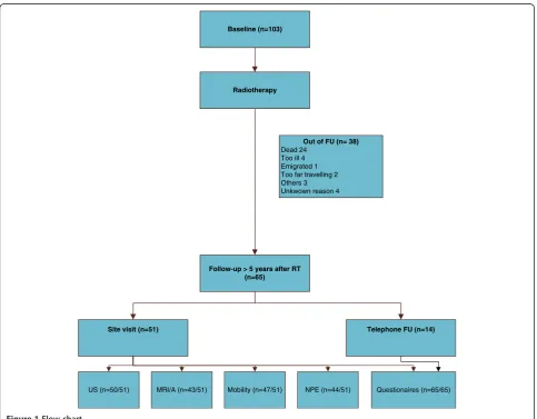
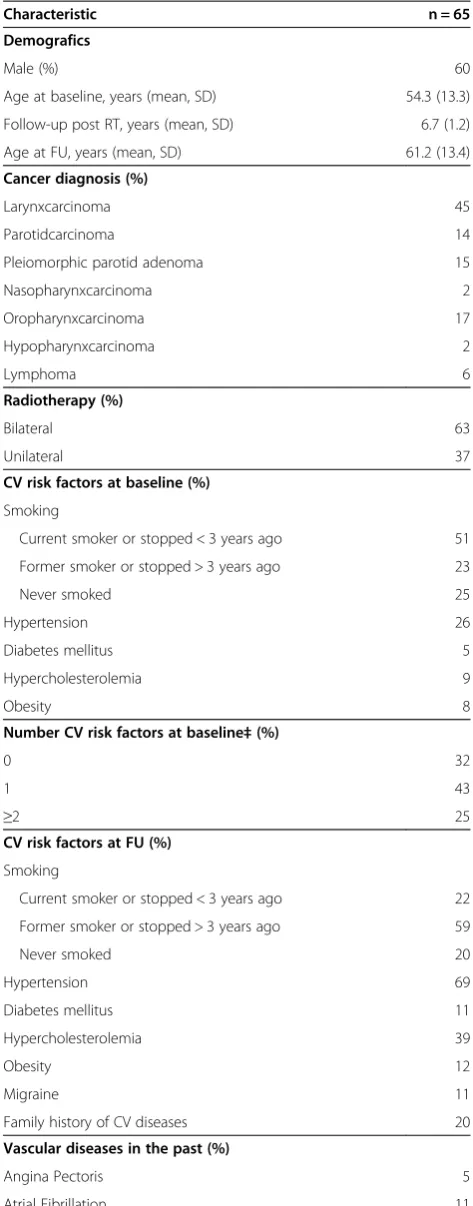
103 patients who had sought medical attention at one of the two University Medical Centers between 2002 and 2008 fulfilled inclusion and exclusion criteria for our study. The number of patients lost to follow-up is reported in Figure 1. 51 patients have been seen for the follow-up protocol more than 5 years after RT between November 2012 and November 2013. Another 14 patients underwent a telephone follow-up. Characteristics of our population at baseline and follow-up are reported in Table 2.
Discussion
The number of cancer survivors is still growing and long-term complications after cancer treatment are becoming a serious problem for the society. However, prospective cohort studies with a long follow-up period after RT of the neck are scarce. More understanding about the underlying pathophysiology of RT induced vasculopathy is needed to develop preventive strategies, such as the use of a statin or thrombocyt aggregation inhibitors. These long-term complications of RT induced carotid artery vasculopathy are not studied in prior studies, because of relatively small patient groups and short follow-up period.
We therefore performed the current prospective cohort study, to investigate the cerebral and vascular long-term complications after RT of the neck. Strong elements of our study are the unique population and study design. This cohort is not only the largest, but also has the longest Table 2 Patient and treatment related characteristics at
baseline and FU
Characteristic n = 65
Demografics
Male (%) 60
Age at baseline, years (mean, SD) 54.3 (13.3)
Follow-up post RT, years (mean, SD) 6.7 (1.2)
Age at FU, years (mean, SD) 61.2 (13.4)
Cancer diagnosis (%)
Larynxcarcinoma 45
Parotidcarcinoma 14
Pleiomorphic parotid adenoma 15
Nasopharynxcarcinoma 2
Oropharynxcarcinoma 17
Hypopharynxcarcinoma 2
Lymphoma 6
Radiotherapy (%)
Bilateral 63
Unilateral 37
CV risk factors at baseline (%)
Smoking
Current smoker or stopped < 3 years ago 51
Former smoker or stopped > 3 years ago 23
Never smoked 25
Hypertension 26
Diabetes mellitus 5
Hypercholesterolemia 9
Obesity 8
Number CV risk factors at baseline‡(%)
0 32
1 43
≥2 25
CV risk factors at FU (%)
Smoking
Current smoker or stopped < 3 years ago 22
Former smoker or stopped > 3 years ago 59
Never smoked 20
Hypertension 69
Diabetes mellitus 11
Hypercholesterolemia 39
Obesity 12
Migraine 11
Family history of CV diseases 20
Vascular diseases in the past (%)
Angina Pectoris 5
Atrial Fibrillation 11
Acute Myocardial Infarction 3
Table 2 Patient and treatment related characteristics at baseline and FU(Continued)
Peripheral arterial disease 2
Weight circumference, cm (mean, SD) 95.5 (16.6) Number CV risk factors at FU* (%)
0 9
1 28
≥2 63
‡Cerebrovascular (CV) risk factors at baseline consists of hypertension diabetes, hypercholostrolemia, overweight and current smoking.
follow-up period (more than 5 years after RT). Furthermore, we performed a complete assessment with radiological and clinical outcome measurements. We use newly devel-oped innovative ultrasonography and MRI techniques, to investigate the underlying pathofysiology of RT induced vasculopathy. Also, we examined cognitive function with sensitive neuropsychological tests covering all cognitive domains, rather than just relying on cognitive screens aimed at the detection of dementia.
We feel that the current study helps us in the under-standing of the causes and consequences of long-term cerebral and vascular changes after RT of the neck. These data will be the source to make a protocol for diagnostic and preventive strategies for long term neurological com-plications in future HNC patients.
Additional file
Additional file 1:Non-invasive carotid ultrasound elastography - data analysis [38-40].
Competing interests
The authors declare that they have no competing interests.
Authors’contributions
JW/LV/RH contribution to conception and design; acquisition of data; involvement in drafting the manuscript; final approval of the version to be published. JW/LV/RH/RK/CK/SS/FAM/AK/EvD/RK/FH/WB/EW/LD contribution to conception and design; revising the manuscript critically; final approval of the version to be published. All authors read and approved the final manuscript.
Acknowledgements
We thank M. van Zanten and S. Bossmann for data collection, W. Raymann for performing the ultrasonographies and M. Massa and H. van Dijk for the computer assisted analysis of the IMT measurements.
Author details
1Department of Neurology, Radboud University Nijmegen Medical Center, Donders Institute for Brain, Cognition and Behaviour Centre for Neuroscience, PO box 9101, 6500 HB Nijmegen, The Netherlands. 2
Department of Radiation Oncology Radboud University Nijmegen Medical Center, PO box 9101, 6500 HB Nijmegen, The Netherlands.3Department of Radiology, Radboud University Medical Center, PO box 9101, 6500 HB Nijmegen, The Netherlands.4Department of medical psychology, Radboud University Medical Center, Donders Institute for Brain, Cognition and Behaviour, Radboud University Nijmegen, 6500 HE Nijmegen, The Netherlands.5Department of Neurology, Netherlands Cancer Institute/ Antoni van Leeuwenhoek Hospital, PO box 90203, 1006 BE Amsterdam, The Netherlands.6Department of Radiotherapy, Netherlands Cancer Institute, Antoni van Leeuwenhoek Hospital, PO Box 90203, 1006 BE Amsterdam, The Netherlands.7Department of Biometrics, Netherlands Cancer Institute/ Antoni van Leeuwenhoek Hospital, PO box 90203, 1006 BE Amsterdam, The Netherlands.8Maastricht University Medical Center, Department of Radiation Oncology (MAASTRO clinic), GROW School for Oncology and Developmental Biology, PO box 3035, 6202 NA Maastricht, The Netherlands.9Department of Neurology, Medisch Spectrum Twente, PO Box 50000, 7500 KA Enschede, The Netherlands.10Medical Ultrasound Imaging Center (MUSIC), Department of Radiology and Nuclear Medicine, Radboud University Medical Center, Geert Grooteplein zuid 10, 6525 GA Nijmegen, The Netherlands.
Received: 19 December 2013 Accepted: 28 May 2014 Published: 19 June 2014
References
1. Dorresteijn LD, Kappelle AC, Scholz NM, Munneke M, Scholma JT, Balm AJ, Bartelink H, Boogerd W:Increased carotid wall thickening after radiotherapy on the neck.Eur J Cancer2005,41(7):1026–1030. 2. Dorresteijn LD, Kappelle AC, Boogerd W, Klokman WJ, Balm AJ, Keus RB,
van Leeuwen FE, Bartelink H:Increased risk of ischemic stroke after radiotherapy on the neck in patients younger than 60 years.J Clin Oncol 2002,20(1):282–288.
3. Wasserman BA:Advanced contrast-enhanced MRI for looking beyond the lumen to predict stroke: building a risk profile for carotid plaque.Stroke 2010,41(10 Suppl):S12–S16.
4. Watanabe Y, Nagayama M:MR plaque imaging of the carotid artery. Neuroradiology2010,52(4):253–274.
5. Hansen HH, Lopata RG, Idzenga T, de Korte CL:Full 2D displacement vector and strain tensor estimation for superficial tissue using beam-steered ultrasound imaging.Phys Med Biol2010,55(11):3201–3218.
6. Vermeer SE, Longstreth WT Jr, Koudstaal PJ:Silent brain infarcts: a systematic review.Lancet Neurol2007,6(7):611–619.
7. de Laat KF, van Norden AG, Gons RA, van Oudheusden LJ, van Uden IW, Bloem BR, Zwiers MP, de Leeuw FE:Gait in elderly with cerebral small vessel disease.Stroke2010,41(8):1652–1658.
8. Manolio TA, Burke GL, O'Leary DH, Evans G, Beauchamp N, Knepper L, Ward B:Relationships of cerebral MRI findings to ultrasonographic carotid atherosclerosis in older adults: the Cardiovascular Health Study. CHS Collaborative Research Group.Arterioscler Thromb Vasc Biol1999,
19(2):356–365.
9. Prins ND, van Dijk EJ, den Heijer T, Vermeer SE, Koudstaal PJ, Oudkerk M, Hofman A, Breteler MM:Cerebral white matter lesions and the risk of dementia.Arch Neurol2004,61(10):1531–1534.
10. van Norden AG, Fick WF, de Laat KF, van Uden IW, van Oudheusden LJ, Tendolkar I, Zwiers MP, de Leeuw FE:Subjective cognitive failures and hippocampal volume in elderly with white matter lesions.Neurology 2008,71(15):1152–1159.
11. Vermeer SE, Prins ND, den Heijer T, Hofman A, Koudstaal PJ, Breteler MM:
Silent brain infarcts and the risk of dementia and cognitive decline. N Engl J Med2003,348(13):1215–1222.
12. van Dijk EJ, Prins ND, Vermeer SE, Koudstaal PJ, Breteler MM:Frequency of white matter lesions and silent lacunar infarcts.J Neural Transm Suppl 2002,62:25–39.
13. Vermeer SE, Hollander M, van Dijk EJ, Hofman A, Koudstaal PJ, Breteler MM:
Silent brain infarcts and white matter lesions increase stroke risk in the general population: the Rotterdam Scan Study.Stroke2003,34(5):1126–1129. 14. Thompson CJ, Ryu JE, Craven TE, Kahl FR, Crouse JR 3rd:Central adipose
distribution is related to coronary atherosclerosis.Arterioscler Thromb 1991,11(2):327–333.
15. Tinetti ME:Performance-oriented assessment of mobility problems in elderly patients.J Am Geriatr Soc1986,34(2):119–126.
16. Podsiadlo D, Richardson S:The timed“Up & Go”: a test of basic functional mobility for frail elderly persons.J Am Geriatr Soc1991,39(2):142–148. 17. Folstein MF, Folstein SE, McHugh PR:“Mini-mental state”. A practical
method for grading the cognitive state of patients for the clinician. J Psychiatr Res1975,12(3):189–198.
18. Dubois B, Slachevsky A, Litvan I, Pillon B:The FAB: a Frontal Assessment Battery at bedside.Neurology2000,55(11):1621–1626.
19. Van der Elst W, van Boxtel MP, van Breukelen GJ, Jolles J:Rey’s verbal learning test: normative data for 1855 healthy participants aged 24-81 years and the influence of age, sex, education, and mode of presentation. J Int Neuropsychol Soc2005,11(3):290–302.
20. Rutten-Jacobs LC, Maaijwee NA, Arntz RM, Van Alebeek ME, Schaapsmeerders P, Schoonderwaldt HC, Dorresteijn LD, Overeem S, Drost G, Janssen MC, van Heerde WL, Kessels RP, Zwiers MP, Norris DG, van der Vlugt MJ, van Dijk EJ, de Leeuw FE:Risk factors and prognosis of young stroke. The FUTURE study: a prospective cohort study. Study rationale and protocol.BMC Neurol 2011,11:109.
21. Wechsler D:Wechsler Adult Intelligence Scale—3rd Edition (WAIS-III).San Antonio, TX: Harcourt Assessment; 1997.
22. Van der Elst W, Van Boxtel MP, Van Breukelen GJ, Jolles J:The Stroop color-word test: influence of age, sex, and education; and normative data for a large sample across the adult age range.Assessment2006,13(1):62–79. 23. Bowie CR, Harvey PD:Administration and interpretation of the Trail
24. van den Berg E, Nys GM, Brands AM, Ruis C, van Zandvoort MJ, Kessels RP:
The Brixton Spatial Anticipation Test as a test for executive function: validity in patient groups and norms for older adults.J Int Neuropsychol Soc2009,15(5):695–703.
25. Houx PJ, Jolles J, Vreeling FW:Stroop interference: aging effects assessed with the Stroop Color-Word Test.Exp Aging Res1993,19(3):209–224. 26. Mahurin RK, Cooke N:Verbal Series Attention Test: Clinical utility in the
assessment of dementia.Clin Neuropsychol1996,10(1):43–53.
27. Van der Elst W, Van Boxtel MP, Van Breukelen GJ, Jolles J:Normative data for the Animal, Profession and Letter M Naming verbal fluency tests for Dutch speaking participants and the effects of age, education, and sex. J Int Neuropsychol Soc2006,12(1):80–89.
28. Schmand B, Groenink SC, van den Dungen M:Letter fluency: psychometric properties and Dutch normative data.Tijdschr Gerontol Geriatr2008,
39(2):64–76.
29. Schaapsmeerders P, Maaijwee NA, van Dijk EJ, Rutten-Jacobs LC, Arntz RM, Schoonderwaldt HC, Dorresteijn LD, Kessels RP, de Leeuw FE:Long-term cognitive impairment after first-ever ischemic stroke in young adults. Stroke2013,44(6):1621–1628.
30. Zigmond AS, Snaith RP:The hospital anxiety and depression scale. Acta Psychiatr Scand1983,67(6):361–370.
31. Vercoulen JH, Swanink CM, Fennis JF, Galama JM, van der Meer JW, Bleijenberg G:Dimensional assessment of chronic fatigue syndrome. J Psychosom Res1994,38(5):383–392.
32. Ware JE Jr, Sherbourne CD:The MOS 36-item short-form health survey (SF-36). I. Conceptual framework and item selection.Med Care1992,
30(6):473–483.
33. Sheehan DV, Lecrubier Y, Sheehan KH, Amorim P, Janavs J, Weiller E, Hergueta T, Baker R, Dunbar GC:The Mini-International Neuropsychiatric Interview (M.I.N.I.): the development and validation of a structured diagnostic psychiatric interview for DSM-IV and ICD-10.J Clin Psychiatry 1998,59(Suppl 20):22–33. quiz 34-57.
34. Fazekas F, Chawluk JB, Alavi A, Hurtig HI, Zimmerman RA:MR signal abnormalities at 1.5 T in Alzheimer’s dementia and normal aging.Am J Roentgenol1987,149(2):351–356.
35. Wahlund LO, Barkhof F, Fazekas F, Bronge L, Augustin M, Sjogren M, Wallin A, Ader H, Leys D, Pantoni L, Pasquier F, Erkinjuntti T, Scheltens P, European Task Force on Age-Related White Matter Changes:A new rating scale for age-related white matter changes applicable to MRI and CT.Stroke 2001,32(6):1318–1322.
36. Pasquier F, Leys D, Weerts JG, Mounier-Vehier F, Barkhof F, Scheltens P:
Inter- and intraobserver reproducibility of cerebral atrophy assessment on MRI scans with hemispheric infarcts.Eur Neurol1996,36(5):268–272. 37. van Norden AG, de Laat KF, Gons RA, van Uden IW, van Dijk EJ, van
Oudheusden LJ, Esselink RA, Bloem BR, van Engelen BG, Zwarts MJ, Tendolkar I, Olde-Rikkert MG, van der Vlugt MJ, Zwiers MP, Norris DG, de Leeuw FE:Causes and consequences of cerebral small vessel disease. The RUN DMC study: a prospective cohort study. Study rationale and protocol.BMC Neurol2011,11:29.
38. Hansen HH, Lopata RG, de Korte CL:Noninvasive carotid strain imaging using angular compounding at large beam steered angles: validation in vessel phantoms.IEEE Trans Med Imaging2009,28(6):872–880.
39. Lopata RG, Nillesen MM, Hansen HH, Gerrits IH, Thijssen JM, de Korte CL:
Performance evaluation of methods for two-dimensional displacement and strain estimation using ultrasound radio frequency data.Ultrasound Med Biol2009,35(5):796–812.
40. Lopata RG, Hansen HH, Nillesen MM, Thijssen JM, De Korte CL:Comparison of one-dimensional and two-dimensional least-squares strain estimators for phased array displacement data.Ultrason Imaging2009,31(1):1–16.
doi:10.1186/1471-2377-14-132
Cite this article as:Wilberset al.:Long term cerebral and vascular complications after irradiation of the neck in head and neck cancer patients: a prospective cohort study: study rationale and protocol.BMC Neurology201414:132.
Submit your next manuscript to BioMed Central and take full advantage of:
• Convenient online submission
• Thorough peer review
• No space constraints or color figure charges
• Immediate publication on acceptance
• Inclusion in PubMed, CAS, Scopus and Google Scholar
• Research which is freely available for redistribution