Open Access
Research
Weight loss and brown adipose tissue reduction in rat model of
sleep apnea
Denis Martinez, Luiz FT Vasconcellos, Patricia G de Oliveira* and
Signorá P Konrad
Address: Division of Cardiology, Hospital de Clinicas de Porto Alegre, Universidade Federal do Rio Grande do Sul, Brazil
Email: Denis Martinez - [email protected]; Luiz FT Vasconcellos - [email protected]; Patricia G de Oliveira* - [email protected]; Signorá P Konrad - [email protected]
* Corresponding author
Abstract
Background -: Obesity is related to obstructive sleep apnea-hypopnea syndrome (OSAHS), but its roles in OSAHS as cause or consequence are not fully clarified. Isocapnic intermittent hypoxia (IIH) is a model of OSAHS. We verified the effect of IIH on body weight and brown adipose tissue (BAT) of Wistar rats.
Methods: Nine-month-old male breeders Wistar rats of two groups were studied: 8 rats submitted to IIH and 5 control rats submitted to sham IIH. The rats were weighed at the baseline and at the end of three weeks, after being placed in the IIH apparatus seven days per week, eight hours a day, in the lights on period, simulating an apnea index of 30/hour. After experimental period, the animals were weighed and measured as well as the BAT, abdominal, perirenal, and epididymal fat, the heart, and the gastrocnemius muscle.
Results: Body weight of the hypoxia group decreased 17 ± 7 grams, significantly different from the variation observed in the control group (p = 0,001). The BAT was 15% lighter in the hypoxia group and reached marginally the alpha error probability (p = 0.054).
Conclusion: Our preliminary results justify a larger study for a longer time in order to confirm the effect of isocapnic intermittent hypoxia on body weight and BAT.
Introduction
Sleep apnea is a threatening aftermath of the obesity epi-demics [1]. Obstructive apneas start with the occlusion of the pharynx after sleep onset, progress during 10 to 40 sec-onds with increasing asphyxia, and end with an arousal [2]. The episodes can be repeated hundreds of times lead-ing to consequences via sleep fragmentation and intermit-tent hypoxia (IH). IH can trigger inflammation and the release of free radicals [3,4]. Although the data are con-flicting [5,6], hypoxia seems to be the main factor causing
leptin increase in obstructive sleep apnea-hypopnea syn-drome (OSAHS) patients [7]; since leptin is an appetite-suppressing and metabolism-enhancing protein hor-mone, the expected effect of such finding would be weight loss in OSAHS. Weight reduction is associated with improvements in sleep apnea pointing towards an etio-logic role of obesity [8,9]. Obesity may be dually associ-ated to obstructive apneas, acting as a cause, by narrowing the pharynx [10,11] and as a consequence of apnea-related sleep deprivation [12,13].
Published: 31 July 2008
Lipids in Health and Disease 2008, 7:26 doi:10.1186/1476-511X-7-26
Received: 19 May 2008 Accepted: 31 July 2008 This article is available from: http://www.lipidworld.com/content/7/1/26
© 2008 Martinez et al; licensee BioMed Central Ltd.
The brown adipose tissue (BAT) [14] is an organ with the specialized function of nonshivering thermogenesis, through intracellular fat oxidation. Recently, it was described that a significant percentage of adult human population display activity compatible with the existence of brown adipose tissue potentially significant for meta-bolic physiology [15]. The uncoupling protein 1 (UCP1) existent in BAT cells makes possible for the mitochondria to release energy directly in heat form [16]. The metabolic activity necessary for the muscles to produce the same heat would require larger blood flow and oxygen con-sumption rate. The UCP1 expression is regulated by cate-cholamines, thyroid hormones and leptin and occurs, for example, during the exposure to cold [17], in the postna-tal period, in febrile stages or during hibernation [18].
Hypoxia induces body temperature reduction in mam-mals mediated by the central nervous system, bringing about decrease of oxygen consumption and increase of hemoglobin-oxygen affinity. This adaptation may affect the BAT activity. In rats, this body temperature decrease could be mediated by the inhibition of cutaneous vaso-constriction or of nonshivering thermogenesis [19]. The metabolism and heat production in BAT are primarily controlled by sympathetic impulses [20] and, during non-shivering thermogenesis, more than a half of all the oxy-gen captured is consumed by BAT [18].
Hypoxia stimulates the carotid body arterial chemorecep-tors, whose fibers found in the carotid sinus activate neu-rons of the solitary tract nucleus, enabling the hypothalamus to control temperature through the auto-nomic nervous system. Activation of the afferent arterial chemoreceptors during brief hypoxia episodes increases the sympathetic outflow [21].
Models of intermittent hypoxia are used in animals and in cell cultures to demonstrate anatomic and functional alterations, in sympathetic reactivity to hypoxia and hipercapnia [22,23]. Studies that submit animals to short cycles, alternating hypoxia and normoxia, preferably maintaining isocapnic condition, at every 30 to 90 sec-onds, during sleep, between 7 and 8 daily hours are exper-imental models of sleep apnea. To the best of our knowledge, no study using a model that simulates OSAHS has investigated the effect of isocapnic intermittent hypoxia (IIH) on the adipose tissue. Our study verified the effect of IIH, simulating sleep apnea, on the adipose tissue of adult Wistar rats.
Methods
Nine-month-old male breeders Wistar rats, recently removed from breeding, were separated in two groups: 8 rats submitted to 21 days of IIH and 5 control rats, sub-mitted to 21 days of sham IIH, both groups housed under
22°–24°C and ad libitum fed. The protocol was approved by the local Ethics Committee. The rats were weighed at the baseline and at the end of 21 days, after anesthesia, prior to sacrifice. The nose-anus length was measured with a ruler and rounded to the next centimeter. The Lee index was calculated as the division of the body weight cubic root in grams, by the nose-anus length in millimeters, multiplied by 10 [24].
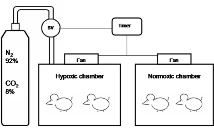
During three weeks, 7 days per week, for 8 hours a day, from 9 a.m. to 5 p.m., in the lights on period, the rat cages were covered with transparent acrylic lids connected to the IIH system. Food and water supplies were kept unchanged during the experimental procedure. A timer activated a solenoid valve to release a mixture with 92% of nitrogen and 8% of CO2 in the hypoxia chamber, for 60 seconds. At the end of this time interval, the nitrogen flow in the cages had reduced the oxygen fraction from 21% to approximately 7 ± 1% and increased the CO2 fraction to approximately 5 ± 1%, lasting the nadir 12 ± 3 seconds. At the end of the hypoxia cycle, the timer activated a fan for 60 seconds insufflating room air into the chamber, restor-ing the oxygen fraction to 21%, in 30 ± 5 seconds, and conducting the chamber air out of the room. In 120 sec-onds, there was a full hypoxia/normoxia cycle; in 8 hours, 240 IIH periods occurred, the equivalent to an apnea index of 30 per hour, a figure commonly observed in patients with severe OSAHS.
In order to confirm the arterial blood oxygen saturation (SaO2) level reached in the animals submitted to this
equipment, two anesthetized rats had the SaO2 measured through a pulse oximeter for veterinary use (NONIN Med-ical, Inc., Plymouth, Minnesota, USA). Repeated arterial saturation falls were verified to values around 70%, SaO2 level usually observed in patients with severe OSAHS.
Statistical Analysis
Results are expressed as mean ± SD. To compare paired observations of weights before and after 21 days we used Willcoxon exact test. To compare IIH versus sham IIH we employed Mann-Whitney's U with Fisher's exact one-tailed test.
Results
As shown in Table 1, the average body weight of the ani-mals from the IIH group decreased after 21 days, while it did not change in the sham IIH group. We did not find difference in heart dimensions. The weight loss and the
reduction in the Lee index in the IIH group were signifi-cantly different from the variation observed in the sham IIH group (p = 0,001). The BAT was lighter in the IIH group and marginally reached the alpha error probability (p = 0.05) (Table 1).
Discussion
The results of the present study suggest that 30 IIH cycles per hour, 8 hours a day, during 21 days cause a reduction in body weight and brown adipose tissue of Wistar mid-dle-age retired breeder rats. The finding of body mass loss and BAT reduction in rats submitted to IIH is original.
Diagram of the hypoxic and normoxic chambers Figure 1
Diagram of the hypoxic and normoxic chambers. Solenoid valve (SV).
Table 1: Mean and standard deviations of body weight, Lee Index and body fat distribution in the two groups of animals prior and after the isocapnic intermittent hypoxia (IIH) or sham IIH exposure
IIH
(n = 8)
Sham IIH
(n = 5)
P
Initial body weight 446.75 ± 50.31 448.40 ± 62.67 0.362*
Final body weight 429.50 ± 52.88 451.60 ± 60.71 0.262*
Initial – final weight 17.25 ± 6.69 -3.20 ± 8.17 0.001**
Nasal – anal length 23.8 ± 0.8 23.4 ± 0.9 0.267*
Initial Lee index 0.320 ± 0.014 0.326 ± 0.015 0.111*
Final Lee index 0.315 ± 0.013 0.327 ± 0.014 0.015*
Initial-final Lee index 0.00428 ± 0.00187 -0.00079 ± 0.00188 0.001**
Gastrocnemius 1.04 ± 0.27 1.07 ± 0.17 0.308*
Heart 1.23 ± 0.137 1.24 ± 0.132 0.472*
Brown adipose tissue 0.51 ± 0.079 0.6 ± 0.075 0.054*
Epididymal fat 8.44 ± 2.558 9.65 ± 3.807 0.311*
Abdominal fat 19.58 ± 8.247 24.08 ± 16.048 0.528*
Despite the reduced number of cases, the BAT difference almost reaches statistic significance. Prior to reaching any definitive conclusions, this study should be repeated in a larger number of animals exposed to intermittent hypoxia for longer periods.
Before engaging in the present study, we performed a pilot study to collect evidence of an effect of IH on BAT. We measured the wet weight of BAT of mice being eutha-nasied after five weeks in IIH for another research proto-col (unpublished data) to serve as a pilot. We found in 8 controls an average BAT wet weight of 0.0655 ± 0.005 g and in the 16 mice of the IIH group an average of 0.0334 ± 0.001 g, approximately half of the control group value (p < 0.0001). Because mice have a low total weight and minuscule BAT, we were concerned about errors occurring during dissection of BAT and decided to do this protocol with rats that are substantially larger. Speculating on what could account for a more prominent effect of IH in the mouse model (50% reduction) than in the rat (15% reduction), two possibilities are of interest: first, mice were submitted to 35 days of IIH against 21 days of the rats; second, the easier dissection of the BAT in rats made the results more exact. The latter is less likely since the var-iation coefficient is less important in the mice. Our initial experiences suggest that future research concentrate on species differences and on the effects of IIH duration on BAT.
The noradrenergic stimulus caused by IIH is the possible determinant of BAT consumption, considering that IIH raises the serum levels of sympathetic neurotransmitters [25]. Interescapular BAT in the rat is densely innervated by the sympathetic nervous system for the control of the tem-perature in cold [26] and diet-induced thermogenesis. Nonshivering thermogenesis performed by BAT is stimu-lated by hypoxia [27]. In acute hypoxic conditions, the O2 consumption response in cold is reduced or thoroughly suppressed by the shivering or nonshivering thermogene-sis hypoxic depression. The BAT activity hypoxic depres-sion mechanisms are not elucidated, although it is known that, even in young animals, the thermogenesis inhibition does not result from oxygen limitation, but from the response regulated by hypoxia [28].
Wet weight is a more relevant physiological parameter for white adipose tissue than for BAT. BAT wet weight may decrease both in face of increased and decreased tissue activity. To elucidate what causes the decrease in BAT it will be necessary to measure the amount of UCP1 or at least the protein amount in the BAT in future experiments.
Sleep was not recorded in this experiment; therefore, it is difficult to ascertain that the animals were sleep deprived. The overall effect of sleep fragmentation on total sleep
time of OSAHS patients is still obscure. In a IH study by Veasey et al., in which sleep was recorded, mice increased sleep time significantly, indicating sleepiness; while the group submitted to two weeks of sham IH slept around 680 minutes per day, the IH group slept 900 minutes [29]. For the sake of reasoning, assuming that sleep time increased, the probability that leptin was increased is min-imal. It is necessary, therefore, that leptin levels are meas-ured to control for this confounding factor and to elucidate the effect of sleep fragmentation on leptin pro-duction.
We chose 9-month old retired breeder male rats as sub-jects in our protocol because these animals better simulate the middle-aged, sexually active men who suffer from OSAHS. There is some evidence that breeder male rats show responses to oxidative stress that are different from males that did not engage in reproductive activity [30].
Obesity is closely related to OSAHS, but until the present moment, obesity roles in OSAHS are not fully clarified: as predisponent factor, as consequence, or as both. If this study's findings are confirmed, the significant weight loss observed in the IIH group may be mediated by leptin increase and is in keeping with findings of correlation between hypoxia and leptin levels [7]. One interesting model to further the understanding of the effect of IIH on leptin is under way in our laboratory, repeating this exper-iment in leptin knockout obese mice.
This is the first report of weight loss and reduction of BAT under intermittent hypoxia. This line of research is war-ranted since the findings raised numerous questions that may be of relevance for sleep apnea patients. We recog-nized the need of additional studies in different species with larger number of animals and with longer exposi-tions in order to confirm the effect of isocapnic intermit-tent hypoxia on body weight and BAT.
Abreviations
BAT: Brown adipose tissue; IH: Intermittent hypoxia; IIH: Isocapnic intermittent hypoxia; OSAHS: Obstructive sleep apnea-hypopnea syndrome; SV: Solenoid valve; UCP1: Uncoupling protein 1.
Competing interests
The authors declare not having any personal or financial support or involvement with organizations with financial interest in the subject matter or any actual or potential conflict of interest.
Acknowledgements
Publish with BioMed Central and every scientist can read your work free of charge
"BioMed Central will be the most significant development for disseminating the results of biomedical researc h in our lifetime."
Sir Paul Nurse, Cancer Research UK
Your research papers will be:
available free of charge to the entire biomedical community
peer reviewed and published immediately upon acceptance
cited in PubMed and archived on PubMed Central
yours — you keep the copyright
Submit your manuscript here:
http://www.biomedcentral.com/info/publishing_adv.asp
BioMedcentral
Project supported by a research grant from FIPE – Hospital de Clinicas de Porto Alegre.
References
1. Somers VK: Sleep- a new cardiovascular frontier. N Engl J Med
2005, 353(19):2070-3.
2. Patil SP, Schneider H, Schwartz AR, Smith PL: Adult obstructive sleep apnea: pathophysiology and diagnosis. Chest 2007,
132(1):325-37.
3. Carpagnano GE, Foschino-Barbaro MP, Resta O, Barnes PJ: Oxida-tive stress and cardiovascular complications in sleep apnea.
Chest 2005, 127(6):2294.
4. Yamauchi M, Nakano H, Maekawa J, Okamoto Y, Ohnishi Y, Suzuki T, Kimura H: Oxidative stress in obstructive sleep apnea. Chest
2005, 127(5):1674-9.
5. Schäfer H, Pauleit D, Sudhop T, Gouni-Berthold I, Ewig S, Berthold HK: Body fat distribution, serum leptin, and cardiovascular risk factors in men with obstructive sleep apnea. Chest 2002,
122(3):829-39.
6. Ip MS, Lam KS, Ho C, Tsang KW, Lam W: Serum leptin and vas-cular risk factors in obstructive sleep apnea. Chest 2000,
118(3):580-6.
7. Tatsumi K, Kasahara Y, Kurosu K, Tanabe N, Takiguchi Y, Kuriyama T: Sleep oxygen desaturation and circulating leptin in obstructive sleep apnea-hypopnea syndrome. Chest 2005,
127(3):716-21.
8. Guardiano SA, Scott JA, Ware JC, Schechner SA: The long-term results of gastric bypass on indexes of sleep apnea. Chest 2003,
124(4):1615-9.
9. Busetto L, Enzi G, Inelmen EM, Costa G, Negrin V, Sergi G, Vianello A: Obstructive sleep apnea syndrome in morbid obesity: effects of intragastric balloon. Chest 2005, 128(2):618-23. 10. Bradley TD, Brown IG, Grossman RF, Zamel N, Martinez D,
Phillip-son EA, Hoffstein V: Pharynx in snorers, nonsnorers, and patients with obstructive sleep-apnea. N Engl J Med 1986,
315(21):1327-31.
11. Mohsenin V: Gender differences in the expression of sleep-dis-ordered breathing: role of upper airway dimensions. Chest
2001, 120(5):1442-7.
12. Spiegel K, Tasali E, Penev P, Van Cauter E: Brief communication: Sleep curtailment in healthy young men is associated with decreased leptin levels, elevated ghrelin levels, and increased hunger and appetite. Ann Intern Med 2004,
141(11):846-50.
13. Knutson KL, Spiegel K, Penev P, Van Cauter E: The metabolic con-sequences of sleep deprivation. Sleep Med Rev 2007,
11(3):163-78.
14. Cannon B, Nedergaard J: Brown adipose tissue: function and physiological significance. Physiol Rev 2004, 84(1):277-359. 15. Nedergaard J, Bengtsson T, Cannon B: Unexpected evidence for
active brown adipose tissue in adult humans. Am J Physiol Endo-crinol Metab 2007, 293(2):E444-52.
16. Kajimoto K, Yamazaki N, Kataoka M, Terada H, Shinohara Y: Identi-fication of possible protein machinery involved in the ther-mogenic function of brown adipose tissue. J Med Invest 2004,
51(1–2):20-8.
17. Golozoubova V, Cannon B, Nedergaard J: UCP1 is essential for adaptive adrenergic nonshivering thermogenesis. Am J Physiol Endocrinol Metab 2006, 291(2):E350-7.
18. Cannon B, Nedergaard J: Brown adipose tissue: function and physiological significance. Physiol Rev 2004, 84(1):277-359. 19. Gautier H, Bonora M, M'Barek SB, Sinclair JD: Effects of hypoxia
and cold acclimation on thermoregulation in the rat. J Appl Physiol 1991, 71(4):1355-63.
20. Lafontan M, Berlan M: Fat cell adrenergic receptors and the control of white and brown fat cell function. J Lipid Res 1993,
34(7):1057-91.
21. Guyenet PG: Neural structures that mediate sympathoexcita-tion during hypoxia. Respir Physiol 2000, 121(2–3):147-62. 22. Boa G, Metreveli N, Li R, Taylor A, Fletcher EC: Blood pressure
response to chronic episodic hypoxia: role of the sympa-thetic nervous system. J Appl Physiol 1997, 83:95-101.
23. Greenberg HE, Sica A, Batson D, Scharf SM: Chronic intermittent hypoxia increases sympathetic responsiveness to hypoxia and hypercapnia. J Appl Physiol 1999, 86:298-305.
24. Lee MO: Determination of the surface area of the white rat with its application to the expression of metabolic results.
Am J Physiol 1928, 89:24-31.
25. Hui AS, Striet JB, Gudelsky G, Soukhova GK, Gozal E, Beitner-John-son D, Guo SZ, Sachleben LR Jr, Haycock JW, Gozal D, Czyzyk-Krzeska MF: Regulation of catecholamines by sustained and intermittent hypoxia in neuroendocrine cells and sympa-thetic neurons. Hypertension 2003, 42:1130.
26. Himms-Hagen J: Lipid metabolism during cold-exposure and during cold-acclimation. Lipids 1972, 7(5):310-23.
27. Mortola JP, Merazzi D, Naso L: Blood flow to the brown adipose tissue of conscious young rabbits during hypoxia in cold and warm conditions. Pflugers Arch 1999, 437(2):255-60.
28. Rohlicek CV, Saiki C, Matsuoka T, Mortola JP: Oxygen transport in conscious newborn dogs during hypoxic hypometabolism. J Appl Physiol84:763-768.
29. Veasey SC, Davis CW, Fenik P, Zhan G, Hsu YJ, Pratico D, Gow A:
Long-term intermittent hypoxia in mice: protracted hyper-somnolence with oxidative injury to sleep-wake brain regions. Sleep 2004, 27(2):194-201.