of Biological Research
European Journal of Biological Research 2018; 8 (4): 232-242
Biochemical composition and antioxidant properties
of some seaweeds from Red Sea coast, Egypt
Rasha M. El-Shazoly
1, Mustafa A. Fawzy*
21
Botany and Microbiology Department, Faculty of Science, New Valley University, 72511, Al-Kharja, New Valley, Egypt
2
Botany and Microbiology Department, Faculty of Science, Assiut University, Assiut, 71516, Egypt *Corresponding author: Mustafa A. Fawzy, E-mail: [email protected]
ABSTRACT
The current study investigated the biochemical composition and antioxidant properties of four seaweeds: Laurencia sp. (Rhodophyta), Cystoseira
myrica, Hydroclathrus clathratus and Padina pavonica (Ochrophyta). The highest amount of
carbohydrates was (215.78 mg/g dry wt.) in
Laurencia sp. and proteins content was maximum
(50 mg/g dry wt.) in Laurencia sp. and Cystoseira
myrica. The highest values of free amino acid
content were recorded in the brown seaweed species
Cystoseira myrica (4.01 mg/g dry wt.). The
pressurized hot water extract of Cystoseira myrica has the highest total phenolic content (1.61 mg GAE/g dry wt.). Cystoseira myrica contained the highest amounts of flavonoids (3.35 mg/g dry wt.), ascorbic acid (9.07 mg/g dry wt.) and α-tocopherol (27.25±0.00 abs. at 520 nm/g dry wt.). Furthermore, the ethyl alcohol extract of Cystoseira myrica showed high antioxidant capacities (541.6 µg/g dry wt.) and achieved the most powerful reducing ability among all of the different extracts of algal species. Statistical evaluation by Spearman correlation between the TAC assay and the total phenolic contents was found to be significant, but the correlation was nonsignificant between FRAP assay and the total phenolic contents. The composition of elements of the studied seaweed species was also
analyzed. The most significant macro-elements present in the studied seaweeds were K, Na and Ca, representing that the seaweeds are good sources of these elements. Since, these seaweeds are widespread in the Egyptian waters, their biochemical composition and antioxidant capacities made them promising candidates for industrial, nutritional and pharmaceutical applications.
Keywords: Seaweeds; Biochemical composition; Phenolic compounds; Ascorbic acids; Antioxidant activity; Elemental analysis.
1. INTRODUCTION
Seaweeds belong to a group of plants known as algae. Seaweeds are classified as Chlorophyta, Phaeophyta and Rhodophyta based on their chemi-cal and nutrient composition. Seaweeds, besides their very significant ecological role in the nature, have come up step by step starting with using them as food, later as raw material for cosmetic, pharmaceutical, medicinal and industrial purposes [1], related to their high contents of vitamins, carotenoids, essential fatty acids, minerals, poly-saccharides and proteins [2, 3]. Polypoly-saccharides are generally the main component of brown, green and red algae [4, 5]. Several polysaccharides constitute the major structure of the algal cell walls. The major Received: 16 August 2018; Revised submission: 23 September 2018; Accepted: 03 November 2018
Copyright: © The Author(s) 2018. European Journal of Biological Research © T.M.Karpiński 2018. This is an open access article licensed under the terms of the Creative Commons Attribution Non-Commercial 4.0 International License, which permits
unrestricted, non-commercial use, distribution and reproduction in any medium, provided the work is properly cited.
European Journal of Biological Research 2018; 8 (4): 232-242
polysaccharides in the algae include fucoidan, ulvans, laminarans, carrageenans, galactans,
algi-nates and agar. Therefore, polysaccharides play the role of storage function and structural support in algae. Generally, macromolecules of algae are formed with several monosaccharides linked by glucosidic bonds, as well as some have linear backbones that contain repeating disaccharide units [6]. The total amounts of polysaccharides in the seaweeds ranged between 4% and 76% of the dry weight [7] where fucoidans, carrageenans, aligns and agar are some of the polysaccharides usually used by man, some with different biological activities [8, 9]. Protein contents are also variable and the highest amounts are commonly found in red and green seaweeds (10-30% of the dry weight) compared to the brown seaweeds (5-15% of the dry weight) [7, 10]. Moreover, seaweeds are considered as a source of the bioactive components, whereas they can produce a high variety of secondary metabolites such as alkaloids, terpenoids, phenolic compounds and flavonoids characterized by a wide spectrum of biological activities [11]. Compounds with antimicrobial, antifungal, antiviral and anti-oxidant activities have been observed in green, red and brown algae [12, 13]. Therefore, more attentions have been paid to their applications in nutraceu-ticals, cosmeceunutraceu-ticals, pharmaceuticals and functio-nal foods [14]. The metabolism of seaweeds can be influenced by several factors such as nutrients, light, salinity and water temperature, being forced to rapidly adapt to the new environmental conditions and to survive, they produce a high variety of biologically active secondary metabolites as pre-viously mentioned [15]. These conditions, as well can lead to the production of free radicals and other oxidizing agents, however seaweeds rarely suffer any severe photodynamic damage through the metabolism. This fact suggests that the cells of seaweed have protective mechanisms. Reactive oxygen species such as superoxide, hydroxyl and peroxyl radicals are formed in the cells of human by endogenous factors and cause wide oxidative damage which can lead to cancer, age related degenerative conditions and a wide range of other human diseases [16]. Seaweeds are also known as sources rich in numerous elements such as Na, Mg, Ca, K and P or trace elements such as Mn, I or Zn. This elemental richness is due to their ability to
preserve inorganic compounds up to 36% of dry weight in some species [17]. Marine algae comprise more than 60 trace elements in a concentration, which are greatly higher than that present in the terrestrial plants and have several pharmacological activities [18]. The Red Sea is a diverse and rich ecosystem, and more than 500 species of seaweeds have been recorded in it [19]. The studies on the antioxidant properties of the extracts of seaweed in Egypt are very scarce. Therefore, the main objective of the present investigation was to analyze the influence of the different extraction methods: cold water, boiling water, pressurized hot water and ethyl alcohol on the total phenolic content and antioxidant activity of four different edible species namely; Laurencia sp. (Red alga),
Cysto-seira myrica, Hydroclathrus clathratus and Padina pavonica (brown algae), to identify the new
resour-ces of natural antioxidant compounds, as well as, to evaluate the correlation between the total pheno-lic content and antioxidant capacities. Flavonoids, vitamin (C and E) contents and other biochemical compounds such as soluble protein, carbohydrate, free amino acids, were also determined in the algal species.
2. MATERIAL AND METHODS
2.1. Samples collection
European Journal of Biological Research 2018; 8 (4): 232-242
2.2. Extraction methods of compounds from seaweeds
Some extraction methods were achieved by using different solvents. Conventional cold and hot water extraction besides extraction with ethanol was used. In addition, pressurized hot water extraction (PHWE) was tested also as extraction method. PHWE is a technique of extraction that uses water as extractant (extraction solvent) at temperatures above the boiling point of the water (100°C/273 K, 0.1 MPa), but below the critical point of water (374°C/647 K, 22.1 MPa).
2.2.1. Extract preparation
Cold water: a known algal tissue weight (0.1 g) was homogenized and suspended in 5 ml cold water for 2 h.
Boiling water: a known algal weight (0.1 g) was boiled in 5 ml distilled water for 2 h.
Pressurized hot water: a known algal weight (0.1 g) was suspended in 15 ml distilled water and autoclaved for 2 h at 121oC.
Ethyl alcohol: a known weight of algal tissue (0.1 g) was homogenized in 20 ml ethyl alcohol 80% and shaken for 2 h.
The resultant extracts from each extraction process was separately filtered through a Whatman No. 1 filter paper, then it was stored in a closed bottle away from the light. The extracts were used for determination of total phenolic contents, total antioxidant activity and reducing power.
2.3. Determination of soluble carbohydrates
The contents of soluble carbohydrate in the crude extract of boiling water were determined by the anthrone sulphuric acid method [25, 26].
2.4. Determination of soluble proteins
The determination of soluble protein in the crude extract of boiling water was performed by Folin reagent according to Lowry et al. [27]. A calibration curve was made by bovine serum albumin (BSA) and the results were expressed as mg BSA/g dry wt.
2.5. Determination of free amino acids
Free amino acid content in the crude extract of boiling water was estimated according to the method of Moore and Stein [28]. A calibration curve was made by glycine, and free amino acid concen-tration was calculated as mg/g dry wt.
2.6. Determination of total phenolics
Total phenolics were estimated according to Kofalvi and Nassuth [29]. Free phenolics were estimated by the Folin-Ciocalteu's phenol reagent by using gallic acid as a standard.
2.7. Total flavonoid content
The measurement of total flavonoid content in the crude extract of ethanol was made according to the method described by Moreno et al. [30] and the results are expressed as quercetin equivalents.
2.8. Determination of ascorbate (vitamin C) content
The ascorbic acid content in the crude extract of trichloroacetic acid was estimated according to Jagota and Dani [31].
2.9. Determination of α-tocopherol (vitamin E) content
α-tocopherol content in the crude extract of petroleum ether was determined by the method of Pearson [32].
2.10. Antioxidant assay
2.10.1. Determination of the total antioxidant activity
European Journal of Biological Research 2018; 8 (4): 232-242
2.10.2. Reducing power assay
The reducing power of the algal samples was estimated according to the methods described by Oyaizu [34] and the results were expressed as µg/g dry wt.
2.11. Estimation of mineral content
2.11.1. Total phosphorus
It was estimated using a Unico UV-2100 spectrophotometer by the chlorostannus-phospho-molybdic acid method in a sulfuric acid system [35].
2.11.2. Total potassium and sodium
They were determined by the flame photo-meter methods (Dr Lange Flame Photometer M 71 D type Nr/ LPG 075) according to Page et al. [36].
2.11.3. Total calcium and magnesium
They were determined by the flame photo-meter methods described by Page et al. [36].
2.12. Statistical analysis
All results obtained were subjected to one-way analysis variance (ANOVA), by using the SPSS statistical package. For comparison of the means, the Duncanʼs multiple range tests (p< 0.05) were used. Analysis of correlation (Spearman correlation) was performed to obtain the relation between the phenolic compounds, total antioxidants and the reducing power.
3. RESULTS AND DISCUSSION
Carbohydrates, proteins and amino acids are the most vital biochemical components of algae. Carbohydrates are considered the major significant biochemical component in algae since they represent the main source of energy for the metabolic activities. In the current study, the red seaweed;
Laurencia sp. appeared to be the highest (215.78
mg/g dry wt.) in the soluble carbohydrate content compared to the brown algae, Cystoseira myrica (47.45 mg/g dry wt.), Hydroclathrus clathratus
(39.55 mg/g dw.) and Padina pavonica (28.40 mg/g dry wt.) (Fig. 1). The amount obtained from
Lauren-cia sp. was much higher than that present in another
red alga, Grateloupia turuturu (41.57 ± 0.66 mg/g dry wt.) [37]. Rhodophycean species presented high content of carbohydrate than the Phaeophycean members. The high carbohydrate content in the red algae might be due to the higher content of phyco-colloid in their cell walls [18]. Seaweeds contain high contents of polysaccharides as cell wall structural components that were already captured by the industry of hydrocolloid. Water soluble polysac-charides of seaweeds were related to hypoglycemic activities and hypocholesterolemia, however inso-luble polysaccharides were related to digestive tract transit time reduction [38].
Proteins content, one of the most biochemical components of seaweeds, ranged from 50 mg/g dry wt. in Laurencia sp. and Cystoseira myrica to 36.35 mg/g dry wt. and 29.05 mg/g dry wt. in Padina
pavonica and Hydroclathrus clathratus, respectively
(Fig. 1). In this respect, Ismail [39] showed that, the protein content in Sargassum linifolium (Phaeo-phyceae) (14.89%) was higher than that of Corallina
officinalis (Rhodophyceae) (5.91%) of dry wt.
According to Ibañez and Cifuentes [40], the content of protein varies between algal species and gene-rally, Rhodophycean and Chlorophycean species are characterized by the larger content of proteins compared with Phaeophycean species. However, differences in the content of proteins may be also related to the seasonal period, temperature values or to its consumption by seaweeds in the growth and reproduction. Also might be associated with the variances between species, the surrounding environ-mental conditions of seaweeds and the geographical locations [41].
The highest contents of free amino acid were present in the Phaeophycean species Cystoseira
myrica (4.01 mg/g dry wt.) followed closely by the
Rhodophycean species Laurencia sp. (3.99 mg/g dry wt.), while the lowest free amino content was present in Padina pavonica and Hydroclathrus
clathratus (2.93 and 2.80 mg/g dry wt., respectively)
European Journal of Biological Research 2018; 8 (4): 232-242
Figure 1. Soluble carbohydrates, soluble proteins and free amino acids (mg/g dry wt.) of crude extract of marine algal species with boiling water.
The studied seaweeds showed a significant content of total phenolic compounds that were significantly different at p ≤ 0.05 (Table 1). Total phenolic content in the algal crude extracts was effected by the algal species and various extraction processes. Data in table 1 indicated that, the pressurized hot water extract of Cystoseira myrica has the highest total phenolic content (1.61 mg GAE/g dry wt.), followed by the pressurized hot water extract of Laurencia sp. and boiling water extract of Cystoseira myrica (1.49 and 1.40 mg GAE/g dry wt., respectively), as compared with the other algae. The pressurized hot water extract has been largely used to extract comparatively polar bioactive molecules such as phenolic compounds [42]. Moreover, PHWE was considered as an effec-tive process for extracting the phenolic compounds in comparison with the other applied extraction methods. In general, the crude extract of cold water from all algal species indicated the lowest total polyphenol content (Table 1). Various algal products provided varied total phenolic contents because of many influencing conditions, such as algal species and, geographical origin or the region of cultivation, environmental, physiological, and seasonal varia-tions [43]. According to Chakraborty et al. [44] phenolic compounds can chelate the metal ions and inhibit the formation of free radical, thus improving the antioxidant intrinsic coordination. In this respect, phenols transfer hydrogen atoms to peroxyl in the lipid peroxidation cycle to form the aryloxyls that
are unable to act as chain carriers for free radicals and therefore delaying the peroxidation process. Based on the research papers dealing with the phenolic contents in fresh algae, the obtained algal product results cannot be suitably compared because of the different extraction methods used; just for clarification, the ethanol extract of Eisenia bicyclis contained about 319 mg.g-1 GAE [45], aqueous extract of Porphyra tenera 10.1 mg/g GAE [46], ethanol extract of Palmaria palmata 10.3 mg/g GAE [47], aqueous extract of Undaria pinnatifida contained 3.8 mg/g GAE [48], methanol-chloroform extract of Laminaria japonica 0.3 mg/g GAE [49] and aqueous extract of Hizikia fusiformis contained 4.1 mg/g GAE [48].
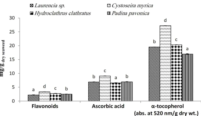
The content of flavonoids, ascorbic acid and
European Journal of Biological Research 2018; 8 (4): 232-242
Table 1. Total phenolic contents (mg GAE/g dry wt.) of crude extract of marine algal species with various extraction processes.
Algal species Different extraction processes
Pressurized hot water Cold water Boiling water Ethyl alcohol
Laurencia sp. 1.49±0.00c 0.97±0.02b 1.11±0.07b 0.92±0.01b
Cystoseira myrica 1.61±0.03c 0.90±0.04b 1.40±0.07c 1.06±0.02c
Hydroclathrus clathratus 0.75±0.02a 0.61±0.05a 0.65±0.04a 0.82±0.01a
Padina pavonica 1.17±0.08b 0.67±0.01a 0.80±0.02a 1.13±0.02d For each treatment the means within the column by different letters are significantly different at P < 0.05 Each value is expressed as the means ±SE (n=3).
Figure 2. Flavonoids, ascorbic acid (mg/g dry wt.) and α-tocopherol (abs. at 520 nm/g dry wt.) of marine algal species.
Ascorbic acid was also significantly different between algal species at p≤ 0.05, whereas, it ranged between 6.57 mg/g dry wt. in Hydroclathrus
clath-ratus and 9.07 mg/g dry wt. in Cystoseira myrica
(Fig. 2). α-tocopherol content varied from 27.25± 0.00 (Cystoseira myrica) to 16.99±0.14 (Padina
pavonica) abs. at 520 nm/g dry wt. seaweed (Fig. 2).
The α-tocopherol content in the Cystoseira myrica extract was significantly different (p < 0.05) compa-red to those of the other algal species. Natural antioxidants such as polyphenols and vitamins in the higher plants were used to capture the reactive oxygen species that lead to lipid peroxidation. Thus, these compounds became used in the food industry to protect human body from the free radicals and prevent expansion of various chronic diseases. Several studies strongly suggested natural sources of polyphenols and vitamins to inhibit the free
radical-damage that lead to aging progression and to prevent initiations of cancer [38, 52].
As a result of the variation of the oxidation methods, the use of one antioxidant process to assess the antioxidant capacity cannot reveal a clear view of their actual antioxidant activities. Thus, the antioxidant capacity of various algal extracts was estimated by two antioxidant methods: ferric redu-cing antioxidant power, and total antioxidant capacity.
European Journal of Biological Research 2018; 8 (4): 232-242
that, the ethyl alcohol extract of Cystoseira myrica showed high antioxidant capacities (541.6 µg/g dry wt.) followed by an ethyl alcohol extract of
Hydro-clathrus clathratus and Padina pavonica (383.02
and 339.92 µg/g dry wt., respectively). On the other hand, the cold water extract of all tested algae showed absolutely the lowest values among all different extraction methods, followed by the boiling water extract.
The determination of reducing power of compounds may serve as an important indicator for their potential antioxidant capacity. In the ferric reducing antioxidant power method, the antioxidants reduce Fe3+-ferricyanide complex to its Fe2+ form. Thus, Fe2+ can be checked by determining the formation of Perl's Prussian blue. The ethyl alcohol extract from Cystoseira myrica followed by
Hydro-clathrus clathratus achieved the most powerful
reducing capacity among all of the different extracts of algal species (Table 3). In contrast, the cold water extract showed the lowest activity for all algal species except in the case of Laurencia sp. These results showed that the ethyl alcohol extract may contain most of the antioxidative substances in these algae.
Statistical evaluation by Spearman correlation
between the total antioxidant capacity assay and the total phenolic contents was found to be significant (r = 0.60, p = 0.014). On the other hand, nonsigni-ficant correlation was found between ferric reducing antioxidant power assay and the total phenolic contents (r = 0.23, p = 0.39). This shows that pheno-lic compounds might be a major contributor to the antioxidant abilities for either of these macroalgae. A high correlation between the total phenolic content and antioxidant capacity has been described by several authors [13]. However, other reports showed that this correlation doesn’t occur and it was concluded that the phenolic compounds are not responsible for the antioxidant capacity [53]. Although phenolic compounds are found to be the most important contributor to the antioxidant capacities in various higher-order species as plants, this might hold real for macroalgae. Dixon and Palva [54] revealed that the antioxidants such as phenolic compounds are in plants part of a complex defense mechanism against a broad range of stresses, and therefore accumulate as a result to these stresses. The macroalgae in this investigation might not have been exposed to stresses, causing less phenolic compounds to be formed, as compared to the plants.
Table 2. Total antioxidants (µg/g dry wt.) of crude extract of marine algal species with various extraction processes.
Algal species Different extraction processes
Pressurized hot water Cold water Boiling water Ethyl alcohol
Laurencia sp. 335.3±8.5d 167.4±3.7c 229.7±2.8d 338.2±2.6a
Cystoseira myrica 212.9±8.4c 111.9±2.5b 159.1±1.2c 541.6±38.7b
Hydroclathrus clathratus 139.9±3.6a 73.1±2.3a 88.3±0.2a 383.1±10.7a
Padina pavonica 170.1±0.7b 69.4±0.8a 98.3±1.1b 339.9±12.5a For each treatment the means within the column by different letters are significantly different at P < 0.05 Each value is expressed as the means ±SE (n=3).
Table 3. Reducing power (µg /g dry wt.) of crude extract of marine algal species with various extraction processes.
Algal species Different extraction processes
Pressurized hot water Cold water Boiling water Ethyl alcohol
Laurencia sp. 185.8±1.7a 501.5±9.8b 116.6±0.7a 629.2±6.0a
Cystoseira myrica 1181.4±16.4c 688.5±22.7c 803.6±10.7d 3208.3±135.8c
Hydroclathrus clathratus 245.2±5.6b 225.9±22.6a 526.7±14.1c 1194.9±162.9b
European Journal of Biological Research 2018; 8 (4): 232-242
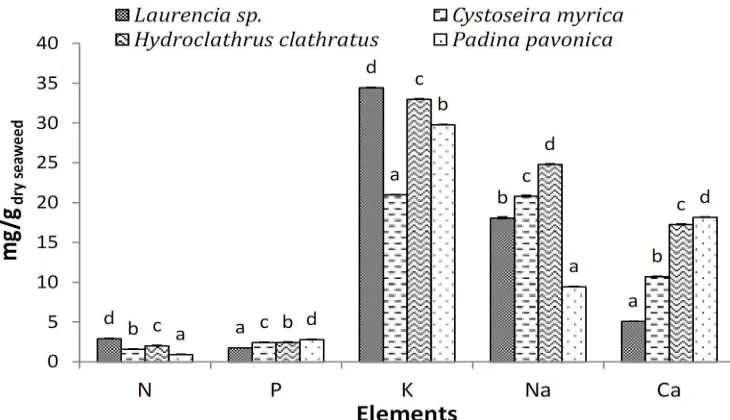
Seaweeds are rich in macroelements with some content in trace elements. The macroelements such as Na, Ca, K and Mg are among the elements which are found in significant quantities in seaweeds [55]. The composition of elements of the four studied species was evaluated and is shown in Fig. 3. The most important macro-elements found in the studied seaweeds were K, Na and Ca, representing that the seaweeds are a good source of these elements. Phosphorous was present in a slightly fairly constant value (between 1.771 and 2.791 mg/g dry seaweed) among all studied seaweeds, but of a much lower order of magnitude than the three other minerals that previously mentioned (Fig. 3). In what concerns the distribution of mineral per species, the differences were detected between all algal species. For instance, potassium was the most dominant element present in all the four studied species with amounts ranging from 21.00 mg/g dry seaweed in
Cystoseira myrica to 34.45 mg/g dry seaweed in Laurencia sp. being statistically different (p < 0.05)
among all the four studied species (Fig. 3). Potassium, the macro element preserves the ionic exchange, osmotic gradients and normal neural functions. Potassium is a very significant element for the appropriate function of all cells, tissues, and organs in the human body. It has a significant role in regulation of water balance of the body [56].
Slight variations were detected among the four studied species for total nitrogen (Fig. 3).
Laurencia sp. and Hydroclathrus clathratus tended
to indicate higher amounts of nitrogen (2.93 and 2.02 mg/g, respectively), while Cystoseira myrica showed lower values (1.6 mg/g) followed by Padina
pavonica (0.933 mg/g). The nitrogen contents varied
maybe due to the algal species, site, season and environment [57]. Sodium content also varied signi-ficantly between the different algal species. The high value of sodium was recorded by Hydroclathrus
clathratus (24.81 mg/g dry seaweed) (Fig. 3).
Calcium was the most significant element that accumulated in the seaweeds at much higher content than in the terrestrial food stuffs [58]. Calcium values ranged from 5.9 mg/g dry seaweed in
Laurencia sp. to 18.18 mg/g dry seaweed in Padina pavonica (Fig. 3). The content of seaweeds mineral
differs according to species, seasons, wave expo-sure, physiological and environmental conditions, type of processing and method of mineralization [59]. Exogenous and endogenous factors have contributed to the variability of the composition of seaweeds mineral. The variations in mineral in seaweed tissues were detected also with the same seaweed species influenced by the stage of the living cycle and the age of seaweed [60].
European Journal of Biological Research 2018; 8 (4): 232-242
4. CONCLUSION
Seaweeds: Laurencia sp., Cystoseira myrica,
Hydroclathrus clathratus and Padina pavonica,
have high contents of proteins, carbohydrates, vitamins and minerals suggesting a promising role in pharmaceutical and nutritional applications. Moreover, these seaweeds offer new potential sources of bioactive compounds such as flavonoids, phenols, α-tocopherol and ascorbic acid. These compounds showed a high antioxidant capacities with wide nutraceutical, pharmaceutical, biomedical and prospected applications. Thus, intensive future studies should be made to develop these naturally economical resources.
CONFLICTS OF INTEREST
The authors declare that they have no competing interests.
AUTHORS’ CONTRIBUTIONS
RME-S and MAF: designed research, analyzed the data, wrote and revised the manuscript, performed the extraction methods of compounds from seaweeds and determined the different parameters, carried out the research point by point. MAF: collected and identified the seaweeds. Both authors read and approved the final manuscript.
REFERENCES
1. Chennubhotla VSK, Rao MU, Rao KS. Commercial
importance of marine macro algae. Seaweed Res Utiln. 2013; 35(2): 118-128.
2. Jeeva S, Marimuthu J, Domettila C, Anantham B,
Mahesh M. Preliminary phytochemical studies on some selected seaweeds from Gulf of Mannar, India. Asian Pac J Trop Biomed. 2012; 2(1): 30-33.
3. Polat S, Ozogul Y. Seasonal proximate and fatty
acid variations of some seaweeds from the northeastern Mediterranean coast. Oceanologia. 2013; 55(2): 375-391.
4. Goo BG, Baek G, Jin Choi D, Il Park Y, Synytsya
A, Bleha R, et al. Characterization of a renewable
extracellular polysaccharide from defatted
microalgae Dunaliella tertiolecta. Biores Technol. 2013; 129: 343-350.
5. Kurniawati HA, Ismadji S, Liu JC. Microalgae
harvesting by flotation using natural saponin and chitosan. Biores Technol. 2014; 166: 429-434.
6. Pérez MJ, Falqué E, Domnguez H. Antimicrobial
action of compounds from marine seaweed. Mar Drugs. 2016; 14(3): 52.
7. Holdt SL, Kraan S. Bioactive compounds in
seaweeds: functional food applications and
legislation. J Appl Phycol. 2011; 23: 543-597.
8. Mak W, Hamid N, Liu T, Lu J, White WL.
Fucoidan from New Zealand Undaria pinnatifida:
Monthly variations and determination of
antioxidantactivities. Carb Polym. 2013; 95: 606-614.
9. Mak W, Wang SK, Liu T, Hamid N, Li Y, Lu J, et
al. Antiproliferation potential and content of fucoidan extracted from sporophyll of New Zealand
Undaria pinnatifida. Front Nutr. 2014; 1: 1-10.
10. Zhou AY, Robertson J, Hamid N, Ma Q, Lu J.
Changes in total nitrogen and amino acid composition of New Zealand Undaria pinnatifida with growth, location and plant parts. Food Chem. 2015; 186: 319-325.
11. Rodriguez EB, Rodriguez-Amaya DB. Formation of
apocarotenals and epoxycarotenoids from β-carotene
by chemical reactions and by autoxidation in model systems and processed foods. Food Chem. 2007; 101(2): 563-572.
12. Yuan YV, Carrington MF, Walsh NA. Extracts from
dulse (Palmaria palmata) are effective antioxidants and inhibitors of cell proliferation in vitro. Food Chem Toxicol. 2005; 43: 1073-1081.
13. Chew YL, Lim YY, Omar M, Khoo, KS.
Antioxidant activity of three edible seaweeds from two areas in South East Asia. LWT. 2008; 41: 1067-1072.
14. Fernando IS, Kim M, Son KT, Jeong Y, Jeon YJ.
Antioxidant activity of marine algal polyphenolic compounds: a mechanistic approach. J Med Food. 2016; 19(7): 615-628.
15. Plaza M, Cifuentes A, Ibáñez E. In the search of
new functional food ingredients from algae. Trends Food Sci Technol. 2008; 19: 31-39.
16. Aruoma IO. Antioxidant action of plant foods. Use
of oxidative DNA damage, as a tool for studying antioxidant efficacy. Free Radical Res. 1999; 30: 419-427.
17. Lordan S, Ross RP, Stanton C. Marine bioactives
European Journal of Biological Research 2018; 8 (4): 232-242
18. Dhargalkar VK. Uses of seaweeds in the Indian diet
for sustenance and well-being. Sci Cult. 2014; 80: 192-202.
19. Gouda EA. Obstacles to sustainable tourism
development on the Red Sea Coast. Int J Innov Educ Res. 2015; 3: 165-176.
20. Guiry MD, Guiry GM. AlgaeBase. National
University of Ireland, Galway [accessed 15 August 2013]. http:// www.algaebase.org
21. Meñez EG, Mathieson AC. The marine algae of
Tunisia. Smithsonian Contributions to the Marine Sciences, Washington. 1981.
22. Nizamuddin M. The green marine algae of Libya.
Elga, Bern. 1991; 227 pp.
23. Aleem AA. The marine algae of Alexandria, Egypt.
Alexandria. 1993.
24. Jha B, CRK, Reddy MC, Thakur MU, Rao.
Seaweeds of India: the diversity and distribution of seaweeds of the Gujarat Coast (Vol. 3). Springer, New York. 2009.
25. Fales FW. The assimilation and degradation of
carbohydrates by yeast cells. J Biol Chem. 1951; 193: 113-118.
26. Schlegel HG. Die Verwertung organischer sauren
duch Chlorella in licht. Planta. 1956; 47: 510-526.
27. Lowry OH, Rosebrough NJ, Farr AL, Randall RJ.
Protein measurement with Folin phenol reagent. J Biol Chem. 1951; 193: 265-275.
28. Moore S, Stein WH. Photometric ninhydrin method
for use in the ehromatography of amino acids. J Boil Chem. 1948; 176: 367-388.
29. Kofalvi SA, Nassuth A. Influence of wheat streak
mosaic virus infection phenylpropanoid metabolism and the accumulation of phenolics and lignin in wheat. Physiol Mol Plant Pathol. 1995; 47: 365-377.
30. Moreno MIN, Isla MI, Sampietro AR, Vattuone
MA. Comparison of the free radical scavenging activity of propolis from several regions of Argentina. J Ethnopharmacol. 2000; 71: 109-114.
31. Jagota SK, Dani HM. A New colorimetric technique
for the estimation of vitamin C using Folin phenol reagent. Anal Biochem. 1982; 127: 178-182.
32. Pearson DC. The chemical analysis of food. 7th edn.
Churchill Livingstone Edinburgh, London, 1976: 19-35.
33. Prieto P, Pineda M, Aguilar M. Spectrophotometric
quantitation of antioxidant capacity through the formation of a phosphomolybdenum complex: specific application to the determination of vitamin E. Anal Biochem. 1999; 269: 337-341.
34. Oyaizu M. Studies on product of browning effect
reaction prepared from glucose amine. J Nutr. 1986; 44: 307-315.
35. Jackson ML. Soil chemical analysis. Prentice-Hall,
Inc. Englewood Cliffs, N.J. New Delhi, India. 1973.
36. Page AL, Miller RH, Keeney DR. Methods of soil
analysis. Part 2: Chemical and microbiological
properties. 2nd edn. Am Soc Agron Inc Soil Sci Soc
Am. Madison, Wisconsin, USA. 1982.
37. Munier M, Dumay J, Morançais M, Jaouen P,
Fleurence J. Variation in the biochemical
composition of the edible seaweed Grateloupia
turuturu Yamada harvested from two sampling sites
on the Brittany coast (France): The influence of storage method on the extraction of the seaweed
pigment R-phycoerythrin. J Chem. 2013: ID
568548.
38. Kolanjinathan K, Ganesh P, Saranraj P.
Pharmacological importance of seaweeds: a review. World J Fish Mar Sci. 2014; 6(1): 1-15.
39. Ismail GA. Biochemical composition of some
Egyptian seaweeds with potent nutritive and antioxidant properties. Food Sci Technol Campinas. 2017; 37(2): 294-302.
40. Ibañez E, Cifuentes A. Benefits of using algae as
natural sources of functional ingredients. J Sci Food Agricult. 2013; 93: 703-709.
41. Fleurence J. Seaweed proteins: biochemical,
nutritional aspects and potential uses. Trends Food Sci Technol. 1999; 10(1): 25-28.
42. Plaza M, Turner C. Pressurized hot water extraction
of bioactives. TrAC Trends Anal Chem. 2015; 71; 39-54.
43. Marinho-Soriano E, Fonseca PC, Carneiro MAA,
Moreira WSC. Seasonal variation in the chemical composition of two tropical seaweeds. Biores Technol. 2006; 97: 2402-2406.
44. Chakraborty K, Praveen N, Vijayan KK, Rao GS.
Evaluation of phenolic contents and antioxidant activities of brown seaweeds belonging to
Turbinaria spp. (Phaeophyta, sargassaceae) collected from Gulf of Mannar. Asian Pac J Trop Biomed. 2013; 3(1): 8-16.
45. Kim SM, Kang K, Jeon JS, Jho EH, Kim CY, Nho
CW, Um BH. Isolation of phlorotannins from
Eisenia bicyclis and their hepatoprotective effects
European Journal of Biological Research 2018; 8 (4): 232-242
46. Heo SJ, Cha SH, Lee KW, Jeon YJ. Antioxidant
activities of red algae from Jeju Island. Algae. 2006; 21: 149-156.
47. Yuan YV, Bone DE, Carrington MF. Antioxidant
activity of dulse (Palmaria palmata) extract evaluated in vitro. Food Chem. 2005; 91: 485-494.
48. Heo SJ, Cha SH, Lee KW, Cho SK, JeonYJ.
Antioxidant activities of Chlorophyta and
Phaeophyta from Jeju Island. Algae. 2005; 20: 251-260.
49. Zhang WW, Duan XJ, Huang HL, Zhang Y, Wang
BG. Evaluation of 28 marine algae from the Qingdao coast for antioxidative capacity and determination of antioxidant efficiency and total phenolic content of fractions and subfractions
derived from Symphyocladia latiuscula
(Rhodomelaceae). J Appl Phycol. 2007; 19: 97-108.
50. Sarojini Y, Lakshminarayana K, Seshagiri Rao P.
Variations in distribution of flavonoids in some seaweed of Visakhapatnam coast of India. Pharma Chemica. 2012; 4(4): 1481-1484.
51. Kokilam G, Vasuki S. Biochemical and
phytochemical analysis on Ulva fasciata and
Caulerpa taxifolia. Int J Pharm Phar Sci Res. 2014;
4(1): 7-11.
52. Collins KG, Fitzgerald GF, Stanton C, Ross RP.
Looking beyond the terrestrial: the potential of
seaweed derived bioactives to treat
non-communicable diseases. Mar Drugs. 2016; 14(60): 1-31.
53. Rapisarda P, Bianco ML, Pannuzzo P, Timpanaro
N. Effect of cold storage on vitamin C, phenolics and antioxidant activity of five orange genotypes [Citrus sinensis (L.) Osbeck]. Post Harv Boil Technol. 2008; 49(3): 348-354.
54. Dixon RA, Paiva NL. Stress induced
phenylpropanoid metabolism. Plant Cell. 1995; 7: 1085-1097.
55. Nisizawa, K. Seaweeds Kaiso: Bountiful Harvest
from the Seas. Japan Seaweeds Assoc. 2006: 106.
56. Anderson J, Young L, Long E. Potassium and
Health in Food and Nutrition Series, Colorado State University, Colorado. 2008.
57. Ito K, Hori K. Seaweed: chemical composition and
potential uses. Food Rev Int. 1989; 5: 101-144.
58. Khairy HM, El-Sheikh MA. Antioxidant activity
and mineral composition of three Mediterranean common seaweeds from Abu-Qir Bay, Egypt. Saudi J Biol Sci. 2015; 22(5): 623-630.
59. Mabeau S, Fleurence J. Seaweed in food products:
biochemical and nutritional aspects. Trends Food Sci Technol. 1993; 4: 103-107.
60. Mišurcová L, Machů L, Orsavová J. Seaweed