Open Access
Review
Multi-photon excitation microscopy
Alberto Diaspro*
1,2,5, Paolo Bianchini
1, Giuseppe Vicidomini
1,
Mario Faretta
3, Paola Ramoino
4and Cesare Usai
5Address: 1LAMBS-MicroScoBio Research Center, Department of Physics, University of Genoa, Via Dodecaneso 33, 16146 Genova, Italy, 2IFOM
The FIRC Institute for Molecular Oncology Foundation, Via Adamello, 16, 20139 Milan, Italy, 3IFOM-IEO Consortium for Oncogenomics
European Institute of Oncology, via Ripamonti 435, 20141 Milan, Italy, 4DIPTERIS – Department for the Study of the Territory and its Resources,
University of Genoa, Corso Europa 26, 16132 Genova, Italy and 5CNR- National Research Council, Institute of Biophysics, Via De Marini, 6, 16149
Genova, Italy
Email: Alberto Diaspro* - [email protected]; Paolo Bianchini - [email protected]; Giuseppe Vicidomini - [email protected]; Mario Faretta - [email protected]; Paola Ramoino - [email protected]; Cesare Usai - [email protected]
* Corresponding author
Abstract
Multi-photon excitation (MPE) microscopy plays a growing role among microscopical techniques utilized for studying biological matter. In conjunction with confocal microscopy it can be considered the imaging workhorse of life science laboratories. Its roots can be found in a fundamental work written by Maria Goeppert Mayer more than 70 years ago. Nowadays, 2PE and MPE microscopes are expected to increase their impact in areas such biotechnology, neurobiology, embryology, tissue engineering, materials science where imaging can be coupled to the possibility of using the microscopes in an active way, too. As well, 2PE implementations in noninvasive optical bioscopy or laser-based treatments point out to the relevance in clinical applications. Here we report about some basic aspects related to the phenomenon, implications in three-dimensional imaging microscopy, practical aspects related to design and realization of MPE microscopes, and we only give a list of potential applications and variations on the theme in order to offer a starting point for advancing new applications and developments.
1. Introduction
There have been a variety of reasons for the continuing growth of interest in optical microscopy in spite of the low resolution with respect to modern scanning probe or elec-tron microscopy [1]. The main reason lies in the fact that optical microscopy is still considered unique in allowing the 4D (x-y-z-t) examination of biological systems in a hydrated state in living samples or under experimental conditions that are very close to living or physiological states. This evidence coupled to fluorescence labelling and other advances in molecular biology permits to attack in an effective way the complex and delicate problem of the
connection between structure and function in biological systems [2-6]. Within this framework, inventions in microscopy were stimulated, and contributed to the evo-lution of the optical microscope in its modern forms [7,8]. Multiphoton excitation microscopy is an important part of this progress in the field of microscopy applied to the study of biological matter from the inventions of the confocal microscope [9] and of the atomic force scope [10]. Nowadays, confocal and multiphoton micro-scopes can be considered the imaging workhorses of life science laboratories [11].
Published: 06 June 2006
BioMedical Engineering OnLine 2006, 5:36 doi:10.1186/1475-925X-5-36
Received: 11 March 2006 Accepted: 06 June 2006
This article is available from: http://www.biomedical-engineering-online.com/content/5/1/36
© 2006 Diaspro et al; licensee BioMed Central Ltd.
Multiphoton excitation microscopy (MPE) has its roots in two-photon excitation (2PE) microscopy whose story dates back more than 70 years. In 1931, Maria Goeppert-Mayer published her brilliant doctoral dissertation on the theory of two-photon quantum transitions in atoms and established the theoretical basis behind 2PE [12]. This photophysical effect was experienced after the develop-ment of laser sources, as well, other non-linear related effects were also observed in the 60s and 70s [13,14]. In 1976, Berns reported about a probable two-photons effect as a result of focusing an intense pulsed laser beam onto chromosomes of living cells [15], and such interactions form the basis of modern nanosurgery [16] and targeted transfection [17]. One has to wait until 1978 to find the description of the first nonlinear scanning optical micro-scope with depth resolution. The reported microscopic imaging was based on second-harmonic generation (SHG) and the possibility of performing 2PE microscopy was outlined [18]. Unfortunately, applications in biology were hampered due by the high peak intensities required for priming 2PE fluorescence. This obstacle was overcome with the advent of ultrashort and fast pulsed lasers in the 80s [19]. Denk and colleagues in a seminal paper on 2PE laser scanning fluorescence microscopy clearly demon-strated the capability of 2PE microscopy for biology [20]. This fact brought a "new deal" in fluorescence microscopy [21-23]. Such a leap in scientific technology stimulated disparate disciplines and several variations on the theme extending studies from the tracking of individual mole-cules within living cells to the observation of whole organ-isms [24-28].
2. Foundations of 2PE microscopy
Two-photon excitation of fluorescent molecules is a non-linear process related to the simultaneous absorption of two photons whose total energy equals the energy required for the more familiar one-photon excitation (1PE) [29]. The excitation process of a fluorescent mole-cule under 1PE typically requires photons in the ultravio-let or blue/green spectral range. Under sufficiently intense illumination, usually provided by a laser source, the very same process, i.e. excitation of a fluorescent molecule from the ground to the excited state, can take place in the infrared spectral range. This situation is illustrated in fig-ure 1 using a Perrin-Jablonski diagram: the sum of the energies of the "two" infrared photons colliding with the very same molecule has to be greater than the energy gap between the molecule's ground and excited states. Since 2PE excitation requires at least two statistically "inde-pendent" photons for each excitation process, its rate depends on the square power of the instantaneous inten-sity. 3PE and higher photon excitation is also possible. This implies that deep ultraviolet (UV) microscopy can be performed without having the disadvantages related to UV-matter interactions, i.e. polymerization or
tempera-ture effects. However, our treatment will be mainly con-ducted in terms of 2PE for sake of simplicity but any step can be extended to MPE.
The most popular relationship about 2PE is related to the practical situation of a train of beam pulses focused through a high numerical aperture (NA) objective, with a duration τp and fprepetition rate. The advantage of using a train of repeated beam pulses is given by the fact that high peak power can be used for priming the process resulting on an average power tolerated by the biological system under investigation. Under controlled conditions, the probability, na, that a certain fluorophore simultaneously absorbs two photons during a single pulse, in the paraxial approximation is given by [20]
where Pave is the average power of the illumination beam,
δ2 is the two-photon cross section of the fluorescent mol-ecule, and λ is the excitation wavelength. Introducing 1 GM (Goppert-Mayer) = 10-58 [m4 s], for a δ
2 of approxi-mately 10 GM, focusing through an objective of NA >1, an average incident laser power of ≈ 1–50 mW, operating at a wavelength ranging from 680 to 1100 nm with 80–150 fs pulsewidth and 80–100 MHz repetition rate, one should get fluorescence without saturation. It is conven-ient that for optimal fluorescence generation, the
desira-n P
f
NA hc
a ave p p
∝ ⋅ ⋅ ⎛
⎝
⎜⎜ ⎞⎠⎟⎟
( )
δ
τ λ
2 2 2
2 2
2 1
Perrin-Jablonski fluorescence diagram Figure 1
ble repetition time of pulses should be on the order of typical excited-state lifetime, which is a few nanoseconds for commonly used fluorescent molecules. For this reason the typical repetition rate is around 100 MHz, i.e. one order of magnitude slower than typical fluorescence life-time. As well, during the pulse time (10-13 s of duration and a typical lifetime in the 10-9 s range) the molecule has insufficient time to relax to the ground state. This can be considered a prerequisite for absorption of another pho-ton pair. Therefore, whenever na approaches unity satura-tion effects start occurring. Such a considerasatura-tion allows one to optimize optical and laser parameters in order to maximize the excitation efficiency without saturation. In case of saturation the resolution is declining and worsen-ing the image. It is also evident that the optical parameter for enhancing the process in the focal plane is the lens numerical aperture, NA, even if the total fluorescence emitted is independent from this parameter. This value is usually kept around 1.3–1.4. As example, for fluorescein that possesses a δ2 ≅38 GM at 780 nm, using NA = 1.4, a repetition rate of 100 MHz and pulse width of 100 fs within a range of Pave assumed 1, 10, 20 and 50 mW, from equation (1) one has na≈ 5930 (Pave)2. This means that, as function of the average excitation power 1, 10, 20, 50 mw one gets 5.93 10-3, 5.93 10-1, 1.86, 2.965 respectively, with saturation starting at 10 mW. The related rate of photon emission per molecule, at a non saturation excitation level, in absence of photobleaching, is given by na multi-plied by the repetition rate of the pulses, i.e. approxi-mately 5·107 photons s-1. It is worth noting that when considering the effective fluorescence emission one should consider a further factor given by the so-called quantum efficiency of the fluorescent molecules. It is worth noting that the fluorophore emission spectrum results independent of the excitation mode from 1PE to MPE like the quantum efficiency.
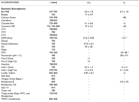
Now, even if the quantum-mechanical selection rules for MPE differ from those for one-photon excitation, several common fluorescent molecules can be used. Unfortu-nately, the knowledge of 1PE cross-section for a specific fluorescent molecule does not allow any quantitative pre-diction of the two-photon trend. The only "rule of thumb" that one can use states that a 2PE cross-section peak can be expected at a 2 folds wavelength with respect to the 1PE case. Table 1 summarises the excitation proper-ties of some popular fluorescent molecules under 2PE regime.
3. Optical implications of 2PE
3.1 Optical sectioning and confocal imaging
The possibility of the three-dimensional reconstruction of the volume distribution of intensive parameters, as fluo-rescence emission, from biological systems is one of the most powerful properties of the optical microscope. To
collect optical slices from a three-dimensional object the so-called optical sectioning [3,11] technique is used as depicted in figure 2. It is essentially based on a fine z step-ping either of the objective or of the sample stage, coupled with the usual x-y image capturing. The synchronous x-y-z scanning allows the collection of a set of two-dimen-sional images, which are somehow affected by signal cross talk from other planes from the sample. In fact, the observed image Oj, obtainable when positioning the geo-metrical focus of the lens at a certain plane" j "within the specimen, is produced by the true fluorescence distribu-tion Ij at plane "j", distorted by the microscope in some way that can be described by a function S, plus differently distorted contributions from adjacent "k" planes posi-tioned above and below the actual plane, and noise N. Using a convenient and appropriate formalism one has:
Oj = IjSj + ∑k≠j IkSk + N. (2)
Equation (2) reflects the fact that when a set of two-dimensional images is acquired at various focus position and under certain conditions, in principle, one can recover the 3D shape of the object, described by the inten-sive parameter I, by solving the above set of equations and finding the best estimate for I, slice by slice. By this proce-dure, unwanted light can be computationally removed combining the image data from a stack of "k" images. Such an operation can be optically performed using some physical stratagems that are behind confocal and MPE/ 2PE scanning microscopy.
3.2 The 2PE optical case
In terms of optical implications the two-photon effect has the important consequence of limiting the excitation region to within a sub-femtoliter volume. This means that the emission region is intrinsically confocal. The resulting 3D confinement in terms of image formation process can be described by means of consolidated optical considera-tions [30]. Using a certain excitation light at a wavelength
λ, the intensity distribution within the focal region of an objective having numerical aperture NA = n sin (α) is given, in the paraxial regime, by
where J0 is the 0th order Bessel function, ρ is a radial coor-dinate in the pupil plane, n is the refractive index of the medium between the lens and the specimen, (α) is the semi-angle of aperture of the lens [31],
and
are dimensionless axial and radial coordinates, respec-tively, normalized to the wavelength. Now, the intensity of fluorescence distribution within the focal region has a I(u, v) behaviour for the 1PE case [31]. In case of 2PE one has to consider a double wavelength and a square behav-iour, i.e. I2(u/2, v/2). As compared with the 1PE case, the 2PE emission intensity distribution is axially confined.
In fact, considering the integral over ν, keeping u constant, its behaviour is constant along z for one-photon and has a half-bell shape for 2PE. This behaviour is responsible of the 3D discrimination property of 2PE, i.e. of the optical sectioning properties of the 2PE microscope.
I u v( , )= 2
∫
J0( )
v e− u d( )
30
1 12
2
2
ρ ρ ρ ρ
u= 8
(
2)
z2
π α
λ
sin /
v= 2π
( )
α r λ sin Table 1: 2PE excitation parameters.FLUOROPHORES λ (nm) ηδ2 δ2
Extrinsic fluorophores
Bis-MSB 691/700 6.0 ± 1.8 6.3 ± 1.8
Bodipy 920 17 ± 4.9
-Calcium Green 740–990 - ~80
Calcofluor 780/820 -
-Cascade blue 750–800 2.1 ± 0.6 ~3
Coumarin 307 776, 700–800 19 ± 5.5 ~20
CY2 780/800 -
-CY3 780 -
-CY5 780/820 -
-DAPI (free) 700/720 0.16 ± 0.05 ~3.5 *
Dansyl 700 1
-Dansyl Hydrazine 700 0.72 ± 0.2
-Dil 700 95 ± 28
-Filipin 720 -
-FITC 740–820 - ~25–38 *
Fluorescein (pH ~11) 780 - 38 ± 9.7
Fura-2 (free) 700 11
-Fura-2 (high Ca) 700 12
-Hoechst 780/820 -
-Indo-1 (free) 700 4.5 ± 1.3 12 ± 4
Indo-1 (high Ca) 590/700 1.2 ± 0.4 2.1 ± 0.6
Lucifer Yellow 840–860 0.95 ± 0.3 ~2
Nile Red 810 -
-Oregon Green Bapta 1 800 -
-Rhodamine B 840 - 210 ± 55
Rhodamine 123 780–860 -
-Syto 13 810 -
-Texas red 780 -
-Triple probe (Dapi, FITC, and Rhodamine)
720/740 -
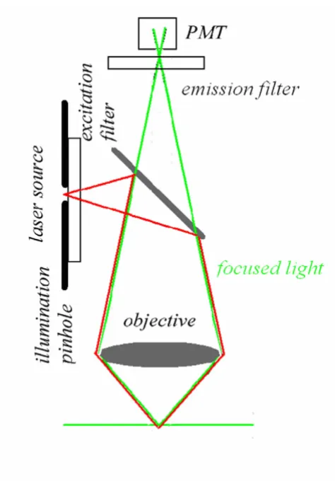
-Now, the most interesting aspect is that the excitation power falls off as the square of the distance from the lens focal point, within the approximation of a conical illumi-nation geometry [31]. In practice this means that the square relationship between the excitation power and the fluorescence intensity brings about the fact that 2PE falls off as the fourth power of distance from the focal point of the objective. This fact implies that those molecules away from the focal region of the objective lens do not contrib-ute to the image formation process and are not affected by photobleaching or phototoxicity. Since these molecules are not involved in the excitation process, a confocal-like effect is obtained without the necessity of a confocal pin-hole. It is immediately evident that in this case the optical sectioning effect is obtained in a physically different way with respect to the confocal case. Accordingly the optical set-up is simplified, under some aspects, see figure 4.
Figure 5 and figure 6 show the differences in terms of exci-tation-emission process between confocal and multipho-ton schemes, respectively. The consequences of the spatial confinement of the MPE result in a consequent confine-ment of the emitted fluorescence: in the confocal case all the molecules within the double cone of excitation are involved in the light-matter interaction while in the MPE case such interaction is restricted to a small volume cen-tred at the geometrical focus of the objective. The imme-diate consequence is that a 2PE microscope is an intrinsically three-dimensional image formation system.
This fact has also very important consequences on the photobleaching processes. So far, in the 2PE case no fluo-rescence has to be removed from the detection pathway since fluorescence can exclusively come from a small focal volume that has a capacity of the order of fraction of fem-toliter. In fact, in 2PE over 80% of the total intensity of flu-orescence comes from a 700–1000 nm thick region about the focal point for objectives with numerical apertures in the range from 1.2 to 1.4 [30]. The fact that the back-ground signal coming from adjacent planes tends to zero produces a sort of compensation for the reduced spatial resolution due to the utilization of a wavelength that is twice with respect to the 1PE case, as shown in figure 7. On the other hand, the utilisation of infrared wavelengths Confocal optical pathways
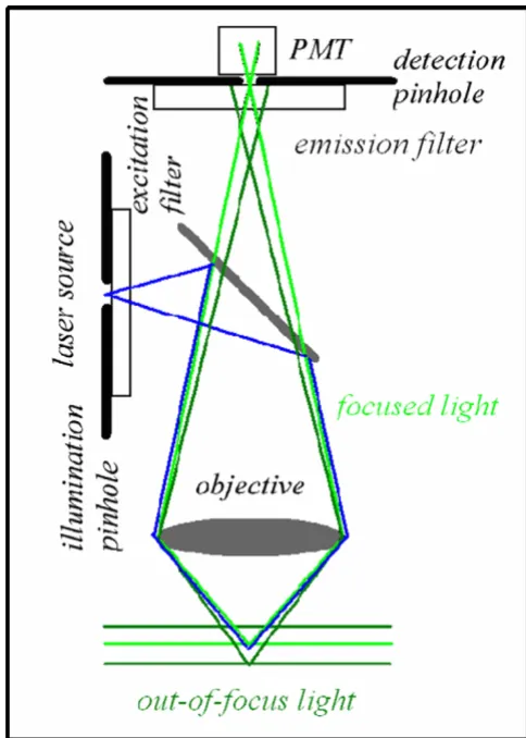
Figure 3
Confocal optical pathways. An illumination and a detec-tion pinhole are placed in the optical pathway. The detecdetec-tion pinhole – the mask – is placed in front of the detector at a plane that is conjugate to the in-focus or "j" plane, such that the illumination spot and the pinhole aperture are simultane-ously focused at the same specimen volume. This coinci-dence of the illumination and detected volume is responsible for confocality. The illumination pinhole allows to perform pointlike scanning.
Optical sectioning scheme Figure 2
instead of UV-visible ones allows achieving a deeper pen-etration than in conventional case [32]. This is due to the fact that the scattering effect is proportional to the inverse fourth power of the wavelength. Thus the longer wave-lengths used in 2PE, or in general in MPE, will be scattered less than the ultraviolet-visible wavelengths used for con-ventional excitation allowing to reach fluorescence targets in depth within thick samples (approx. 1 mm). It has been shown that two-photon fluorescence images can be obtained throughout almost the entire grey matter of the mouse neocortex by using optically amplified femtosec-ond pulses. The achieved imaging depth approaches the theoretical limit set by excitation of out-of-focus fluores-cence [33]. The fluoresfluores-cence light emitted, on the way back, can be more efficiently collected using a large area detector since it can uniquely come from the sub-femto-liter 2PE volume of event.
4. Practical aspects for the realization of a 2PE
microscope
The main elements to realize a 2PE/MPE architecture, including confocal modality, are the following: high peak-power laser delivering moderate average peak-power (fs or ps pulsed at relatively high repetition rate) emitting infrared or near infrared wavelengths (650–1100 nm), laser sources for confocal 1PE, a laser beam scanning system, high numerical aperture objectives (>1), a high-through-put microscope pathway, a spectral separation module for the emitted signal discrimination, and a high-sensitivity detection system [34]. Figure 8 shows a general scheme for a MPE microscope also illustrating two popular approaches that can be used for image formation, namely: de-scanned and non de-scanned mode. The former uses Confocal fluorescence emission distribution
Figure 5
Confocal fluorescence emission distribution. The emission process, in green, under blue 1PE excitation is broadened in the whole double cone excitation shape within the analyzed cell.
MPE simplified optical pathways Figure 4
the very same optical pathway and mechanism employed in confocal laser scanning microscopy. The latter mainly optimises the optical pathway by minimising the number of optical elements encountered on the way from the sam-ple to detectors, and increases the detector area. MPE non-descanned mode allows very good performances provid-ing superior signal-to-noise ratio inside strongly scatterprovid-ing
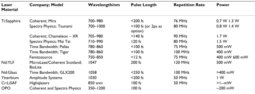
samples [32,33]. In the de-scanned approach pinholes are removed or set to their maximum aperture and the emis-sion signal is captured using the very same optical scan-ning pathway used for excitation. In the latter, the aim is to optimize the collection efficiency: pinholes are removed and the radiation emitted without passing through the laser beam scanning mirrors. Photomultiplier tubes are the most popular detectors in MPE microscopy. Avalanche photodiodes are also excellent in terms of sen-sitivity exhibiting quantum efficiency close to 70%–80% in the visible spectral range. Unfortunately they are high cost and the small active photosensitive area could intro-duce drawbacks in the detection scheme and require spe-cial de-scanning optics. CCD cameras are generally used in video rate multifocal imaging. Laser sources represent the core element for the 2PE/MPE microscope since for MPE high photon flux densities are required, > 1024 pho-tons cm-2s-1. Using radiation in the spectral range of 650– 1100 nm for MPE, excitation intensities in the MW-GW cm-2 have to be produced. Nowadays, laser sources suita-ble for 2PE can be described as "turnkey" systems, and Ti Sapphire lasers are the most utilized due to the high coin-cidence with the 2PE wavelengths needed for the majority of the commonly used fluorescent molecules. Other laser sources used for 2PE are Cr-LiSAF, pulse-compressed YLF in the femtosecond regime, and mode-locked Nd-YAG and picosecond Ti-Sapphire lasers in picosecond regime. Moreover the absorption coefficients of most bio-logical samples, cells and tissues are minimised within this spectral window. Table 2 reports data on the currently available laser sources for applications in MPE micros-copy and spectrosmicros-copy. The parameters that are more rel-evant in the selection of the laser source are average power, pulsewidth and repetition rate, and wavelength also accordingly to equation (1). The most popular fea-tures for an infrared pulsed laser are 700mW-1W average power, 80–100 MHz repetition rate, and 100–150 fs pulse width. So far, the use of short pulses and high repetition rates are mandatory to allow image acquisition in a rea-sonable time while using power levels that are biologi-cally tolerable. In order to minimise pulse width dispersion problems it should be considered to operate with pulses around 150 nm. This constitutes a very good compromise both for pulse stretching and sample viabil-ity. One should always remind that a shorter pulse broad-ens more than a longer one. Advances in laser sources are going on considering more compact sources, large tuna-bility range, high average power, and special designs for tailored needs at lower prices [35]. Objective lenses influ-ence the performances of any optical microscope, and for a MPE system special considerations have to be done tak-ing into the proper account both equations (1) and (3). New technological requisites have to be considered with respect to conventional excitation fluorescence micro-scope. An adequate transmission in the IR regime has to MPE fluorescence emission distribution
Figure 6
be coupled with good collection efficiency towards the ultraviolet region. Moreover, the number of components should be minimised without affecting resolution proper-ties in order to reduce pulse widths distortions. Although the collection efficiency of the time averaged photon flux is dependent on the numerical aperture of the collecting lens, the total fluorescence generation is independent of the numerical aperture of the focusing lens when imaging thick samples. Figure 9 shows an example of optical sec-tioning performed through a thick sample that exploits the autofluorescence signal. This is due to the fact that the increase of intensity, obtained by a sharper focusing (high NA), is counterbalanced by the shrinking of the excitation volume. Thus the total amount of fluorescence summed over the entire space remains constant. The very relevant practical consequence of this fact is that in 2PE
measure-ments on thick samples, assuming no aberrations, the generated fluorescence is insensitive to the size of the focal spot. As a positive consequence, a moderate variation of the laser beam size would not affect the measurements. This is a very efficient condition due to the fact that using an appropriate (non-descanned) acquisition scheme it is possible to collect all the generated fluorescence. In terms of pulse broadening, a 100 fs pulse can result between 1.14 and 1.23 times at the sample using a good lens. Once a MPE architecture has been realized, one should consider to keep under control the following parameters: power and pulse width at the sample focal plane checking for the square intensity/power behaviour, spectral separation of the emitted fluorescence including removal of the possi-ble excitation reflections that could be particularly subtle, z-axis precise control and laser-scanning system align-Pointlike emitter optical response
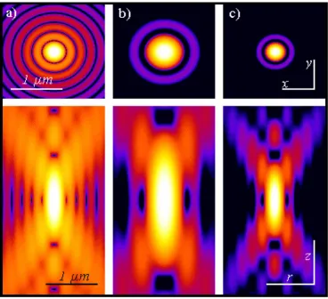
Figure 7
ment [34]. Figure 10 and figure 11 demonstrate the mul-tiple fluorescence imaging capability and 3D mulmul-tiple fluorescence imaging, respectively.
5. Application trends and conclusions
2PE and MPE microscopes are expected to increase their impact in areas such biotechnology, neurobiology, embryology, tissue engineering, materials science where imaging can be coupled to the possibility of using the microscopes in an active way, too. Clinically, 2PE may find applications in non-invasive optical bioscopy, while in cell biology the imaging abilities are coupled to the possibility of producing localized chemical reactions. Potential applications to integrative cardiac physiology or the possibility of tracking for long time biological events in living systems point out to the ability of making direct observations of phenomena and circumstances that before could only be inferred using other approaches. The myriad of new investigation possibilities offered by 2PE/ MPE microscopy enlarges so much the fields of applica-tion that it is not possible to outline in a complete way all the variations that can take place. For this reason, we give
in this last paragraph asummary of main properties of MPE and a limited overview of paramount trends.
The great impact of 2PE in optical microscopy is related to the fact that it couples a three-dimensional intrinsic abil-ity with almost five other interesting capabilities [21]. First, 2PE greatly reduces photo-interactions and allows imaging of living specimens on long time periods. Sec-ond, it allows operating in a high-sensitivity background-free acquisition scheme. Third, 2PE microscopy can pene-trate turbid and thick specimens down to a depth of a few hundreds micrometers. Fourth, due to the distinct charac-ter of the multiphoton absorption spectra of many of the fluorophores 2PE allows simultaneous excitation of dif-ferent fluorescent molecules reducing colocalization errors. Fifth, 2PE can prime photochemical reactions within subfemtoliter volumes inside solutions, cells and tissues.
Moreover, the advances in the field of fluorescent markers added value and potential to MPE microscopy. It is worth mentioning: the design of application suited chromo-MPE simplified optical schemes
Figure 8
phores [36]; the development and utilization of the so-called quantum dots [37]; the use of visible [38] and pho-toactivatable [39,40] fluorescent proteins from the green fluorescent protein (GFP) and its natural homologues to specifically engineered variants [6], the use of pho-toswitchable proteins to break the diffraction barrier in fluorescence microscopy at low light intensities [41].
Furthermore, this form of non-linear microscopy also supported the development and application of several investigation techniques, among them: three-photon excited fluorescence [42], second harmonic generation [43], third-harmonic generation [44], fluorescence corre-lation spectroscopy [45], image correcorre-lation spectroscopy [46], single molecule detection [47,48]; photodynamic therapies [49], and flow cytometry [50]. There is also an ongoing research activity to use 2PE and MPE in new fields where its special features can be advantageously applied to improve and to optimise existing schemes [51]. This covers new online detection systems like endoscopic imaging based on gradient refractive index fibres [52], the development of new substrates with higher fluorescence output [53] as well as the use of 2PE to systematically crosslink protein matrices and control the diffusion [54] and to perform localized uncaging [55]. When consider-ing the growconsider-ing interest for detection/sensconsider-ing technology in medical diagnostics and biotechnology, one should not ignore the recent explosion in the use of metallic
nanos-tructures to favourably modify the spectral properties of fluorophores and to alleviate some fluorophore photo-physical constraints. Within the framework the fusion of MPE with metal-enhanced fluorescence has a powerful potential in biotechnology: from immunoassay to enhanced ratiometric sensing and DNA detection [56]. A further mention is due to biomolecular tracking in real time and in vivo. Here 2PE and MPE can be considered as the dominant technologies. One mention is for in vivo brain imaging realized by means of ea newly designed compact and portable 2PE micro endoscope recently used to visualize hippocampal blood vessels in the brains of live mice [57]. As well a first partial view into the dynam-ics of developmentally programmed, long-range cell migration in the mammalian thymus was obtained using in a 4D (x-y-z-t) manner 2PE. So, the movement of thy-mocytes was followed in real time through the cortex within intact thymic lobes [58]. All these facts point out to the consideration that MPE makes possible to perform a 7D exploration of living cells due to its inherent ability in (x-y-z-t), FLIM (Fluorescence Lifetime Imaging Micros-copy), FRAP (Fluorescence Recovery After Photobleach-ing), FRET (Forster-fluorescence Resonance Energy Transfer) and SHG (Second Harmonic Generation) [24]. Additionally, regardless of the fact that all far field light microscopes are limited in the achievable diffraction-lim-ited resolution, MPE is pushing modern light microscopy towards fluorescence optical nanoscopy [59,60].
Table 2: MPE laser table.
Laser Material
Company; Model Wavelength/nm Pulse Length Repetition Rate Power
Ti:Sapphire Coherent; Mira 700–980 <200 fs 76 MHz 0.7 W 1.3 W Spectra Physics; Tsunami 700–1000 <100 fs (or 2ps as
option)
80 MHz 0.8 W 1.4 W
Coherent; Chameleon – XR 705–980 <140 fs 90 MHz 1.7 W Spectra Physics; Mai Tai 710–990 120 fs 80 MHz 1.5 W Time Bandwidth; Pallas 780–860 <100 fs 75 MHz 500 mW Time Bandwidth; Tiger 780–860 <100 fs 100 MHz 400 mW Femtosource 750–850 <12 fs 75 MHz 400 mW 600 mW Nd:YLF MicroLase/Coherent Scotland;
BioLite
1047 200 fs 120 MHz 500 mW
Nd:Glass Time Bandwidth; GLX200 1058 <250 fs 100 MHz >400 mW Ytterbium Amplitude Systems 1030 <200 fs 50 MHz 1 W Cr:LiSAF Highqlasers 850 snm 100 fs 50 MHz >1--mW OPO Coherent and Spectra Physics 350–1200 100 fs ~200 mW
Optical sectioning using 2PE autofluorescence Figure 9
3D and 2D fluorescence projections Figure 11
3D and 2D fluorescence projections. Pictorial represen-tation of the 3D and 2D projections of multiple fluorescence from a marine sponge, Chondrilla nucula. The specimen has been loaded with Alexa 488 fluorescent molecules specific aminobutirric acid (GABA) emitting in green, DAPI for nuclear DNA for the blu component. Red signals are due to the autofluorescence from symbiontic bacteria contamina-tion. Imaging has been perfomed using a Chameleon XR ultrafast Ti-Sapphire laser (Coherent Inc., USA) coupled at LAMBS-MicroScoBio with a Spectral Confocal Laser Scanning Microscope, Leica SP2-AOBS. (Sample availability and prepa-ration, courtesy of Renata Manconi, University of Sassari, Roberto Pronzato and Lorenzo Gallus, University of Genoa).
Multiple fluorescence 2PE imaging Figure 10
Multiple fluorescence 2PE imaging. 2PE multiple fluo-rescence image from a 16 μm cryostat section of mouse intestine stained with a combination of fluorescent stains (F-24631, Molecular Probes). Alexa Fluor 350 wheat germ agglutinin, a blue-fluorescent lectin, was used to stain the mucus of goblet cells. The filamentous actin prevalent in the brush border was stained with red-fluorescent Alexa Flu or 568 phalloidin. Finally, the nuclei were stained with SYTOX ®
Acknowledgements
The authors dedicate this work to Osamu Nakamura, a pioneer in 2PE, who passed away on 23 Jan 2005 at Handai Hospital, Japan. AD dedicates this work to Mario Arace and is still using his oscilloscope, purchased in 1978, in the lab. The first Italian 2PE architecture realized at LAMBS of the University of Genoa has been supported by INFM grants. LAMBS-Micro-ScoBio is currently granted by IFOM (Istituto FIRC di Oncologia Moleco-lare, FIRC Institute of Molecular Oncology, Milan, Italy) and Fondazione San Paolo (Torino, Italy). LAMBS joins the NANOMED Italian Research Pro-gram. MicroScoBio is a Research Center of the University of Genoa on Correlative Microscopy and Spectroscopy with applications in Biomedicine and Oncology, Genoa, Italy. IFOM is the FIRC Foundation on Molecular Oncology, Milan, Italy. LAMBS is a multicenter Laboratory for Advanced Microscopy, Bioimaging and Spectroscopy http://www.lambs.it.
References
1. Diaspro A: New World Microscopy. IEEE Engineering In Medicine And Biology Magazine 1996, 15:29-100.
2. Beltrame F, Bianco B, Castellaro G, Diaspro A: Fluorescence, Absorption, Phase-contrast, Holographic and Acoustical Cytometries of Living Cells. In Interactions between Electromag-netic Fields and CellsVolume 97. Edited by: Chiabrera A. NATO ASI Series, Plenum Press Publishing, New York and London; 1985:483-498.
3. Arndt-Jovin DJ, Nicoud RM, Kaufmann J, Jovin TM: Fluorescence digital-imaging microscopy in cell biology. Science 1985, 230:13330-13335.
4. Fay FS, Carrington W, Fogarty KE: Three-dimensional molecular distribution in single cells analyzed using the digital imaging microscope. J Microsc 1989, 153:133-149.
5. Periasamy A: Methods in Cellular Imaging. Oxford University Press, New York; 2000.
6. Zhang J, Campbell RE, Ting A, Tsien RY: Creating new fluorescent probes for cell biology. Nature Review Molecular Biology 2002, 3:906-918.
7. Amos B: Lessons from the history of light microscopy. Nat Cell Biol 2000, 2(8):E151-152.
8. Bastiaens PI, Hell SW: Recent Advances in Light Microscopy.
Journal of Structural Biology 2004, 147:1-89.
9. Pawley JB: Handbook of Biological Confocal Microscopy. 3rd edition. Plenum Press- Springer, New York; 2006.
10. Jena BP, Horber JHK: Atomic Force Microscopy in Cell Biology. In Methods in Cell BiologyVolume 68. Academic Press; 2002. 11. Amos WB, White JG: How the Confocal Laser Scanning
Micro-scope entered Biological Research. Biology of the Cell 2003, 95:335-342.
12. Göppert-Mayer M: Über Elementarakte mit zwei Quan-tensprüngen. Ann Phys 1931, 9:273-295.
13. Masters BR, So PT: Antecedents of two-photon excitation laser scanning microscopy. Microsc Res Tech 2004, 63:3-11.
14. Esposito A, Federici F, Usai C, Cannone F, Chirico G, Collini M, Dias-pro A: Notes on theory and experimental conditions behind two-photon excitation microscopy. Microsc Res Tech 2004, 63:12-17.
15. Berns MW: A possible two-photon effect in vitro using a focused laser beam. Biophys J 1976, 16:973-977.
16. Konig K: Multiphoton microscopy in life sciences. J Microsc
2000, 200:83-104.
17. Tirlapur UK, Konig K: Cell biology: Targeted transfection by femtosecond laser. Nature 2002, 418:290-291.
18. Sheppard CJ, Kompfner R: Resonant scanning optical micro-scope. Appl Opt 1978, 17:2879-2885.
19. Girkin JM: Optical physics enables advances in multiphoton imaging. Journal of Physics D: Applied Physics 2003, 36:R250-R258. 20. Denk W, Strickler JH, Webb WWW: Two-photon laser scanning
fluorescence microscopy. Science 1990, 248:73-76.
21. Diaspro A: Confocal and two-photon microscopy : founda-tions, applicafounda-tions, and advances. Wiley-Liss, New York; 2002. 22. Pennisi E: Biochemistry: Photons Add Up to Better
Micros-copy. Science 1999, 275:480-481.
23. Diaspro A: Rapid dissemination of two-photon excitation microscopy prompts new applications. Microsc Res Tech 2004, 63:1-2.
24. Zoumi A, Yeh A, Tromberg BJ: Imaging cells and extracellular matrix in vivo by using second-harmonic generation and two-photon excited fluorescence. Proc Natl Acad Sciences (USA)
2002, 99:11014-11019.
25. Zipfel WR, Williams RM, Webb WW: Nonlinear magic: multi-photon microscopy in the biosciences. Nature Biotechnology
2003, 21:1369-1377.
26. Helmchen F, Denk W: Deep tissue two-photon microscopy. Nat Methods 2005, 2:932-940.
27. Rubart M: Two-Photon Microscopy of Cells and Tissue. Circ Res 2004, 95:1154-1166.
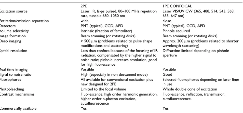
28. Cruz HG, Lüscher C: Applications of two-photon microscopy in the neurosciences. Frontiers in Bioscience 2005, 10:2263-2278. Table 3: 2PE vs. 1PE optical microscopy.
2PE 1PE CONFOCAL
Excitation source Laser, IR, fs-ps pulsed, 80–100 MHz repetition rate, tunable 680–1050 nm
Laser VIS/UV CW (365, 488, 514, 543, 568, 633, 647 nm)
Excitation/emission separation wide close
Detectors PMT (typical), CCD, APD PMT (typical), CCD, APD Volume selectivity Intrinsic (fraction of femtoliter) Pinhole required
Image formation Beam scanning (or rotating disks) Beam scanning (or rotating disks) Deep imaging > 500 μm (problems related to pulse shape
modifications and scattering)
Approx. 200 μm (problems related to shorter wavelength scattering)
Spatial resolution Less than confocal because of the focusing of IR radiation, compensated by the higher signal to noise ratio; pinhole increases resolution, good for high fluorescence
Diffraction limited depending on pinhole aperture
Real time imaging Possible Possible Signal to noise ratio High (especially in non descanned mode) Good Fluorophores All available for conventional excitation plus
new designed for 2PE
Selected fluorophores depending on laser lines in use
Photobleaching Limited to the focal volume Whole double cone of excitation Contrast mechanisms Fluorescence, high order harmonic generation,
higher order n-photon excitation, autofluorescence
Fluorescence, reflection, transmission, autofluorescence.
Commercially available Yes Yes
Publish with BioMed Central and every scientist can read your work free of charge "BioMed Central will be the most significant development for disseminating the results of biomedical researc h in our lifetime."
Sir Paul Nurse, Cancer Research UK
Your research papers will be:
available free of charge to the entire biomedical community
peer reviewed and published immediately upon acceptance
cited in PubMed and archived on PubMed Central
yours — you keep the copyright
Submit your manuscript here:
http://www.biomedcentral.com/info/publishing_adv.asp
BioMedcentral 29. Callis PR: Two-photon-induced fluorescence. Ann Rev Phys Chem
1997, 48:271-297.
30. Nakamura O: Three-dimensional imaging characteristics of laser scan fluorescence microscopy: Two-photon excitation vs. single-photon excitation. Optik 1993, 93:39-42.
31. Bianco B, Diaspro A: Analysis of the three dimensional cell imaging obtained with optical microscopy techniques based on defocusing. Cell Biophys 1989, 15:189-200.
32. Centonze VE, White JG: Multiphoton excitation provides opti-cal sections from deeper within scattering specimens than confocal imaging. Biophys J 1998, 75:2015-2024.
33. Theer P, Hasan MT, Denk W: Two-photon imaging to a depth of 1000 μm in living brains by use of a Ti:Al2O3 regenerative amplifier. Optics Lett 2003, 28:1022-1024.
34. Diaspro A: Building a two-photon microscope using a laser scanning confocal architecture. In Methods in Cellular Imaging
Edited by: Periasamy A. Oxford University Press, New York; 2001:162-179.
35. Girkin J, McConnell G: Advances in Laser Sources for Confocal and Multiphoton Microscopy. Microsc Res Tech 2005, 67:8-14. 36. Abbotto A, et al.: Dimethyl-pepep: a DNA probe in two-photon
excitation cellular imaging. Biophys Chem 2005, 114: :35-41. 37. Jaiswal JK, Simon S: Potentials and pitfalls of fluorescent
quan-tum dots for biological imaging. Trends Cell Biol 2004, 14:497-504.
38. Matz MV, Lukyanov KA, Lukyanov SA: Family of the green fluo-rescent protein: journey to the end of the rainbow. Bioessays
2002, 24:953-959.
39. Schneider M, Barozzi S, Testa I, Faretta M, Diaspro A: Two-photon activation and excitation properties of PA-GFP in the 720– 920-nm region. Biophys J 2005, 89:1346-1352.
40. Post JN, Lidke KA, Rieger B, Arndt-Jovin DJ: One- and two-photon photoactivation of a paGFP-fusion protein in live Drosophila embryos. FEBS Lett 2005, 579:325-330.
41. Hofmann M, Eggeling C, Jakobs S, Hell SW: Breaking the diffrac-tion barrier in fluorescence microscopy at low light intensi-ties by using reversibly photoswitchable proteins. Proc Natl Acad Sci U S A 2005, 102:17565-17569.
42. Hell SW, Bahlmann K, Schrader M, Soini A, Malak H, Gryczynski I, Lakowicz JR: Three-photon excitation in fluorescence micros-copy. J Biomedical Optics 1996, 1:71-74.
43. Campagnola PJ, Loew LM: Second-harmonic imaging micros-copy for visualizing biomolecular arrays in cells, tissues and organisms. Nat Biotechnol 2003, 21:1356-1360.
44. Mueller M, Squier J, Wilson KR, Brakenhoff GJ: 3D microscopy of trasparent objects using third-harmonic generation. J Microsc
1998, 191:266-274.
45. Schwille P: Fluorescence correlation spectroscopy and its potential for intracellular applications. Cell Biochem Biophys
2001, 34:383-408.
46. Wiseman PW, et al.: Spatial mapping of integrin interactions and dynamics during cell migration by image correlation microscopy. J Cell Sci 2004, 117:5521-5534.
47. Sonnleitner M, Schutz GJ, Schmidt T: Imaging individual mole-cules by two-photon excitation. Chem Phys Lett 1999, 300:221-226.
48. Chirico G, Cannone F, Beretta S, Diaspro A: Single molecule stud-ies by means of the two-photon fluorescence distribution.
Microsc Res Techniq 2001, 55:359-364.
49. Bhawalkar JD, Kumar ND, Zhao CF, Prasad PN: Two-photon pho-todynamic therapy. J Clin Laser Med Surg 1997, 15:201-204. 50. Diaspro A: Two-photon fluorescence excitation. A new
potential perspective in flow cytometry. Minerva Biotecnologica
1998, 11:87-92.
51. McConnell G, Riis E: Two-photon laser scanning fluorescence microscopy using photonic crystal fibre. J Biomed Opt 2004, 9:922-927.
52. Jung JC, et al.: In vivo mammalian brain imaging using one- and two-photon fluorescence microendoscopy. J Neurophysiol 2004, 92:3121-3133.
53. Kappel C, et al.: Giant enhancement of two-photon fluores-cence induced by resonant double grating waveguide struc-tures. Applied Physics B-Lasers and Optics 2004, 79:531-534. 54. Basu S, Campagnola PJ: Properties of crosslinked protein
matri-ces for tissue engineering applications synthesized by multi-photon excitation. J Biomed Mater Res A 2004, 71:359-368.
55. Diaspro A, et al.: Two-Photon Photolysis of 2-Nitrobenzalde-hyde Monitored by Fluorescent-Labeled Nanocapsules. J Phys Chem B 2003, 107:11008-11012.
56. Aslan K, et al.: Metal-enhanced fluorescence: an emerging tool in biotechnology. Curr Opin Biotechnol 2005, 16:55-62.
57. Flusberg BA, et al.: In vivo brain imaging using a portable 3.9 gram two-photon fluorescence microendoscope. Opt Lett
2005, 30:2272-2274.
58. Witt CM, et al.: Directed migration of positively selected thy-mocytes visualized in real time. PLoS Biol 2005, 3:e160. 59. Hell SW: Toward fluorescence nanoscopy. Nat Biotechnol 2003,
21:1347-1355.