VIEWS AND REVIEWS OPEN ACCESS
HSAN-VI
A spectrum disorder based on dystonin isoform expression
Anisha Lynch-Godrei, PhD, and Rashmi Kothary, PhD
Neurol Genet2020;6:e389. doi:10.1212/NXG.0000000000000389
Correspondence Dr. Kothary [email protected]
Abstract
Hereditary sensory and autonomic neuropathy (HSAN-VI) is a recessive genetic disorder that arises because of mutations in the human dystonin gene (DST, previously known asbullous pemphigoid antigen 1). Although initial characterization of HSAN-VI reported it as a sensory neuropathy that was lethal in infancy, we now know of a number of heterozygous mutations in DSTthat result in milder forms of the disease. Akin to what we observe in the mouse model dystonia musculorum(Dstdt), we believe that the heterogeneity of HSAN-VI can be attributed to a number of dystonin isoforms that the mutation affects. Lack of neuronal isoform dystonin-a2 is likely the universal determinant of HSAN-VI because all reported human cases are null for this isoform, as are all Dstdt mouse alleles. Compensatory mechanisms by intact dystonin-a isoforms dystonin-also likely pldystonin-ay dystonin-a role in reguldystonin-ating disedystonin-ase severity, dystonin-although we hdystonin-ave yet to determine what specific effect dystonin-a1 and dystonin-a3 have on the pathogenesis of HSAN-VI.
From the Regenerative Medicine Program (A.L.-G., R.K.), Ottawa Hospital Research Institute; Department of Cellular and Molecular Medicine (A.L.-G., R.K.) and Department of Biochemistry, Microbiology, and Immunology, Faculty of Medicine, University of Ottawa; Department of Medicine (R.K.), University of Ottawa; and Centre for Neuromuscular Disease (R.K.), University of Ottawa, Canada.
Go to Neurology.org/NG for full disclosures. Funding information is provided at the end of the article.
The Article Processing Charge was funded by the authors.
The human dystonin gene (DST, also known as bullous pemphigoid antigen 1 [BPAG1]) consists of 496 kb located on the short arm of chromosome 6. TheDST gene is fairly complex in that various tissue-specific promoters yield epi-thelial- (dystonin-e/BPAG1e), neuronal- (dystonin-a/ BPAG1a), and muscle-specific (dystonin-b/BPAG1b) iso-forms, whereas alternative splicing of the neuronal and muscle isoforms further produce 3 unique proteins termed dystonin-a1/b1, dystonin-a2/b2, and dystonin-a3/b3 (figure).1Since the dystonin proteins belong to the spectraplakin family of proteins,2,3 they function as cytoskeletal linkers responsible for maintaining structural integrity and mediating processes such as intracellular trafficking.4–6Considering the large size of the humanDSTgene, it should be susceptible to mutations over time, which would eventually manifest to some observ-able phenotype. However, the complete lack of any reported human cases had suggested that anyDSTmutation was likely embryonic lethal.
In 2004, thefirst instance of aDSTgene disruption associated with a disease phenotype was described in a female child with a 6; 15 chromosomal translocation.7The breakage point oc-curred toward the 39end ofDSTand was predicted to affect only dystonin-a and dystonin-b isoforms. The patient pre-sented with esophageal atresia, and through development she would exhibit severe motor and intellectual disability, non-progressive encephalopathy, and delayed visual maturation. Her second chromosome 6, however, was unaffected by the translocation and would still express full-length dystonin-a dystonin-and dystonin-b trdystonin-anscripts. In dystonin-an dystonin-attempt to expldystonin-ain her clinical presentation, it was suggested that either hap-loinsufficiency ofDSTwas enough to cause pathology or that truncated dystonin-a/b interrupted function of the full-length protein leading to manifestation of symptoms.8
A few years later, a case of epidermolysis bullosa simplex was discovered to be caused by homozygous mutations affecting BPAG1/DST.9 The mutation resulted in a disruption of the coiled-coil rod domain of the protein, which is specific to only the skin dystonin-e isoform. This individual was reported to have spontaneous skin blistering and erosion, as well as mild neuro-logic features (weakness, numbness, and headaches). Subsequent reports of individuals with the sameDSTmutations would show that only skin defects were common between these patients.10 The neurologic features described in the initial case were instead proposed to be caused by heterozygousNOTCH3mutations.
In 2012, homozygous mutations in the humanDSTgene were discovered to be associated with a severe phenotype that shared many features with a subset of genetic disorders termed hereditary sensory and autonomic neuropathies
(HSANs).11,12Three infants from 2 consanguineous families from Ashkenazi Jewish background presented with dysauto-nomic symptoms, distal contractures, motionless open-mouthed facies, and severe psychomotor retardation (tables 1 and 2).13Ultimately, all 3 patients would die around the age of 2 years from cardiopulmonary events, likely related to poor autonomic control. Also of note was that a second pregnancy for 1 of the families was aborted at 21 weeks because of signs of the same disease as its sibling. The underlying mutation in these patients was determined to be a frameshift occurring at Glu4955, which leads to the loss of 502 amino acids at the C-termini microtubule-binding domain. Because this domain is common to all dystonin-a and dystonin-b splice variants, it effectively ablates expression of the predominant neural and muscle isoforms. Considering that the disease presentation shared many clinical features such as HSAN-III (also named familial dysautonomia), although more severe, this newly identified disorder was termed HSAN-VI.
In 2017, we then learned that homozygousDSTmutations do not exclusively produce a disease phenotype that is lethal in infancy since 2 separate studies described DST mutations in adolescent and adult patients. Thefirst of these studies that was published identified 3 siblings from a nonconsanguineous family from southern Italy as having 2 heterozygous com-pound mutations in the DST gene, which affected the ex-pression of dystonin-a2 and -b2 isoforms.14 These patients exhibited impaired pain sensitivity and distal ulcerations from infancy, weakness of intrinsic foot muscles, and a number of autonomic disturbances including heat intolerance, problems with sweating, pupillary abnormalities, chronic diarrhea, and sexual dysfunction (tables 1 and 2). The other study identified a female patient with both skin and neuronal phenotype, which resulted from compound heterozygous mutations in DSTaffecting exon 7 (specific to dystonin-a1,and -a2) and exon 29 (common to e, a, and dystonin-b isoform).15 She presented with chronic diarrhea, iris het-erochromia, bilateral cataracts, syringomyelia from D3-D8, bilateral sensorineural hearing loss, pain insensitivity, skin blistering, and behavioral problems such as avoidant/ restrictive food intake disorder, obsessive compulsive disor-der, and anxiety (tables 1 and 2).
Most recently, a 2018 study identified 3 elderly siblings from an Italian family with biallelic DST mutations ablating dystonin-a2/b2 expression also causing dystonin-a1/b1 and dystonin-a3/b3 haploinsufficiency.16 These patients recall experiencing dysautonomic symptoms in childhood, and be-tween the ages of 20–40 years, they would present with painless fractures, osteomyelitis, joint deformities, and di-abetes mellitus type II (tables 1 and 2).
Glossary
It has only been over the past few years that we have observed thefirst human cases ofDSTmutation, leading to its
classi-fication as HSAN-VI, a lethal form of sensory neuropathy. Through the identification of adult patients with deleterious DST mutations, this definition would expand to categorize HSAN-VI as a spectrum disorder, with severity being de-termined by which isoforms are affected. With a growing awareness of HSAN-VI, we can expect to observe the number of reported cases to increase in the coming years.
Discovery of neuronal
dystonin isoforms
Before the identification of the human disease HSAN-VI,13 the dystonin gene and its resulting protein had long since been studied in a murine model known as the dystonia
musculorum(Dstdt) mouse.17–21In 1963, theDstdtmouse that arose by spontaneous mutation at The Jackson Laboratory wasfirst described.18,21 Severe ataxia and dystonic postures were the major phenotypic characteristics, which were also associated with significant sensory neuron degeneration. Al-though the underlying genetic mutation was not identified at the time, the disease was predicted to be caused by autosomal recessive mutation. In 1995, 2 separate lines of work led to the identification of theBPAG1/Dstgene as the causative agent for theDstdtdisease.20,22While studying the hemidesmosomal skin protein BPAG1, generation of a BPAG1 knockout mouse unexpectedly produced the ataxic and dystonic phenotype associated with theDstdtmouse.22In parallel, definitive evi-dence came from cloning experiments identifying the gene that was disrupted by the insertion of anhsp68-LacZ trans-gene, which resulted in mice bearing theDstdtphenotype.19,20 Ultimately, crossbreeding heterozygous mice from this line
FigureSchematic of the predominant DST isoforms
and the BPAG1 knockout line onto The Jackson Laboratory Dstdtline revealed that these mice were allelic, and thus, their mutations mapped to the same genetic locus. This indicated that the gene responsible for producing the BPAG1 skin protein was also responsible for producing the neuronal dystonin protein underlyingDstdtpathology.19,23
Subsequent studies would later propose splice variants of neuronal dystonin that were predicted to interact with in-termediatefilaments (termed BPAG1n), much like how the epithelial isoform dystonin-e interacts with keratinfilaments in skin. It was hypothesized that loss of these BPAG1n isoforms was responsible for the neurologic phenotype of the Dstdt mice.24However, in 2001, a study evaluating BPAG1 isoform expression in mice found that BPAG1n messenger RNA went completely undetected in neural tissue.1This led to the dis-covery of the larger, more prominently expressed neuronal and muscle isoforms: dystonin-a and dystonin-b, respectively.
In the years to come, these neuronal- and muscle-specific splice variants would also be identified (figure). Elucidating the roles of the neuronal-specific splice variants would be-come the focus, considering that sensory neurons are the major cell type affected in theDstdtmice. However, the major challenge of studying dystonin and its various isoforms lies in the fact that the protein is remarkably large (dystonin-a = 615 kDa, dystonin-b = 834 kDa) and shares high amino acid se-quence similarity between the isoforms.1,25,26This has made the development of Dst antibodies an incredibly arduous task, and as such, there are currently no reliable isoform-specific antibodies available. Much of what we have learned about the functions of the individual isoforms has come from in vitro
experiments using small interfering RNA knockdowns and fusion protein constructs, as well as in vivo examination of the Dstdtalleles that differ in the nature of their mutations and thus in the isoforms affected.
What the
Dst
dtalleles can tell us about
HSAN-VI heterogeneity
By comparing theDSTmutations identified in the individuals with HSAN-VI, the commonality among the cases is that dystonin-a2 is absent. From what we know through studies on Dstdtmice and through knockdown experiments on immor-talized cells, dystonin-a2 is the most crucial isoform for neu-ronal functioning since its loss is associated with the most profound and lasting defects to intracellular pathways.4,25,27,28 We have also observed a moderate rescuing effect when dystonin-a2 is partially restored to neurons inDstdt-Tg4mice, which lack both dystonin-a/b1 and -a/b2.25These mice have significantly longer lifespans and show improvements in many pathways that are normally defective in theDstdt-Tg4mice.29 The results provided by theDstdtmouse experiments strongly suggest that dystonin-a2 is the major determinant for disease. Supportive evidence for this comes from the patients de-scribed in the earlier 2017 HSAN-VI study.14 The affected siblings have a milder form of HSAN-VI that continues into adulthood and is associated with mutations affecting only the neuronal dystonin-a2 isoform (dystonin-b2 is also likely af-fected, although muscle defects are not primary disease fea-tures). It therefore seems highly likely that disrupted expression of dystonin-a2 is both necessary and sufficient to produce HSAN-VI. However, mutations affecting only
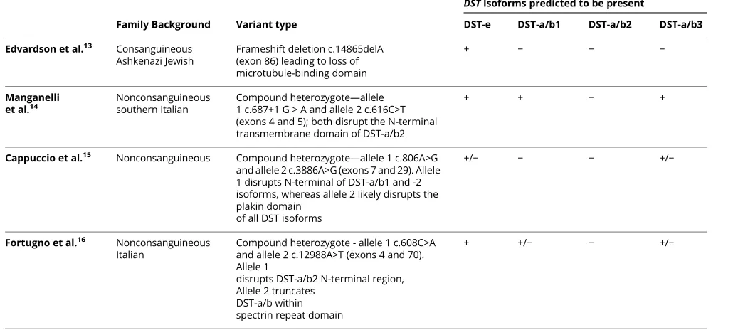
Table 1Genetic comparison of the various patients with HSAN-VI
Family Background Variant type
DSTIsoforms predicted to be present
DST-e DST-a/b1 DST-a/b2 DST-a/b3
Edvardson et al.13 Consanguineous Ashkenazi Jewish
Frameshift deletion c.14865delA (exon 86) leading to loss of microtubule-binding domain
+ − − −
Manganelli et al.14
Nonconsanguineous southern Italian
Compound heterozygote—allele 1 c.687+1 G > A and allele 2 c.616C>T (exons 4 and 5); both disrupt the N-terminal transmembrane domain of DST-a/b2
+ + − +
Cappuccio et al.15 Nonconsanguineous Compound heterozygote—allele 1 c.806A>G and allele 2 c.3886A>G (exons 7 and 29). Allele 1 disrupts N-terminal of DST-a/b1 and -2 isoforms, whereas allele 2 likely disrupts the plakin domain
of all DST isoforms
+/− − − +/−
Fortugno et al.16 Nonconsanguineous Italian
Compound heterozygote - allele 1 c.608C>A and allele 2 c.12988A>T (exons 4 and 70). Allele 1
disrupts DST-a/b2 N-terminal region, Allele 2 truncates
DST-a/b within spectrin repeat domain
+ +/− − +/−
Table 2 Symptom comparison of the various HSAN-VI patients Gender Skin defects Bone/joint defects Neurologic symptoms Dysautonomic
symptoms Other Cause of death
Age at onset
Edvardson et al.13
Patient 1 F Blotching Distal contractures Severe psychomotor retardation, absent deep tendon reflex Absent tearing, feeding and breathing difficulties Motionless, open mouth facies; early death (<2 y/o)
Cardiopulmonary arrest
Birth
Patient 2 M Severe apneic
episode
Patient 3 F Cardiopulmonary
arrest
Fetus 1 N/A N/A Bilateral club
feet and hand clenching
N/A N/A Aborted N/A N/A
Manganelli et al.14
Patient 4 M Distal
ulcers Joint deformities, distal amputations Pain insensitivity, impaired touch and vibration sensitivity, altered deep tendon reflex Hypohidrosis and heat intolerance, chronic diarrhea Weakness in intrinsic foot muscles N/A Infancy
Patient 5 M Joint
deformities, distal amputations Hypohidrosis and heat intolerance, chronic diarrhea, pupillary abnormalities, erectile dysfunction
Patient 6 M Distal
amputations Hypohidrosis and heat intolerance, chronic diarrhea, pupillary abnormalities Cappuccio et al.15
Patient 7 F Recurrent
blistering, peeling, ulcers, atrophic scars Osteoporosis Bilateral sensorineural hearing loss, pain insensitivity, headaches, behavioural disorders Chronic diarrhea, abdominal pain Iris heterochromia, cataract, syringomelia
N/A 4 mo
Fortugno et al.16
Patient 8 M Ulcers Painless
fractures, recurrent septic osteoarthritis, joint deformities, toe amputations Severe pain insensitivity, symmetric sensorineural hearing loss, altered deep tendon reflex Hyperhidrosis and heat intolerance, pupillary abnormalities, urinary incontinence
Type II diabetes, general muscle weakness
N/A 37 y/o
Patient 9 M Painless
dystonin-a1 or dystonin-a3 have not been described in humans or in theDstdtmouse models, and we therefore do not know to what extent these isoforms contribute to disease etiology. It may be that mutations in either of these isoforms do not result in any major clinical pathology, which could explain the absence in reported cases. In addition, isoform compensation may be a mechanism that could be involved in masking these mutations, as we recently observed this phe-nomenon in theDstdt-Tg4mice.30Because these mice retain dystonin-a3, we saw a significant upregulation of this isoform in neural tissues most affected by dystonin loss of function. This upregulation was associated with maintenance of mi-crotubule stability in sensory neurons (a function previously unknown to this isoform), which was reversed on loss of transcript overexpression.30 This pattern of upregulation is consistent with dystonin-a3 compensating for the loss of dystonin-a1 and -a2. Thus, it is reasonable to believe that each of the dystonin-a isoforms possesses the ability to modify its normal function and substitute for an absent or nonfunctional isoform. Nevertheless, future work involving isoform specific knockouts is still needed to conclusively determine the role of each isoform in HSAN-VI pathogenesis.
Seeing as DST mutations can result in a milder form of HSAN-VI expanding into adulthood, HSAN-VI should be recognized as a spectrum disorder whereby severity of the disease may be predicted based on the isoforms affected. Of interest, theDstdt−27Jmice, which are completely null for all neuronal isoforms of dystonin, present with the most severe phenotype (ataxic affecting all limbs and dystonic postures) and have the shortest lifespan.26,30ThisDstdt−27Jallele would most closely resemble thefirst group of very severe patients with HSAN-VI.13The adolescent girl described in the later 2017 HSAN-VI study may be the most severe one among the adult patients with HSAN-VI.15Her genotype would mostly resemble Dstdt-Tg4 or DstGt(E182H05)Wrst alleles because she lacks both dystonin-a1 and -a226,31but is haploinsufficient for dystonin-a3. Although her symptoms are not identical to the other adult patients with HSAN-VI, her younger age may indicate that these symptoms are yet to develop. She does, however, have a number of unique symptoms including iris heterochromia, cataracts, syringomyelia from D3-D8, osteo-porosis, headaches, and behavioral problems (anxiety, ob-sessive compulsive disorder, and avoidant/restrictive food
intake disorder), suggesting potential roles for dystonin-a1 and -a2 in these systems. Retention of the dystonin-a/b3 isoforms might also contribute to her longer lifespan com-pared with the dystonin-a/b null infants. She also experienced skin blistering, which was hypothesized to be because of dystonin-e haploinsufficiency. However, considering the presence of skin ulceration and a lack of dystonin-e isoform involvement in the other adult patients with HSAN-VI, it is likely that her skin blisters are due to peripheral neuropathy leading to skin damage going unnoticed. Furthermore, het-erozygous Dsttm1EFu mice (dystonin knockouts) do not display skin symptoms and have intact hemidesmosomes at the dermoepidermal junction.22Thus far, the 3 siblings de-scribed in the earlier 2017 study likely represent the mildest form of HSAN-VI because they only have dystonin-a2/b2 isoforms affected. With coming age, patients with HSAN-VI may develop new symptoms indicating novel roles of dys-tonin isoforms that would never have been characterized in theDstdtmice due to limitations such as short lifespan and objective measures. As more individuals withDSTmutations are identified and as we continue our investigation of af-fected tissues and mechanisms in theDstdtmice, we hope to gain a better understanding of how each isoform contributes to disease.
Moving forward
With the recent identification of adult patients with deleterious DSTmutations, it has become clear that HSAN-VI can present on a spectrum based on which neuronal dystonin isoforms are affected. Current evidence indicates that dystonin-a2 is the most important factor dictating development of HSAN-VI. Although to further advance our knowledge of what roles the other neuronal dystonin isoforms play in the development of HSAN-VI, if any, dystona1 and -a3 should be in-dependently assessed. As isoform compensation is a mecha-nism that potentially modulates disease presentation, expression levels of remaining isoforms should also be evaluated. Although many HSAN-VI symptoms have been accurately predicted by theDstdtmice, the short lifespan of these mice is a major limitation for addressing how dystonin loss of function affects adult patients with HSAN-VI. In addition, considering the diverse expression pattern of dystonin-a across tissues, this suggests there are many more
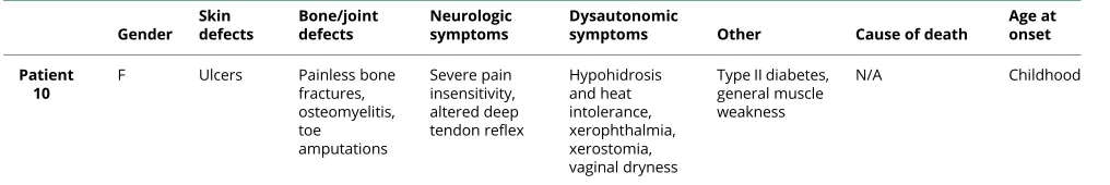
Table 2Symptom comparison of the various HSAN-VI patients(continued)
Gender Skin defects Bone/joint defects Neurologic symptoms Dysautonomic
symptoms Other Cause of death
Age at onset
Patient 10
F Ulcers Painless bone fractures, osteomyelitis, toe amputations Severe pain insensitivity, altered deep tendon reflex Hypohidrosis and heat intolerance, xerophthalmia, xerostomia, vaginal dryness
Type II diabetes, general muscle weakness
N/A Childhood
roles for the dystonin isoforms that have yet to be charac-terized. Further evaluation of these patients with HSAN-VI and identification of novelDSTmutations will be pivotal in our understanding of the biological roles of the dystonin isoforms and how they relate to pathogenesis.
Study funding
This work was funded by grants from the Canadian Institutes of Health Research to RK (# MOP-126085). ALG is sup-ported by an Ontario Graduate Scholarship.
Disclosure
Disclosures available: Neurology.org/NG.
Publication history
Received byNeurology: GeneticsJune 25, 2019. Accepted infinal form November 19, 2019.
References
1. Leung CL, Zheng M, Prater SM, Liem RK. The BPAG1 locus: alternative splicing produces multiple isoforms with distinct cytoskeletal linker domains, including pre-dominant isoforms in neurons and muscles. J Cell Biol 2001;154:691–697. 2. Suozzi KC, Wu X, Fuchs E. Spectraplakins: master orchestrators of cytoskeletal
dynamics. J Cell Biol 2012;197:465–475.
3. Kunzli K, Favre B, Chofflon M, Borradori L. One gene but different proteins and diseases: the complexity of dystonin and bullous pemphigoid antigen 1. Exp Dermatol 2016;25:10–16.
4. Ryan SD, Bhanot K, Ferrier A, et al. Microtubule stability, Golgi organization, and transportflux require dystonin-a2-MAP1B interaction. J Cell Biol 2012;196:727–742. 5. Liu JJ, Ding J, Kowal AS, et al. BPAG1n4 is essential for retrograde axonal transport in
sensory neurons. J Cell Biol 2003;163:223–229.
6. De Repentigny Y, Deschenes-Furry J, Jasmin BJ, Kothary R. Impaired fast axonal transport in neurons of the sciatic nerves from dystonia musculorum mice. J Neurochem 2003;86:564–571.
7. Giorda R, Cerritello A, Bonaglia MC, et al. Selective disruption of muscle and brain-specific BPAG1 isoforms in a girl with a 6;15 translocation, cognitive and motor delay, and tracheo-oesophageal atresia. J Med Genet 2004;41:e71.
8. Goryunov D, Adebola A, Jefferson JJ, Leung CL, Messer A, Liem RK. Molecular characterization of the genetic lesion in dystonia musculorum (dt-Alb) mice. Brain Res 2007;1140:179–187.
9. Groves RW, Liu L, Dopping-Hepenstal PJ, et al. A homozygous nonsense mutation within the dystonin gene coding for the coiled-coil domain of the epithelial isoform of BPAG1 underlies a new subtype of autosomal recessive epidermolysis bullosa sim-plex. J Invest Dermatol 2010;130:1551–1557.
10. Liu L, Dopping-Hepenstal PJ, Lovell PA, et al. Autosomal recessive epidermolysis bullosa simplex due to loss of BPAG1-e expression. J Invest Dermatol 2012;132: 742–744.
11. Axelrod FB. Hereditary sensory and autonomic neuropathies. Familial dysautonomia and other HSANs. Clin Auton Res 2002;12(suppl 1):I2–I14.
12. Axelrod FB, Gold-von Simson G. Hereditary sensory and autonomic neuropathies: types II, III, and IV. Orphanet J Rare Dis 2007;2:39.
13. Edvardson S, Cinnamon Y, Jalas C, et al. Hereditary sensory autonomic neuropathy caused by a mutation in dystonin. Ann Neurol 2012;71:569–572.
14. Manganelli F, Parisi S, Nolano M, et al. Novel mutations in dystonin provide clues to the pathomechanisms of HSAN-VI. Neurology 2017;88:2132–2140.
15. Cappuccio G, Pinelli M, Torella A, et al. Expanding the phenotype of DST-related disorder: a case report suggesting a genotype/phenotype correlation. Am J Med Genet A 2017;173:2743–2746.
16. Fortugno P, Angelucci F, Cestra G, et al. Recessive mutations in the neuronal isoforms of DST, encoding dystonin, lead to abnormal actin cytoskeleton organization and HSAN type VI. Hum Mutat 2019;40:106–114.
17. Duchen LW. Dystonia musculorum—an inherited disease of the nervous system in the mouse. Adv Neurol 1976;14:353–365.
18. Duchen LW, Strich SJ, Falconer DS. Clinical and pathological studies of an hereditary neuropathy in mice (dystonia musculorum). Brain 1964;87:367–378.
19. Kothary R, ClapoffS, Brown A, Campbell R, Peterson A, Rossant J. A transgene containing lacZ inserted into the dystonia locus is expressed in neural tube. Nature 1988;335:435–437.
20. Brown A, Bernier G, Mathieu M, Rossant J, Kothary R. The mouse dystonia mus-culorum gene is a neural isoform of bullous pemphigoid antigen 1. Nat Genet 1995; 10:301–306.
21. Duchen LW, Strich SJ, Falconer DS. Dystonia musculorum. A hereditary neuropathy of mice affecting mainly sensory pathways. J Physiol 1963;165:7–9.
22. Guo L, Degenstein L, Dowling J, et al. Gene targeting of BPAG1: abnormalities in mechanical strength and cell migration in stratified epithelia and neurologic de-generation. Cell 1995;81:233–243.
23. Brown A, Copeland NG, Gilbert DJ, Jenkins NA, Rossant J, Kothary R. The genomic structure of an insertional mutation in the dystonia musculorum locus. Genomics 1994;20:371–376.
24. Yang Y, Dowling J, Yu QC, Kouklis P, Cleveland DW, Fuchs E. An essential cyto-skeletal linker protein connecting actin microfilaments to intermediatefilaments. Cell 1996;86:655–665.
25. Ferrier A, Boyer JG, Kothary R. Cellular and molecular biology of neuronal dystonin. Int Rev Cel Mol Biol 2013;300:85–120.
26. Pool M, Boudreau Lariviere C, Bernier G, Young KG, Kothary R. Genetic alterations at the Bpag1 locus in dt mice and their impact on transcript expression. Mamm Genome 2005;16:909–917.
27. Ryan SD, Ferrier A, Sato T, et al. Neuronal dystonin isoform 2 is a mediator of endoplasmic reticulum structure and function. Mol Biol Cell 2012;23:553–566. 28. Young KG, Kothary R. Dystonin/Bpag1 is a necessary endoplasmic reticulum/
nuclear envelope protein in sensory neurons. Exp Cel Res 2008;314:2750–2761. 29. Ferrier A, Sato T, De Repentigny Y, et al. Transgenic expression of neuronal dystonin
isoform 2 partially rescues the disease phenotype of the dystonia musculorum mouse model of hereditary sensory autonomic neuropathy VI. Hum Mol Genet 2014;23: 2694–2710.
30. Lynch-Godrei A, De Repentigny Y, Gagnon S, Trung MT, Kothary R. Dystonin-A3 upregulation is responsible for maintenance of tubulin acetylation in a less severe dystonia musculorum mouse model for hereditary sensory and autonomic neuropathy type VI. Hum Mol Genet 2018;27:3598–3611.
31. Horie M, Watanabe K, Bepari AK, et al. Disruption of actin-binding domain-containing dystonin protein causes dystonia musculorum in mice. Eur J Neurosci 2014;40:3458–3471.
AppendixAuthors
Name Location Role Contribution
Anisha Lynch-Godrei, PhD
The Ottawa Hospital Research Institute; the University of Ottawa, ON, Canada
Author Drafted the manuscript for intellectual content Rashmi Kothary, PhD
The Ottawa Hospital Research Institute; the University of Ottawa, ON, Canada
DOI 10.1212/NXG.0000000000000389
2020;6;
Neurol Genet
Anisha Lynch-Godrei and Rashmi Kothary
HSAN-VI: A spectrum disorder based on dystonin isoform expression
This information is current as of January 2, 2020
reserved. Online ISSN: 2376-7839.
Published by Wolters Kluwer Health, Inc. on behalf of the American Academy of Neurology.. All rights an open-access, online-only, continuous publication journal. Copyright Copyright © 2020 The Author(s).
Services
Updated Information &
http://ng.neurology.org/content/6/1/e389.full.html including high resolution figures, can be found at:
References
http://ng.neurology.org/content/6/1/e389.full.html##ref-list-1 This article cites 31 articles, 6 of which you can access for free at:
Citations
http://ng.neurology.org/content/6/1/e389.full.html##otherarticles This article has been cited by 1 HighWire-hosted articles:
Subspecialty Collections
http://ng.neurology.org//cgi/collection/peripheral_neuropathy Peripheral neuropathy
http://ng.neurology.org//cgi/collection/gene_expression_studies Gene expression studies
http://ng.neurology.org//cgi/collection/autonomic_diseases Autonomic diseases
http://ng.neurology.org//cgi/collection/association_studies_in_genetics Association studies in genetics
http://ng.neurology.org//cgi/collection/anterior_nerve_cell_disease Anterior nerve cell disease
http://ng.neurology.org//cgi/collection/all_genetics All Genetics
following collection(s):
This article, along with others on similar topics, appears in the
Permissions & Licensing
http://ng.neurology.org/misc/about.xhtml#permissions its entirety can be found online at:
Information about reproducing this article in parts (figures,tables) or in
Reprints
http://ng.neurology.org/misc/addir.xhtml#reprintsus Information about ordering reprints can be found online:
reserved. Online ISSN: 2376-7839.
Published by Wolters Kluwer Health, Inc. on behalf of the American Academy of Neurology.. All rights an open-access, online-only, continuous publication journal. Copyright Copyright © 2020 The Author(s).