DOI
10.17219/acem/68363
Copyright
© 2018 by Wroclaw Medical University This is an article distributed under the terms of the Creative Commons Attribution Non-Commercial License (http://creativecommons.org/licenses/by-nc-nd/4.0/)
Address for correspondence
Ewa Woźnica
E-mail: [email protected]
Funding sources
None declared
Conflict of interest
None declared
Received on October 12, 2016 Reviewed on December 29, 2016 Accepted on January 9, 2017
Abstract
Despite continuous progress in medicine, sepsis remains the main cause of deaths in the intensive care unit. Liver failure complicating sepsis/septic shock has a significant impact on mortality in this group of patients. The pathophysiology of sepsis-associated liver dysfunction is very complicated and still not well understood. According to the Surviving Sepsis Campaign (SSC) Guidelines, the diagnosis of liver dysfunction during sepsis is based on the increase in bilirubin concentration >2 mg/dL and the occurrence of coagulation disorders with INR > 1.5. The lack of specificity and ability to distinguish acute liver failure from previous liver dysfunction disqualifies bilirubin as a single parameter reflecting the complex liver function. Clinical manifestations of sepsis-associated liver dysfunction include hypoxic hepatitis, sepsis-induced cholestasis and dysfunction of protein synthesis manifesting with, e.g., coagulopathies. Detoxifying liver dysfunction, which is associated with an increase in serum ammonia concentration, manifesting with e.g., confusion, loss of consciousness and hepatic encephalopathy, may be disguised by analgosedation used in the intensive care unit. To determine a liver dysfunction in a critically ill patient, the concept of shock liver may be used. It is a complex syndrome of hemodynamic, cellular, molecular and immunologic changes leading to severe liver hypoxia. In clinical practice, there is no standardized diagnostic panel that would allow for an early, clear diagnosis of acute liver dysfunction, and there is no therapeutic panel enabling the full restoration of damaged liver function. The aim of the article is to present the pathophysiology and clinical manifestations of sepsis-associated liver dysfunction.
Key words: sepsis, MODS, liver dysfunction, shock liver
Liver dysfunction in sepsis
Ewa A. Woźnica
1,A–D, Małgorzata Inglot
2,C,E, Ryszard K. Woźnica
3,C,E, Lidia Łysenko
1,C,E,F1 Department of Anaesthesiology and Intensive Therapy, Wroclaw Medical University, Poland
2 Department of Infectious Diseases, Liver Diseases and Acquired Immune Deficiences, Wroclaw Medical University, Poland 3 Altnagelvin Area Hospital, Londonderry, UK
A – research concept and design; B – collection and/or assembly of data; C – data analysis and interpretation; D – writing the article; E – critical revision of the article; F – final approval of the article
Introduction
Despite the continuous progress in medicine, sepsis re-mains the leading cause of deaths in the intensive care units (ICUs). American data estimate the incidence of sep-sis to be about 300 cases/100,000 people, and the mortality rate ranges between 30% and 50%.1–3 That is more than
the number of deaths caused by prostate cancer, breast cancer and AIDS all together.4
The prevalence of sepsis in Poland according to recently published data is estimated to be about 25% of patients hos-pitalized in the ICUs. Among patients diagnosed with severe sepsis, almost half of them (44%) developed septic shock.5
The development of multiple organ dysfunction syn-drome (MODS) is one of the complications of sep-sis. A study conducted by Kubler et al, which analyzed the course and outcome of severe sepsis in Poland, revealed that patients admitted to ICUs were severely ill. Dysfunc-tion of 1 or 2 organs was diagnosed in 9–12% of patients at the time of admission, whereas most of the patients (89%) developed dysfunction of 3 or more organs during their stay in ICUs.6
The level of organ dysfunction, including liver failure, may vary from a mild organ dysfunction to life-threatening fulminant organ failure.
During sepsis, not only infection itself, but also hyper-activity of the inflammatory response, microcirculatory failure, and side effects of the therapy are responsible for liver injury.
The liver plays a pivotal role in maintaining homeostasis. Its functions include: metabolism of carbohydrates, lipids, proteins and hormones; biosynthesis of blood components, enzymes and clotting factors; production of bile; detoxi-fication; metabolism of nitrogen compounds – synthesis of urea; storage of glycogen, cholesterol, vitamins (A, D, B12), iron, and many more.
The incidence of sepsis-associated liver dysfunction (SALD) is hard to establish due to the lack of a homogeneous definition, and consequently lack of a thorough registry. What is more, the incidence of SALD varies, as currently there are no specific diagnostic tools available, especially ones that could detect liver injury in the early stages.
In a study performed by Birrer R. et al., the presence of hepatic injury was identified in 1.1% of admissions.7
The causes of hepatic injury in the group of patients were: hypotension, congestive heart failure (secondary liver hy-poperfusion), sepsis, respiratory failure resulting in hy-poxia, and other causes resulting in hypoxemia. Sepsis was diagnosed in 16.1% of patients who developed hypoxic hepatitis.
Another study performed by Kobashi H. et al. observed SALD in 34.7% of the patients.8 Among those who
devel-oped SALD, the authors distinguished 3 groups: “hepa-tocellular” (21.8%), “cholestatic” (48.1%) and “shock liver” (30.1%). In each group, jaundice as a complication was observed in 17.6%, 33% and 8.5% of cases, respectively.
The aim of this article is to explain the pathophysiology and review the clinical manifestations of sepsis-associated liver dysfunction.
The liver in sepsis
The liver is a parenchymatous organ composed of 3 types of cells: hepatocytes (HCs), Kupffer cells (KCs), and liver sinusoidal endothelial cells (LSECs).
In the course of sepsis/septic shock, the metabolism of HCs is modified towards the inflammatory response. The main cytokine of the liver inflammatory response is in-terleukin-6 (IL-6), which is responsible for synthesizing acute phase proteins such as C-reactive protein (CRP), α-1 antitrypsin, fibrinogen, prothrombin, and haptoglobin.9
The increase in acute phase protein concentrations leads to inhibition of the protein C pathway, and thus it is respon-sible for the increase of coagulation factor activity. Secretion of IL-6 is induced by endotoxin (lipopolysaccharide – LPS) and tumor necrosis factor-α (TNF-α).
Lipopolysaccharide also stimulates secretion of TNF-α, interleukin-1β(IL-1β), interleukin-12 (IL-12) and interleu-kin-18 (IL-18) by KCs.10 Il-18 is the main factor responsible
for LPS-induced liver damage. IL-18 leads to interferon-γ (IFNγ) secretion, which results in hepatocyte apoptosis, an increase in TNF-α concentration and upregulation of CD14 expression. CD14 is a monocyte/macrophage surface receptor responsible for binding the lipopolysac-charide binding protein (LPS/LBP) complex.
Kupffer cells are macrophages of the liver, which are re-sponsible for scavenging portal vein blood from bacteria and endotoxins. As a response to LPS stimulation, KCs release TNF-α, IL-1β, IL-6, IL-12 and IL-18, reactive oxygen spe-cies (ROS), and nitric oxide (NO), which induce endothelial cell and hepatocyte injury. During the early stages of sep-sis, as a response to KCs’ release of TNF-α and leukotriene B4, neutrophils are recruited to the liver.11 Cytokines
pro-duced by neutrophils are responsible for further damage of hepatocytes.
Endothelial cell (EC) dysfunction contributes to the de-velopment of MODS.12 Liver sinusoidal endotheliac cells
are also involved in cytokine release in response to LPS stimulation, and they are the main hepatic source of en-dothelin-1 (ET-1), which is a strong vasoconstrictor.13,14
During sepsis, ET-1 is secreted in response to NO release from inducible nitric oxide synthase (iNOS).15
Endothelin-1 has also been found to have a strong corre-lation with the inflammatory response. It involves the ex-pression of cytokines such as TNF-α, IL-1 and IL-6, as well as the activation of transcription factors such as nuclear factor kappa B (NF-κB).16 Endothelin-1 also increases
syn-thesis of TNF-α in monocytes and macrophages.17 In their
In order to maintain a proper hepatic perfusion, a bal-ance of vasoactive effects of ET-1 and NO is needed.
Nitric oxide is responsible for the relaxation of vascular smooth muscle cells, the regulation of hepatic blood flow and the inhibition of platelet aggregation and the adhesion of leukocytes to endothelium.19 The impact of NO on liver
depends on its source. The endogenous NO released from endothelial nitric oxide synthase (eNOS) helps protect the liver cells from damage caused by vasoconstriction induced by endothelin-1 (ET-1) release, whereas iNOS promotes microvascular dysfunction and thereby SALD.15
The other components that regulate the vasculature tone are carbon monoxide (CO) and hydrogen sulfide (H2S).
One of the products of cysteine metabolism is H2S, which
is synthesized in the brain, in the liver, and in vessels.20
H2S may also by synthesized by microflora
of the gastro-intestinal tract and transferred to the liver via the portal circulation.21 Synthesis of H2S is increased during sepsis.22
Hydrogen sulfide relaxes vascular smooth muscle cells and inhibits their proliferation and platelet aggregation.23
Finally, H2S oxidation may contribute to exacerbation
of sepsis-associated tissue hypoxia.24
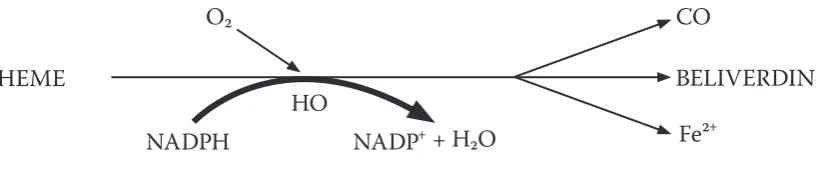
Carbon monoxide (CO) is one of the products of heme degradation by heme oxygenases (HO) (Fig. 1). Carbon monoxide is responsible for maintaining the liver’s regional perfusion, resulting in the activation of leucocytes.25,26
Fur-thermore, HO-1/CO prevents EC apoptosis via suppressing inflammatory reactions contributing to EC apoptosis. CO generated through heme catabolism by HO has an anti-apoptotic effect on ECs through activation of mitogen-ac-tivated protein kinases (MAPK).27 It is still not clear which
of the above-mentioned properties of CO has a hepato-protective influence in sepsis.
In a study on rodent model ischemia-reperfusion in-duced systemic inflammation, exogenous CO was shown to have a hepatoprotective effect via improving liver cell integrity and the redox state as well as protecting the liver microcirculation.28
Clinical manifestations of SALD
In septic patients, the spectrum of liver dysfunction may vary from subclinical to symptomatic liver failure. In criti-cally ill patients the concept of “shock liver” may be used. “Shock liver” is a syndrome of hemodynamic, cellular, im-munologic and molecular disorders.29 SALD can manifestin 2 clinical forms – jaundice/sepsis-induced cholestasis, and hypoxic hepatitis (HH).19 Coagulopathy may be another
symptom of SALD. These processes are still not well un-derstood due to the complexity of their pathomechanisms.
Jaundice/sepsis-induced cholestasis
The synthesis of bile is a complex process, requiring prop-er enprop-ergy input and normal function of transmembrane
proteins.30 Energy shortage caused by hypoxemia/liver
hypoperfusion may impair most bile synthesis steps.31
Liver histological examinations in patients with jaundice occurring during bacterial infection revealed the pres-ence of intrahepatic cholestasis. The impairment of bile transport is a result of alterations in genes activating tran-scription and modifying posttranslational treatment of bile acids transporting proteins caused by LPS and proinflam-matory cytokines.32
Laboratory test abnormalities include an increase of to-tal bilirubin (>2 mg/dL), alkaline phosphatase, ALT, and AST.33
Hypoxic hepatitis
Hypoxic hepatitis (HH) may be the cause of fulminant hepatitis. In septic shock, an increase in blood flow and cardiac output is not enough to compensate increased hepatic oxygen demand.34 Decreased hepatic blood flow
in shock does not always cause HH; it may occur in patients with normal blood pressure.
Other risk factors causing HH are LPS and inflammatory cytokines. Hypoxic hepatitis may also be a result of the re-oxygenation phase in the course of the ischemia/reperfu-sion phenomenon.
In the course of HH, except for a rapid (24 h from the onset of shock) substantial increase in aminotransferases and lactate dehydrogenase activity, an early decrease in serum prothrombin concentration is observed.35
Coagulopathy
A wide range of coagulopathy may be observed in sep-sis, starting with mild deviation in laboratory results (prolonged clotting time, decreased number of platelets) to severe coagulopathy and/or disseminated intravascular coagulation (DIC).
The main cause of coagulopathy in sepsis is microvas-cular endothelial injury resulting in an imbalance between fibrinolysis and coagulation.36 The changes seen
in en-dothelial injury include loss of vascular tone, capillary obstruction by platelet or fibrin clots, as well as the degra-dation of heparan sulfate leading to a pro-coagulant state.37
Coagulopathy may be another symptom of liver disease. Several factors may contribute to hemostatic changes in liver disease (Table 1).38
Assessment of liver function
Diagnosis of liver dysfunction in sepsis, according to the Surviving Sepsis Campaign (SSC) Guidelines, is based on an increase in serum bilirubin concentration >2 mg/dL (34.2 μmol/L) and occurrence of coagulopathy (INR > 1.5).39
damage in the course of sepsis/septic shock and distin-guishing it from a pre-existing liver pathology. Liver func-tion can be assessed using static and dynamic parameters.
Static parameters include: – secretory capacity – bilirubin;
– parameters of cholestasis – alkaline phosphatase, γ-glutamyltransferase;
– intracellular enzymes activity – alanine amino-transferase, aspartate aminoamino-transferase, glutamate dehydrogenase;
– synthesizing capacity – albumin, clotting factors V and VII.40
However, these parameters cannot be used for contin-uous and rapid monitoring of liver function in patients treated in ICU, nor are they diagnostic or prognostic in this group of patients.40,41 As mentioned above,
accord-ing to the SSC Guidelines, serum bilirubin concentration (>2 mg/dL or >34.2 μmol/L) is used as a single marker guideline to diagnose liver dysfunction.39 Due to a number
of drawbacks limiting its application, serum bilirubin is not an appropriate marker to reflect complex liver function. Increase in serum bilirubin concentration is neither spe-cific nor does it allow acute liver dysfunction to be distin-guished from a pre-existing liver pathology.41 Table 2 shows
causes of hyperbilirubinemia in sepsis.33
Dynamic parameters assessing liver function include: – indocyanine green (ICG), caffeine and
bromosul-fophthalein clearance;
– liver detoxification capacity – measuring the concen-tration of 14CO2 in exhaled air (measuring
the con-centration of [14C]aminopyrine, [14C]methacetin,
[14C]erythromycin metabolites) and measuring
the concentration of lidocaine/midazolam serum metabolites;
– ability to eliminate galactose.40
Maximal liver function capacity
Kaffarnik et al., in their study published in Critical Care in 2013, attempted to assess the maximal liver func-tion capacity (LiMax) test as a useful tool for early di-agnosis of sepsis-associated liver dysfunction.42 LiMaxis a non-invasive breath test using 13C-labeled
methace-tin, which is exclusively metabolized by cytochrome P450 (1A2) to 13CO2 and acetaminophen. The idea of the test
is to measure the amount of exhaled 13CO2. The result
is given as the LiMax value in μg per kg of body weight per hour. It shows the speed of substrate metabolism, thus allowing one to evaluate the metabolic capacity of the liver.
Table 1. Factors contributing to hemostatic changes in liver dysfunction
Hemostatic changes in liver dysfunction promoting hemostasis impairing hemostasis
• low plasminogen activity • decreased protein
S, protein C and antithrombin activity • increased VWF activity • increased factor VIII serum
concentration
• reduced hematocrit • thrombocytopenia
• production of nitric oxide and prostacyclin
• low serum concentration of coagulation factors II, V, VII, IX, X, and XI
• dysfibrinogenemia • vitamin K deficiency • elevated activity of tPA
• low serum concentrations of plasmin inhibitor, factor XII and TAFI
VWF – von Willebrand factor; TAFI – thrombin activatable fibrinolysis inhibitor; tPA – tissue plasminogen activator.
Table 2. Causes of hyperbilirubinemia in sepsis
1. Cholestasis
2. Hemolysis – drug-induced/infection-related • in normal RBCs
• in RBCs with enzyme defects
3. Hepatic dysfunction • ischemia: hypotension, hypoxia
• hepatocellular damage
• bilirubin transport dysfunction: decreased uptake, canalicular transport, clearance
RBCs – red blood cells.
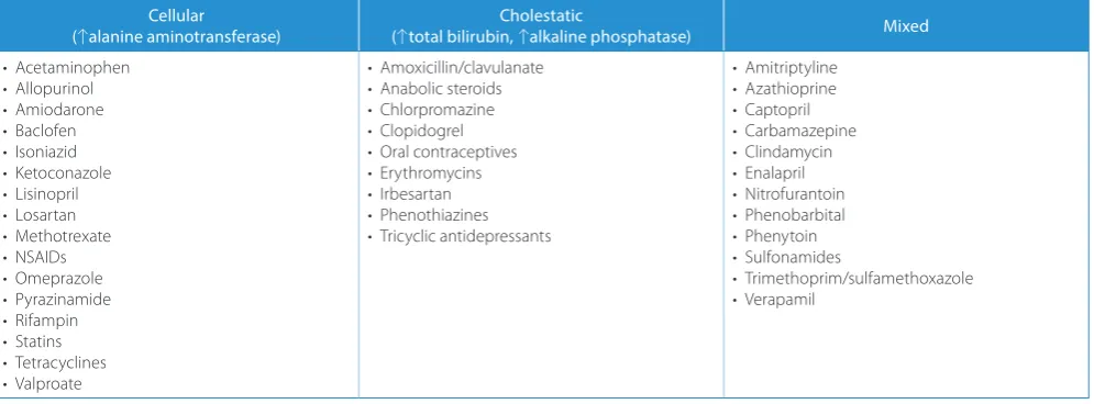
Table 3. Types of drug-induced liver injury. Examples of hepatotoxic drugs
Cellular
(↑alanine aminotransferase) (↑total bilirubin, Cholestatic↑alkaline phosphatase) Mixed
• Acetaminophen • Allopurinol • Amiodarone • Baclofen • Isoniazid • Ketoconazole • Lisinopril • Losartan • Methotrexate • NSAIDs • Omeprazole • Pyrazinamide • Rifampin • Statins • Tetracyclines • Valproate
• Amoxicillin/clavulanate • Anabolic steroids • Chlorpromazine • Clopidogrel • Oral contraceptives • Erythromycins • Irbesartan • Phenothiazines • Tricyclic antidepressants
• Amitriptyline • Azathioprine • Captopril • Carbamazepine • Clindamycin • Enalapril • Nitrofurantoin • Phenobarbital • Phenytoin • Sulfonamides
In the study, a pathologic deterioration of LiMax values in patients with septic shock was observed within 2 days after the onset of sepsis. Among patients with LiMax, <100 μg/kg/h the mortality rate was 55%, and with LiMax >100 μg/kg/h the mortality rate was 0%. The authors’ conclusion was that LiMax values <100 μg/kg/h could be a good predictor of morbidity and mortality.
Indocyanine green clearance
Indocyanine green (ICG) is a non-toxic, water-soluble fluorescent dye. Its spectrophotometric evaluation does not depend on oxygen saturation and serum bilirubin concentration. Indocyanine green clearance (ICG PDR) can be used to reflect the liver function. Because it is not metabolized, it is secreted almost exclusively by the liver and it is not subject to enterohepatic circulation.43
A significant limitation in using ICG PDR is the hemo-dynamic condition of the patient, as ICG PDR depends on hepatic blood flow.44 Additional parameters limiting
the use of this test are the serum bilirubin concentration, serum albumin concentration, body weight, and patient’s age.45 Studies have shown that ICG PDR may be
a diag-nostic and proga diag-nostic tool in monitoring acute liver fail-ure in critically ill patients in the ICU, but there are still no randomized control trials clearly confirming the utility of ICG PDR in daily clinical practice.43
It is worth underlining that in clinical practice there are no standardized diagnostic panels allowing for an early, clear diagnosis of acute liver dysfunction. Until now only a few studies have been published, and their results remain equivocal.
Therapeutic considerations
Currently, there is no specific therapeutic treatment available for the full restoration of damaged liver func-tion. The therapy, according to the SSC Guidelines, should focus on eradicating infection and treating sepsis and its complications.39 Furthermore, there are tools available
that could reduce the risk of further damage to this organ.
These include:
1. avoiding potentially hepatotoxic drugs;
2. early enteral feeding of hemodynamically stable patients; 3. glucose concentration monitoring and adequate
glu-cose supply if necessary;
4. extracorporeal liver support – Molecular Adsorbent Recirculating System (MARS) albumin dialysis, single-pass albumin dialysis (SPAD).19,46,47
Drugs are an important cause of liver injury. Examples of potentially hepatotoxic drugs are given in Table 3.48
La Mura proposed simvastatin administration for the pre-vention of LPS-induced intrahepatic endothelial dysfunc-tion. He found that prophylactic simvastatin prevents endotoxemia-induced liver injury, reduces liver inflamma-tion, and prevents microvascular dysfunction in rodents.49
In other studies, prophylactic simvastatin has also been reported as being able to correct endothelial dysfunction.50
Summary
The incidence of sepsis-associated liver failure is hard to estimate, but it is incontestable that liver failure as a complication of sepsis dramatically worsens the out-come of the patients. It is important to remember that dur-ing sepsis not only the infection itself is responsible for liver dysfunction, but also hyperreactivity of the inflamma-tory response, microcirculaof the inflamma-tory failure, and side effects of the therapy. Only an early diagnosis of sepsis and its complications as well as quick implementation of thera-peutic bundles allow reducing the incidence of severe organ complications, to shorten the hospitalization time and improve patients’ quality of life.
References
1. Blanco J, Muriel-Bombín A, Sagredo V, et al. Incidence, organ dys-function and mortality in severe sepsis: A Spanish multicentre study.
Crit Care. 2008;12:R158.
2. Martin GS, Mannino DM, Eaton S, Moss M. The epidemiology of sepsis in the United States from 1979 through 2000. N Engl J Med. 2003;348:1546–1554.
3. Angus DC, Linde-Zwirble WT, Lidicker J, Clermont G, Carcillo J, Pin-sky MR. Epidemiology of severe sepsis in the United States: Anal-ysis of incidence, outcome, and associated costs of care. Crit Care Med.2001;29:1303–1310.
Fig. 1. Heme degradation products
HO – heme oxygenase; CO – carbon monoxide; NADPH – r educed form of nicotinamide adenine dinucleotide phosphate; NADP+ – nicotinamide adenine dinucleotide phosphate.
HEME
NADPH
HO
O₂
CO
BELIVERDIN
Fe²
+4. National Institute of General Medical Sciences – Sepsis Fact Sheet. http://www.nigms.nih.gov/Education/Pages/factsheet_sepsis.aspx Accessed on December 28, 2016.
5. Kübler A, Adamik B, Ciszewicz-Adamiczka B, Ostrowska E. Severe sep-sis in intensive care units in Poland – Point prevalence study in 2012 and 2013. Anaesthesiol Intensive Ther. 2015;47:315–319.
6. Kübler A, Adamik B, Durek G, et al. Results of the severe sepsis reg-istry in intensive care units in Poland from 2003–2009. Anaesthesiol Intensive Ther. 2015;47:7–13.
7. Birrer R, Takuda Y, Takara T. Hypoxic hepatopathy: Pathophysiology and prognosis. Intern Med. 2007,46(14):1063–1070.
8. Kobashi H, Toshimori J, Yamamoto K. Sepsis-associated liver injury: Incidence, classification and the clinical significance. Hepatol Res. 2013;43(3):255–266.
9. Aninat C, Seguin P, Descheemaeker P, Morel F, Malledant Y, Gullou-zo A. Catecholamines induce an inflammatory response in human hepatocytes. Crit Care Med. 2008,36:848–854.
10. Kolios G, Valatas V, Manousou P, Xidakis C, Notas G, Kouroumalis E. Nitric oxide and MCP-1 regulation in LPS activated rat Kupffer cells.
Mol Cell Biochem. 2008;319:91–98.
11. Doi F, Goya T, Torisu M. Potential role of hepatic macrophages in neutrophil-mediated liver injury in rats with sepsis. Hepatology. 1993;17:1086–1094.
12. Aird WC. The role of the endothelium in severe sepsis and multiple organ dysfunction syndrome. Blood. 2003;101(10):3765–3777. 13. Wang D, Yin Y, Yao Y. Advances in sepsis-associated liver
dysfunc-tion. Burns Trauma. 2014;2:97–105.
14. Kwok W, Lee SH, Culberson C, Korneszczuk K, Clemens M. Caveolin-1 mediates endotoxin inhibition of endothelin-1-induced endothelial nitric oxide synthase activity in liver sinusoidal endothelial cells. Am J Physiol Gastrointest Liver Physiol. 2009;297(5): G930–G939. 15. Hyun-Ae Eum, Sang-Won Park, Sun-Mee Lee. Role of nitric oxide
in the expression of hepatic vascular stress genes in response to sep-sis. Nitric Oxide. 2007;17:126–133.
16. Yeager ME, Belchenko DD, Nguyen CM, Colvin KL, Ivy DD, Stenmark KR. Endothelin-1, the unfolded protein response, and persistent inflammation: Role of pulmonary artery smooth muscle cells. Am J Respir Cell Mol Biol. 2012;46:14–22.
17. Bellisai F, Morozzi G, Scaccia F, et al. Evaluation of the effect of bosen-tan treatment on proinflammatory cytokine serum levels in patients affected by systemic sclerosis. Int J Immunopathol Pharmacol. 2011;24:261–264.
18. Brauner JS, Rohde LE, Clausell N. Circulating endothelin-1 and tumor necrosis factor-a: Early predictors of mortality in patients with sep-tic shock. Intensive Care Med. 2000;26:305–313.
19. Nesseler N, Launey Y, Aninat C, Morel F, Mallédant Y, Seguin P. Clini-cal review: The liver in sepsis. Critical Care. 2012;16:235.
20. Bukovska G, Kery V, Kraus JP. Expression of human cystathionine beta-synthase in Escherichia coli: Purification and characterization.
Protein Expr Purif. 1994;5:442–448.
21. Blachier F, Davila AM, Mimoun S, et al. Luminal sulfide and large intes-tine mucosa: Friend or foe? Amino Acids. 2010;39:335–347. 22. Zhang H, Zhi L, Moore PK, Bhatia M. Role of hydrogen sulfide in cecal
ligation and puncture induced sepsis in the mouse. Am J Physiol Lung Cell Mol Physiol. 2006;290:L1193–1201.
23. Altaany Z, Moccia F, Munaron L, Mancardi D, Wang R. Hydrogen sul-fide and endothelial dysfunction: Relationship with nitric oxide. Curr Med Chem. 2014;21(32):3646–3661.
24. EJ Norris, CR Culberson, S Narasimhan, MG Clemens. The liver as a central regulator of hydrogen sulfide. Shock. 2011;36(3):242–250. 25. Pannen BHJ, Köhler N, Hole B, Bauer M, Clemens MG, Geiger KK. Pro-tective role of endogenous carbon monoxide in hepatic microcir-culatory dysfunction after hemorrhagic shock in rats. J Clin Invest. 1998,102:1220–1228.
26. Hoetzel A, Dolinay T, Schmidt R, Choi AMK, Ryter SW. Carbon mon-oxide in sepsis. Antioxid Redox Signal. 2007;11(9):2013–2026. 27. Brouarda S, Otterbeinb LE, Anratherc J, et al. Carbon monoxide
gen-erated by heme oxygenase 1 suppresses endothelial cell apoptosis.
JEM. 2000;192(7):1015–1026.
28. Wunder C, Brock RW, Frantz S, et al. Carbon monoxide, but not endothelin-1, plays a major role for the hepatic microcirculation in a murine model of early systemic inflammation. Crit Care Med. 2005;33:2323–2331.
29. Strassburg CP. Gastrointestinal disorders of the critically ill. Shock Liver. Best Pract Res Clin Gastroenterol. 2003;17:369–381.
30. Trauner M, Meier PJ, Boyer JL. Molecular pathogenesis of cholesta-sis. N Engl J Med. 1998;339:1217–1227.
31. Fuchs M, Sanyal AJ. Sepsis and cholestasis. Clin Liver Dis. 2008;12:151– 172. ix.
32. Moseley RH. Sepsis and cholestasis. Clin Liver Dis. 2004;8:83–94. 33. Chand N, Sanyal AJ. Sepsis-induced cholestasis. Hepatology.
2007;45(1):230–241.
34. Dahn MS, Lange P, Lobdell K, Hans B, Jacobs LA, Mitchell RA. Splanch-nic and total body oxygen consumption differences in septic and injured patients. Surgery. 1987;101:69–80.
35. Henrion J. Hypoxic hepatitis. Liver Int. 2012;32(7):1039–1052. 36. Taylor FB Jr, Toh CH, Hoots WK, Wada H, Levi M. Scientific
Subcom-mittee on Disseminated Intravascular Coagulation (DIC) of the Inter-national Society on Thrombosis and Haemostasis (ISTH): Towards definition, clinical and laboratory criteria, and a scoring system for disseminated intravascular coagulation. Thromb Haemost. 2001;86:1327–1330.
37. Lipinska-Gediga M. Sepsis and septic shock-is a microcirculation a main player? Anaesthesiol Intensive Ther. September 23, 2016. doi: 10.5603/AIT.a2016.0037. [Epub ahead of print]
38. Lisman T, Leebeek FWG. Hemostatic alterations in liver disease: A review on pathophysiology, clinical consequences, and treatment.
Dig Surg. 2007;24:250–258.
39. Dellinger RP, Levy MM, Rhodes A, et al. Surviving Sepsis Campaign: International Guidelines for Management of Severe Sepsis and Sep-tic Shock: 2012. Crit Care Med. 2013;41(2):580–637.
40. Sakka S. Assessing liver function. Curr Opin Crit Care. 2007;13:207–214. 41. Marshall JC, Cook DJ, Christou NV, Bernard GR, Sprung CL, Sibbald
WJ. Multiple organ dysfunction score: A reliable descriptor of a com-plex clinical outcome. Crit Care Med. 1995;23:1638–1652.
42. Kaffarnik, Lock JF, Vetter H, et al. Early diagnosis of sepsis-related hepatic dysfunction and its prognostic impact on survival: A pro-spective study with the LiMAx test. Crit Care. 2013;17(5):R259. 43. Vos JJ, Wietasch JKG, Absalom AR, Hendriks HGD, Scheeren TWL.
Green light for liver function monitoring using indocyanine green? An overview of current clinical applications. Anaesthesia. 2014;69:1364–1376.
44. Janssen MW, Druckrey-Fiskaaen KT, Omidi L, et al. Indocyanine green R15 ratio depends directly on liver perfusion flow rate. J Hepatobili-ary Pancreat Sci. 2010;17:180–185.
45. Kim GY, Bae KS, Noh GJ, Min WK. Estimation of indocyanine green elimination rate constant k and retention rate at 15 min using patient age, weight, bilirubin and albumin. J Hepatobiliary Pancreat Sci. 2009;16:521–528.
46. Sauer IM, Goetz M, Steffen I, et al. In vitro comparison of the Molec-ular Adsorbent Recirculation System (MARS) and single-pass albu-min dialysis (SPAD). Hepatology. 2004;39(5):1408–1414.
47. Woznica R. Single Pass Albumin Dialysis for treatment of the acute liver failure – A case report. Med Intens Ratunk. 2007;10(4):233–237. 48. Herrine SK. Liver injury caused by drugs.
http://www.merckmanu- als.com/professional/hepatic-and-biliary-disorders/drugs-and-the-liver/liver-injury-caused-by-drugs Accessed on December 28,2016. 49. La Mura V, Pasarín M, Meireles CZ, et al. Effects of simvastatin admin-istration on rodents with lipopolysaccharide-induced liver microvas-cular dysfunction. Hepatology. 2013;57:1172–1181.