K
INGAM
USIAŁ, D
ANUTAZ
WOLIŃSKAAdhesion Molecules, Cytokines
and Endothelial Dysfunction in Atherosclerosis
Molekuły adhezyjne, cytokiny i zaburzenie funkcji śródbłonka
w miażdżycy
Department of Pediatric Nephrology, Silesian Piasts University of Medicine in Wrocław, Poland
Adv Clin Exp Med 2006, 15, 6, 971–978 ISSN 1230−025X
EDITORIAL
Accumulating evidence suggests that athero− sclerosis should be regarded as a complex set of interactions among endothelium, circulating cells and particles, soluble mediators, and chemotactic agents. The processes leading to the creation of atherosclerotic lesions may also be seen as imbal− ances between pro− and anti−atherogenic factors, pro− and anti−inflammatory agents, and vasocon− strictors and vasodilators. However, irrespective
of the pathogenesis, multiple cross−reacting com− pounds influence both the structure and function of vessels, resulting in their dysfunction and dam− age. The endothelium thus seems to be a target and a focus for all the reactions mentioned above. Therefore, a detailed analysis of its structure and function may clarify the relationships between the various agents responsible for the progression of atherosclerosis.
© Copyright by Silesian Piasts University of Medicine in Wrocław
Abstract
The aim of this review is to present current knowledge of atherosclerosis. Recent investigations point to chronic inflammation as a key player in the functional impairment of the endothelium. Enhanced cell migration and diape− desis lead to monocyte recruitment and their change into macrophages, thus initiating atherosclerotic plaque for− mation. The involvement of adhesion molecules, interleukins, growth factors, and enzymes in the adhesion casca− de is tightly connected with dyslipidemia and the overproduction of reactive oxygen species. The latter may be a causative factor for the propagation of endothelial damage due to pleiotropic activities, including the impact on lipid metabolism, adhesion cascade functioning, cytokine activity, and nitric oxide bioavailability. The over−activi− ty of pro−atherogenic agents results in a vicious circle of tissue destruction. These self−perpetuating reactions pro− gressively impair the function of vessels, resulting in lumen occlusion, local ischemia, and clinical complications. Therefore, clarifying the mechanisms responsible for atherosclerosis seems to be the key target for future investi− gations (Adv Clin Exp Med 2006, 15, 6, 971–978).
Key words: adhesion molecules, cytokines, nitric oxide, reactive oxygen species, endothelium.
Streszczenie
Celem pracy jest przedstawienie obecnego stanu wiedzy na temat miażdżycy. Badania ostatnich lat wskazują na rolę przewlekłego procesu zapalnego jako kluczowego czynnika zaburzającego funkcję śródbłonka. Nasilona mi− gracja i diapedeza komórek prowadzi do gromadzenia monocytów i przekształcania w makrofagi, co jest począt− kiem procesu tworzenia blaszki miażdżycowej. Udział molekuł adhezyjnych, interleukin, czynników wzrostu i en− zymów w kaskadzie adhezji jest ściśle związany z zaburzeniami gospodarki lipidowej i wydzielaniem wolnych ro− dników tlenowych. Procesy wolnorodnikowe mogą powodować postępującą destrukcję śródbłonka przez wielokierunkowe oddziaływania, m.in. na metabolizm lipidów, kaskadę adhezji, aktywność cytokin i biodostęp− ność tlenku azotu. Wzmożona aktywność czynników proaterogennych uruchamia błędne koło niszczenia tkanek. Nieodwracalne reakcje stopniowo zaburzają funkcję naczyń, powodując zwężenie ich światła, niedokrwienie i po− wikłania. Wyjaśnienie mechanizmów odpowiedzialnych za rozwój miażdżycy wymaga więc dalszych badań (Adv Clin Exp Med 2006, 15, 6, 971–978).
The Natural History
of Atherosclerosis
Atherosclerotic lesions, although asympto− matic, start already in childhood. Atherogenesis covers a wide range of abnormalities, including fatty streaks, lipid cores, and fibrous caps. Early lesions, complicated by thrombi, calcifications, and ulcerations, develop into plaques that change the geometry of vessels and lead to their stenosis or even occlusion. All these disturbances are con− ditioned by microscopic changes within the endothelium caused by inadequate responses to various stimuli from the serum and surrounding tissues.
Classification
of Atherosclerotic Lesions
In the early stages of atherosclerosis, leuko− cyte recruitment and lipid deposition dominate (Table 1). Lipid−laden macrophages (foam cells) accumulate in the intima and form regions of thickening, called fatty streaks [1]. The next com− ponents of atherosclerotic lesions are lipid−con− taining vascular smooth muscle cells (VSMCs) and pools of extracellular lipids. The migration and proliferation of VSMCs coexists with extra− cellular matrix synthesis [1]. Until this moment, the changes are potentially reversible and elimi−nation of the factors destroying the endothelium, as well as lipid−lowering therapy, may prevent plaque formation.
Further lesion progression is characterized by continuous lipid accumulation, leading to the for− mation of lipid cores. These appear due to the fusion of lipid deposits, necrotic foam cells, and cellular debris [2]. Initially, the layer surrounding the lipid core consists only of the intima. Then the lipid core becomes encapsulated by collagen. The fibrous cap may also contain calcifications [2]. Extensive VSMC proliferation may result in the overproduction of collagen fibers and lipid core destruction. Shear stress disturbs blood flow in the regions of the modified vessel geometry and may break down the fibrous caps [2]. Erosive athero− sclerotic plaques are places of thrombi formation (Table 1).
The initial lesion and plaque formation results in stenosis of the vessel lumen and hypertrophy of the entire vessel wall. Such remodeling aims at maintaining the ability of the vessel wall to con− tract and relax. When compensatory mechanisms are depleted, the vessel stenosis becomes irre− versible and the final occlusion progresses.
Adhesion Molecules
in Atherosclerosis
Current data emphasize the role of chronic inflammation in the pathogenesis of atherosclero−
Histological type Cells and structures involved Functional changes
(Typ histologiczny) (Zaangażowane komórki i struktury) (Zmiany czynnościowe)
I; intima monocyte recruitment clinically silent
macrophage formation reversible
lipid deposition
II; intima, media foam cell accumulation →fatty streaks clinically silent lipid−containing vascular smooth muscle cells (VSMCs) reversible III; intima, media VSMC proliferation and migration clinically silent
extracellular matrix synthesis potentially reversible small pools of extracellular lipids
IV; intima, media fusion of lipid deposits, necrotic foam cells and cellular vessel wall hypertrophy
debris, lipid cores vascular remodeling
Va; intima, media collagen−containing fibrous cap modified vessel geometry vessel stenosis
Vb; intima, media calcifications in fibrous cap modified vessel geometry vessel stenosis
Vc; intima, media, lumen collagen fiber overproduction vessel stenosis, occlusion
lipid core destruction clinical manifestations
VI; intima, media, lumen plaque rupture, formation of thrombi, erosive plaque vessel stenosis, occlusion clinical manifestations
Table 1. Classification of atherosclerotic lesions
sis. Endothelial response to injury is a prerequisite for initiating this reaction. Although laminar shear stress is an atheroprotective factor, even slight changes in blood flow may stimulate defense mechanisms [3]. Endothelial activation may also be provoked by other unspecific stimuli, such as hypoxia, hypercholesterolemia, histamine or thrombin, or specific agents, such as immune com− plexes [4]. Endothelial activation causes structural changes within the innermost layer of the endothe− lium and enables the rolling, activation, firm adhe− sion, and migration of cells from the vessels to the inflamed or injured tissues. In particular it allows cells to migrate towards atherosclerotic lesions, thus aggravating the inflammatory reactions in situ. The sequence of these reactions, defined as the leukocyte adhesion cascade, is regulated by adhesion molecules, such as immunoglobulin superfamily members (ICAM−1, VCAM−1, platelet−endothelial cell adhesion molecule (PECAM)−1), selectins (E−, L−, P−selectin), and integrins [4].
Circulating cells do not adhere to endothelium under normal conditions. However, unfavorable hemorheology in lesion−prone areas of vessels (e.g. flow turbulence in the regions of bifurca− tions) may stimulate the adhesion cascade by increasing ICAM−1 and inducing VCAM−1 expression on the endothelium [4]. Ox−LDL, IL−1, IL−4, TNF−α, and monocyte chemoattractant pro−
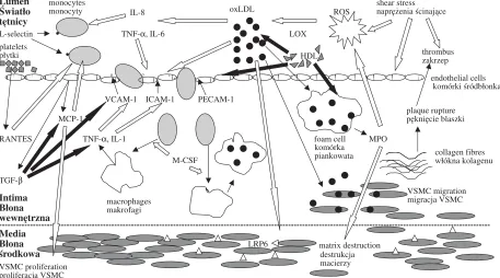
tein (MCP)−1 are other potent ICAM−1 and VCAM−1 activators, whereas HDL, transforming growth factor (TGF)−β, fibroblast growth factor (FGF)−2, and IL−10 inhibit their cytokine−induced expression (Fig. 1). Meanwhile, P−selectin is released upon activation from Weibel−Palade bod− ies in endothelial cells and translocated to the plas− ma membrane. Then it can react with a counter− receptor on leukocytes, i.e. sialyl Lewis x antigen [4]. Simultaneously, L−selectin, constitutively expressed on leukocytes, binds to endothelial receptors [4]. During the phase of selectin ligation, cells transiently adhere and detach, depending on local shear forces and the variable endothelial dis− tribution of P−selectin [5]. These reactions dimin− ish the velocity of rolling and facilitate cell contact with chemotactic agents and other cytokines [4]. The subsequent firm adhesion of leukocytes to the endothelium, conditioned by the local expressions of ICAM−1 and VCAM−1, is a prerequisite for monocyte transmigration through the endothelial layer into the intima.
The migration of monocytes has been impli− cated as a key player in atherogenesis. Their recruitment is mainly governed by MCP−1, a member of the CC family of chemokines (Fig. 1). The pro−atherogenic activity of MCP−1 has been observed in apoE–/– mice [6], whereas an investigation on rats revealed a growth−promoting effect of MCP−1 on VSMCs, which may suggest
Lumen Światło tętnicy
monocytes
monocyty ROS L−selectin
·
TNF−α, IL−6 LOXplatelets
płytki
·
HDL·
°
°
· ° · · · ° ° endothelial cells VCAM−1 ICAM−1 PECAM−1komórki śródbłonka MCP−1
plaque rupture pęknięcie blaszki RANTES TNF−α, IL−1 foam cell MPO
M−CSF TGF−β
collagen fibres włókna kolagenu
Intima Błona wewnętrzna
−−−−−−−−−−−−−−−−−−−−−−−−−−−−−−−−−−−−−−−−−−−−−−−−−−−−−−−−−−−−−−−−−−−−−−−−−−−−−−−−−−−−−−−−−−−−−−−−−−−−−−−−−−−−−−−−−−−−−−−−−−−−−−−−−−−−−−−−−−−−−−−−−−−−−−−−−−−−−−−−−−−−−−−−−−−−−−−
Media Błona
środkowa matrix destruction
VSMC proliferation proliferacja VSMC
destrukcja macierzy komórka piankowata oxLDL
IL−8
shear stress naprężenia ścinające
thrombus zakrzep
VSMC migration migracja VSMC macrophages
makrofagi
LRP6
Fig. 1. Network of interactions among adhesion molecules, cytokines, and ROS in atherogenesis; stimulation B, suppression ¨
a potent role of this chemokine in vessel wall remodeling [7]. Another potent chemoattractant for monocytes is the soluble form of fractalkine (FK), the only member of the CX3C chemokine family, found in atherosclerotic plaques [8]. The next chemokine with well−established pro−athero− genic activity is IL−8. Its expression is stimulated by oxLDL and its chemoattractant action mainly concerns monocytes [9]. Platelet−derived RANTES, delivered to the endothelium by platelet microparticles, is also known for its chemotactic impact on monocytes [10]. However, their activity may be suppressed by TGF−β, down−regulating MCP−1 and IL−8. Macrophage colony−stimulating factor (M−CSF) also plays an important role in cell migration. M−CSF influences monocyte differenti− ation into macrophages, which is the next step to foam cell formation (Fig. 1).
Cytokine stimulation activates membrane− bound adhesion molecules and induces their prote− olytic shedding from cells into the circulation [11]. Soluble forms (s) appear to be biologically active and influence leukocyte attachment to the vascular endothelium, thus playing an important role in atherogenesis. Additional support for this notion is the fact that the levels of circulating adhesion mol− ecules correlate with intima−media thickness and plaque score [12]. Moreover, sICAM−1 serum con− centration has been established as a risk factor for future myocardial infarction in apparently healthy men and might predict the progression of carotid atherosclerosis independently of traditional risk factors [13]. sVCAM−1 and sE−selectin are also potential serum markers of atherosclerosis [14].
Cytokine Activity
in Atherosclerosis
Most of the cytokines have been proposed to be involved in atherogenesis, and all cells found in the atherosclerotic plaques produce cytokines and respond to cytokine mediators. Migrating cells, such as monocytes, neutrophils, T cells, and platelets, accumulate due to the adhesion cascade on the vessel wall and release numerous inter− leukins (IL), proteins, growth factors, and enzymes upon activation.
The globally accepted classification of inter− leukins as either pro− or anti−inflammatory, or rather pro− or anti−atherogenic, can only partly sys− tematize this heterogeneous and ever−expanding group of molecules. Apart from those with well− established pro−atherogenic (IL−1α/β, IL−2, IL−6, IL−8, IL−12) and anti−atherogenic (IL−1ra, IL−9, IL−10) roles, there are still those with unknown impact on atherogenesis (IL−19 to IL−27). Certain
cytokines may also exert pro− and anti−inflamma− tory activity simultaneously. For example, IL−13 can induce VCAM−1, but has no impact on ICAM− 1 or E−selectin [15]. Disturbed balance among dif− ferent cytokines may represent another candidate mechanism underlying atherogenesis. Such inter− actions have been noted between IL−10 and IL−12, IL−4 and IL−12, and IL−6 and IL−10 [16]. In vitro
studies have also shown that different combina− tions of several cytokines may act either synergis− tically or antagonistically towards adhesion mole− cule expression [11]. The changes in gene expres− sion induced by certain cytokines are attracting increasing attention due to their potent role in ath− erosclerotic lesion formation. Up−regulation of genes encoding VCAM−1, MCP−1, and IL−6 in human vascular endothelial cells treated with IL−4 may be an example of pleiotropic cytokine activi− ty [17]. Therefore, the cytokine contribution to atherosclerosis seems to be a set of actions among various signals, creating a self−perpetuating mech− anism of lesion formation. Its complexity is aggra− vated by the fact that migrating cells are not the only source of cytokines.
Adipose tissue produces a recently discovered group of cytokines called adipocytokines. Apart from the well−characterized representatives IL−6 and TNF−α, there are new proteins, leptin and adiponectin, of great interest due to their role in atherogenesis. In vitrostudies have demonstrated that leptin stimulates MCP−1 expression and ROS production in cultured endothelial cells, thus act− ing pro−atherogenically [18]. Adiponectin has been suggested to have anti−atherogenic proper− ties. Together with a decrease in oxLDL uptake by macrophages and inhibition of VCAM−1, ICAM− 1, and E−selectin expression, it also stimulates NO production [19]. Therefore it is connected with major pathways of atherosclerotic disturbances, i.e. dyslipidemia, inflammation, and endothelial dysfunction.
Lipid Disturbances
in Atherosclerosis
ular, when LDL concentrations in the blood are increased, cholesterol synthesis in situis blocked and only LDL influx takes place, thus playing a defensive role against hyperlipidemia [20]. Additional protection results from HDL−related cholesterol efflux. However, even slight modifica− tions in LDL structure, e.g. caused by oxidative stress or hyperglycemia, render LDL receptors on the endothelium unable to recognize the lipopro− tein particles. Therefore, oxidized, glycated, or glycoxidized LDLs are caught by scavenger recep− tors present on endothelial cells and macrophages. The density of these receptors is independent of intracellular cholesterol concentrations. Moreover, glycoxidized LDLs induce their specific class A scavenger receptors (SR−A) and suppress class B type I scavenger receptors (SR−BI) for HDL, thus acting pro−atherogenically [21]. Due to these facts, macrophages and endothelial cells can be easily laden with cholesterol, thus transforming them into foam cells and initiating early athero− sclerotic lesions. Their subsequent development into atherosclerotic plaques requires immune sys− tem activation. One of the possible ways of such stimulation is the expression of CD1 proteins on lipid−laden macrophages. CD1+ foam cells present lipid antigens to T cells, thus driving T cell−depen− dent inflammatory reactions within atherosclerotic lesions [22]. In particular, natural killer T (NKT) cell activation via the CD1d receptor in apolipoprotein E−deficient (apo E–/–) mice caused a burst of cytokines and increased atherosclerotic lesion size [23]. Another mechanism responsible for the secretion of proinflammatory cytokines may be the induction of tumor necrosis factor (TNF)−α and interleukin (IL)−6 mRNA by mini− mally oxidized LDL and free cholesterol accumu− lated in macrophages [24].
The above−mentioned investigations suggest that lipids do not only regulate their own metabo− lism, but may also influence other compounds responsible for atherosclerosis (Fig. 1). Therefore, lipid pleiotropic activities have been brought into focus. It has been shown that oxLDLs and lysophospholipids induce the expressions of inter− cellular adhesion molecule−1 (ICAM−1) and vascu− lar cell adhesion molecule−1 (VCAM−1) on endothelial cells [25], thus activating cell migration towards inflamed tissues. OxLDLs can also induce interleukin (IL)−12 production in peripheral blood mononuclear cells (PBMCs), whereas PBMC incu− bation with oxLDL in the presence of IL−10 decreased the number of interferon (IFN)−γ−produc− ing cells [26]. In addition, LDL receptor−related protein LRP6 has been identified on vascular smooth muscle cells (VSMCs) and proposed as a mediator of VSMC proliferation critical for
atherogenesis [27]. Finally, free fatty acids stimulate the production of reactive oxygen species (ROS), reduce nitric oxide (NO) bioavailability, and cause endothelial dysfunction, which is thought to be a major contributor in atherogenesis [28].
Endothelial Dysfunction
in Atherosclerosis
Impairment of endothelium−dependent vasore− laxation is a characteristic feature of atherosclero− sis. It strongly depends on the bioactivity of the endothelium−derived relaxing factor nitric oxide (NO˙). Decreased NO˙ bioavailability, typical of atherosclerosis, may result from a decline in its synthesis by endothelial NO˙ synthase (eNOS) or accelerated degradation by reactive oxygen species (ROS).
ROS are a group of oxygen−containing mole− cules responsible for the oxidation of lipids, pro− teins, carbohydrates, and DNA. Some of these molecules, such as superoxide anion (O2–˙), hydroxyl radical (HO˙), and NO˙, are free radicals due to unpaired electrons (˙), and such a chemical structure provokes chain reactions occurring at extremely fast rates. The end products of the initial reactions usually attack adjacent molecule side chains. This perpetual process causes the accumu− lation of free radicals. The aggravated regional toxic effects are partly suppressed by defense mechanisms which decrease the ROS concentra− tion. These native protectors include anti−oxida− tive enzymes, such as copper/zinc−superoxide dis− mutase (Cu,Zn−SOD) and manganese−superoxide dismutase (Mn−SOD), metal−binding proteins (transferrin, ceruloplasmin, ferritin, metalloth− ionein), and vitamins (A, C, E) [29]. Recent inves− tigations have shown that imbalance between pro− and antioxidant mechanisms is of paramount importance in the pathogenesis of lipid oxidation and NO˙−related endothelial dysfunction.
gene polymorphism should be regarded as a risk factor for endothelial dysfunction [30].
L−arginine is the substrate for NO˙ synthesis by eNOS and tetrahydrobiopterin (BH4) is the cofactor of this reaction. In the absence of one of these, eNOS becomes uncoupled, thus producing superoxide anion (O2–˙) and hydrogen peroxide (H2O2) instead of NO˙. eNOS activity can also be suppressed by hypercholesterolemia and, in partic− ular, by oxLDL [31]. The latter inhibits dimethy− larginine dimethylaminohydrolase, which is the enzyme inactivating asymmetric dimethylarginine (ADMA) [32]. Subsequently, increased concentra− tions of ADMA, which is in competition with L− arginine, inhibit NO˙ synthesis and increase O2–˙ production via eNOS uncoupling. When NO˙ and O2–˙ are produced simultaneously, they react with each other. The reaction rate for NO˙ and O2–˙ is three times faster than that for O2–˙ and superoxide dismutase (SOD), which can stabilize NO˙. Therefore, the first reaction dominates and NO˙ becomes inactivated. Moreover, other free radicals are generated. One of them is peroxynitrite (ONOO–), which at low concentrations has a mild− ly relaxing activity, but at high levels destroys the vessel wall. NO˙ can also react with hydroxyl rad− ical (HO˙) and products of lipid oxidation [33].
Oxidized lipids can be generated in the vascu− lature by several enzymatic and non−enzymatic mechanisms. Animal experiments have provided evidence that lipoxygenases (LOXs) play a major role in atherosclerosis progression [34]. Enzyme− catalyzed oxidation takes place in leukocytes (iso− form 5−LOX), platelets (isoform 12−LOX), and reticulocytes (isoform 15−LOX). During these reactions, enzyme−bound radical intermediates are generated, such as lipid alkyl (L˙), alkoxyl (LO˙), and peroxyl (LOO˙). All of these react with NO˙ and decrease its bioavailability. Moreover, 15−LOX is cytokine inducible and may show pro−athero− genic activity [35]. Another enzyme prone to cytokine stimulation is prostaglandin endoperoxide H synthase (PGHS), the expression of which is raised in the presence of IL−1 and TNF−α. It acts similarly to LOX, generating lipid radicals reacting with NO˙ [36]. Increased activity of myeloperoxi− dase (MPO), the protease which catalyzes hypochlorous acid (HOCl) synthesis, may also play a causative role in lipid oxidation. MPO is produced by foam cells and its expression has been demonstrated in human atherosclerotic plaques. Nitrating and chlorinating products of reactions
catalyzed by MPO damage tissues due to their toxic properties [37]. Sub−endothelial matrix destruction further increases ROS production and initiates smooth muscle cell migration, thus propa− gating the formation of atherosclerotic lesions. Moreover, MPO influences NO˙ bioavailability, thus showing additional pro−atherogenic properties [37]. A non−enzymatic candidate mechanism for lipid oxidation is the Fenton reaction, in which free metal ions (Fe2+ and Fe3+) react with lipid hydroper− oxide (LOOH), thus producing LO˙ and LOO˙.
ROS overproduction and lipid disturbances are tightly connected with macrophage activity. Oxidative stress is responsible for impaired macrophage phagocytosis of apoptotic cells in ath− erosclerotic plaques. Apart from the already men− tioned LOX and MPO, there are other macrophage− derived enzymes and substances influencing atherogenesis. Recently developed animal models revealed pro−atherogenic features of lipoprotein lipase (LPL) and anti−atherogenic activities of lysophospholipase 3 and lysophosphatidylcholine [38, 39]. Macrophages are also a source of resistin, a newly discovered adipocytokine inducing insulin resistance, correlating with inflammatory markers and predicting atherosclerosis [40].
References
[1] Stary HC, Chandler AB, Glagov S, Guyton JR, Insull W Jr., Rosenfeld ME, Schaffer SA, Schwartz CJ, Wagner WD, Wissler RW: A definition of initial, fatty streak, and intermediate lesions of atherosclerosis. A report from the Committee on Vascular Lesions of the Council on Arteriosclerosis, American Heart Association. Arterioscler Thromb 1994, 14, 840–856.
[2] Stary HC, Chandler AB, Dinsmore RE, Fuster V, Glagov S, Insull W Jr., Rosenfeld ME, Schwartz CJ, Wagner WD, Wissler RW:A definition of advanced types of atherosclerotic lesions and a histological classifi− cation of atherosclerosis. A report from the Committee on Vascular Lesions of the Council on Arteriosclerosis, American Heart Association. Circulation 1995, 92, 1355–1374.
[3] Dardik A, Chen L, Frattini J, Asada H, Aziz F, Kudo FA, Sumpio BE:Differential effects of orbital and lam− inar shear stress on endothelial cells. J Vasc Surg 2005, 41, 869–880.
[4] Carlos TM, Harlan JM: Leukocyte−Endothelial Adhesion Molecules. Blood 1994, 84, 2068–2101.
[5] Kim MB, Sarelius IH: Role of shear forces and adhesion molecule distribution on P−selectin−mediated leukocyte rolling in postcapillary venules. Am J Physiol Heart Circ Physiol 2004, 287, H2705–H2711.
[6] Aiello RJ, Bourassa PAK, Lindsey S, Weng W, Natoli E, Rollins BJ, Milos PM: Monocyte chemoattractant protein−1 accelerates atherosclerosis in apolipoprotein E−deficient mice. Arterioscler Thromb Vasc Biol 1999, 19, 1518–1525.
[7] Parenti A, Bellik L, Brogelli L, Filippi S, Ledda F: Endogenous VEGF−A is responsible for mitogenic effects of MCP−1 on vascular smooth muscle cells. Am J Physiol Heart Circ Physiol 2004, 286, H1978–H1984.
[8] Lesnik P, Haskell CA, Charo IF: Decreased atherosclerosis in CX3CR1–/– mice reveals a role for fractalkine in
atherogenesis. J Clin Invest 2003, 111, 333–340.
[9] Gerszten RE, Garcia−Zapeda EA, Lim YC, Yoshida M, Ding HA, Gimbrone MA Jr., Luster AD, Luscinskas FW, Rosenzweig A: MCP−1 and IL−8 trigger firm adhesion of monocytes to vascular endothelium under flow conditions. Nature 1999, 398, 718–723.
[10] Mause SF, von Hundelshausen P, Zernecke A, Koenen RR, Weber C:Platelet microparticles. A transcellular delivery system for RANTES−promoting monocyte recruitment on endothelium. Arterioscler Thromb Vasc Biol 2005, 25, 1512–1518.
[11] Raab M, Daxecker H, Markovic S, Karimi A, Griesmacher A, Mueller MM: Variation of adhesion molecule expression on human umbilical vein endothelial cells upon multiple cytokine application. Clin Chim Acta 2002, 321, 11–16.
[12] Hashimoto H, Kitigawa K, Kuwabara K, Hougaku H, Ohtsuki T, Matsumoto M, Hori M: Circulating adhe− sion molecules are correlated with ultrasonic assessment of carotid plaques. Clin Sci 2003, 104, 521–527.
[13] Kondo K, Kitigawa K, Nagai Y, Yamagami H, Hashimoto H, Hougaku H, Hori M: Associations of soluble intercellular adhesion molecule−1 with carotid atherosclerosis progression. Atherosclerosis 2005, 179, 155–160.
[14] Hwang SJ, Ballantyne CM, Sharrett AR, Smith LC, Davis CE, Gotto AM, Boerwinkle E: Circulating adhe− sion molecules VCAM−1, ICAM−1, and E−selectin in carotid atherosclerosis and incident coronary heart disease cases. The Atherosclerosis Risk In Communities (ARIC) Study. Circulation 1997, 96, 4219–4225.
[15] Bochner BS, Klunk DA, Sterbinsky SA, Coffman RL, Schleimer RP:IL−13 selectively induces vascular cell adhesion molecule−1 expression in human endothelial cells. J Immunol 1995, 154, 799–803.
[16] Davenport P, Tipping PG:The role of interleukin−4 and interleukin−12 in the progression of atherosclerosis in apolipoprotein E−deficient mice. Am J Pathol 2003, 163, 1117–1125.
[17] Lee YW, Eum SY, Chen KC, Hennig B, Toborek M:Gene expression profile in interleukin−4−stimulated human vascular endothelial cells. Mol Med 2004, 10, 19–27.
[18] Yamagishi SI, Edelstein D, Du XL, Kaneda Y, Guzman M, Brownlee M:Leptin induces mitochondrial super− oxide production and monocyte chemoattractant protein−1 expression in aortic endothelial cells by increasing fatty acid oxidation via protein kinase A. J Biol Chem 2001, 276, 25096–25100.
[19] Chen H, Montagnani M, Funahashi T, Shimomura I, Quon MJ:Adiponectin stimulates production of nitric oxide in vascular endothelial cells. J Biol Chem 2003, 278, 45021–45026.
[20] Brown MS, Goldstein JL: A receptor−mediated pathway for cholesterol homeostasis. Science 1986, 232, 34–47.
[21] Lam MCW, Tan KCB, Lam KSL: Glycoxidized low−density lipoprotein regulates the expression of scavenger receptors in THP−1 macrophages. Atherosclerosis 2004, 177, 313–320.
[22] Melian A, Geng YJ, Sukhova GK, Libby P, Porcelli SA:CD1 expression in human atherosclerosis. A potential mechanism for T cell activation by foam cells. Am J Pathol 1999, 155, 775–786.
[23] Tupin E, Nicoletti A, Elhage R, Rudling M, Ljunggren HG, Hansson GK, Pulsson Berne G: CD1d−depen− dent activation of NKT cells aggravates atherosclerosis. J Exp Med 2004, 199, 417–422.
[24] Li Y, Schwabe RF, Devries−Seimon T, Yao PM, Gerbod−Giannone MC, Tall AR, Davis RJ, Flavell R, Brenner DA, Tabas I:Free cholesterol−loaded macrophages are an abundant source of Tumor Necrosis Factor− alpha and Interleukin−6. Model of NF−kappa B− and MAP kinase−dependent inflammation in advanced athero− sclerosis. J Biol Chem 2005, 280, 21763–21772.
[25] Lee H, Lin CI, Liao JJ, Lee YW, Yang HY, Lee CY, Hsu HY, Wu HL:Lysophospholipids increase ICAM−1 expression in HUVEC through a Gi− and NF−κB−dependent mechanism. Am J Physiol Cell Physiol 2004, 287,
C1657–C1666.
[27] Wang X, Adhikari N, Li Q, Hall JL:LDL receptor−related protein LRP6 regulates proliferation and survival through the Wnt cascade in vascular smooth muscle cells. Am J Physiol Heart Circ Physiol 2004, 287, H2376–H2383.
[28] Sainsbury CAR, Sattar N, Connell JMC, Hillier C, Petrie JR: Non−estrified fatty acids impair endothelium− dependent vasodilation in rat mesenteric resistance vessels. Clin Sci 2004, 107, 625–629.
[29] Koga T, Kwan P, Zubik L, Ameho C, Smith D, Meydani M: Vitamin E supplementation suppresses macrophage accumulation and endothelial cell expression of adhesion molecules in the aorta of hypercholes− terolemic rabbits. Atherosclerosis 2004, 176, 265–272.
[30] Heltianu C, Costache G, Gafencu A, Diaconu M, Bodeanu M, Cristea C, Azibi K, Poenaru L, Simionescu M:
Relationship of eNOS gene variants to diseases that have in common an endothelial cell dysfunction. J Cell Mol Med 2005, 9, 135–142.
[31] Vergnani L, Hatrik S, Ricci F, Passaro A, Manzoli N, Zuliani G, Brovkovych V, Fellin R, Malinski T:Effect of native and oxidized low−density lipoprotein on endothelial nitric oxide and superoxide production: key role of L−arginine availability. Circulation 2000, 101, 1261–1266.
[32] Ito A, Tsao PS, Adimoolam S, Kimoto M, Ogawa T, Cooke JP: Novel mechanism for endothelial dysfunction: dysregulation of dimethylarginine dimethylaminohydrolase. Circulation 1999, 99, 3092–3095.
[33] O’Donnell VB, Chumley PH, Hogg N, Bloodsworth A, Darley−Usmar VM, Freemen BA:Nitric oxide inhi− bition of lipid peroxidation: kinetics of reaction with lipid peroxyl radicals and comparison with α−tocopherol. Biochemistry 1997, 36, 15216–15223.
[34] Cyrus T, Witztum JL, Rader DJ, Tangirala R, Fazio S, Linton MF, Funk CD: Disruption of the 12/15−lipoxy− genase gene diminishes atherosclerosis in apo E−deficient mice. J Clin Invest 1999, 103, 1487–1488.
[35] Conrad DJ, Kühn H, Mulkins M, Highland E, Sigal E: Specific inflammatory cytokines regulate the expres− sion of human monocyte 15−lipoxygenase. Proc Natl Acad Sci USA 1992, 89, 217–221.
[36] O’Donnell VB, Coles B, Lewis MJ, Crews BC, Marnett LJ, Freeman BA: Catalytic consumption of nitric oxide by prostaglandin H synthase−1 regulates platelet function. J Biol Chem 2000, 275, 38239–38244.
[37] Eiserich JP, Baldus S, Brennan ML, Ma W, Zhang C, Tousson A: Myeloperoxidase, a leukocyte−derived vas− cular NO oxidase. Science 2002, 296, 2391–2394.
[38] Ichikawa T, Liang J, Kitajima S, Koike T, Wang X, Sun H, Morimoto M, Shikama H, Watanabe T, Yamada N, Fan J: Macrophage−derived lipoprotein lipase increases aortic atherosclerosis in cholesterol−fed Tg rabbits. Atherosclerosis 2005, 179, 87–95.
[39] Taniyama Y, Fuse H, Satomi T, Tozawa R, Yasuhara Y, Shimakawa K, Shibata S, Hattori M, Nakata M, Taketomi S:Loss of lysophospholipase 3 increases atherosclerosis in apolipoprotein E−deficient mice. Biochem Biophys Res Commun 2005, 29, 104–110.
[40] Reilly MP, Lehrke M, Wolfe ML, Rohatgi A, Lazar MA, Rader DJ: Resistin is an inflammatory marker of ath− erosclerosis in humans. Circulation 2005, 111, 932–939.
Address to correspondence:
Kinga Musiał
Department of Pediatric Nephrology Silesian Piasts University of Medicine Skłodowskiej−Curie 50/52
50−369 Wrocław Poland
Conflict of interest: None declared
Received: 17.02.2006 Revised: 7.04.2006 Accepted: 2.07.2006
Praca wpłynęła do Redakcji: 17.02.2006 r. Po recenzji: 7.04.2006 r.