Introduction
The red eye is the most common ocular complaint in patients seeking medical assistance at primary healthcare level. The differential diagnosis of the red eye varies from relatively innocuous and trivial conditions to those that are more devastating and potentially blinding. The purpose of this article is to help identify certain key clinical features of the discussed conditions, to help avoid pitfalls in diagnosis and to convey appropriate treatment modalities.
Conjunctivitis
Conjunctivitis is defined as inflammation of the conjunctival membrane that covers the ocular surface. Potential causes are bacterial, viral and allergic.
Viral conjunctivitis
The most common responsible viruses include adenovirus, herpes simplex virus and molluscum contagiosum. Typically, viral infection is characterised by an acute follicular conjunctival reaction (Figure 1), as well as pre-auricular or submandibular lymphadenopathy. Although usually benign and self-limiting, generally the course is longer than that of bacterial conjunctivitis and lasts roughly 2-4 weeks. Every necessary precaution to prevent the spread of infection should be taken. It is extremely important that instruments should be cleaned, ophthalmic drops changed frequently, and hands thoroughly sanitized before seeing every patient. Education of the patient, as well as other staff members, also plays a role.
manifestations. These infections induce an acute follicular conjunctival reaction which is often bilateral and associated with pre-auricular lymphadenopathy. The incubation period is usually 7-9 days before the onset of symptoms. Patients report ocular itching, tearing, redness and photophobia. In severe cases, subconjunctival bleeding may occur and the cornea may become affected in the second week.
Treatment of adenoviral conjunctivitis is mainly supportive. Patients should be instructed to use cold compresses and lubricating eye drops for comfort. Topical antihistamine eye drops, such as Spersallerg®, may be used to alleviate severe itching.
Herpes simplex conjunctivitis
Primary ocular herpes simplex virus infection affects infants and young children predominantly. Patients may present with a follicular conjunctivitis that manifests as a red, teary eye, associated with typical vesicular eruption on the eyelids (Figure 2). Treatment is the same as that for herpes simplex keratitis. The treatment of skin lesions may also include the topical administration of 3% acyclovir ointment.
Molluscum contagiosum keratoconjunctivitis
Molluscum contagiosum can cause follicular conjunctivitis in association with an eyelid lesion. Usually, the lesion is a small, pearly umbilicated nodule on or near the lid margin (Figure 3). Multiple lesions may be present, especially in a patient with human immunodeficiency virus (HIV). Treatment involves incision and curettage of the symptomatic lesions. Asymptomatic lesions, if left long enough, are usually self-limiting.
Bacterial conjunctivitis
Acute bacterial conjunctivitis
Acute bacterial conjunctivitis is a very common ocular condition which is primarily caused by Staphylococcus, Haemophilus and
Streptococcus species. These organisms may be spread by hand-to-eye contact or by colonisation of adjacent mucosal tissues, such as the nasal or sinus mucosa. Initially, acute bacterial conjunctivitis presents unilaterally. The second eye is often affected soon Figure 2: Vesicular eruption on the eyelids
Figure 4: Acute bacterial conjunctivitis with injection of the conjunctiva
Figure 3: Lesions caused by molluscum contagiosum
Gonococcal conjunctivitis
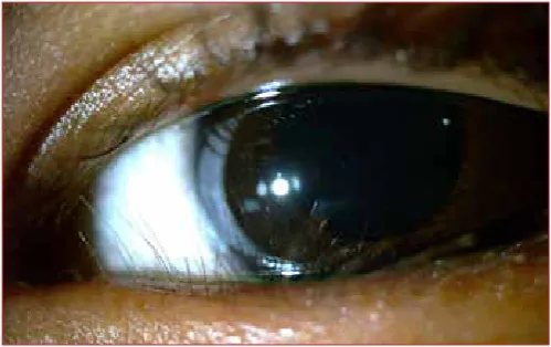
Typically, this ocular disease presents as a hyperacute purulent conjunctivitis. A profuse purulent discharge is often present and is associated with periorbital oedema, marked conjunctival hyperaemia and chemosis (severe conjunctival swelling) (Figure 5), as well as pre-auricular lymphadenopathy. Prompt treatment of gonococcal conjunctivitis is essential since this organism can rapidly cause corneal ulceration and perforation. Ideally, because of the rapid progression of this condition, patients should be referred to an eye specialist after commencing initial management with saline irrigation to clear the purulent material from the lids and conjunctiva. Hourly topical fluoroquinolone drops (ofloxacin,ciprofloxacin, moxifloxacin or gatifloxacin) should be given, as well as an intra-muscular loading dose of ceftriaxone 1 g. Oral treatment with either erythromycin 250-500 mg four times a day, azithromycin 1g as a single dose or doxycycline 100 mg twice daily for possible coexisting chlamydial infection, is also advised.
Chlamydial conjunctivitis
Chlamydia is a common cause of chronic conjunctivitis and is responsible for two clinical entities namely adult inclusion conjunctivitis and trachoma.
• Adult inclusion conjunctivitis
This is a sexually transmitted infection that typically affects young, sexually active adults. It presents as a bilateral follicular conjunctivitis with a mucopurulent discharge and assumes a chronic course if left untreated. Non-specific urethritis or cervicitis is commonly associated with this condition. Management involves oral administration of doxycycline 100 mg twice daily for 3 weeks, as well as screening for HIV and syphilis. Erythromycin
National governments implement trachoma control programmes using the World Health Organization’s recommended ‘SAFE’ strategy, which includes:
• Surgery to correct the advanced stages of the disease.
• Antibiotics, such as doxycycline or azithromycin, to treat the active stage of the disease.
• Face washing to reduce disease transmission.
• Environmental changes to improve access to clean water and sanitation.
Allergic conjunctivitis
Acute allergic rhinoconjunctivitis
Acute allergic rhinoconjunctivitis (hayfever) is the most common form of ocular and nasal allergy.
affects children and teenagers, especially boys, and tends to worsen seasonally. There is often associated atopy with a history of asthma and eczema in infancy. Characteristic symptoms include
intense itching associated with tearing, photophobia and the sensation of a foreign body in the eye. Secondary skin changes of the eyelids are a common consequence of persistent eye rubbing. Traditionally, this condition is classified as either a palpebral, limbal or mixed type. Palpebral disease is typified by the presence of diffuse papillary hypertrophy of the superior tarsal conjunctiva, associated with thick mucoid deposition between the papillae (Figure 7). Limbal disease is characterised by the presence of gelatinous papillae on the limbal conjunctiva (Figure 8). In severe cases, the cornea may also be affected with erosions and dry white plaques called shield ulcers (Figure 9). Treatment may be complex. Most patients require ophthalmic assessment. Mild cases require the use of a mast cell stabiliser, which is the cornerstone of treatment, and antihistamine drops. Topical steroids are effective, but should only be given under medical supervision.
Orbital cellulitis
Orbital cellulitis is an infection of the soft tissues extending beyond the orbital septum. Differentiation from preseptal cellulits
(Figure 10) is crucial as orbital cellulitis requires aggressive in-hospital treatment to prevent sight and life-threatening complications. Any age group may be affected but this disease occurs more commonly in children. Implicated organisms include
Staphylococcus aureus, Streptococcus pyogenes, Streptococcus pneumoniae and Haemophilus influenza.
Local spread from a sinusitis, dacryocystitis, mid facial skin infection and tooth abscesses often occurs. Other causes include extension of a preseptal cellulitis, remote haematogenous spread, orbital trauma (Figure 11) and surgery.
Rapid onset of pain, double vision with visual loss and malaise are the presenting symptoms. Signs include significant pyrexia and severely swollen, tender, firm and erythematous eyelids. Impaired visual acuity, proptosis, conjunctival chemosis, a relative afferent pupillary defect and decreased, painful eye movements are characteristic of orbital cellulitis and are not present in preseptal cellulitis.
Referral for hospital admission is required and intravenous ceftazidime with oral metronidazole, for anaerobic cover, should be initiated promptly.
Figure 7: Vernal keratoconjunctivitis: papillary hypertrophy in the palpebral type
Figure 8: Vernal keratoconjunctivitis: limbal type
Figure 9: Vernal keratoconjunctivitis: shield ulcer
Keratitis
Keratitis may be non-infective or infective in origin. Infective keratitis is often a unilateral condition and may be caused by a variety of organisms, including bacteria, viruses and fungi. Usually, the symptoms are of a red eye associated with pain, photophobia, decreased vision and a discharge occur.
Bacterial keratitis
Bacterial keratitis is uncommon in a normal eye as the corneal epithelium provides a barrier against many organisms. Most central corneal ulcers follow a breach in this layer, secondary to either trauma or pre-existing ocular surface disease. Other risk factors include a history of contact lens wear and systemic immunosuppression. The most common implicated organisms are Pseudomonas, Staphylococcus and Streptococcus species.
Signs include an epithelial defect which stains with fluorescein, stromal infiltrates and hypopyon (pus in the anterior chamber) (Figure 12). Initial treatment in a primary care setting may include discontinuation of wearing contact lenses and the use of a plastic eye shield for protection. Since bacterial keratitis has the potential to progress rapidly to corneal perforation, these patients
Typical skin eruption is obvious, but a valuable indicator of potential ocular involvement is the presence of skin lesions that affect the tip of the nose. This is referred to as Hutchinson’s sign (Figure 14). Management consists of local wound care, systemic acyclovir in high doses (800mg five times daily for 7-10 days) and early referral. Oral acyclovir has been shown to shorten the duration of signs and symptoms and appears to be most beneficial if instituted within 72 hours of the onset of skin lesions.
Fungal keratitis
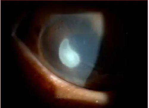
Fungal keratitis remains a significant cause of blindness in the developing world. Local predisposing factors include trauma, especially with vegetable or organic matter, contact lenses and topical steroids. Usually, fungal ulcers are much less aggressive than bacterial ulcers. Typically, presentation is a red, painful eye associated with a decrease in visual acuity. These ulcers are slow-spreading and are dull grey in appearance, with feather-like extensions. Generally, satellite lesions and hypopyon are also evident (Figure 15). Successful resolution of the disease depends on a high index of suspicion and early institution of therapy. Thus, immediate referral of these patients is necessary. Management consists of topical and systemic antifungals and cycloplegic drops, as well as treating co-existent bacterial infection that is often present.
Acute angle closure
Acute angle closure is a condition in which the intraocular pressure rises rapidly to very high levels. It represents an ophthalmic emergency and requires immediate referral for specialist intervention. Acute angle closure classically occurs in older, far-sighted patients. Symptoms include severe unilateral eye pain associated with a headache on the same side as the painful eye, Figure 12: Signs of keratitis include an epithelial defect which stains with
Anterior uveitis
Uveitis is defined as inflammation that affects one or more parts of the uveal tract. The uvea is the vascular coat of the eye and consists of the iris, ciliary body and the choroid. Anterior uveitis is the most common form of uveitis and predominantly affects the iris (iritis).2 Usually, presentation occurs with the acute onset of unilateral pain, redness, lacrimation, photophobia and a moderate reduction in visual acuity. The redness is often more marked in the circumcorneal region. The pupil is miotic and irregular due to adhesions that formbetween the pupil margin and the anterior surface of the lens (posterior synechiae) (Figure 17).
Clusters of cells on the corneal endothelium, called keratic precipitates (Figure 18), may also be seen, and if the uveitis is severe, a hypopyon may form. Diagnosis is dependent on slit-lamp examination of the eye so most patients require referral. Initially, topical steroids are used frequently, then tapered over several days to weeks, depending on the severity of the inflammation. Adequate cycloplegia using atropine 1% twice daily is maintained during the course of the treatment to relieve pain and also to dilate the pupil which prevents the formation of posterior synechiae. Figure 13: Dendritic ulcer stained with fluorescein in herpes simplex
keratitis
Figure 14: Herpes zoster ophthalmicus with potential ocular involvement, indicated by the presence of lesions on the tip of the nose (green arrow)
Figure 15: Fungal keratitis with satellite lesions and a hypopyon
Figure 16: In acute angle closure, examination reveals diffuse conjunctival injection, a hazy cornea and a fixed, mid-dilated pupil
Penetrating trauma
Penetrating injuries to the eye are serious as they are potentially blinding. However, life-threatening conditions should be recognised and immediately treated as these take precedence over any associated ocular injuries. Most of these injuries are obvious (Figure 19), although a high index of suspicion is needed to diagnose an occult injury. Signs suggestive of this type of penetrating globe injury include severe conjunctival swelling, an anterior chamber that looks deeper than the other eye, and a soft eye. These patients require urgent referral as surgical repair is mandatory. Manipulation of an open globe can result in exacerbation of the initial injury, as even the smallest amount of pressure placed on the eye can lead to extrusion of the ocular contents, and thus should be avoided. The eye should be covered with a hard, protective plastic shield, not an eye pad, and use of topical medication must be avoided. If a plastic shield is not available, the bottom half of a clean polystyrene cup can be used
eye is most important to limit the extent of damage from such burns. A few litres of sterile isotonic saline should be rinsed through the palpebral opening for 15-30 minutes with the aid of an eyelid speculum and topical anaesthetic to prevent reflex blepharospasm. Complete removal of the offending agents, by sweeping under the eyelids with a wet cotton bud, should also be attempted. The end-point of this initial management is to achieve a pH of between 7.3-7.7 measured with the appropriate block on a urine dipstick. Systemic analgesia, as well as topical dilating drops, also induce comfort. The patient should be referred for further evaluation by an ophthalmologist.
Thermal burns
Usually, these injuries are less severe than their chemical counterparts. Topical antibiotics, dilating drops, as well as systemic analgesics, constitute a good initial empirical approach. The patient should be referred for further evaluation and treatment of possible long-term complications.
Conclusion
This article has outlined a wide variety of conditions with which patients may present. A red eye is implicated in all of them. If the diagnosis is approached systematically by taking a thorough history from the patient and knowing what to look for during the examination, in many cases, it is possible to make the correct diagnosis. Once a diagnosis has been made, it is very important Figure 20: Chemical injury with conjunctival injection, small
subconjunctival haemorrhages and a large staining corneal epithelial defect


![Figure 12: Signs of keratitis include an epithelial defect which stains with fluorescein (yellow arrow), stromal infiltrates (blue arrow) and a hypopyon [pus in the anterior chamber (green arrow)]](https://thumb-us.123doks.com/thumbv2/123dok_us/8782395.1762349/5.595.43.290.67.144/figure-keratitis-epithelial-fluorescein-infiltrates-hypopyon-anterior-chamber.webp)


