Leyla Guler
1, A, C, Fusun Bozkirli
1, A, Nurdan Bedirli
1, A, Yusuf Unal
1, B,
Adem Guler
2, C, Yesim Oztas
3, Sevket Balta
4, D, Mustafa Cakar
5, C,
Sait Demirkol
4, E, Zekeriya Arslan
4, F, Murat Unlu
4, FComparison of the Effects of Dexmedetomidine
vs. Ketamine in Cardiac Ischemia/Reperfusion Injury
in Rats – Preliminary Study
1 Department of Anesthesiology, Gazi University, Ankara, Turkey
2 Department of Cardiovascular Surgery, Gulhane Medical Academy, Etlik-Ankara, Turkey 3 Department of Clinical Biochemistry, Hacettepe University, Ankara, Turkey
4 Department of Cardiology, Gulhane Medical Academy, Etlik-Ankara, Turkey 5 Department of Internal Medicine, Gulhane Medical Academy, Etlik-Ankara, Turkey
A – research concept and design; B – collection and/or assembly of data; C – data analysis and interpretation;
D – writing the article; E – critical revision of the article; F – final approval of article; G – other
Abstract
Objectives. Following ischemia/reperfusion injury, antioxidant defense mechanisms may remain insufficient depending on the duration of ischemia which is caused by any reason (MI, after percutaneous coronary inter-vention, during cardiac surgery). After that, free oxygen radicals increasing within the cell cause structural dete-rioration. Cytokines which activate a series of reactions that cause tissue damage and inflammatory response are released during reperfusion of ischemic tissues. In this study, we aimed to compare the effects of dexmedetomidine and ketamine in cardiac ischemia/reperfusion injury.
Material and Methods. The study included 18 rats randomly divided into three groups. Group I/R (n = 6): con-trol, Group I/R-K (n = 6): ketamine, and Group I/R-D (n = 6): dexmedetomidine. Before the 10 min surgery, after the 20 min ischemia and 20 min reperfusion period, hemodynamic parameters were compared among the three groups. After the 45 min ischemia and 120 min reperfusion period, tissue samples were obtained from the rat hearts, and MDA, SOD, GSH-Px, IL-1β and TNF-α levels were compared.
Results. MDA and GSH-Px levels were significantly higher in the control group compared to the ketamine and dexmedetomidine groups. However, both levels were similar in the ketamine and dexmedetomidine groups. SOD levels were significantly lower in the ketamine and dexmedetomidine groups compared to the control group, but they were similar in the ketamine and dexmedetomidine groups. IL-1β levels were similar in all groups. TNF-α levels were significantly lower in the ketamine and dexmedetomidine groups compared to the control group. They were similar in the ketamine and dexmedetomidine groups.
Conclusions. According to our study, it can be concluded that dexmedetomidine and ketamine have similar effects on reducing myocardial ischemia reperfusion injury. Dexmedetomidine provides better heart rate control but causes hypotension, so, because of cardiac depression, we think that its clinical use may necessitate further inves-tigation (Adv Clin Exp Med 2014, 23, 5, 683–689).
Key words: dexmedetomidine, ketamine, cardiac ischemia/reperfusion injury.
Adv Clin Exp Med 2014, 23, 5, 683–689 ISSN 1899–5276
ORIGINAL PAPERS
© Copyright by Wroclaw Medical University
Coronary artery disease (CAD) is one of the most common causes of morbidity and mortali-ty at the present time and is also responsible for about half of the deaths in developed countries. CAD is the result of the accumulation of athero-matous plaques within the walls of the coronary arteries. The deposition of the plaque in the lumen
of an artery causes a narrowing of the lumen of the artery by decreasing its diameter. Atherosclerotic plaques reduce the blood flow due to narrowing of the lumen and coronary artery vasospasm.
ischemia, thrombotic stroke, hemorrhagic shock, and surgical procedures such as organ transplan-tation as well as thrombolytic therapy. When these tissues are trying to provide the necessary ener-gy for vital functions via anaerobic metabolism in an ischemia area, increased metabolic residues and metabolic asidosis occur in this area due to re-duced perfusion. Cell membrane permeability is increased and then cell swelling occurs. The oxi-dation of metabolites is spread to the whole body by systemic circulation after reperfusion flow [1].
Free oxygen radicals are the most important toxic substance as a result of the oxygen to reach the site of ischemia [2]. In normal healthy conditions, formation of free oxygen radicals and protective an-tioxidant mechanisms to clean the free oxygen rad-icals are in a state of equilibrium. Antioxidant de-fense systems try to eliminate the excessive amount of free oxygen radical production. If the amount of free oxygen radicals is increased, antioxidant de-fense systems may be inadequate and severe reper-fusion injury may occur. Various markers are used for detection of the damage. For the determination of the level of lipid peroxidation, malondialdehyde was measured in this study. On the other hand, in order to determine the degree of response of the su-peroxide radicals and H2O2, superoxide dismutase
and glutathione peroxidase levels were also mea-sured in heart tissue in this study.
We aimed to investigate the association between the effects of adjuvant analgesics in balanced anes-thesia with ketamine and dexmedetomidine and sys-temic cytokines such as interleukin-1 beta (IL-1β), tumor necrosis factor-alpha (TNF-α), malondialde-hyde (MDA), glutathione peroxidase (GSH-Px) and superoxide dismutase (SOD) levels in a model of rat cardiac ischemia/reperfusion (I/R).
Material and Methods
Sample Collections
All animal procedures were approved by the institutional committee on the care and use of ani-mals of our institution. Eighteen male Wistar rats (275–335 g) provided by the animal laboratory of our institute were used in the experiments. There were three groups in the study: I/R-K, I/R-D and control groups. Before the experiments, the animals were fed on standard rat chow and water ad libi-tum and housed in identical cages with controlled temperature and 12-hour light/dark cycle for at least one week. The study was based on an in vi-vo, randomized, controlled, single-blind, prospec-tive, experimental I/R model. The study included 18 rats divided into 3 groups randomly. Group I/R
(n = 6): control, Group I/R-K (n = 6): ketamine, and Group I/R-D (n = 6): dexmedetomidine. We performed temporary occlusion of the left anteri-or descending artery with a snare. After the 45 min ischemia, a 120 min reperfusion period was per-formed by loosening the snare. One mg/kg/min of intravenous ketamine (Ketalar® 500 mg vial of
in-jectable Pfizer, Turkey) was infused in the I/R-K group. After 1 µ/kg intravenous dexmedetomidine loading dose in 10 min, 1 µ/kg/h infusion of dex-medetomidine (Precedex® IV concentrate for
solu-tion for infusion vial containing 200 mg/2 mL, Ab-bott, Turkey) was given in the standard volume to the I/R-D group [3].
Before the 10 min surgery, after the 20 min ischemia and 20 min reperfusion period, hemody-namic parameters were compared among the three groups. After the 45 min ischemia, 120 min reper-fusion period, tissue samples were obtained from the rat hearts, and MDA, SOD, GSH-Px, IL-1β, TNF-α levels were compared.
Biochemical Analysis
The determination of the level of lipid perox-idation was assessed with MDA, the extent of an-tioxidant response against superoxide radical was determined by SOD levels and GSH-Px levels, in fighting with H2O2, were measured in the heart
tis-sue. In addition, tissue protein levels were also calcu-lated in order to express these results numerically.
Measurements of MDA, SOD
and GSH-Px Levels
The MDA levels were calculated using the method described by Draper HH [4]. We calculat-ed superoxide dismutase activity using the method described by Sun et al. [5]. We measured the activ-ity of the GSH-Px enzyme according to the meth-ods described by Paglia and Valen [6].
Measurements of TNF-α
and IL-1β Levels
Proinflammatory tissue cytokines (TNF-α, IL-1β) levels were measured using commer-cial ELISA kits according to the manufacturer’s instructions.
Statistical Analysis
Chicago, IL, USA) was used for statistical analyses. Continuous variables were given as mean ± stan-dard deviation and categorical variables were de-fined as percentages. The data was tested for nor-mal distribution using the Kolmogorov-Smirnov test. To compare continious variables, a one-way analysis of variance test or Kruskall-Wallis test was used as appropriate. When a significant difference was observed between the 3 groups, post hoc tests (Tukey test or Mann-Whitney U) were used for the determination of the difference between couples. A Pearson correlation test was used to evaluate the degree of association between examined variables. To compare categorical variables, a chi-square test was used. Statistical significance was defined as p < 0.05.
Results
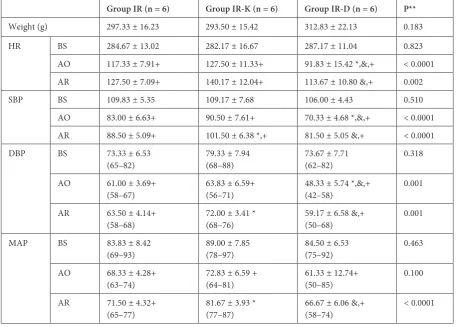
There was no statistically significant difference between the weights of the rats (p > 0.05). There was no statistically significant difference between the heart rate of the rats 10 min before the surgery (Table 1). In all groups, average values of heart rate (HR), SBP (systolic blood pressure) and DBP (di-astolic blood pressure) were lower in the 20 min after LAD occlusion and 20 min after reperfusion than 10 min before surgery. After 20 min of LAD occlusion, HR, SBP and DBP were compared to the average values of these groups. There were statisti-cally significantly lower values in the I/R-D group (p < 0.005). In addition, 20 min after reperfusion, HR, SBP, mean arterial pressure (MAP) and the mean values of DBP were statistically significant in the I/R-D group compared to the I/R-K group (p < 0.005) (Table 1).
Table 1. The comparison of the weights of the rats and hemodynamic parameters of both groups
Group IR (n = 6) Group IR-K (n = 6) Group IR-D (n = 6) P**
Weight (g) 297.33 ± 16.23 293.50 ± 15.42 312.83 ± 22.13 0.183
HR BS 284.67 ± 13.02 282.17 ± 16.67 287.17 ± 11.04 0.823
AO 117.33 ± 7.91+ 127.50 ± 11.33+ 91.83 ± 15.42 *,&,+ < 0.0001 AR 127.50 ± 7.09+ 140.17 ± 12.04+ 113.67 ± 10.80 &,+ 0.002
SBP BS 109.83 ± 5.35 109.17 ± 7.68 106.00 ± 4.43 0.510
AO 83.00 ± 6.63+ 90.50 ± 7.61+ 70.33 ± 4.68 *,&,+ < 0.0001 AR 88.50 ± 5.09+ 101.50 ± 6.38 *,+ 81.50 ± 5.05 &,+ < 0.0001
DBP BS 73.33 ± 6.53
(65–82) 79.33 ± 7.94(68–88) 73.67 ± 7.71(62–82) 0.318 AO 61.00 ± 3.69+
(58–67) 63.83 ± 6.59+(56–71) 48.33 ± 5.74 *,&,+(42–58) 0.001 AR 63.50 ± 4.14+
(58–68) 72.00 ± 3.41 *(68–76) 59.17 ± 6.58 &,+(50–68) 0.001
MAP BS 83.83 ± 8.42
(69–93) 89.00 ± 7.85(78–97) 84.50 ± 6.53(75–92) 0.463 AO 68.33 ± 4.28+
(63–74) 72.83 ± 6.59 +(64–81) 61.33 ± 12.74+(50–85) 0.100 AR 71.50 ± 4.32+
(65–77) 81.67 ± 3.93 *(77–87) 66.67 ± 6.06 &,+(58–74) < 0.0001
IR – ischemia/reperfusion; K – ketamine; D – dexmedetomidine; SBP – systolic blood pressure; DBP – diastolic blood pres-sure; MAP – mean arterial prespres-sure; HR – heart rate; BS – 10 min before surgery; AO – 20 min after left anterior descending coronary artery occlusion; AR – 20 min after reperfusion.
Values are mean ± SD.
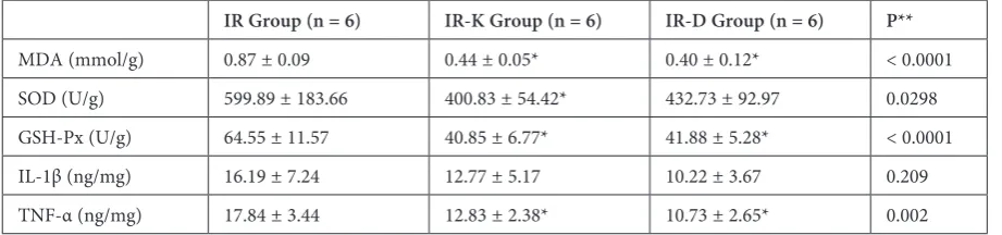
In our study, MDA levels in the I/R group were significantly higher than the I/R-K and I/R-D groups (p < 0.0001). MDA levels were not statis-tically significant in the I/R-K and I/R-D groups. The levels of GSH-Px in the I/R group were sig-nificantly higher than the I/R-K and I/R-D groups (p < 0.0001). The levels of GSH-Px were similar in the I/R-K and I/R-D groups. The levels of SOD in the I/R group were significantly higher than the I/R-K and I/R-D groups. The levels of SOD in the I/R-K and I/R-D groups were similar. The levels of IL-1β were similar in all groups in our study (p = 0.209). In contrast, the levels of TNF-α in the I/R group were significantly higher than the I/R-K and I/R-D groups. The levels of TNF-α in the I/R-K and I/R-D groups were not different (p = 0.002) (Table 2).
Discussion
In order to maintain a healthy life, pro-oxidant and antioxidant defense mechanisms should work in a balanced way due to possible development of serious damage to any cell. Ischemia is seen for many reasons such as MI, percutaneous coronary intervention and cardiac surgery. After ischemia to reperfusion, antioxidant defense mechanisms may be insufficient depending on the duration and in-crease of free oxygen radicals in cells, so it can re-sult in damage to various degrees [7]. Depending on reperfusion injury in myocardial cells, some-times the damage is caused as much as by isch-emia, sometimes it is seen frequently. Because free oxygen radicals are more responsible for reperfu-sion injury, tissue damage may be reduced by de-creasing free oxygen radicals in the ischemic area.
Although there are significant improvements to protect the body from ischemia/reperfusion injury in the myocardial tissue, the ideal drug, technique,
solution and methods for this period have not been clearly defined during reperfusion yet. This situa-tion may be due to the complexity or the patho-physiology of ischemia-reperfusion injury.
Studies have focused on the inhibition of the production of free oxygen radicals in the patho-genesis of the cell injury, determining the role of antioxidant mechanisms and the trials of antioxi-dant free radical scavenger therapies [8].
Vitamins, ACE inhibitors, NO donors, ade-nosine, Na+-H+ exchanger inhibitors, glutamate, aspartate, aprotinin, methylprednisolone, Ca++ channel blockers, ATP-sensitive K+ channel open-ers and glucose-insulin-K+ solutions, prostaglan-dins, glutathione, N-acetylcysteine, pentoxifylline, C1 esterase inhibitors, endothelin-1 receptor an-tagonists and various anesthetic agents have been used in previous studies for this purpose.
There are limited studies on the effect of ket-amine and dexmedetomidine on cardiac I/R injury. We aimed to contribute to the topic in our exper-imental study. We aimed to provide a temporary occlusion of the LAD artery that gives the dom-inant myocardial perfusion, then started reperfu-sion by eliminating the provireperfu-sion of temporary oc-clusion, meanwhile hemodynamic and biochemical changes in rats following drugs was established for the purpose of identification. The same anesthet-ic medanesthet-ications and surganesthet-ical techniques are used in this study for all groups, the hemodynamic and ar-terial pressure and heart rate did not differ between the records 10 min before surgery.
This result is also an indication that the prac-tices are the same on all the subjects. After 20 min of LAD occlusion, HR, SBP and DBP were com-pared to the average values of these groups, there were statistically significantly lower values in the I/R-D group. In addition, 20 min after reperfu-sion, HR, SBP, MAP and the mean values of DAB were statistically significant in the I/R-D group
Table 2. The comparison of oxidative status parameters of both groups
IR Group (n = 6) IR-K Group (n = 6) IR-D Group (n = 6) P**
MDA (mmol/g) 0.87 ± 0.09 0.44 ± 0.05* 0.40 ± 0.12* < 0.0001
SOD (U/g) 599.89 ± 183.66 400.83 ± 54.42* 432.73 ± 92.97 0.0298
GSH-Px (U/g) 64.55 ± 11.57 40.85 ± 6.77* 41.88 ± 5.28* < 0.0001
IL-1β (ng/mg) 16.19 ± 7.24 12.77 ± 5.17 10.22 ± 3.67 0.209
TNF-α (ng/mg) 17.84 ± 3.44 12.83 ± 2.38* 10.73 ± 2.65* 0.002
IR – ischemia/reperfusion; K – ketamine; D – dexmedetomidine; MDA – malondialdehyde; SOD – superoxide dismutase; GSH-Px – glutathione peroxidase; IL-1β – interleukin-1 beta; TNF-α – tumor necrosis factor-alpha.
Values are mean ± SD.
compared to the I/R-K group. These results sup-ported that dexmedetomidine is an effective agent for controlled hypotension as it reduces HR and MAP, does not cause reflex tachycardia and blocks sympathetic system response [9]. The mean val-ues of HR, SBP, MAP and DBP were significant-ly higher in the I/R-K group than the I/R-D group. These findings support the stimulant effect of ket-amine on the cardiovascular system. In all groups, the average values of HR, SBP and DBP were low-er in 20 min aftlow-er LAD occlusion and 20 min af-ter reperfusion than 10 min before surgery. We be-lieve that the main reason for this condition was the unmet oxygen and energy demand of the myo-cardium by the occlusion of the LAD and CAD and arterial pressures were lower due to myocar-dial depression.
Ketamine increases arterial blood pressure and heart rate by approximately 30% with a stimulat-ing effect on the cardiovascular system and this condition returns to normal within 20–30 min. Sloan et al. [10] reported that the infarct area mea-sured in the group treated with high-dose ket-amine was significantly smaller than in the group receiving low-dose ketamine. The ECG and hemo-dynamic parameters were similar in both groups after 20 min ischemia and 2-h reperfusion. Reg-ueiro et al. [11], compared sevoflurane, ketamine, midazolam and atropine after 75 min LAD occlu-sion. In the sevoflurane group, hemodynamic sta-bility was better and total mortality and ventricu-lar arrhythmias were less reported. In their study assessing the infarct area and cardiac output, Mül-lenheim et al. [12] reported that ketamine potenti-ated the protective effect of ischemic precondition-ing in the heart. The authors demonstrated that dexmedetomidine may rarely cause bradycardia and sinusal arrest. It also decreased oxygen con-sumption and reduced the level of serum lactate in an ischemic heart. Dexmedetomidine led to blood flow from the non-ischemic zone to the ischemic zones during acute occlusion. It also causes an in-crease in the endocardial-epicardial blood flow ratio, shown to be 35% in previous experimental studies [13, 14].
In the event of intraoperative tachycardia re-fractory to esmolol during revascularization, dex-medetomidine has been reported to decrease heart rate [15]. Dexmedetomidine supresses central ad-renergic hormone secretion, and thus reduces the levels of plasma catecholamines causing the sup-pression of cardiac stimulant effects during the stress response in the surgery. Dexmedetomidine is also shown to have strong anesthetic and anal-gesic effects [16, 17].
In myocardial I/R studies, it has been shown that free oxygen radicals increased significantly in
myocardial cells during reperfusion. An increase of free oxygen radicals in the cell membrane is one of the most important cell injuries affecting the lipids of the cell structure. Some authors believe that lip-id peroxlip-idation plays a key role in I/R injury [18]. Free oxygen radicals initiate the lipid peroxidation of polyunsaturated fatty acids by a hydrogen atom ultimately making hydroperoxides. As a result of these reactions, the cell membrane loses fluidity and the membrane loses its integrity. This condi-tion leads to a release of cell fraccondi-tions into the en-vironment and then leads to cell death. On the oth-er hand, the subcellular structures released to the environment trigger inflammatory events, and the cellular damage gets worse [19]. One of the most important markers of lipid peroxidation in tissues used as an indicator is determination of the level of MDA [20]. High levels are an indicator of higher lipid peroxidation.
In our study, MDA levels in the I/R group were significantly higher than in the I/R-K and I/R-D groups. In the I/R-K and I/R-D groups, the MDA levels were compared with each other and the I/R-D group was found to be lower, but they were statis-tically similar. This result is an indicator that ket-amine and dexmedetomidine are effective in reduc-ing I/R injury. Salman et al. [21] found that after I/R with ketamine infusion, plasma MDA levels de-crease and it can reduce lipid peroxidation in mus-cle tissue. Öksüz et al. [22] compared the cardiopro-tective effect of propofol and ketamine, and MDA levels were lower in both groups than in the control group. Our results are similar to the literature.
One of the major markers that can be used in decreasing ischemia/reperfusion injury hypothesis is glutathione peroxidase. Glutathione acts as a nat-ural cleaner against superoxide anions and tries to protect the integrity of the cell against oxidation. A decrease in glutathione peroxidase activity leads to severe cell damage with an increase in hydro-gen peroxide [23]. The increase in the activity of glutathione peroxidase is an indicator of clean-ing hydrogen peroxide. So there is a reduction in the probability of occurrence of damage to the cell membrane due to superoxide radicals. In our study, the levels of GSH-Px in the I/R group were signifi-cantly higher than in the I/R-K and I/R-D groups. The levels of GSH-Px were similar in the I/R-K and I/R-D groups. These results show that ketamine and dexmedetomidine reduces oxidative stress.
One of the other hypotheses that can be used in ischemia/reperfusion injury is superoxide dis-mutase. Superoxide dismutase catalyzes the con-version of free oxygen radicals and H2O2. It is
oxygen toxicity in the body [24]. The increase in activity of this enzyme indicates that there are large amounts of superoxide radicals and a power-ful cleaning action in this area.
In our study, the levels of SOD in the I/R group were significantly higher than in the I/R-K and I/R-D groups. The levels of SOD in the I/R-K and I/R-D groups were similar. Ketamine and dexme-detomidine reduced the oxidative stress in the en-vironment, and associated with this, SOD enzyme activity had a lower rate of increase.
When activated, tissue macrophages release free oxygen radicals, lysosomal enzymes, myelo-peroxidase, TNF-α, IL-2 and IL-6, and these proin-flammatory cytokines can be used as indicators of I/R injury [25, 26]. The levels of IL-1β were not dif-ferent between the groups in our study. In contrast, the levels of TNF-α in the I/R group were signifi-cantly higher than in the I/R-K and I/R-D groups. The levels of TNF-α in the I/R-K and I/R-D groups were not different. This result may indicate a de-creased triggering role of neutrophil activation in reperfusion injury.
Riha et al. [27] demonstrated that cardiac tro-ponin-I and CK-MB levels were found to be lower in dexmedetomidine and ketamine anesthesia. En-gelhard et al. [3] found that the induction of apop-tosis began after I/R 30 min of ischemia and af-ter 240 min reperfusion. This effect is reduced by dexmedetomidine and ketamine anesthesia. A ket-amine-medetomidine combination can provide hemodynamic control during hemorrhagic shock because of the high MAP and low HR.
The main limitation of our study is the rela-tively small sample sizes in each animal group. This may increase the questionability of the study results.
In this experimental study, dexmedetomidine and ketamine reduced myocardial ischemia reper-fusion injury, which is similar to previous findings. In addition, dexmedetomidine provides a better control of heart rate but may cause hypotension. This anesthetic agent should be used with caution in clinical practice because of this effect. We be-lieve that these findings should be supported with larger series of clinical studies.
References
[1] Duckrow RB, LaManna JS, Rosenthal M: Disparate recovery of resting and stimulated oxidative metabolism fol-lowing transient ischemia. Stroke 1981, 12, 677–686.
[2] Oeseburg H, De Boer RA, Buikema H, Van der Harst P, Van Gilst WH, Silljé HHW: Glucagon-like peptide 1 prevents reactive oxygen species-induced endothelial cell senescence through the activation of protein kinase A. Arteriosclerosis Thromb Vasc Biol 2010, 30, 1407–1414.
[3] Engelhard K, Werner C, Eberspächer E, Bachl M, Blobner M, Hildt E: The effect of the alpha 2-agonist dexme-detomidine and the N-methyl-D-aspartate antagonist S(+)-ketamine on the expression of apoptosis-regulating proteins after incomplete cerebral ischemia and reperfusion in rats. Anest Analg 2003, 96, 524–531.
[4] Draper HH, Hadley M: Malondialdehyde determination as index of lipid peroxidation. Methods Enzymol 1990, 186, 421–431.
[5] Sun Y, Oberley LW, Li Y: A simple method for clinical assay of superoxide dismutase. Clin Chem 1988, 34, 497–500.
[6] Paglia DE, Valentine WN: Studies on the quantitative and qualitative characterization of erythrocyte glutathione peroxidase. J Lab Clin Med 1967, 70, 158–169.
[7] Robicsek F, Schaper J: Reperfusion injury: fact or myth? J Card Surg 1997, 12, 133–137, discussion 138.
[8] Dhalla NS, Elmoselhi AB, Hata T, Makino N: Status of myocardial antioxidants in ischemia-reperfusion injury. Cardiovasc Res 2000, 47, 446–456.
[9] Gerlach AT, Dasta JF: Dexmedetomidine: an updated review. Ann Pharmacother 2007, 41, 245–252.
[10] Sloan RC, Rosenbaum M, O’Rourke D, Oppelt K, Frasier CR, Waston CA: High doses of ketamine-xylazine anesthesia reduce cardiac ischemia-reperfusion injury in guinea pigs. JAALAS 2011, 50, 349–354.
[11] Regueiro-Purriños M, Fernández-Vázquez F, De Prado AP, Altónaga JR, Cuellas-Ramón C, Ajenjo-Silverio JM: Ventricular arrhythmias and mortality associated with isoflurane and sevoflurane in a porcine model of myo-cardial infarction. JAALAS 2011, 50, 73–78.
[12] Müllenheim J, Frässdorf J, Preckel B, Thämer V, Schlack W: Ketamine, but not S(+)-ketamine, blocks ischemic preconditioning in rabbit hearts in vivo. Anesthesiology 2001, 94, 630–636.
[13] Willigers HM, Prinzen FW, Roekaerts PM, De Lange S, Durieux ME: Dexmedetomidine decreases perioperative myocardial lactate release in dogs. Anesth Analg 2003, 96, 657–664.
[14] Roekaerts PM, Prinzen FW, Willigers HM, De Lange S: The effects of alpha 2-adrenergic stimulation with mivaz-erol on myocardial blood flow and function during coronary artery stenosis in anesthetized dogs. Anesth Analg 1996, 82, 702–711.
[15] Ruesch S, Levy JH: Treatment of persistent tachycardia with dexmedetomidine during off-pump cardiac surgery. Anesth Analg 2002, 95, 316–318.
[16] Ramsay MAE, Luterman DL: Dexmedetomidine as a total intravenous anesthetic agent. Anesthesiology 2004, 101, 787–790.
[18] Tappel AL: Lipid peroxidation damage to cell components. Federation Proceedings 1973, 32, 1870–1874.
[19] Jaeschke H, Smith CW, Clemens MG, Ganey PE, Roth RA: Mechanisms of inflammatory liver injury: adhesion molecules and cytotoxicity of neutrophils. Toxicol Appl Pharmacol 1996, 139, 213–226.
[20] Xu Y, Liu B, Zweier JL, He G: Formation of hydrogen peroxide and reduction of peroxynitrite via dismutation of superoxide at reperfusion enhances myocardial blood flow and oxygen consumption in postischemic mouse heart. J Pharmacol Exp Ther 2008, 327, 402–410.
[21] Salman AE, Dal D, Salman MA, Iskit AB, Aypar U: The effect of ketamine on acute muscular ischaemia reperfu-sion in rats. Eur J Anaesthesiol 2005, 22, 712–716.
[22] Oksuz H, Senoglu N, Yasim A, Turut H, Tolun F, Ciralik H: Propofol with N-acetylcysteine reduces global myo-cardial ischemic reperfusion injury more than ketamine in a rat model. Invest Surg Res 2009, 22, 348–352.
[23] Wheeler CR, Salzman JA, Elsayed NM, Omaye ST, Korte DW: Automated assays for superoxide dismutase, catalase, glutathione peroxidase, and glutathione reductase activity. Anal Biochem 1990, 184, 193–199.
[24] Johnson F, Giulivi C: Superoxide dismutases and their impact upon human health. Mol Aspects Med 2005, 26, 340–352.
[25] Hillegass LM, Griswold DE, Brickson B, Albrightson-Winslow C: Assessment of myeloperoxidase activity in whole rat kidney. J Pharmacol Methods 1990, 24, 285–295.
[26] Siegers CP, Riemann D, Thies E, Younes M: Glutathione and GSH-dependent enzymes in the gastrointestinal mucosa of the rat. Cancer Lett 1988, 40, 71–76.
[27] Ríha H, Kotulák T, Březina A, Hess L, Kramář P, Szárszoi O: Comparison of the effects of ketamine-dexmedeto-midine and sevoflurane-sufentanil anesthesia on cardiac biomarkers after cardiac surgery: an observational study. Physiol Res/Academia Scientiarum Bohemoslovaca 2012, 61, 63–72.
Address for correspondence
Sevket Balta
Department of Cardiology Gulhane School of Medicine Tevfik Saglam St.
06018 Etlik-Ankara Turkey
Tel: +90 312 304 42 81 E-mail: [email protected]
Conflict of interest: None declared