BIROn - Birkbeck Institutional Research Online
Dumontheil, Iroise and Hillebrandt, H. and Apperly, I.A. and Blakemore, S.J.
(2012) Developmental differences in the control of action selection by social
information. Journal of Cognitive Neuroscience 24 (10), pp. 2080-2095.
ISSN 0898-929X.
Downloaded from:
Usage Guidelines:
Please refer to usage guidelines at or alternatively
Developmental Differences in the Control of Action
Selection by Social Information
Iroise Dumontheil
1*, Hauke Hillebrandt
1*, Ian A. Apperly
2,
and Sarah-Jayne Blakemore
1Abstract
■ Our everyday actions are often performed in the context of a social interaction. We previously showed that, in adults, selecting an action on the basis of either social or symbolic cues was asso-ciated with activations in the fronto-parietal cognitive control net-work, whereas the presence and use of social versus symbolic cues was in addition associated with activations in the temporal and medial prefrontal cortex (MPFC) social brain network. Here we investigated developmental changes in these two networks. Fourteen adults (21–30 years of age) and 14 adolescents (11– 16 years) followed instructions to move objects in a set of shelves. Interpretation of the instructions was conditional on the point of view of a visible“director”or the meaning of a symbolic cue (Director Present vs. Director Absent) and the number of poten-tial referent objects in the shelves (3-object vs. 1-object). 3-object
trials elicited increased fronto-parietal and temporal activations, with greater left lateral prefrontal cortex and parietal activations in adults than adolescents. Social versus symbolic information led to activations in superior dorsal MPFC, precuneus, and along the superior/middle temporal sulci. Both dorsal MPFC and left temporal clusters exhibited a Director × Object interaction, with greater activation when participants needed to consider the di-rectorsʼviewpoints. This effect differed with age in dorsal MPFC. Adolescents showed greater activation whenever social infor-mation was present, whereas adults showed greater activation only when the directorsʼviewpoints were relevant to task per-formance. This study thus shows developmental differences in domain-general and domain-specific PFC activations associated with action selection in a social interaction context. ■
INTRODUCTION
How is an appropriate action selected among many pos-sibilities? How do we decide to pick a particular fruit out of many on offer at the supermarket? The PFC is thought to support the integration of information from the current environment (the smell, color, appearance, and identity of melons on stand) and internal information generated more or less remotely in time (the plan to invite friends for dinner, the decision to have melon as a starter, but also the value associated with melons compared with other fruits); to orient attention appropriately (toward the stand and the most appetizing looking melon); and to select an action appropriate with the current goals (picking up the selected melon; e.g., Burgess, Gilbert, & Dumontheil, 2007; Koechlin & Summerfield, 2007; Fuster, 2000; Baron-Cohen, Leslie, & Frith, 1986). Using visual search paradigms, previous research has investigated the selection of targets combining different properties in arrays of simple stimuli (Humphreys, Allen, & Mavritsaki, 2009; Davis & Palmer, 2004; Booth et al., 2003). Results suggest a dissociation between bottom–up (spontaneous orientation toward a stimulus) and top–down (intentionally driven by knowl-edge, expectations, and goals) processes of visuospatial
selective attention (Beck & Kastner, 2009; Hahn, Ross, & Stein, 2006). However, few studies have attempted to use more complex stimuli.
A large number of our everyday actions are performed in a social interactive context. If you are paying at a shop checkout, you need to perform a series of actions that en-sures the shopkeeper knows whether you wish to pay by cash or credit card, and so on. These interactions are sometimes based on verbal communication and, at other points, on communicative gestures. For example, making sure the shopkeeper can see you have taken your credit card out of your wallet informs the shopkeeper that you want to pay by card. This type of interaction relies on an understanding of other peopleʼs mental states, also called theory of mind or mentalizing (Frith & Frith, 2007). Thus, we often need to use the theory of mind to make top– down decisions and select actions that are appropriate to the inferred mental state of the people we interact with in complex real-world situations. The combination of domain-general processes supporting selective attention, action selection and cognitive control, and cognitive pro-cesses that may be specific to social information and men-talizing, is the focus of this study. PFC is involved in both top–down action selection (Burgess et al., 2007) and men-talizing (Frith & Frith, 2007), in particular, in ill-structured or novel situations (Apperly, 2011). Following previous
1
work in adults (Dumontheil, Küster, Apperly, & Blakemore, 2010), the aim of the current study was to investigate the development of these domain-general and domain-specific processes during adolescence, using fMRI and a paradigm that permits the comparison of action selection in a social versus nonsocial context.
Adolescence is a period of social and psychological de-velopment during which social awareness and behavior undergo profound change (Brown, 2004; Eisenberg & Morris, 2004). At the same time, higher cognitive control and reasoning abilities mature both in terms of behavior and brain function (Dumontheil & Blakemore, 2012; Crone & Ridderinkhof, 2010; Luna, Padmanabhan, & OʼHearn, 2010; Crone, 2009). As well as alterations in hor-mone levels and social environment, a possible cause of these developmental changes are structural changes taking place in brain areas involved in social cognition, including the medial PFC (MPFC), the superior temporal cortex, and the TPJ (Saxe, 2006; Frith & Frith, 2003; Gallagher & Frith, 2003), and in brain regions involved in cognitive control tasks, in particular, lateral parts of PFC. Notably, frontal and temporal lobes undergo protracted structural develop-ment in humans (Shaw et al., 2008; Gogtay et al., 2004; Giedd et al., 1999; Sowell, Thompson, Holmes, Jernigan, & Toga, 1999). Developmental functional imaging studies of mental state attribution have consistently shown that the MPFC activity during a variety of mentalizing tasks (e.g., understanding irony or thinking about oneʼs intentions) decreases between adolescence and adulthood (see Burnett, Sebastian, Cohen-Kadosh, & Blakemore, 2011; Blakemore, 2008, for review). In addition, there is evidence of develop-mental changes in functional connectivity between MPFC and other parts of the mentalizing network during adoles-cence (Burnett & Blakemore, 2009). The age-related changes lie in the part of the MPFC superior toz= 0 la-beled anterior rostral medial frontal cortex (MFC), which is recruited by tasks involving self-knowledge, person per-ception, and mentalizing, in contrast to a more ventral
“orbital MFC”region and a more posterior “posterior rostral MFC”region (Amodio & Frith, 2006). Other groups use slightly different subdivisions of the MPFC, with a more superior border atz= 20 (Van Overwalle, 2009) to distin-guish between “ventral MPFC”and“dorsal MPFC.”This dissociation has been related to a distinction between men-talizing judgments made toward the self or similar others versus on dissimilar others (Tamir & Mitchell, 2010; Van Overwalle, 2009; Jenkins, Macrae, & Mitchell, 2008; Mitchell, Macrae, & Banaji, 2006). The developmental cog-nitive control literature has shown less consistent findings, with studies showing both decreases and increases in lateral PFC activations as well as qualitative changes with age during adolescence (see Crone & Ridderinkhof, 2010; Luna et al., 2010; Astle & Scerif, 2009; Casey, Jones, & Hare, 2008; Bunge & Wright, 2007).
The ability to infer mental states, or mentalizing, devel-ops in a step-wise fashion during the first 5 years of life (Frith & Frith, 2003) at which point children are able to
reason explicitly about theory of mind. However, this abil-ity does not always lead to an automatic, on-line use of theory of mind information even in adolescents and adults. For instance, Keysar and colleagues (Keysar, Lin, & Barr, 2003; Keysar, Barr, Balin, & Brauner, 2000) de-signed a task in which participants were faced with a set of shelves containing objects that were either visible or not visible from the viewpoint of a“Director”(a confed-erate). The director asked participants to move objects in the shelves, and critical instructions required participants to use information about the directorʼs viewpoint to cor-rectly interpret their instructions. In this Director task, adults did not reliably use their theory of mind knowledge to interpret the intentions of others (Keysar et al., 2000, 2003). Around 50% of the time, they failed to use infor-mation about the directorʼs perspective and instead erro-neously used their own (egocentric) viewpoint only when trying to follow the directorʼs instruction. These results were replicated using a computerized version of the para-digm and controlling for the inhibitory control demands of the task with a matched no-director condition (Apperly et al., 2010). We have further found that egocentric errors in this task are accentuated in adolescence, with an even stronger egocentric bias observed in 14- to 17-year-old adolescents than in young adults (Dumontheil, Apperly, & Blakemore, 2010). These results suggest that the ability to take another personʼs perspective to select appropriate actions is still undergoing development at this relatively late stage. The Director task differs from other theory of mind tasks in that it requires participants to have a func-tioning theory of mind, to compute the perspective and intentions of another person (the director), and use this theory of mind information in concert with other cogni-tive processes such as execucogni-tive functions to overcome their egocentric bias and select the appropriate response quickly and accurately (Apperly et al., 2010). It is proposed that it is this interaction between theory of mind and ex-ecutive functions that continues to develop in late adoles-cence and is still prone to errors in adults (see Dumontheil, Apperly, et al., 2010).
for the selection of the appropriate action from the alter-native options. Note that, although beyond the scope of the current study, the computation of the value of different objects or actions may be another important aspect of action selection and may be affected by personal and social factors developing during adolescence.
If mentalizing is a specialized and domain-specific cog-nitive process (e.g., Leslie, 2005; Sperber & Wilson, 2002; Stone, Baron-Cohen, & Knight, 1998; see Apperly, 2011, for discussion), then we might expect differential per-formance and the recruitment of regions of the social brain network (Frith & Frith, 2007; Brothers, 1990) when the guiding information is of social nature compared with more arbitrary symbolic stimuli. This social stimuli con-dition might thus recruit both domain-general action se-lection resources and domain-specific resources that are involved in processing social information and mentaliz-ing. Such a pattern has previously been observed in adults, with nonoverlapping brain regions implicated in response selection and belief attribution during a belief attribution task (Saxe, Schulz, & Jiang, 2006; see also Saxe, Carey, & Kanwisher, 2004).
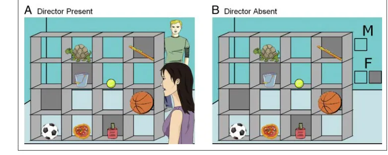
Participants followed auditory instructions to move ob-jects in a set of shelves. A 2 × 2 factorial design with the factors Director (Director Present vs. Director Absent) and Object (3-object vs. 1-object) was employed. In the Director Present condition, two directors were shown on the display, one female and one male. One of them stood behind the shelves, facing the participant, whereas the other stood on the same side of the shelves as the participant. In the 3-object condition, participants needed to use the social cues, that is, the position of the speaking director, to select and move the appropriate object (see Figure 1A). The instructions in these blocks referred to an object that was one of three exemplars in the shelves (the ball). Two of these objects could correspond to the heard instruction (“Move the large ball up”)
de-pending on the directorʼs viewpoint. The largest ball (or equivalent) was always located in a closed shelf (not visible from the back), whereas the second largest ball was located in an open shelf. Importantly, when consider-ing the identity of the director (male voice), only one of the objects was the appropriate response (the football); the other object corresponded to the other viewpoint and was thus a distractor (the basketball). The third object was another type of distractor that did not fit any of the perspectives (the tennis ball). On half of the Director Present 3-object trials, the perspective of the director issu-ing the instruction was different from that of the partici-pant; on the other half, the directorʼs and participantʼs perspectives were the same. This varied on a trial-by-trial basis; thus, participants needed to take into account the directorʼs perspective on every trial. In Director Present 1-object trials, there was no need to take into account the directorʼs perspective to identify the correct object (e.g.,“Move the turtle left”), as there were no distractors or other referents, and so this resembled a bottom–up, visual pop-out as opposed to a visual search (Buschman & Miller, 2007). The Director Absent condition was logically equivalent, but the directors were replaced by symbolic cues (see Figure 1B).
[image:4.612.160.551.537.694.2]We previously obtained results on this task in a group of adult participants (Dumontheil, Küster, et al., 2010). Selec-tion of an appropriate acSelec-tion when faced with alternatives (3-object vs. 1-object contrast, collapsed across Director Present and Director Absent conditions) was associated with domain-general bilateral brain activations located pri-marily in the fronto-parietal cortex, with additional activa-tions in the inferior temporal cortex. Processing of social (Director Present) versus symbolic information (Director Absent) was associated with specific activations in the superior dorsal MPFC and STS. Finally, using perspective taking in this communicative context (Director × Object interaction), which required participants to think not only
Figure 1. Examples of a 3-object trial in the Director Present (A) and the Director Absent (B) conditions. In both conditions in this example, participants hear the instruction:“Move the large ball up”in either a male or female voice. In both examples, if the voice is female, the object to be moved would be the basketball, because in the Director Present condition (A) the female Director is standing in front of the shelves and can see all the
about what the other person sees, but also about his or her intentions, led to further recruitment of superior dorsal MPFC and the left middle temporal gyri, extending into the temporal pole.
The focus of the current study was the difference be-tween the adolescent and adult groups in these three con-trasts of interests: (1) 3-object > 1-object; (2) Director Present > Director Absent; and (3) Director × Object. In line with the adult data, we expected activation in the cognitive control fronto-parietal regions for the first com-parison and activation in the social brain network in the latter two comparisons, which contrast the presence of social versus symbolic information and the specific per-spective taking requirement. In terms of developmental effects, for the first comparison, as the direction of changes in parietal and frontal cortex activations with age in cogni-tive control tasks is inconsistent in the literature and may be task dependent, we tested for both developmental increases and decreases in the 3-object vs. 1-object com-parison. Regarding the second and third comparisons, the social brain has shown more reliable decreases be-tween adolescence and adulthood in MPFC activations (Blakemore, 2008) and increases in temporal cortex activa-tions (Burnett et al., 2011; Blakemore, 2008) in a variety of social cognition tasks. Our predictions thus aligned with this previous literature: We predicted decreased MPFC activation and increased temporal cortex activa-tion with age in the Director Present versus Director Absent comparison and/or more specifically in the Director Present 3-object condition, which requires on-line use of perspective taking information. Regarding the MPFC, our prediction was focused on the dorsal MPFC on the basis of our previous results in adults as well as the associa-tion between this region and mentalizing judgments per-formed toward others (in this case, the Directors) rather than the self.
METHODS
Participants
Fourteen adult (mean age = 24.9 years,SD= 3.0 years, range = 21.3–30.6 years) and 14 adolescent (mean age = 14.0 years, SD = 1.6 years, range = 11.6–16.8 years) right-handed female volunteers were included in the analyses (two additional adolescent participants per-formed poorly on the task due to malfunctioning head-phones and were excluded). All participants spoke English fluently and had no history of psychiatric or neurological disorder. Adult participants or the parents of the adolescent participants gave informed consent, and the study was approved by the local ethics com-mittee. General ability was assessed using the two sub-testsʼformat ( Vocabulary and Matrix Reasoning) of the Wechsler Abbreviated Scale of Intelligence (Wechsler, 1999). Estimated IQ normalized for age did not signifi-cantly differ between the adolescents (mean IQ = 122,
SD = 12, range = 106–140) and adults (mean IQ = 110,SD= 10, range = 100–134;t(26) = 0.735,p= .47).
Design and Stimulus Material
Stimuli consisted of sets of 4 × 4 shelves with objects lo-cated in half of the shelves. Five of the shelves had a gray background (Figure 1). On each trial, participants were given instructions via headphones, by either a male or a fe-male voice, to move one of the eight objects in the shelves to a different slot, either up, down, left, or right (note that this was the participantʼs left or right). A 2 × 2 factorial within-subject design was used with the factors Director (Present vs. Absent) and Object (1-object vs. 3-object) vary-ing between blocks.
Director Factor
In the Director Present (DP) condition, the display in-cluded two directors, one female and one male. In the Di-rector Absent (DA) condition, there were no diDi-rectors in the display (Figure 1). Instead, the letters“F”for female and“M”for male were shown beside the shelves. Below each of the letters, there was either one transparent box, which indicated to participants that only objects in open shelves should be moved, or two boxes, one gray and one transparent, which indicated that there was no restric-tion on the participantʼs choice and all objects (both in open shelves and occluded shelves) could be moved. For example, in Figure 1B, if participants heard the male voice say,“Move the large ball up,”they would need to reason that, because the M is above one clear box, they could only pick objects in clear shelves and thus should ignore the basketball in the gray slot and move the football. These rules had precisely the same consequences as the position of the director in the DP blocks. In DP blocks, the physical position of the director issuing the instruction varied on a trial-by-trial basis; similarly, in DA blocks, the M/F rules changed on a trial-by-trial basis.
Object Factor
50% of the trials) to know which was the correct object to move. In DP 1-object blocks, the directorʼs perspective made no difference to the correct interpretation of his or her instructions, and thus, participants could use their own perspective to select the appropriate object on all trials. In the DA condition, perspective taking was not involved.
There were 48 object-shelf configurations, each con-taining eight objects. Sets of three exemplars of the same object were used for 3-object trials (e.g., three drums). These objects differed in either size (large/small) or position (top/bottom) and were distributed so that the smallest/ largest or topmost/ bottommost object identified in the instruction was in a closed shelf and the second smallest/ largest or topmost/bottommost object and the remaining object were in open shelves. Five additional unique objects were distributed in three gray-backed closed shelves and two open shelves. Those objects in the open shelves could be used for 1-object trials.
To move objects, participants used a trackball mouse, rolling the trackball with their thumb and pressing the left mouse button with the index finger of their right hand. On each trial, participants first moved the mouse cursor from the middle of the screen to the selected ob-ject, then clicked on the obob-ject, and dragged it to the appropriate slot, before releasing the mouse button. RTs were calculated as the delay between the presentation of the visual stimulus and the pressing of the mouse button. Accuracy was measured on the basis of which object was moved.
On each trial, the visual stimulus and the auditory in-struction were presented over a period of 2.2 sec, after which the display remained on the screen for another 3.8 sec. Between trials, a blank screen was shown for 200 msec. The task was programmed with Cogent 2000 and Cogent Graphics (www.vislab.ucl.ac.uk/cogent.php) implemented in Matlab 6.5 (Mathworks, Inc., Sherborn, MA). Standardized instructions were read to participants and included example stimuli in which they had to state which objects should be moved for the different directors and voices. A practice session including one block of each of the four conditions was run outside the scanner to ensure that participants understood the task and could perform it correctly. If a participant did not take into account the directorʼs perspective appropriately, this was highlighted and the task requirements were ex-plained again. Participants also practiced using the track-ball mouse ahead of scanning and in the scanner (see Dumontheil, Kuster, et al., 2010).
Participants performed three scanning sessions (two adolescent participants performed two sessions only be-cause of time constraints). Each session consisted of 16 task blocks with four trials in each block. There were four types of experimental block: DA 1-object; DA 3-object; DP 1-object; DA 3-object. Each of the 48 object-shelf con-figurations was shown once in each block type; thus, there were 12 blocks of four trials for each block type.
Task blocks lasted 24.8 sec and were preceded by an in-struction screen presented for 2 sec, which indicated whether the upcoming block was a DP or DA block. The order of the four block types was counterbalanced within and between sessions. A fixation baseline block lasting 20 sec was included after each set of four task blocks.
fMRI Data Acquisition
3-D T1-weighted fast-field echo structural images and
multislice T2-weighted echo-planar volumes with BOLD
contrast (repetition time = 3 sec; echo time = 50 msec; acquisition time = 2.9143 sec) were obtained using a 1.5-T MRI scanner (Siemens TIM Avanto, Erlangen, Germany). Functional imaging data were acquired in two or three scanning sessions lasting approximately 8 min 40 sec each in which 174 volumes were obtained. The first two volumes of each session were discarded to allow for T1equilibrium
effects. Each functional brain volume was composed of 35 axial slices with an in-plane resolution of 3 × 3 × 3 mm, positioned to cover the whole brain. A T1-weighted
anatomical image lasting 5 min 30 sec was acquired after the first two functional sessions for each participant.
Data Analysis
Behavioral Data
RTs and accuracy in all four conditions were recorded and analyzed using a 2 (Age Group: Adolescents vs. Adults) × 2 (Director: DA vs. DP) × 2 (Object: 1-object vs. 3-object) mixed model repeated-measures ANOVA to investigate the effects of each task factor, the interaction between task factors and their interaction with age.
fMRI Data
fMRI image preprocessing and analysis were carried out using SPM8 ( Wellcome Department of Imaging Neuroscience, London, UK), implemented in MATLAB 7.8 (Mathworks, Inc., Sherborn, MA). To correct for movement effects, images were realigned with a fourth-degree B-spline interpolation. These realigned images were corrected for differences in acquisition times and were then normalized to a standard EPI template based on the Montreal Neurological Institute (MNI) reference brain. The resulting 3 × 3 × 3 mm im-ages were finally spatially smoothed with an 8-mm FWHM Gaussian kernel. Analyses of the movement parameters showed that translation within each session was <3 mm in all participants. Mean movement per session was cal-culated for translations and rotations in each direction for each subject. Independentttests were used to compare the means of these values over the sessions between the two age groups. There was no significant difference for translations [allt(26) < .43,p> .67] nor for rotations [all
For each participant, the scanning sessions were treated as separate time series; statistical parametric maps were created and estimated using a general linear model for each time series (Friston et al., 1995). Included in the model were six boxcar regressors, modeling the instruction, fixation and four types of task blocks, plus one event-related regres-sor representing error trials. All regresregres-sors were convolved with a canonical hemodynamic response function and, to-gether with regressors representing residual movement-related artifacts and the mean over scans, comprised the full model for each session. The data and model were high-pass filtered to a cut-off of 1/128 Hz. Parameter estimates calcu-lated from the least mean squares fit of the model to the data were used in four pairwise contrasts comparing each block type with the fixation baseline. These contrasts were entered into a Condition × Age Group × Participant flex-ible factorial design second-level analysis. The factor partici-pant was included to model the repeated aspect of the data. Main effects of Object (3-object > 1-object) and Director (DP > DA and DA > DP) and the interaction between the two factors and with age group were determined using thetstatistic on a voxel-by-voxel basis. Statistical contrasts were used to create SPM{Z} maps thresholded atp< .001 at the voxel level and at family-wise error (FWE)-corrected
p< .05 at the cluster level (corresponding to a minimum cluster size of 82 voxels determined with SPM8). Activations that survived whole-brain FWE correction atp< .05 are indicated. Analyses performed with age as a continuous regressor did not highlight any regions not observed with the age group analyses and are thus not reported. All coor-dinates are given in MNI space. ROI analyses based on the main effect of DP > DA were further performed to test for orthogonal interaction effects between the Director, Ob-ject, and Age Group factors in the brain regions identified as responding to the social stimuli. Additional analyses ex-plored possible continuous effects of age and differences between the seven youngest and seven oldest adolescent
participants and the adults. Note that these analyses are limited by the age range gap between 17 and 21 and under-powered by the smallNof the adolescent subgroups. Sta-tistical threshold for the ROI analyses performed in SPSS wasp< .05 (two-tailed).
RESULTS
Behavioral Results
Accuracy was calculated for each of the four conditions in each session. Four sessions (from three participants) were discarded from further analyses because of poor perfor-mance in one of the conditions (accuracy < 50%). In-cluded in the analyses were thus 14 adults (13 with three sessions, 1 with two sessions) and 14 adolescents (10 with three sessions, 3 with two sessions, 1 with one session). Accuracy and median RTs in correct trials were analyzed using a 2 (Age Group: Adolescent, Adult) × 2 (Director: DP vs. DA) × 2 (Object: 1-object vs. 3-object) mixed model repeated-measures ANOVA.
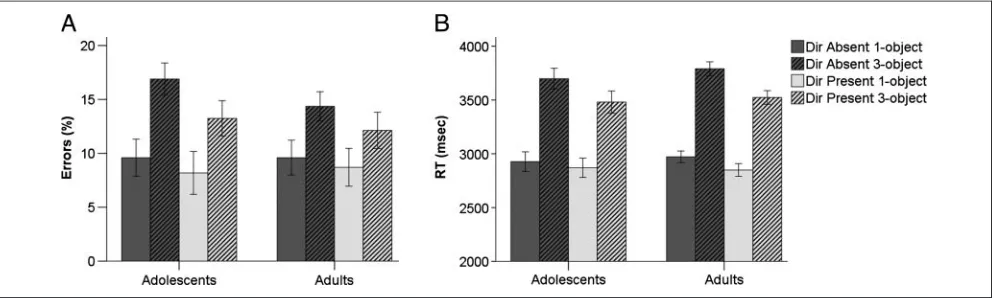
Accuracy was higher in the DP than in the DA condition [main effect of Director,F(1, 26) = 6.29,p= .019] and higher in 1-object than 3-object blocks [main effect of Object,F(1, 26) = 38.29, p< .001; Figure 2A]. There was no main effect of age group (p> .6) and no action between age group and the task factors (all inter-actionpvalues > .22). A similar pattern of performance was observed in terms of RTs. Participants were faster in the DP than DA condition,F(1, 26) = 48.36,p< .001, and in 1-object than 3-object blocks,F(1, 26) = 465.16, p< .001, and there was no main effect of age and no inter-action with age group (allps > .24). However, there was a significant interaction between Director and Object,
[image:7.612.64.561.531.680.2]F(1, 26) = 15.69,p< .001, reflecting a greater effect of Object in the DA condition (Figure 2B). Thus, effects of Director and Object were observed on both measures
of performance, but the adolescentsʼ and adultsʼ perfor-mance did not differ.
fMRI Results
The four first-level contrasts comparing each block type [Director (2) × Object (2)] to fixation were entered in a flexible factorial second-level analysis, including age group and participant as factors.
Object Factor
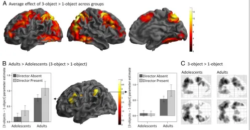
A broad bilateral network of fronto-parietal, occipital, and inferior temporal regions showed increased BOLD signal in 3-object compared with 1-object blocks (Table 1 and
Figure 3A), that is, when the participants had to identify a specific object to move among three exemplars of the same object type (e.g., one of several balls on Figure 1) as opposed to when there was only one exemplar of the object (e.g., the turtle on Figure 1). Greatest increases in BOLD signal were observed bilaterally in superior and inferior parietal lobules, superior frontal sulci, and precu-neus. Other regions included the medial superior frontal gyrus and anterior parts of PFC in the middle frontal gyri bilaterally.
[image:8.612.53.551.97.482.2]Object and Age Group factors significantly interacted in fronto-parietal regions in the left hemisphere. Adults showed increased BOLD signal in the 3-object vs. 1-object blocks, compared with adolescents, in the intraparietal sulcus and in a cluster extending from the precentral gyrus to the inferior frontal gyrus and insula (Table 1 and
Table 1. Coordinates andZValues for Regions of Significant Differences in BOLD Signal in the Main Effect Contrast of Object [3-Object > 1-Object] and the Interaction between Object and Age Group [(Adults 3-Object > 1-Object) > (Adolescents 3-Object > 1-Objects)] (p< .001 Uncorrected at the Voxel-level,p< .05 FWE Corrected at the Cluster Level)
L/R Brodmannʼs Area MNI (x y z) Z Score p(FWE)* Cluster Size
Main Effect of Object: 3-Object> 1-Object
Superior parietal lobule R 7 27−64 52 >8 <.001 14181
IPS L 40 −39−49 46 >8 <.001
MTG/occipital gyrus R 39/19 42−79 22 >8 <.001
SFS R 6 30−4 64 >8 <.001
Precuneus R 7 6−64 49 >8 <.001
SFS L 6 −21−4 52 >8 <.001
IFG R 44 48 8 34 >8 <.001
MFG R 9 45 26 31 >8 <.001
Supramarginal gyrus R 40 39−40 40 >8 <.001
Precentral gyrus L 6 −42 2 37 >8 <.001
ITS R 37 48−55−14 7.61 <.001
Occipital gyrus L 19 −39−73−11 6.75 <.001
MFG L 9 −45 23 40 6.32 <.001
MFG L 10/46 −42 47 7 6.22 <.001
Medial SFG R 6 6 14 49 6.20 <.001
Thalamus L −12−16 10 4.93 .010 117
Age Group× Object Interaction: Adults > Adolescents (3-Object > 1-Object)
Precentral gyrus L 6 −45 5 31 4.96 .009 368
Inferior frontal gyrus L 45 −42 23 22 4.60
Insula L −39 17 7 4.12
IPS/supramarginal gyrus L 40 −45−49 43 4.27 .018 110
IFG = inferior frontal gyrus; IPS = intraparietal sulcus; ITS = inferior temporal sulcus; MFG = middle frontal gyrus; MTG = middle temporal gyrus; SFG = superior frontal gyrus; SFS = superior frontal sulcus; L/ R: left/right.
Figure 3B). This significant interaction between Object and Age Group reflected more bilateral activations in frontal and parietal regions in adults than adolescents (Fig-ure 3C). Note that these two clusters remained significant when mean RT and accuracy for each condition and par-ticipant were entered as covariates in the second-level analyses [left frontal cluster: Z = 4.84,p(FWE) < .001, 326 voxels; left parietal cluster:Z= 4.27,p(FWE) = .029, 96 voxels].
Both younger and older adolescent subgroups showed weaker 3-object than 1-object fronto-parietal activation than adults (ps < .05) and did not differ from each other (parietal region: p> .5; frontal region: p= .066, trend for greater activation in the younger adolescents). The activation in 3-object vs. 1-object trials also significantly increased with age entered as a continuous variable (ps < .01), although this effect was less significant than the Age Group effect.
Director Factor
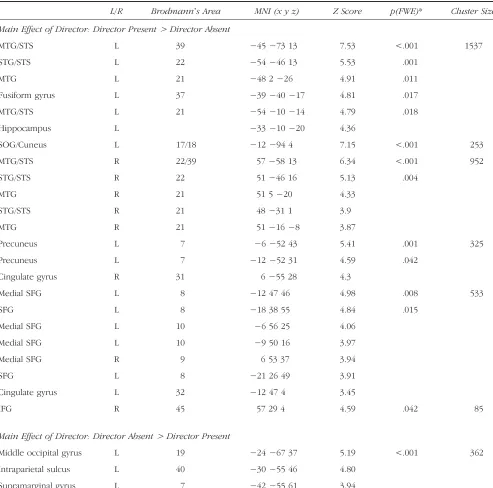
When comparing the Director Present condition to the Director Absent condition, that is, when the cues were social stimuli rather than symbols, increased BOLD signal was observed in bilateral superior and middle temporal cortex regions along the STS and extending into the
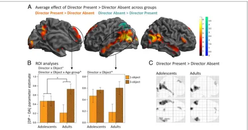
ante-rior temporal cortex, as well as in the right infeante-rior frontal gyrus, dorsal MPFC, precuneus, and occipital gyrus (Table 2 and Figure 4A). The reverse contrast, that is, when the cues were not social but symbolic and rule-based, revealed increased BOLD signal in the left parietal cortex only (Table 2 and Figure 4A). There was no significant inter-action between Director and Age Group factors.
Director × Object Interaction
Whole-brain analyses at the cluster FWE-corrected thresh-old p < .05 showed no brain regions with significant Director × Object or Director × Object × Age Group interactions. An ROI approach (which is potentially less robust because it is biased toward particular clusters) was thus used as follows: mean parameter estimates were calcu-lated for all clusters of the DP > DA contrast and analyzed in SPSS using a mixed model repeated-measures ANOVA. The ROI clusters were in the left temporal cortex, right tem-poral cortex, occipital gyrus, dorsal MPFC, precuneus, and right inferior frontal gyrus (see Table 2 and Figure 4A).
[image:9.612.63.561.68.327.2]the Directorsʼperspective to perform the task correctly. A significant Director × Object interaction was observed in the left temporal cortex cluster,F(1, 26) = 6.12,p= .020, and in the dorsal MPFC cluster,F(1, 26) = 4.87,p= .036. In both cases, the difference in BOLD signal between DP and DA was greater in 3-object than 1-object trials. In addition, the Director × Object × Age Group interaction was significant in the dorsal MPFC cluster,F(1, 26) = 4.76,p= .038. This
[image:10.612.54.547.212.700.2]three-way interaction reflected the fact that the difference between DP and DA was greater in 3-object than 1-object trials in adults (p= .014) but not in adolescents (p> .9; Figure 4B). Adolescents thus showed greater BOLD sig-nal in DP than DA in both 1-object and 3-object trials, whereas adults showed a greater DP than DA activation in 3-object trials specifically, that is, when the perspective of the directors needed to be taken into account.
Table 2. Coordinates andZValues for Regions of Significant Differences in BOLD Signal in the Main Effect Contrasts of the Director Factor (p< .001 Uncorrected at the Voxel Level,p< .05 FWE Corrected at the Cluster Level)
L/R Brodmannʼs Area MNI (x y z) Z Score p(FWE)* Cluster Size
Main Effect of Director: Director Present> Director Absent
MTG/STS L 39 −45−73 13 7.53 <.001 1537
STG/STS L 22 −54−46 13 5.53 .001
MTG L 21 −48 2−26 4.91 .011
Fusiform gyrus L 37 −39−40−17 4.81 .017
MTG/STS L 21 −54−10−14 4.79 .018
Hippocampus L −33−10−20 4.36
SOG/Cuneus L 17/18 −12−94 4 7.15 <.001 253
MTG/STS R 22/39 57−58 13 6.34 <.001 952
STG/STS R 22 51−46 16 5.13 .004
MTG R 21 51 5−20 4.33
STG/STS R 21 48−31 1 3.9
MTG R 21 51−16−8 3.87
Precuneus L 7 −6−52 43 5.41 .001 325
Precuneus L 7 −12−52 31 4.59 .042
Cingulate gyrus R 31 6−55 28 4.3
Medial SFG L 8 −12 47 46 4.98 .008 533
SFG L 8 −18 38 55 4.84 .015
Medial SFG L 10 −6 56 25 4.06
Medial SFG L 10 −9 50 16 3.97
Medial SFG R 9 6 53 37 3.94
SFG L 8 −21 26 49 3.91
Cingulate gyrus L 32 −12 47 4 3.45
IFG R 45 57 29 4 4.59 .042 85
Main Effect of Director: Director Absent> Director Present
Middle occipital gyrus L 19 −24−67 37 5.19 <.001 362
Intraparietal sulcus L 40 −30−55 46 4.80
Supramarginal gyrus L 7 −42−55 61 3.94
IFG = inferior frontal gyrus; MTG = middle temporal gyrus; SFG = superior frontal gyrus; SOG = superior occipital gyrus; STG = superior temporal gyrus; STS = superior temporal sulcus; L/ R = left/right.
Although the Object × Age Group interactions of DP vs. DA did not reach significance when comparing the younger and older adolescent subgroups to the adults (p= .071 and
p= .152, respectively), the same pattern of no difference in DP vs. DA activation between 3-object and 1-object trials (ps > .7) was observed in both adolescent groups. Simi-larly, the interaction between Object and Age Group as a continuous variable for DP vs. DA activation (p= .2) did not reach significance, suggesting the developmental effect was best accounted for by group (adolescents vs. adults).
DISCUSSION
This study investigated the development of the neural substrates of action selection when the information guiding the choice is symbolic or social in nature, which enabled us to investigate domain-general processes (common to the symbolic and social conditions) and domain-specific pro-cesses (specific to the social or symbolic conditions). The paradigm we used required participants to take the per-spective of another person in an implicit manner and re-spond appropriately in a communicative context. First, we showed that the fronto-parietal and temporal brain net-work showing increased BOLD signal when participants
[image:11.612.63.561.61.322.2]Present than Director Absent both in 1-object and 3-object trials (Figure 4).
Development of the Integration of Information to Guide Action Selection
The comparison between trials requiring the identifica-tion and selecidentifica-tion of one of three objects compared with a single target object highlights brain regions recruited in top–down control of attention and goal-directed action. Participants needed to remember the instruction and integrate it with the social or symbolic rule-based cues to identify which of the three exemplar of the target object (e.g., ball) is the correct one to move. All par-ticipants were slower and less accurate in 3-object than 1-object trials, but the age groups did not differ (Figure 2). Over the whole group of participants, a large bilateral network of brain regions showed greater BOLD signal in 3-object compared with 1-object blocks of trials (Figure 3A). Frontal and parietal cortices have been proposed to be the source of spatial attentional modulation of the ventral visual system during object recognition or discrimina-tion (Beck & Kastner, 2009; Tong, 2003; Corbetta, 1998; Corbetta & Shulman, 1998). This network has also been shown to drive nonspatial, feature-based (e.g., color) selec-tive attention (Giesbrecht, Woldorff, Song, & Mangun, 2003). The network observed in the current study in-cludes the fronto-parietal cortex clusters (see Table 1) identified as supporting top–down or endogeneous con-trol of selective attention in a spatial cueing paradigm (Hahn et al., 2006) and also those observed in a spatial and feature cueing paradigm (Giesbrecht et al., 2003) and in a visual search paradigm (Booth et al., 2003). Dif-ferences in the location of occipital activations and in the spread of activations between these paradigms and the current study may relate to the cueing in this study being partly based on auditory verbal stimuli and/or to the greater complexity of the visual stimuli in this study.
There is little previous research regarding neural changes associated with the development of selective attention. Booth et al. (2003) report greater activations in the left thalamus and the right anterior cingulate in children (aged 9–11 years old) compared with adults (aged 20– 30 years) when contrasting a nine-stimuli array visual con-junction search to a simple stimulus detection response; no brain region showed greater activation in adults than in children. In the present study, the target stimuli varied on a trial-by-trial basis, thus BOLD signal changes during the task reflect the encoding and integration of the auditory and visual target information, in addition to the simpler visual detection of the appropriate target shape (e.g., a ball). The developmental results obtained in the current study revealed increased BOLD signal in adults compared with adolescents in the left precentral gyrus extending into the inferior frontal gyrus and in the left intraparietal sulcus/ supramarginal gyrus when comparing 3-object with 1-object trials (Figure 3B). The literature on the development of
the neural substrates of attention and cognitive control has not shown a consistent direction of changes in BOLD signal with age (Luna et al., 2010). However, lateral PFC and parietal cortex are key regions that consistently show developmental changes. Overall, the pattern of results in the literature suggests that, although core regions of the circuitry underlying cognitive control are on-line early in development, the network of brain regions underlying, for example, working memory is still developing during adolescence (Crone & Ridderinkhof, 2010; Luna et al., 2010). The current 3-object versus 1-object comparison contains a combination of attention (visual search), working memory (remembering the rule, instruction and already attended aspects of the stimuli), and inhibition demands (inhibiting attention toward the distractor stimuli and its associated response). In this context, the results show that adolescent development is associated with increasingly bilateral frontal and parietal activations.
In summary, this study shows fronto-parietal and tem-poral cortex activations when participants are required to integrate complex visual and auditory information to search and select one of two possible actions versus when the action to perform is more simply identified. Despite similar performance, adolescents show hypoactivation of the left frontal and parietal cortex.
Processing of Social versus Symbolic Information
The instructions were fully matched between the Director Present and Director Absent conditions. However, the vi-sual stimuli differed, with two characters standing in front or at the back of the set of shelves in the Director Present condition, versus the letters F and M and gray or transpar-ent boxes in the Director Abstranspar-ent condition. Main effects of Director were observed for both accuracy and RTs, with better performance in the Director Present condition. Pre-vious research using a behavioral variant of the Director task showed both young and adult participants were much more error prone in the Director Present condition (Apperly et al., 2010; Dumontheil, Apperly, et al., 2010). However, these studies investigated participantsʼ natural tendency to take into account the directorʼs perspective. In this study, participants went through a training session where their performance was corrected if they did not take into account the directorsʼ perspectives. This aspect of the task was stressed as important, and accordingly, we obtained high accuracy rates. The performance benefit associated with the social versus the symbolic stimuli is in line with previous studies showing that participants per-form faster (den Ouden, Frith, Frith, & Blakemore, 2005) and more accurately (Baron-Cohen et al., 1986) on social compared with nonsocial tasks.
stimuli was associated with greater activation in a large bilateral network of temporal, precuneus, and dorsal MPFC regions that mostly did not overlap with those regions more activated in the 3-object than 1-object contrast, ex-cept in the posterior parts of the middle temporal gyrus bilaterally, the precuneus, and part of the left superior occipital gyrus (Figure 4A). Thus, the presence of social stimuli led to increased BOLD signal in a number of regions that form the social brain network, from face and eye gaze-sensitive brain regions along the STS (Haxby, Hoffman, & Gobbini, 2000), body-sensitive regions in the extrastriate body area (Taylor, Wiggett, & Downing, 2007), to mentaliz-ing regions in the posterior STS and the MPFC (Frith & Frith, 2003; Gallagher & Frith, 2003). More specifically, the STS activation observed in this Director Present versus Director Absent contrast extends into a pSTS region pre-viously observed to show increased BOLD when partici-pants planned or recognized each otherʼs communicative intentions (Noordzij et al., 2010). The MPFC activation was dorsal and aligned with the activations observed in tasks requiring mentalizing or trait judgments made on others rather than judgment made on the self (Van Overwalle, 2009; Mitchell et al., 2006). In addition, although the MPFC activation observed in the current study is located in quite a superior dorsal part of the MPFC, recent meta-analyses suggest that mentalizing activations extend over a wide range of coordinates in the MPFC (Van Overwalle, 2009, 2011) and that non-story-based theory of mind studies tend to show more superior activations than story-based theory of mind studies (Mar, 2011). The Director Present con-dition also likely led participants to associate the male or female voice heard with the male or female director char-acter presented visually. Activations along the STS have been reported in a study of the encoding of speaker iden-tity in the cortical surface (Formisano, De Martino, Bonte, & Goebel, 2008), and the STS has been shown to represent the integration of auditory–visual integration of faces and voices (Chandrasekaran & Ghazanfar, 2009).
It is noteworthy that the current Director Present ver-sus Director Absent contrast was collapsed across a con-dition requiring participants to take into account the directorʼs perspective (3-object) and a condition where the directorʼs perspective was not necessary to identify the target object (1-object). Thus, here, the mere pres-ence of the directors and possibly the integration of the auditory instruction and the directorʼs visual representa-tion (Chandrasekaran & Ghazanfar, 2009; Formisano et al., 2008) were sufficient to elicit activations in the mentalizing network. This finding is consistent with the suggestion that MPFC plays a broad role in general social cognition (Saxe & Powell, 2006; see also Saxe, Whitfield-Gabrieli, Scholz, & Pelphrey, 2009). An alternative ac-count is that, although mentalizing was not necessary in the 1-object condition, participants nonetheless com-puted the directorʼs perspective. We did not make a dis-tinction between 1-object and 3-object blocks during the training phase or during scanning; thus; participants may
have computed the directorʼs perspective on all trials rather than deciding whether it was necessary to do so on a block-by-block or trial-by-trial basis. In line with this interpretation, previous research provides evidence that mentalizing can happen even when it is unnecessary (e.g., Back & Apperly, 2010; Kovacs, Teglas, & Endress, 2010) and even when it actively impedes performance on the main task (e.g., Qureshi, Apperly, & Samson, 2010; Samson, Apperly, Braithwaite, Andrews, & Bodley Scott, 2010).
Our second prediction was that the Director Present vs. Director Absent comparison may show age-related decreases in activation in the dorsal MPFC and increases in the temporal cortex, associated with the processing of social information. No significant Age Group × Director interaction was observed, suggesting that the age groups in fact did not differ in their average social brain response to the Director Present stimuli. To summarize, in this study we observed both domain-general activations in cognitive control regions (3-object vs. 1-object contrast) and domain-specific activations associated with the pro-cessing of social cues (Director Present vs. Director Absent contrast).
The Use of Perspective Information to Guide Action Selection
The reliance on social cues was associated with faster RTs than the use of symbolic cues in 3-object trials (compared with responses in 1-object trials; Figure 2B). Whole-brain analyses did not reveal regions exhibiting an interaction between Director and Object factors, and Director, Object, and Age Groups. However, ROI analyses of the clusters ob-tained in the Director Present vs. Director Absent contrast showed that the left temporal and superior dorsal MPFC clusters exhibited increased activation in the DP 3-object condition (Figure 4B). In the superior dorsal MPFC, this Director × Object interaction was further modulated by Age Group, reflecting the fact that the increase in BOLD signal in DP 3-object trials was observed in the adults only (Figure 4B). Our third prediction was that age effects on the social brain activation, in particular, in the MPFC and temporal cortex, if not observed in the Director Present versus Director Absent contrast, might have been more specific to the Director Present 3-object condition, which requires the on-line use of perspective taking information. Our results do show significant age effects in dorsal MPFC (although the pattern in the temporal cortex is qualitatively similar, there was no significant interaction with Age Group); however, the pattern of changes in activation with age is more complex than that predicted from the literature and discussed below. Note that a limitation of this study is that these findings obtained using ROI analyses are con-sequently weaker than the whole-brain findings described in the previous sections.
during adolescence. Previous studies have consistently reported greater MPFC activations in adolescents than adults in a variety of social cognition tasks (see Blakemore, 2008, for a review of the earlier studies; Gunther Moor et al., 2012; Sebastian et al., 2012; Pfeifer et al., 2009; Burnett, Bird, Moll, Frith, & Blakemore, 2008). The tasks used typically required participants to make an explicit judgment regarding the mental states of a character in a scenario presented in animations (Moriguchi, Ohnishi, Mori, Matsuda, & Komaki, 2007), drawings (Sebastian et al., 2012; Wang, Lee, Sigman, & Dapretto, 2006), or text (Burnett et al., 2008; Blakemore, den Ouden, Choudhury, & Frith, 2007). Two studies required participants to judge how much a phrase (e.g.,“I am popular”) described them-selves (Pfeifer et al., 2009; Pfeifer, Lieberman, & Dapretto, 2007), and one study involved participants judging a per-sonʼs emotion from photos of their eyes (Gunther Moor et al., 2012). Thus, participants were asked to reflect on their own or someoneʼs thoughts or emotions in an explicit and somewhat detached manner, and the results showed that in such situations there are greater BOLD signal increases in adolescents than adults in the MPFC. In this study, participants were required to use social cues regarding the perspective and knowledge of another per-son in an on-line manner and in a communicative context and then to perform the appropriate action. Interestingly, our results show that the adolescents did not recruit dorsal MPFC specifically in the perspective taking condition (Di-rector Present 3-object) but more generally whenever the stimuli had a social aspect, that is, in the comparison of Director Present versus Director Absent. Adults, however, showed greater dorsal MPFC activations that were specific to the Director Present 3-object trials, that is, when infor-mation about the directorʼs perspective had to be taken into account to respond appropriately.
These results cast some light on the possible inter-pretation of previous findings of greater MPFC activa-tions during mentalizing in adolescents. In previous studies, it is not clear whether this greater MPFC activa-tion is due to adolescents“overmentalizing”in response to the same stimuli, or having to put in more work in terms of neural resources to achieve the same mentaliz-ing computations or to lower signal-to-noise ratio asso-ciated with increased prefrontal gray matter volumes in adolescence compared with adulthood (see Blakemore, 2008). Recent work using other tasks suggests that de-creases in brain activation during adolescence do not necessarily reflect concomitant gray matter volumes de-creases (Dumontheil, Hassan, Gilbert, & Blakemore, 2010; Dumontheil, Houlton, Christoff, & Blakemore, 2010). The current study provides no evidence that ado-lescents use more neural effort to achieve the same men-talizing performance: In the absence of differences in performance, adolescents did not show greater activa-tions than adults in Director Present 3-object trials, which require participants to take the directorʼs perspective into account. Instead, adolescents appeared to show less
specific mentalizing MPFC activations than adults, with MPFC activations observed at a similar level in Director Present 1-object and 3-object trials. These findings are thus more consistent with the overmentalizing interpre-tation of the greater MPFC activations observed during adolescence in previous studies. The pattern of results observed in the dorsal MPFC in the current study is similar to the finding of a lack of specificity of right TPJ activation in early childhood in a verbal story-based task (Saxe et al., 2009). Among children aged 6–11 years old, the right TPJ was similarly recruited when the younger children listened to sections of a story describing a characterʼs thoughts (Mental condition) or appearance and social relationships (People), whereas older children showed right TPJ activa-tion only for the Mental condiactiva-tion (Saxe et al., 2009), thus showing increased right TPJ specificity for theory of mind with age.
To summarize, the current study showed that adoles-cents exhibited dorsal MPFC activation in both social con-ditions and did not show the specific increased dorsal MPFC activation observed in adults when the trial re-quired the participant to take into account the directorʼs perspective to choose the appropriate response.
Ecological Validity of the Task
taking requirements compared with adults. The hypoactiva-tion of cognitive control regions in the adolescents may be behind the greater egocentric bias observed in a similar paradigm during adolescence (Dumontheil, Apperly, et al., 2010). Future studies with greater number of adolescent participants could investigate in more details the observed effects and test their association with pubertal develop-ment as opposed to chronological age only (Blakemore, Burnett, & Dahl, 2010), as well as the connectivity between the cognitive control and social brain networks.
Conclusion
The aim of this study was to investigate the development of the neural substrates associated with the selection of action among distractors and with the use of social cues to guide action selection. We used a novel paradigm that requires participants to use either symbolic rules or per-spective information of other individuals to select an appropriate action in a communicative context. Having previously shown that the on-line use of perspective infor-mation led to the recruitment of superior dorsal MPFC, left STS, and anterior temporal cortex regions in adults, we showed here that adolescents exhibited hypoactivation of domain-general cognitive control regions in the parietal cortex and PFC and hyperactivation of parts of the social brain network, with dorsal MPFC activation observed whether or not the social cues were necessary to perform the action appropriately. These results provide further evidence of the prolonged development of neural sub-strates of social cognition. They suggest that the pattern of increased MPFC activations in adolescence associated with explicit mentalizing judgments (Burnett & Blakemore, 2009) is not found when mentalizing has to be used on-line by taking anotherʼs perspective in an active communi-cative context. Instead, MPFC showed increased activations in adolescence in both the 1- and 3-object conditions, whereas adults engaged MPFC more for 3-object condition. This pattern may reflect overmentalizing in adolescence in conditions where mentalizing is not needed (1-object condition).
Acknowledgments
This study was funded by grants from the Royal Society to S. J. B., a Fellowship from the European Commission to I. D., and a University College London scholarship to H. H.
Reprint requests should be sent to Dr. Iroise Dumontheil, Univer-sity College London Institute of Cognitive Neuroscience, 17 Queen Square, London, WC1N 3AR, UK, or via e-mail: i.dumontheil@ucl. ac.uk.
REFERENCES
Amodio, D. M., & Frith, C. D. (2006). Meeting of minds: The medial frontal cortex and social cognition.Nature Reviews Neuroscience, 7,268–277.
Apperly, I. A. (2011).Mindreaders: The cognitive basis of “theory of mind.”Hove, UK: Psychology Press.
Apperly, I. A., Carroll, D. J., Samson, D., Humphreys, G. W., Qureshi, A., & Moffitt, G. (2010). Why are there limits on theory of mind use? Evidence from adultsʼability to follow instructions from an ignorant speaker.Quarterly Journal of Experimental Psychology, 63,1201–1217.
Apperly, I. A., Samson, D., Chiavarino, C., & Humphreys, G. W. (2004). Frontal and temporo-parietal lobe contributions to theory of mind: Neuropsychological evidence from a false-belief task with reduced language and executive demands. Journal of Cognitive Neuroscience, 16,
1773–1784.
Astle, D. E., & Scerif, G. (2009). Using developmental cognitive neuroscience to study behavioral and attentional control.
Developmental Psychobiology, 51,107–118.
Back, E., & Apperly, I. A. (2010). Two sources of evidence on the non-automaticity of true and false belief ascription.
Cognition, 115,54–70.
Baron-Cohen, S., Leslie, A. M., & Frith, U. (1986). Mechanical, behavioural and intentional understanding of picture stories in autistic children.British Journal of Developmental Psychology, 4,113–125.
Beck, D. M., & Kastner, S. (2009). Top–down and bottom–up mechanisms in biasing competition in the human brain.
Vision Research, 49,1154–1165.
Blakemore, S. J. (2008). The social brain in adolescence.
Nature Reviews Neuroscience, 9,267–277.
Blakemore, S.-J., Burnett, S., & Dahl, R. E. (2010). The role of puberty in the developing adolescent brain.Human Brain Mapping, 31,926–933.
Blakemore, S. J., den Ouden, H., Choudhury, S., & Frith, C. (2007). Adolescent development of the neural circuitry for thinking about intentions.Social Cognitive and Affective Neuroscience, 2,130–139.
Booth, J. R., Burman, D. D., Meyer, J. R., Lei, Z., Trommer, B. L., Davenport, N. D., et al. (2003). Neural development of selective attention and response inhibition.Neuroimage, 20,737–751.
Brothers, L. (1990). The social brain: A project for integrating primate behavior and neurophysiology in a new domain.
Concepts in Neuroscience, 1,27–51.
Brown, B. B. (2004). Adolescentsʼrelationships with peers. In R. M. Lerner & L. Steinberg (Eds.),Handbook of adolescent psychology(pp. 363–394). Hoboken, NJ: Wiley.
Bunge, S. A., & Wright, S. B. (2007). Neurodevelopmental changes in working memory and cognitive control.
Current Opinion in Neurobiology, 17,243–250. Burgess, P. W., Gilbert, S. J., & Dumontheil, I. (2007).
Function and localization within rostral prefrontal cortex (area 10).Philosophical Transactions of the Royal Society of London, Series B, Biological Sciences, 362,
887–899.
Burnett, S., Bird, G., Moll, J., Frith, C., & Blakemore, S.-J. (2008). Development during adolescence of the neural processing of social emotion.Journal of Cognitive Neuroscience, 21,
1736–1750.
Burnett, S., & Blakemore, S.-J. (2009). Functional connectivity during a social emotion task in adolescents and in adults.
The European Journal of Neuroscience, 29,1294–1301. Burnett, S., Sebastian, C., Cohen-Kadosh, K., & Blakemore, S.-J.
(2011). The social brain in adolescence: Evidence from functional magnetic resonance imaging and behavioural studies.Neuroscience and Biobehavioral Reviews, 35,
1654–1664.
Casey, B. J., Jones, R. M., & Hare, T. A. (2008). The adolescent brain.Annals of the New York Academy of Sciences, 1124,
111–126.
Chandrasekaran, C., & Ghazanfar, A. A. (2009). Different neural frequency bands integrate faces and voices differently in the superior temporal sulcus.Journal of Neurophysiology, 101,773–788.
Corbetta, M. (1998). Frontoparietal cortical networks for directing attention and the eye to visual locations: Identical, independent, or overlapping neural systems?Proceedings of the National Academy of Sciences, U.S.A., 95,831–838. Corbetta, M., & Shulman, G. L. (1998). Human cortical
mechanisms of visual attention during orienting and search.Philosophical Transactions of the Royal Society of London, Series B, Biological Sciences, 353,1353–1362. Crone, E. A. (2009). Executive functions in adolescence:
Inferences from brain and behavior.Developmental Science, 12,825–830.
Crone, E. A., & Ridderinkhof, K. R. (2010). The developing brain: From theory to neuroimaging and back.
Developmental Cognitive Neuroscience, 1,101–109. Davis, E. T., & Palmer, J. (2004). Visual search and attention:
An overview.Spatial Vision, 17,249–255.
den Ouden, H. E., Frith, U., Frith, C., & Blakemore, S. J. (2005). Thinking about intentions.Neuroimage, 28,787–796. Dumontheil, I., Apperly, I. A., & Blakemore, S. J. (2010). Online
usage of theory of mind continues to develop in late adolescence.Developmental Science, 13,331–338. Dumontheil, I., & Blakemore, S.-J. (2012). Social cognition
and abstract thought in adolescence: The role of structural and functional development in rostral prefrontal cortex.
British Journal of Educational Psychology Monograph Series II, Number 8—Educational Neuroscience, 1,
99–113.
Dumontheil, I., Hassan, B., Gilbert, S. J., & Blakemore, S.-J. (2010). Development of the selection and manipulation of self-generated thoughts in adolescence.Journal of Neuroscience, 30,7664–7671.
Dumontheil, I., Houlton, R., Christoff, K., & Blakemore, S.-J. (2010). Development of relational reasoning during adolescence.Developmental Science, 13,F15–F24. Dumontheil, I., Küster, O., Apperly, I. A., & Blakemore, S.-J.
(2010). Taking perspective into account in a communicative task.Neuroimage, 52,1574–1583.
Eisenberg, N., & Morris, A. S. (2004). Moral cognitions and prosocial responding in adolescence. In R. M. Lerner & L. Steinberg (Eds.),Handbook of adolescent psychology
(pp. 155–188). Hoboken, NJ: Wiley.
Flavell, J., Abrahams Everett, B., Croft, K., & Flavell, E. R. (1981). Young childrenʼs knowledge about visual perception: Further evidence for Level 1–Level 2 distinction.Developmental Psychology, 17,99–103.
Formisano, E., De Martino, F., Bonte, M., & Goebel, R. (2008).
“Who”is saying“what”? Brain-based decoding of human voice and speech.Science, 322,970–973.
Friston, K. J., Holmes, A. P., Poline, J. B., Grasby, P. J., Williams, S. C., Frackowiak, R. S., et al. (1995). Analysis of fMRI time-series revisited.Neuroimage, 2,45–53.
Frith, C. D., & Frith, U. (2007). Social cognition in humans.
Current Biology, 17,R724–R732.
Frith, U., & Frith, C. D. (2003). Development and neurophysiology of mentalizing.Philosophical Transactions of the Royal Society of London, Series B, Biological Sciences, 358,
459–473.
Fuster, J. M. (2000). Executive frontal functions.Experimental Brain Research, 133,66–70.
Gallagher, H. L., & Frith, C. D. (2003). Functional imaging of
“theory of mind.”Trends in Cognitive Sciences, 7,77–83.
Giedd, J. N., Blumenthal, J., Jeffries, N. O., Castellanos, F. X., Liu, H., Zijdenbos, A., et al. (1999). Brain development during childhood and adolescence: A longitudinal MRI study.
Nature Neuroscience, 2,861–863.
Giesbrecht, B., Woldorff, M. G., Song, A. W., & Mangun, G. R. (2003). Neural mechanisms of top–down control during spatial and feature attention.Neuroimage, 19,496–512. Gogtay, N., Giedd, J. N., Lusk, L., Hayashi, K. M., Greenstein, D.,
Vaituzis, A. C., et al. (2004). Dynamic mapping of human cortical development during childhood through early adulthood.Proceedings of the National Academy of Sciences, U.S.A., 101,8174–8179.
Gunther Moor, B., Op de Macks, Z. A., Güroglu, B., Rombouts, S. A. R. B., Van der Molen, M. W., & Crone, E. A. (2012). Neurodevelopmental changes of reading the mind in the eyes.Social Cognitive and Affective Neuroscience, 7,44–52. Hahn, B., Ross, T. J., & Stein, E. A. (2006). Neuroanatomical
dissociation between bottom–up and top–down processes of visuospatial selective attention.Neuroimage, 32,
842–853.
Haxby, J., Hoffman, E., & Gobbini, M. (2000). The distributed human neural system for face perception.Trends in Cognitive Sciences, 4,223–233.
Humphreys, G. W., Allen, H. A., & Mavritsaki, E. (2009). Using biologically plausible neural models to specify the functional and neural mechanisms of visual search.Progress in Brain Research, 176,135–148.
Jenkins, A. C., Macrae, C. N., & Mitchell, J. P. (2008). Repetition suppression of ventromedial prefrontal activity during judgments of self and others.Proceedings of the National Academy of Sciences, U.S.A., 105,4507–4512.
Keysar, B., Barr, D. J., Balin, J. A., & Brauner, J. S. (2000). Taking perspective in conversation: The role of mutual knowledge in comprehension.Psychological Science, 11,
32–38.
Keysar, B., Lin, S., & Barr, D. J. (2003). Limits on theory of mind use in adults.Cognition, 89,25–41.
Koechlin, E., & Summerfield, C. (2007). An information theoretical approach to prefrontal executive function.
Trends in Cognitive Sciences, 11,229–235.
Kovacs, A. M., Teglas, E., & Endress, A. D. (2010). The social sense: Susceptibility to othersʼbeliefs in human infants and adults.Science, 330,1830–1834.
Leslie, A. M. (2005). Developmental parallels in understanding minds and bodies.Trends in Cognitive Sciences, 9,459–462. Luna, B., Padmanabhan, A., & OʼHearn, K. (2010). What
has fMRI told us about the development of cognitive control through adolescence?Brain and Cognition, 72,
101–113.
Mar, R. A. (2011). The neural bases of social cognition and story comprehension.Annual Review of Psychology, 62,
103–134.
Mitchell, J. P., Macrae, C. N., & Banaji, M. R. (2006). Dissociable medial prefrontal contributions to judgments of similar and dissimilar others.Neuron, 50,655–663.
Moll, H., & Tomasello, M. (2006). Level 1 perspective-taking at 24 months of age.British Journal of Developmental Psychology, 24,603–613.
Moriguchi, Y., Ohnishi, T., Mori, T., Matsuda, H., & Komaki, G. (2007). Changes of brain activity in the neural substrates for theory of mind during childhood and adolescence.
Psychiatry and Clinical Neurosciences, 61,355–363. Noordzij, M. L., Newman-Norlund, S. E., de Ruiter, J. P., Hagoort, P.,
Levinson, S. C., & Toni, I. (2010). Neural correlates of intentional communication.Frontiers in Neuroscience, 4,188.
knowledge retrieval in children and adults.Journal of Cognitive Neuroscience, 19,1323–1337.
Pfeifer, J. H., Masten, C. L., Borofsky, L. A., Dapretto, M., Fuligni, A. J., & Lieberman, M. D. (2009). Neural correlates of direct and reflected self-appraisals in adolescents and adults: When social perspective-taking informs self-perception.
Child Development, 80,1016–1038.
Qureshi, A. W., Apperly, I. A., & Samson, D. (2010). Executive function is necessary for perspective selection, not Level-1 visual perspective calculation: Evidence from a dual-task study of adults.Cognition, 117,230–236.
Samson, D., Apperly, I. A., Braithwaite, J. J., Andrews, B. J., & Bodley Scott, S. E. (2010). Seeing it their way: Evidence for rapid and involuntary computation of what other people see.
Journal of Experimental Psychology: Human Perception and Performance, 36,1255–1266.
Samson, D., Apperly, I. A., Kathirgamanathan, U., & Humphreys, G. W. (2005). Seeing it my way: A case of a selective deficit in inhibiting self-perspective.Brain, 128,1102–1111. Saxe, R. (2006). Uniquely human social cognition.Current
Opinion in Neurobiology, 16,235–239.
Saxe, R., Carey, S., & Kanwisher, N. (2004). Understanding other minds: Linking developmental psychology and functional neuroimaging.Annual Review of Psychology, 55,87–124. Saxe, R., & Powell, L. J. (2006). Itʼs the thought that counts:
Specific brain regions for one component of theory of mind.
Psychological Science, 17,692–699.
Saxe, R., Schulz, L. E., & Jiang, Y. V. (2006). Reading minds versus following rules: Dissociating theory of mind and executive control in the brain.Social Neuroscience, 1,284–298. Saxe, R. R., Whitfield-Gabrieli, S., Scholz, J., & Pelphrey, K. A.
(2009). Brain regions for perceiving and reasoning about other people in school-aged children.Child Development, 80,1197–1209.
Sebastian, C. L., Fontaine, N. M. G., Bird, G., Blakemore, S.-J., De Brito, S. A., McCrory, E. J. P., et al. (2012). Neural
processing associated with cognitive and affective theory of mind in adolescents and adults.Social Cognitive and Affective Neuroscience, 7,53–63.
Shaw, P., Kabani, N. J., Lerch, J. P., Eckstrand, K., Lenroot, R., Gogtay, N., et al. (2008). Neurodevelopmental trajectories of the human cerebral cortex.Journal of Neuroscience, 28,
3586–3594.
Sowell, E. R., Thompson, P. M., Holmes, C. J., Jernigan, T. L., & Toga, A. W. (1999). In vivo evidence for post-adolescent brain maturation in frontal and striatal regions.Nature Neuroscience, 2,859–861.
Sperber, D., & Wilson, D. (2002). Pragmatics, modularity and mind-reading.Mind and Language, 17,3–23.
Stone, V. E., Baron-Cohen, S., & Knight, R. T. (1998). Frontal lobe contributions to theory of mind.Journal of Cognitive Neuroscience, 10,640–656.
Tamir, D. I., & Mitchell, J. P. (2010). Neural correlates of anchoring-and-adjustment during mentalizing.Proceedings of the National Academy of Sciences, U.S.A., 107,10827–10832. Taylor, J. C., Wiggett, A. J., & Downing, P. E. (2007). Functional MRI analysis of body and body part representations in the extrastriate and fusiform body areas.Journal of Neurophysiology, 98,1626–1633.
Tong, F. (2003). Primary visual cortex and visual awareness.
Nature Reviews Neuroscience, 4,219–229.
Van Overwalle, F. (2009). Social cognition and the brain: A meta-analysis. Human Brain Mapping, 30,829–858. Van Overwalle, F. (2011). A dissociation between social
mentalizing and general reasoning.Neuroimage, 54,
1589–1599.
Wang, A. T., Lee, S. S., Sigman, M., & Dapretto, M. (2006). Developmental changes in the neural basis of interpreting communicative intent.Social Cognitive and Affective Neuroscience, 1,107–121.
![Table 1. Coordinates and Z Values for Regions of Significant Differences in BOLD Signal in the Main Effect Contrast of Object[3-Object > 1-Object] and the Interaction between Object and Age Group [(Adults 3-Object > 1-Object) > (Adolescents 3-Object >1-Objects)] ( p < .001 Uncorrected at the Voxel-level, p < .05 FWE Corrected at the Cluster Level)](https://thumb-us.123doks.com/thumbv2/123dok_us/8876376.944180/8.612.53.551.97.482/coordinates-significant-differences-contrast-interaction-adolescents-uncorrected-corrected.webp)