RESEARCH ARTICLE
TGF
β
and FGF promote tendon progenitor fate and act
downstream of muscle contraction to regulate tendon
differentiation during chick limb development
Emmanuelle Havis, Marie-Ange Bonnin, Joana Esteves de Lima, Benjamin Charvet, Cécile Milet and Delphine Duprez*
ABSTRACT
The molecular programme underlying tendon development has not been fully identified. Interactions with components of the musculoskeletal system are important for limb tendon formation. Limb tendons initiate their development independently of muscles; however, muscles are required for further tendon differentiation. We show that both FGF/ERK MAPK and TGFβ/SMAD2/3 signalling pathways are required and sufficient forSCX expression in chick undifferentiated limb cells, whereas the FGF/ERK MAPK pathway inhibitsScxexpression in mouse undifferentiated limb mesodermal cells. During differentiation, muscle contraction is required to maintain SCX,TNMDandTHBS2expression in chick limbs. The activities of FGF/ERK MAPK and TGFβ/SMAD2/3 signalling pathways are decreased in tendons under immobilisation conditions. Application of FGF4 or TGFβ2 ligands prevents SCX downregulation in immobilised limbs. TGFβ2 but not FGF4 preventTNMDandTHBS2 downregulation under immobilisation conditions. We did not identify any intracellular crosstalk between both signalling pathways in their positive effect onSCXexpression. Independently of each other, both FGF and TGFβ promote tendon commitment of limb mesodermal cells and act downstream of mechanical forces to regulate tendon differentiation during chick limb development.
KEY WORDS: Chick, Limb, Tendon, Mechanobiology, Scleraxis
INTRODUCTION
Tendon is a connective tissue that transmits the forces generated by muscle to bone and allows body motion. Type I collagen is the main structural and functional component of tendons. The signals regulating the production and the spatial organisation of type I collagen in developing tendons have not been fully identified. Moreover, because type I collagen is not specific to tendons, it is not possible to follow tendon development by mapping collagen expression (reviewed by Gaut and Duprez, 2016). The basic helix-loop-helix (bHLH) transcription factor Scleraxis (Scx) has been identified as an early tendon marker during development.Scx is expressed in tendon progenitors, developing tendons and adult tendons (Schweitzer et al., 2001; Pryce et al., 2007; Mendias et al., 2012). Scx is not the unique master gene driving tendon development, as tendons are formed in Scx−/− mice, albeit
displaying differentiation defects (Murchison et al., 2007). Moreover, Col1a1 expression is downregulated in tendons of Scx−/− mutant mice (Murchison et al., 2007), consistent with
transcriptional regulation of the mouseCol1a1gene by Scx, via direct binding on the Col1a1 promoter (Lejard et al., 2007). Tenomodulin (Tnmd) andCol14a1expression is lost in developing limb tendons of Scx−/− mice (Murchison et al., 2007). The
transmembrane glycoprotein Tnmd is considered as a late tendon marker (Jelinsky et al., 2010; Sugimoto et al., 2013) and is highly expressed in embryonic day (E) 14.5 mouse limb tendon cells (Havis et al., 2014). Thrombospondin 2 and 4 (THBS2 and THBS4) were also identified in the transcriptome of mouse limb tendon cells (Havis et al., 2014) and have been shown to be involved in tendon development in mouse,Drosophilaand zebrafish (Kyriakides et al., 1998; Subramanian et al., 2007; Subramanian and Schilling, 2014). Two other transcription factors are involved in tendon development: the homeobox protein Mkx (mohawk) (Ito et al., 2010; Liu et al., 2010; Kimura et al., 2011) and the zinc finger transcription factor EGR1 (early growth response factor 1) (Lejard et al., 2011). In contrast toScx,MkxandEgr1are not expressed during early tendon limb development and are not specific to tendons (Anderson et al., 2006; Liu et al., 2006; Ito et al., 2010; Lejard et al., 2011), but they activate Scx and Tnmd expression in various stem cell types and positively regulate type I collagen productionin vivo(Ito et al., 2010; Lejard et al., 2011; Guerquin et al., 2013; Liu et al., 2015). In addition to the tendon-related transcription factors, two signalling pathways have been identified as being involved in tendon development: the transforming growth factor-beta (TGFβ) and fibroblast growth factor (FGF) signalling pathways (reviewed by Huang et al., 2015a; Gaut and Duprez, 2016). The TGFβsignalling pathway positively regulatesScxexpression in early E9/E10 mouse limb explants (Pryce et al., 2009; Havis et al., 2014). TGFβfunction in chick tendon development is less understood. Although TGFβ1 and 2 have been shown to increaseSCXandTNMDexpression in high-density cultures of chick limb cells (Lorda-Diez et al., 2009), TGFβ1 failed to activateSCXexpression in Hamburger Hamilton stage (HH) 20/21 chick limb explants (Lorda-Diez et al., 2010). FGF positively regulatesSCX expression in axial and foetal limb tendons during chick development (Edom-Vovard et al., 2002; Brent et al., 2003; Brent and Tabin, 2004). In contrast to the chick model, FGF has an anti-tenogenic effect in mouse embryonic tendon cells (Brown et al., 2014) and inhibition of the ERK MAPK pathway is sufficient to increase Scx expression in early mouse limb explants (Havis et al., 2014). Although the experimental situations in the reports described above differ between the chick and mouse models, they nevertheless suggest a differential regulation of Scx by FGF in the chick and mouse models.
Received 9 February 2016; Accepted 25 August 2016
Sorbonne Universités, UPMC Univ Paris 06, CNRS UMR 7622, Inserm U1156, IBPS-Developmental Biology Laboratory, Paris F-75005, France.
*Author for correspondence ([email protected])
D.D., 0000-0003-0248-7417
DEVEL
O
Another important aspect of tendon development is its dependency on muscles. Axial, limb and head tendons require the presence of muscles for full development in chick, mouse and zebrafish embryos (reviewed by Gaut and Duprez, 2016). However, in the absence of muscle,Scxexpression is normally initiated (and then lost) in limb and head regions of mouse, chick and zebrafish embryos (Schweitzer et al., 2001; Edom-Vovard et al., 2002; Bonnin et al., 2005; Grenier et al., 2009; Chen and Galloway, 2014; Huang et al., 2015b). The muscle dependency ofScxexpression defines two phases for limb tendon formation: a progenitor, muscle-independent phase and a differentiation, muscle-dependent phase. This muscle dependency applies only to stylopod (arm) and zeugopod (forearm) limb tendons, as autopod (digit) tendons are dependent on cartilage in mouse embryos (Huang et al., 2015b). The molecular mechanisms underlying the muscle dependency of chick tendon development remain elusive. Although one can assume that the muscle dependency ofScxexpression is related to muscle activity, the requirement of mechanical forces for chick limb tendon development has not been addressed and the molecular signals downstream of muscle contraction involved in tendon differentiation have not been identified.
RESULTS
The FGF/ERK MAPK pathway activatesSCXexpression in early chick limb buds
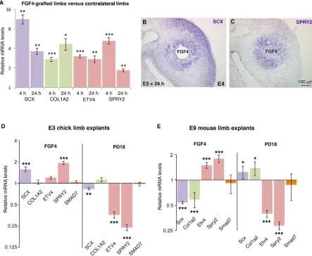
FGF positively regulates SCX expression via the ERK MAPK signalling pathway in chick somites (Brent and Tabin, 2004; Smith et al., 2005) and in foetal chick limbs (Edom-Vovard et al., 2002; Eloy-Trinquet et al., 2009). However, the role of FGF on SCX expression was not determined in chick limb undifferentiated cells during the muscle-independent phase of limb tendon development. SCXexpression is initiated in E3 (HH20) chick limb buds (Brent and Tabin, 2004). At these stages, a source of FGF is observed in the apical ectodermal ridge (Niswander et al., 1994). We implanted FGF4 beads in early chick limb buds (E3 to E4) and analysedSCX expression by RT-q-PCR and in situ hybridisation experiments (Fig. 1A-C). SCX and COL1A2 expression was upregulated as soon as 4 h after FGF4 bead implantation; this upregulation was maintained 24 h after FGF4 bead implantation (Fig. 1A). We used ETV4(also known asPEA3) andSPRY2as transcriptional readouts of ERK MAPK activity (O’Hagan et al., 1996; Mason et al., 2006; Havis et al., 2014). The mRNA levels ofETV4 andSPRY2were increased in FGF4-implanted limbs 4 h and 24 h after grafting (Fig. 1A) andSPRY2expression was activated around FGF4 beads 24 h after grafting (Fig. 1C).TNMDis not expressed before E5 in chick limbs (Shukunami et al., 2006) and FGF4 was not able to activateTNMDprematurely (data not shown). This FGF4 tenogenic effect in chick limb buds contrasted with the previously demonstrated anti-tenogenic effect of FGF4 in mouse limb explants (Havis et al., 2014). We next performed chick limb bud explants in order to exclude differences due to different experimental designs and allow comparison between the chick and mouse models. Consistent with the in vivo FGF4 bead experiments (Fig. 1A-C), we observed that FGF4 increased the mRNA levels ofSCXandSPRY2(Fig. 1D), whereas blockade of ERK MAPK with the inhibitor PD18 decreased SCX,ETV4and SPRY2expression in chick limb bud explants 6 h after treatment (Fig. 1D). In order to allow comparison between species, we performed equivalent mouse limb bud explant experiments and found that FGF4 significantly decreased Scxexpression, whereas PD18 increasedScxexpression in mouse limbs, 6 h after treatment (Fig. 1E), consistent with previously published effects of 24 h FGF4
and PD18 treatments in mouse limbs (Havis et al., 2014). We conclude that the FGF tenogenic effect observed in chick limb cells is opposite to the anti-tenogenic FGF effect observed in mouse limb cells.
We next tested whether the FGF4 effect onSCXin chick cells involved the SMAD2/3 pathway. We applied the SMAD2/3 inhibitor SIS3 in FGF4 gain-of-function experiments in chick limb buds (Fig. S1). Blockade of SMAD2/3 did not block the positive effect of FGF4 onSCXexpression (Fig. S1). This result is consistent with the absence of modification of SMAD7/Smad7 expression upon FGF/ERK MAPK manipulations in both chick and mouse limb explants (Fig. 1D,E). Smad7 is a negative-feedback regulator that is considered to be a general TGFβ/SMAD2/3 transcriptional target gene (Massague, 2012). We conclude that FGF4 positively regulates SCX independently of the SMAD2/3 intracellular pathway in chick limb cells.
The TGFβ/SMAD2/3 pathway activatesSCXexpression in early chick limb buds
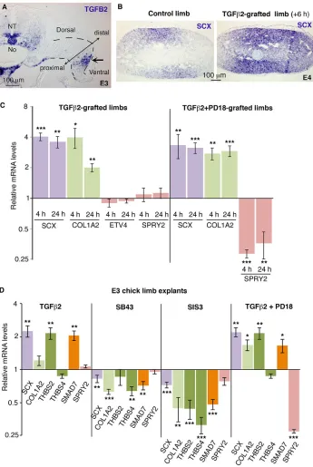
TGFβ2 inducesScxexpression in E10.5 mouse limb explants (Pryce et al., 2009), but TGFβ1 does not modifySCXexpression in E3.5 (HH20/21) chick limb explants (Lorda-Diez et al., 2010). We found TGFB2 to be expressed in ventral parts of E3 chick limb buds (Fig. 2A), as previously described (Lorda-Diez et al., 2010). Application of TGFβ2 beads in E3/E4 (HH19/21) chick limb buds increasedSCXexpression 6 h after grafting (Fig. 2B) and the mRNA levels of SCX and COL1A2 were increased in grafted limbs compared with control limbs (Fig. 2C). TGFβ2 application on chick limb bud explants also increasedSCXexpression in addition to increasingTHBS2(Fig. 2D). TGFβ2 was not able to activateTNMD prematurely in chick limb undifferentiated cells (data not shown), as in mouse limb undifferentiated cells (Havis et al., 2014). Blockade of TGFβreceptors (SB43) and of the SMAD2/3 signalling pathway (SIS3) decreasedSCX expression, in addition to that of CO1A2, THBS2andTHBS4(Fig. 2D). Consistently,SMAD7mRNA levels, the transcriptional readout of the SMAD2/3 intracellular pathway, were increased following TGFβ2 application and decreased with the inhibitors SB43 and SIS3 (Fig. 2D). These results show that TGFβ2 positively regulates SCX expression, and that the SMAD2/3 intracellular pathway is required forSCXexpression in early chick limb undifferentiated cells.
TGFβis known to activate the ERK MAPK pathway as a non-canonical signalling pathway (reviewed by Guo and Wang, 2009; Massague, 2012). The expression of ETV4 and SPRY2 was not modified upon TGFβ2 bead application (Fig. 2C). In order to confirm experimentally that the positive effect of TGFβ2 onSCXexpression did not involve the ERK MAPK signalling pathway, we applied the inhibitor PD18 in TGFβ2 gain-of-function experiments in chick limb buds and limb explants. The blockade of the ERK MAPK pathway did not modify the positive effect of TGFβ2 onSCX expression in chick limbs (Fig. 2C) and in chick limb explants (Fig. 2D). We conclude that TGFβ2 activatesSCXexpression independently of the ERK MAPK signalling pathway in chick limb cells.
FGF4 positively regulatesTNMDandTHBS2expression during tendon differentiation
TNMD is considered as a late tendon marker in chick and mouse embryos, and is expressed during the differentiation and muscle-dependent phase of limb tendon development (reviewed by Dex et al., 2016). Tnmd mutant mice display an altered structure of collagen fibrils, and reduced self-renewal and increased senescence of tendon progenitors, in post-natal tendons (Docheva et al., 2005;
DEVEL
O
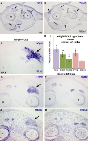
Alberton et al., 2015). TNMD was expressed in SCX-positive tendons in E9 chick limbs (Fig. 3A,B, arrows), but also in dermal regions (Fig. 3B, arrowhead). Retroviral mouse Fgf4 (mFgf4/RCAS) induced ectopicTNMD expression in chick limbs (Fig. 3C-F), in addition to activatingSCXexpression (Edom-Vovard et al., 2002). Consistently, the relative mRNA levels ofTNMDandSCXtendon genes and ETV4were increased in mFgf4/RCAS-limbs compared with control limbs (Fig. 3D). THBS2, another late tendon marker (Havis et al., 2014) was also upregulated in chick limbs upon retroviral Fgf4 (Fig. 3D,G,H). We conclude that FGF4 positively regulates TNMD and THBS2 expression in chick limbs during the differentiation and muscle-dependent phase of limb tendon development.
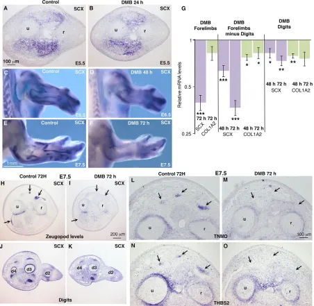
Muscle contraction is required to maintain tendon marker expression in chick limb stylopod/zeugopod tendons
Scx/SCXexpression is lost in stylopod/zeugopod muscleless limbs of mutant mice or experimental chick embryos (Schweitzer et al., 2001; Edom-Vovard and Duprez, 2004), defining the
[image:3.612.83.526.60.424.2]muscle-dependent phase of limb tendon development. In the absence of muscle activity, Scx/GFP expression is diminished but not lost in zeugopod/stylopod regions of forelimbs of E18.5 paralysedmdg mice (Huang et al., 2015b). In order to determine the importance of mechanical signals for chick limb tendon development, we blocked muscle contraction in chick embryos using the drug decamethonium bromide (DMB). DMB acts as an acetylcholine agonist, induces depolarisation in skeletal muscles and ultimately leads to rigid muscle paralysis and to immobilised embryos (Nowlan et al., 2010). We applied DMB or control buffer in E4.5 chick embryos and analysed gene expression either byin situhybridisation on sections and wholemounts or by RT-q-PCR (Fig. 4). In the absence of muscle contraction, muscles, visualised with MYOD expression, were present 2 days after DMB application, but displayed splitting delay 3 days after DMB application (Fig. S2). As previously described, limbs of immobilised embryos were smaller than control limbs (Nowlan et al., 2010). During the muscle-independent phase, SCX expression was not affected in chick limbs of immobilised embryos, 24 h after DMB application (Fig. 4A,B), consistent with Fig. 1. Tenogenic effect of FGF on chick limb cells.(A-C) FGF4 bead application to forelimbs of E3/E4 (HH18/HH22) chick embryos. FGF4-grafted right limbs and control left limbs were processed for RT-q-PCR (n=6 for each time point) or forin situhybridisation (n=3) analyses 4 h or 24 h after bead application. (A) RT-q-PCR analyses of the expression levels ofSCX,COL1A2and readouts of FGF/ERK MAPK activity (ETV4andSPRY2expression) in FGF4-grafted right limbs, 4 h or 24 h after FGF4 application. For each gene, the mRNA levels in FGF4-grafted limbs are expressed relative to those of control left limbs (normalised to 1). (B,C)In situhybridisation forSCXandSPRY2in E4 (E3+24 h) FGF4-grafted limbs. (D) E3 chick limb explant cultures. RT-q-PCR analyses of the relative expression levels ofSCX,COL1A2,ETV4,SPRY2andSMAD7in E3 chick limb explants cultured for 6 h with FGF4 (n=10) or the inhibitor PD18 (n=10). (E) E9 mouse limb explant cultures. RT-q-PCR analyses of the expression levels ofScx,Col1a2,Etv4,Spry2andSmad7in E9 mouse limb explants cultured for 6 h with FGF4 (n=5) or the inhibitor PD18 (n=5). For each gene, the mRNA levels of treated limb explants are expressed relative to those of control limb explants (normalised to 1).P-values were determined by unpaired Student’st-test using Microsoft Excel. *P<0.05; **P<0.01; ***P<0.001. Error bars represent s.e.m.
DEVEL
O
normal SCX expression in muscleless limbs of E6 experimental chick embryos (Edom-Vovard et al., 2002) and E12.5 mousePax3 mutants (Schweitzer et al., 2001).SCXexpression was decreased in limbs of immobilised embryos from E6.5 (Fig. 4C-F). In order to confirm the decrease of SCX expression observed by in situ hybridization, we comparedSCXmRNA levels in paralysed limbs versus control limbs by RT-q-PCR (Fig. 4G). RT-q-PCR analyses of whole forelimbs, forelimbs without digits, or digits only indicated a decrease ofSCXexpression in the absence of muscle contraction (Fig. 4G). The expression of COL1A2 was slightly decreased in limbs of immobilised embryos (Fig. 4G), consistent with the general and non-tendon-specific expression of type I collagen. The decrease of SCX expression was more obvious in stylopod/zeugopod regions compared with digits (Fig. 4C-F),
[image:4.612.50.395.57.572.2]consistent withSCXexpression pattern in muscleless limbs of chick and mouse embryos (Schweitzer et al., 2001; Edom-Vovard et al., 2002; Bonnin et al., 2005) and with the modular development of mouse limb tendons (Huang et al., 2013, 2015b). Similar SCX downregulation was observed in stylopod/zeugopod tendons of hindlimbs in immobilised chick embryos (Fig. S3). In situ hybridisation to forelimb sections at the levels of the zeugopod (Fig. 4H,I) and digits (Fig. 4J,K) confirmed the more pronounced decrease ofSCXexpression in zeugopod compared with digits.SCX was also decreased in stylopod/zeugopod tendons of forelimbs, 3 days after injection of pancuronium bromide (PB), an acetylcholine antagonist, which induced flaccid muscle paralysis (Nowlan et al., 2010) (Fig. S4). The expression of the late tendon markers TNMD and THBS2 was also lost in limb tendons of
Fig. 2. Involvement of the TGFβ/SMAD2/3 pathway in chick limb tendon progenitors. (A)In situhybridisation forTGFB2in E3 (HH19) chick embryos at the level of the forelimbs. TGFB2was expressed in ventral limb regions (arrow), in addition to being expressed in ventral neural tube (NT), notochord (No) and ventral aorta. Dashed lines delineate the limb bud. (B) TGFβ2 beads were grafted to forelimbs of E3.5 (HH21) chick embryos. Six hours later, transverse limb sections of TGFβ2-grafted right limbs and control left limbs were processed for in situhybridisation forSCX(n=6). (C) TGFβ2 or TGFβ2+PD18 beads were grafted to forelimbs of E3/E4 (HH19/HH22) chick embryos. RT-q-PCR analyses of the expression levels of SCX,COL1A2,ETV4andSPRY2in TGFβ 2-and TGFβ2+PD18-grafted right limbs, 4 h or 24 h after bead application. For each gene, the mRNA levels of bead-grafted limbs are expressed relative to those of control left limbs (normalised to 1). (D) RT-q-PCR analyses of mRNA levels for tendon genes (SCX,COL1A2, THBS2,THBS4) and readout of signalling pathways (SMAD7, SPRY2) in E3 chick limb explants cultured for 24 h with TGFβ2 (n=6), SB43 (n=7), SIS3 (n=9) or TGFβ2+PD18 (n=6). For each gene, the mRNA levels of treated explants are expressed relative to those of control limb explants (normalised to 1). P-values were determined by paired Student’s t-test using Microsoft Excel. *P<0.05; **P<0.01; ***P<0.001. Error bars represent s.e.m.
DEVEL
O
immobilised E7.5 embryos (Fig. 4L-O). We conclude that SCX, TNMD and THBS2 expression is sensitive to mechanical signals provided by muscle contraction in stylopod/zeugopod tendons, during chick limb development.
The expression of tendon-related FGF signalling components is downregulated in paralysed limbs
In order to determine whether the FGF signalling pathway is involved in the downregulation of tendon gene expression in the absence of muscle contraction, we analysed the expression of components of the FGF/ERK MAPK signalling pathway related to tendon development, in immobilised chick embryos. During the muscle-dependent phase of limb tendon development,ETV4,SPRY1 andSPRY2are expressed ubiquitously in chick limbs but with a high expression at muscle and tendon interface (Eloy-Trinquet et al., 2009).FGF4is expressed at muscle tips close to tendons (Edom-Vovard et al., 2002), whereasFGF8is expressed in tendons close to muscles (Edom-Vovard et al., 2001). The expression ofETV4and
SPRY2was dramatically decreased in limbs of immobilised chick embryos assessed by RT-q-PCR andin situhybridisation analyses (Fig. 5A-E). The ETV4 and SPRY2 downregulation was more pronounced in forelimbs (digit excluded) compared with digits alone (Fig. 5A). In the absence of muscle contraction, the expression of FGF ligands related to tendon development,FGF4andFGF8, was lost at muscle tips (Fig. 5F,G, arrows) and in tendons (Fig. 5H,I, arrows), respectively. These results showed that the expression of FGF ligands and transcriptional readouts of ERK MAPK activity was downregulated at the muscle/tendon interface in chick limbs, in the absence of muscle contraction.
FGF4 activatesSCXexpression in limbs of immobilised chick embryos
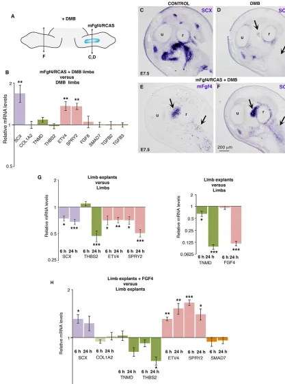
[image:5.612.51.349.55.523.2]In order to determine whether FGF would rescue tendon gene expression in the absence of mechanical signals, we applied mFgf4-expressing retroviruses in chick limbs (mFgf4/RCAS) and injected DMB in order to prevent muscle contraction (Fig. 6A). In the
Fig. 3. FGF4 positively regulates the expression of the tendon differentiation markersTNMDandTHBS2. (A,B) Adjacent transverse sections of forelimbs of E9 chick embryos were hybridised withSCXandTNMDprobes.TNMD was expressed inSCX-positive tendons (arrows).TNMDwas also expressed in the dermis (B, arrowhead). (C-H) mFgf4/ RCAS-producing cells were grafted into forelimb buds of E3.5 (HH21) chick embryos. Embryos (n=3) were fixed at E7.5, and grafted (C,E,G) and control (F,H) forelimbs were transversely sectioned at the level of the zeugopod. (C,E,G) Adjacent sections were hybridised withmFgf4probe to show the extent of virus spread and withTNMDandTHBS2probes to show ectopic expression (E,G, arrows) in mFgf4-positive regions (C, arrow) compared with normalTNMDandTHBS2expression in control left limbs (F,H). (D) RT-q-PCR analyses of mRNA levels in mFgf4/RCAS-infected limbs (4 days after grafting) (n=5). For each gene, the mRNA levels of infected (right) limbs are expressed relative to those of control (left) limbs (normalised to 1).P-values were determined by paired Student’st-test using Microsoft Excel. *P<0.05; **P<0.01. Error bars represent s.e.m. r, radius; u, ulna.
DEVEL
O
absence of muscle contraction,SCXexpression was downregulated (Fig. 6C,D). mFgf4 was able to activate ectopicSCXexpression in limbs of immobilised embryos (Fig. 6D-F). Consistent with this, the relative mRNA levels ofSCX,ETV4andSPRY2were significantly upregulated in mFgf4-paralysed-limbs compared with paralysed limbs (Fig. 6B). Under these experimental conditions,TNMDand THBS2expression was not changed (Fig. 6B). The relative mRNA levels of TGFB2,TGFB3 and SMAD7 were not changed in the presence of mFgf4 in immobilised embryos (Fig. 6B), indicating that TGFβsignalling was not modified under these experimental conditions. We performed a similar FGF rescue experiment in chick limb explants, in which we considered that the E5 limb explant
[image:6.612.83.535.60.501.2]culture system was devoid of mechanical movements. Analysis of the relative mRNA levels in chick limb explants compared with stage-matched limbs originating from in ovo embryos showed a significant diminution ofSCX,TNMD,THBS2,ETV4,SPRY2and FGF4 gene expression (Fig. 6G), similar to that observed in immobilised chick embryos (Figs 4 and 5). Consistent with the in ovo FGF rescue experiments (Fig. 6A-F), the application of recombinant FGF4 in limb explant cultures induced a significant increase of the mRNA levels ofSCX,ETV4andSPRY2, but did not affectCOL1A2andSMAD7expression (Fig. 6H). The expression levels ofTNMDandTHBS2genes were not increased upon FGF4 treatment and even displayed a decrease of expression (Fig. 6H). Fig. 4. Muscle contraction is required to maintainSCXandTNMDexpression in chick forelimbs.DMB reagent was injected into E4.5 (HH24) chick embryos to induce immobilisation. Immobilised embryos were processed forin situhybridisation (A-F,H-O) or RT-q-PCR (G) analyses. (A,B) Forelimb transverse sections of control (n=2) and DMB-treated embryos (n=4) were hybridised withSCXprobe 24 h after treatment. (C-F) Forelimbs of control (n=10) and DMB-treated (n=10) embryos fixed 48 h (n=5) or 72 h (n=5) after application were hybridised withSCXprobe. (G) RT-q-PCR analyses of mRNA levels for tendon genes in forelimbs (n=10), forelimbs with digits removed (n=20) and digits only (n=20) of DMB-treated embryos, 48 h (n=10) and 72 h (n=10) after DMB application. For each gene, the mRNA levels of treated limbs are expressed relative to those of control limbs (normalised to 1).P-values were determined by unpaired Student’st-test using Microsoft Excel. *P<0.05; **P<0.01; ***P<0.001. Error bars represent s.e.m. (H-O) Forelimb (H,I,L-O) and digit (J,K) transverse sections of control (H,J, L,N) and DMB-treated (I,K,M,O) embryos (n=4) were hybridised withSCX(H-K),TNMD(L,M) orTHBS2(N,O) probes 72 h after treatment. r, radius; u, ulna.
DEVEL
O
We conclude that FGF4 activatesSCX but notTNMD orTHBS2 expression in chick limbs in immobilisation conditions.
TGFβ2 maintainsSCX,TNMDandTHBS2expression in immobilised chick limbs
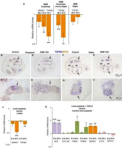
Next, we wanted to determine whether TGFβwas also sensitive to immobilisation. Both Tgfb2 and Tgfb3 have been shown to be involved in mouse limb tendon development (Pryce et al., 2009). In E7.5 limbs, TGFB2 was observed in tendons, in addition to displaying expression in muscles (Fig. S5). TGFB3 was mainly expressed in chick limb muscles, with faint expression in tendons (Fig. S5). In DMB-treated embryos, the mRNA levels ofSMAD7 and TGFB2 were decreased in paralysed limbs compared with control limbs (Fig. 7A).TGFB2expression was lost in limb tendons of the zeugopod regions (Fig. 7B-E, arrows), but not in digits (Fig. 7F-I) of immobilised embryos. The diminution of the relative mRNA levels ofSMAD7and TGFB2was also observed in limb explants compared with stage-matched limbs originating from in ovo embryos (Fig. 7J). These results show that the TGFβ/
SMAD2/3 signalling pathway was decreased in chick limb tendons under immobilisation conditions. Application of TGFβ2 to limb explants increased SCX, TNMD,THBS2 and SMAD7 expression compared with control limb explants (Fig. 7K). The expression levels of the transcriptional readouts of ERK MAPK activity were not modified upon exposure to TGFβ2. We conclude that TGFβ2 is sufficient to maintain the expression of SCX and the tendon differentiation markers TNMD and THBS2 in chick limbs under immobilisation conditions.
DISCUSSION
TGFβfunction in tendon development is similar in chick and mouse limbs
[image:7.612.72.541.58.445.2]Our TGFβ2 gain- and loss-of-function experiments in early chick limbs and explants (Fig. 2) show that TGFβ2 is sufficient and the SMAD2/3 intracellular pathway is required forSCXexpression in undifferentiated limb cells. These results are fully consistent with those obtained in early mouse limb explants (Pryce et al., 2009; Havis et al., 2014). These results highlight a universal role for TGFβ Fig. 5. Muscle contraction is required to maintain active FGF/ERK MAPK signalling in chick limb tendons.(A) RT-q-PCR analyses of mRNA levels for ETV4,SPRY2,FGF4andFGF8in forelimbs (n=9), forelimbs (digits excluded) (n=19) and digits (n=20) of DMB-treated embryos, 48 h (n=20) and 72 h (n=19) after DMB application. For each gene, the mRNA levels of treated limbs are expressed relative to those of control limbs (normalised to 1).P-values were determined by unpaired Student’st-test using Microsoft Excel. *P<0.05; ***P<0.001. Error bars represent s.e.m. (B-I) Forelimb transverse sections at the level of the zeugopod of control (B,D,F,H) and DMB-treated (C,E,G,I) embryos were hybridised withETV4(B,C),SPRY2(D,E),FGF4(F,G) andFGF8(H,I) probes (n=3) 72 h after treatment. Arrows point to gene expression in control limbs (B,D,F,H) and loss of gene expression in DMB-treated limbs (C,E,G,I). r, radius; u, ulna.
DEVEL
O
Fig. 6. FGF4 inducesSCXexpression in limbs of immobilised embryos.(A) mFgf4/RCAS-producing cells were grafted into right forelimbs of E3.5 chick embryos. These embryos were then treated with DMB at E4.5 and fixed 3 days after DMB application at E7.5. (B,D-F) The manipulated embryos were either processed for RT-q-PCR analysis (B) (n=7) or forin situhybridisation to limb sections (D-F) (n=3). (B) RT-q-PCR analyses of mRNA levels for components of tendons (SCX,COL1A2,TNMD,THBS2) and of the FGF/ERK (ETV4, SPRY, FGF8) and of the TGF-β/SMAD2/3 (SMAD7, TGFB2, TGFB3) signalling pathways in mFgf4/RCAS forelimbs of DMB-treated embryos. For each gene, the mRNA levels of mFgf4/RCAS limbs are expressed relative to those of contralateral (DMB-treated only) limbs (normalised to 1). (D-F) Transverse sections of right mFgf4/RCAS forelimbs (E,F) and left forelimbs (D) of DMB-(DMB-treated embryos were hybridised with probes formFgf4(E) andSCX(D,F). (C)In situhybridisation to E7.5 control limbs withSCXprobe. r, radius; u, ulna. (G) The mRNA levels forSCX, ETV4,SPRY2,TNMD,THBS2andFGF4were compared by RT-q-PCR analysis in E5 (HH25/26) limb explants cultured for 6 h (n=5) and 24 h (n=5) versus limbs (n=10) of stage-matched embryos. (H) RT-q-PCR analyses of mRNA levels in E5 limb explants cultured for 6 h (n=7) and 24 h (n=5) in the presence or absence of FGF4. For each gene, the mRNA levels of E5 limb explants cultured for 6 and 24 h with no FGF4 were normalised to 1.P-values were determined by unpaired Student’st-test using Microsoft Excel. *P<0.05; **P<0.01; ***P<0.001. Error bars represent s.e.m.
DEVEL
O
in initiating the commitment of undifferentiated limb mesodermal cells towards the tendon lineage during chick and mouse development (Fig. 8). In zebrafish embryos, blocking the TGFβ pathway (using the chemical drug SB431542) inhibits scxa
[image:9.612.92.519.56.576.2]expression (Chen and Galloway, 2014), suggesting that TGFβ is also important for the initiation of scxa expression in fish. The developmental TGFβtenogenic effect is likely to be related to the recognized effect of TGFβin positively regulatingScxexpression in Fig. 7. TGFβ2 preventsSCXdownregulation in limb explants.(A-I) DMB application into E4.5 chick embryos. E7.5 immobilised embryos were processed for either RT-q-PCR analysis (n=10) orin situhybridisation to limb sections (n=3). (A) RT-q-PCR analyses for components of the TGFβpathway, in paralysed limbs. For each gene, the mRNA levels of limbs of DMB-treated embryos are expressed relative to those of control embryos (normalised to 1). (B-I) Transverse limb sections at the zeugopod (B-E) and digit (F-I) levels of immobilised embryos (D,E,H,I) or control embryos (B,C,F,G) were hybridised with theTGFB2probe and then immunostained with MF20 antibody. C,E,G,I are high magnifications of the boxed regions of B,D,F,H, respectively. Arrows point to zeugopod (B-E) or digit (F-I) tendons in control or DMB embryos. d3, digit 3; d4, digit 4; r, radius; u, ulna. (J) The mRNA levels forSMAD7andTGFB2were compared by RT-q-PCR analysis in E5 (HH25/26) limb explants (n=10) cultured for 6 h (n=5) and 24 h (n=5) versus limbs of stage-matched embryos. (K) RT-q-PCR analyses of mRNA levels in E5 limb explants (n=15) cultured for 6 h (n=8) and 24 h (n=7) in the presence or absence of TGFβ2. For each gene, the mRNA levels of E5 limb explants cultured for 6 h and 24 h with no TGF-β2 were normalised to 1.P-values were determined by unpaired Student’st-test using Microsoft Excel. *P<0.05; **P<0.01; ***P<0.001. Error bars indicate s.e.m.
DEVEL
O
embryonic tendon progenitors (Brown et al., 2014) and stem cell culture systems (Pryce et al., 2009; Barsby and Guest, 2013; Goncalves et al., 2013; Guerquin et al., 2013; Havis et al., 2014). We find that the positive effect of TGFβ2 on chick limb SCX expression is independent of ERK MAPK signalling (Fig. 2C,D), as also observed in mouse limb explants (Pryce et al., 2009; Havis et al., 2014).
Tnmd/TNMDis one of the tendon markers displaying the highest expression levels in E14.5 mouse Scx+cells but is not expressed in
E11.5 mouse limb bud explants (Havis et al., 2014) or E4 chick limbs (Shukunami et al., 2006) or activated by TGFβ2 at these early stages (Havis et al., 2014). However,TNMDexpression is activated upon TGFβexposure in late chick (Fig. 8) and mouse (Havis et al., 2014) limb explants. This is consistent with previous reports showing TNMD upregulation by TGFβ ligands in 3D-culture systems of human tendon cells (Bayer et al., 2014) and of equine embryo-derived stem cells (Barsby et al., 2014), and in high-density cultures of chick limb cells (Lorda-Diez et al., 2009). It is worth mentioning that TGFβdramatically decreasesTnmdexpression (and activatesScx) in 2D-culture systems of embryonic or adult mouse tendon progenitors and in mouse mesenchymal stem cells (Guerquin et al., 2013; Brown et al., 2014; Liu et al., 2015). We believe that the opposite effects of TGFβonTnmdexpression are due to the different cell contact environments in 2D-culture versus 3D-culture systems.
FGF has a tenogenic effect in chick limb undifferentiated cells, but has an anti-tenogenic effect in mouse limb undifferentiated cells
In vivo and ex vivoexperiments demonstrated that FGF activates SCXexpression in early chick limb buds. This is consistent with FGF function in somites of chick embryos (Brent and Tabin, 2004; Smith et al., 2005). This result observed in chick embryos is
opposite to those obtained in mouse limb explants, in which FGF inhibits Scxexpression and ERK MAPK inhibition activatesScx expression (Havis et al., 2014; Fig. 1E). Blockade of SMAD2/3 did not prevent theSCXactivation by FGF4 in chick limbs (Fig. S1). Moreover,Smad7/SMAD7expression was not modified in any of the FGF misexpression experiments (Fig. 1D,E) (Havis et al., 2014), indicating that the TGFβpathway is not involved in the positive or negative effect of FGF onSCX/Scxexpression in chick and mouse, respectively. We believe that FGF has a tenogenic effect in chick undifferentiated limb mesodermal cells, but has an anti-tenogenic effect on mouse undifferentiated limb mesodermal cells. The reasons for the opposite effect of FGF signalling on limb mesodermal cells between the chick and mouse models remain unclear. However, these results are consistent with an absence or deleterious effect of FGF on tendon marker expression in 2D-culture systems of various stem cells, including mouse embryonic tendon progenitors (Brown et al., 2014), mouse mesenchymal stem cells (Havis et al., 2014), canine tendon fibroblasts (Thomopoulos et al., 2010), human amniotic fluid stem cells or adipose-derived stem cells (Goncalves et al., 2013). Consistent with the FGF tenogenic function during chick tendon development, a clear beneficial effect of FGF has been described during tendon repair in a chick digital tendon injury model. The expression of the ligand FGFb is decreased in chick tendons during the process of tendon repair (Chen et al., 2008) and ectopic application of FGF has a beneficial effect on chick tendon repair (Tang et al., 2008, 2014). We conclude that FGF positively regulatesSCX in chick limb undifferentiated cells (Fig. 8).
FGF4 and TGFβ2 have a tenogenic effect, independently of each other, during chick limb development
FGF4 and TGFβ2 activateSCXexpression independently of each other in early chick limb buds (Fig. 2D; Fig. S1). Although intracellular crosstalk has been identified between the ERK MAPK and SMAD2/3 signalling pathways in many biological systems (reviewed by Massague, 2012), our results indicate that these signalling pathways do not interact in the activation ofSCXin chick limb buds. The fact that two signalling pathways activate SCX independently of each other indicates the presence of a safety system for tendon specification in chick limbs. This safety system is classically observed during developmental processes. Another possible hypothesis could be that two pools ofSCX-positive cells co-exist within the chick limb buds, one pool being sensitive to the TGFβ2/SMAD2/3 signalling pathway and another one being sensitive to the FGF ERK/MAPK signalling pathway.
Limb tendon development relies on mechanical forces generated by muscle contraction
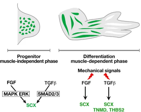
[image:10.612.53.298.55.249.2]Immobilisation following muscle paralysis induces a drastic diminution of SCX, TNMD and THBS2 gene expression in stylopod/zeugopod tendons of chick limbs (Fig. 4). This shows that tendon gene expression is sensitive to mechanical signals in chick limbs. However, in the mdg mouse, which is deprived of muscle activity, Scx/GFP-positive tendons are observed in stylopod/zeugopod limb regions, although they are reduced in size (Huang et al., 2015b). This difference could be due to the possibility that the GFP fluorescence can be still detected even if the Scxpromoter is no longer active or the fact that the mouse embryos are still submitted to mechanical movements from maternal activity, whereas the pharmacologically induced immobilisation used in our experiments is more drastic. However, an alternative and plausible explanation is that these results indicate that tendon development in Fig. 8. FGF4 and TGFβ2 involvement in stylopod/zeugopod limb tendons
during chick development.During the progenitor stage, which is independent of muscle, FGF or TGFβligand, independently of each other, each have a tenogenic effect on limb mesodermal undifferentiated cells. Moreover, the MAPK ERK pathway and SMAD2/3 intracellular pathways are both required forSCXexpression in early chick limbs. During the differentiation step, which is dependent on muscle, muscle contraction is required for FGF/ MAK ERK and TGFβ/SMAD2/3 activity at the muscle/tendon interface. Downstream of mechanical forces, FGF4 and TGFβ2 positively regulateSCX expression, and TGFβ2 (but not FGF4) regulatesTNMDandTHBS2 expression in limb tendons.
DEVEL
O
chick and mouse has differential requirements for mechanical movements. It has been demonstrated in mice that muscles are required for zeugopod tendon elongation, but only tendon size and individuation depend on mechanical forces in mouse limbs (Huang et al., 2015b). The complete loss of zeugopod tendons in chick immobilised embryos shows that mechanical signals are crucial for all the steps of chick zeugopod tendon differentiation. This can be correlated with the fact that tendon cells experience higher levels of mechanical signals in actively moving chick embryos in the egg compared with mouse embryos embedded in uterine membranes and with the faster development of the musculoskeletal system in chick versus mouse embryos.
Although differences exist between the mechanical signal requirement between chick and mouse tendon development, mechanical forces generated by muscle contraction are recognised as being required for skeletal system formation during chick and mouse development (reviewed by Shwartz et al., 2013). Immobilisation affects bone, cartilage and synovial joint morphogenesis, targeting general processes such as proliferation and differentiation leading to shape and size defects (Blitz et al., 2009; Kahn et al., 2009; Roddy et al., 2011a). A correlation has been established between biophysical stimuli patterns and skeletal regions affected upon immobilisation (Roddy et al., 2011b). Tendons that link muscles to bones are expected to experience high mechanical strains and are largely affected in immobilisation conditions. The mechanosensitivity of tendon development is maintained in adult life, as tendon cells are sensitive to mechanical signals generated by tendon loading (reviewed by Nourissat et al., 2015). Scx expression is downregulated in adult tendons in unloading conditions (Maeda et al., 2011), whereasScxandTnmd are activated under overloading conditions in mice (Mendias et al., 2012; Zhang and Wang, 2013). We conclude that tendon cells require appropriate mechanical signals during development and adult life.
FGF4 and TGFβ2 act downstream of mechanical signals to regulate tendon differentiation
In immobilised chick embryos, transcriptional readouts of both FGF/ERK MAPK and TGFβ/SMAD2/3 signalling pathways are downregulated in limb tendons. This shows that both pathways are sensitive to mechanical signals in chick limbs. These pathways are known to be sensitive to mechanical forces in other biological systems (Humphrey et al., 2014). Moreover, transcriptome profiling analyses have identified FGF and TGFβ signalling as being downregulated in developing humerus (limb bone) of immobilised mouse foetuses (Rolfe et al., 2014). In addition to being downregulated in tendons of immobilised embryos, both FGF4 and TGFβ2 prevent the decrease ofSCXexpression in chick limbs in immobilisation conditions. Rescue experiments with FGF4 or TGFβ2 in immobilised limbs do not activate TGFβor FGF transcriptional readout, respectively, indicating that FGF4 and TGFβ2 act independently of each other to activate SCX expression during limb tendon differentiation. The ability of TGFβ2 to rescueSCXexpression in chick limbs in immobilised embryos is reminiscent of the requirement of the SMAD2/3 pathway for Scx induction downstream of mechanical forces in tendon cells (Maeda et al., 2011). However,TNMDandTHBS2 downregulation was only prevented by TGFβ2 and not by FGF4. We hypothesise that TGFβ2 activates another signal required for TNMDandTHBS2expression, which is not regulated by FGF4, in immobilisation conditions. In the presence of muscle contraction, FGF4 activates TNMD and THBS2, whereas in the absence of
muscle contraction FGF4 is not able to activateTNMDorTHBS2 expression, highlighting the independent effects of both pathways in tendon differentiation.
In summary, both FGF4 and TGFβ2 signalling molecules are involved in the commitment of undifferentiated chick limb mesodermal cells towards the tendon lineage and act downstream of mechanical forces to regulate tendon differentiation during chick limb development (Fig. 8). Both FGF4 and TGFβ2 have a tenogenic effect, independently of each other, during both muscle-independent and -dependent phases of chick limb tendon development.
MATERIALS AND METHODS
Chick and mouse embryos
Fertilised chick eggs (JA 57 strain) (EARL Morizeau, Dangers, France) were incubated at 38°C. Embryos were aged according to the number of days of incubation (embryonic day) or staged according to Hamburger and Hamilton (HH) stages (Hamburger and Hamilton, 1992). Swiss mouse embryos (Janvier Labs) were collected after natural overnight matings. For staging, fertilisation was considered to take place at midnight. The manipulation of chick and non-transgenic mouse embryos was performed in accordance with the guidelines of the French National Ethics Committee.
Bead implantation and grafting mFgf4/RCAS-expressing cells to chick limb buds
FGF4, TGFβ2, TGFβ2+PD18, FGF4+SIS3 beads or
mFgf4/RCAS-expressing cells were grafted in limbs of E3/E4 chick embryos as described (Edom-Vovard et al., 2002). Embryos were harvested 4, 6 or 24 h after grafting. Further details are provided in supplementary Materials and Methods.
Chick and mouse limb explant cultures
Chick limb explants were prepared as described by Placzek and Dale (1999) and treated with TGFβ2, FGF4, PD18, SB43, SIS3 or TGFβ2+PD18 as described by Havis et al. (2014). Further details are provided in supplementary Materials and Methods.
DMB or PB application in chick embryos
Decamethonium bromide (DMB) and pancuronium bromide (PB) were prepared in Hank’s solution, at final concentrations of 12 mM. DMB or PB solution (100 µl) was injected daily using a Pipetman pipette (Gilson) into the amniotic fluid next to the embryos after vitelline membrane removal in E4.5, E5.5 and E6.5 chick embryos. Control embryos were injected with Hank’s solution using the same daily protocol. Immobilised or control embryos were analysed at E5.5 (24 h), E6.5 (48 h) or E7.5 (72 h). Forelimbs or hindlimbs were isolated and analysed byin situhybridisation on sections or wholemounts or by RT-q-PCR analysis. For RT-q-PCR analysis, RNAs were prepared from the whole limbs, the limbs without digits or the digits alone.
RNA isolation, reverse transcription and quantitative real-time PCR
RT-q-PCR of experimental or control chick limbs, chick limb explants or mouse limb explants were performed as previously described (Havis et al., 2014). A detailed protocol is provided in supplementary Materials and Methods.
In situhybridisation and immunohistochemistry
Control or manipulated chick limbs were fixed and processed forin situ
hybridisation as previously described by Havis et al. (2014). The probes that were used are described in supplementary Materials and Methods. Differentiated muscle cells were detected afterin situhybridisation with the monoclonal antibody MF20 (non-diluted supernatant) developed by D. A. Fischman and obtained from the Developmental Studies Hybridoma Bank developed under the auspices of the NICHD and maintained by the
University of Iowa.
DEVEL
O
Acknowledgements
We thank laboratory members for comments on the manuscript and Sophie Gournet for illustrations.
Competing interests
The authors declare no competing or financial interests.
Author contributions
D.D. designed the experiments. E.H., M.-A.B., J.E.d.L., B.C. and C.M. performed experiments. E.H. and D.D. analysed the data and D.D. wrote the manuscript. All of the authors have read and approved the final manuscript.
Funding
This work was supported by the Fondation pour la Recherche Médicale (FRM) [DEQ20140329500]; the Agence Nationale de la Recherche (ANR) [ANR-12-BSV1-0038]; the Association Française contre les Myopathies (AFM) [16752/16826]; Institut National de la Santéet de la Recherche Médicale (INSERM); the Centre National de la Recherche Scientifique (CNRS); and the UniversitéPierre et Marie Curie (UPMC).
Supplementary information
Supplementary information available online at
http://dev.biologists.org/lookup/doi/10.1242/dev.136242.supplemental
References
Alberton, P., Dex, S., Popov, C., Shukunami, C., Schieker, M. and Docheva, D. (2015). Loss of tenomodulin results in reduced self-renewal and augmented senescence of tendon stem/progenitor cells.Stem Cells Dev.24, 597-609. Anderson, D. M., Arredondo, J., Hahn, K., Valente, G., Martin, J. F.,
Wilson-Rawls, J. and Wilson-Rawls, A.(2006). Mohawk is a novel homeobox gene expressed in the developing mouse embryo.Dev. Dyn.235, 792-801.
Barsby, T. and Guest, D.(2013). Transforming growth factor beta3 promotes tendon differentiation of equine embryo-derived stem cells.Tissue Eng. A19, 2156-2165.
Barsby, T., Bavin, E. P. and Guest, D. J.(2014). Three-dimensional culture and transforming growth factor beta3 synergistically promote tenogenic differentiation of equine embryo-derived stem cells.Tissue Eng. A20, 2604-2613.
Bayer, M. L., Schjerling, P., Herchenhan, A., Zeltz, C., Heinemeier, K. M., Christensen, L., Krogsgaard, M., Gullberg, D. and Kjaer, M.(2014). Release of tensile strain on engineered human tendon tissue disturbs cell adhesions, changes matrix architecture, and induces an inflammatory phenotype.PLoS ONE
9, e86078.
Blitz, E., Viukov, S., Sharir, A., Shwartz, Y., Galloway, J. L., Pryce, B. A., Johnson, R. L., Tabin, C. J., Schweitzer, R. and Zelzer, E.(2009). Bone ridge patterning during musculoskeletal assembly is mediated through SCX regulation of Bmp4 at the tendon-skeleton junction.Dev. Cell17, 861-873.
Bonnin, M.-A., Laclef, C., Blaise, R., Eloy-Trinquet, S., Relaix, F., Maire, P. and Duprez, D.(2005). Six1 is not involved in limb tendon development, but is expressed in limb connective tissue under Shh regulation.Mech. Dev. 122, 573-585.
Brent, A. E. and Tabin, C. J.(2004). FGF acts directly on the somitic tendon progenitors through the Ets transcription factors Pea3 and Erm to regulate scleraxis expression.Development131, 3885-3896.
Brent, A. E., Schweitzer, R. and Tabin, C. J.(2003). A somitic compartment of tendon progenitors.Cell113, 235-248.
Brown, J. P., Finley, V. G. and Kuo, C. K.(2014). Embryonic mechanical and soluble cues regulate tendon progenitor cell gene expression as a function of developmental stage and anatomical origin.J. Biomech.47, 214-222. Chen, J. W. and Galloway, J. L.(2014). The development of zebrafish tendon and
ligament progenitors.Development141, 2035-2045.
Chen, C. H., Cao, Y., Wu, Y. F., Bais, A. J., Gao, J. S. and Tang, J. B.(2008). Tendon healing in vivo: gene expression and production of multiple growth factors in early tendon healing period.J. Hand Surg.33, 1834-1842.
Dex, S., Lin, D., Shukunami, C. and Docheva, D.(2016). Tenogenic modulating insider factor: systematic assessment on the functions of tenomodulin gene.Gene
587, 1-17.
Docheva, D., Hunziker, E. B., Fassler, R. and Brandau, O.(2005). Tenomodulin is necessary for tenocyte proliferation and tendon maturation.Mol. Cell. Biol.25, 699-705.
Edom-Vovard, F. and Duprez, D.(2004). Signals regulating tendon formation during chick embryonic development.Dev. Dyn.229, 449-457.
Edom-Vovard, F., Bonnin, M.-A. and Duprez, D.(2001). Fgf8 transcripts are located in tendons during embryonic chick limb development.Mech. Dev.108, 203-206.
Edom-Vovard, F., Schuler, B., Bonnin, M.-A., Teillet, M.-A. and Duprez, D. (2002). Fgf4 positively regulates scleraxis and tenascin expression in chick limb tendons.Dev. Biol.247, 351-366.
Eloy-Trinquet, S., Wang, H., Edom-Vovard, F. and Duprez, D.(2009). Fgf signaling components are associated with muscles and tendons during limb development.Dev. Dyn.238, 1195-1206.
Gaut, L. and Duprez, D. (2016). Tendon development and diseases. Wiley Interdiscip. Rev. Dev. Biol.5, 5-23.
Goncalves, A. I., Rodrigues, M. T., Lee, S.-J., Atala, A., Yoo, J. J., Reis, R. L. and Gomes, M. E.(2013). Understanding the role of growth factors in modulating stem cell tenogenesis.PLoS ONE8, e83734.
Grenier, J., Teillet, M.-A., Grifone, R., Kelly, R. G. and Duprez, D.(2009). Relationship between neural crest cells and cranial mesoderm during head muscle development.PLoS ONE4, e4381.
Guerquin, M.-J., Charvet, B., Nourissat, G., Havis, E., Ronsin, O., Bonnin, M.-A., Ruggiu, M., Olivera-Martinez, I., Robert, N., Lu, Y. et al.(2013). Transcription factor EGR1 directs tendon differentiation and promotes tendon repair.J. Clin. Invest.123, 3564-3576.
Guo, X. and Wang, X.-F.(2009). Signaling cross-talk between TGF-beta/BMP and other pathways.Cell Res.19, 71-88.
Hamburger, V. and Hamilton, H. L.(1992). A series of normal stages in the development of the chick embryo. 1951.Dev. Dyn.195, 231-272.
Havis, E., Bonnin, M.-A., Olivera-Martinez, I., Nazaret, N., Ruggiu, M., Weibel, J., Durand, C., Guerquin, M. J., Bonod-Bidaud, C., Ruggiero, F. et al.(2014). Transcriptomic analysis of mouse limb tendon cells during development.
Development141, 3683-3696.
Huang, A. H., Riordan, T. J., Wang, L., Eyal, S., Zelzer, E., Brigande, J. V. and Schweitzer, R.(2013). Repositioning forelimb superficialis muscles: tendon attachment and muscle activity enable active relocation of functional myofibers.
Dev. Cell26, 544-551.
Huang, A. H., Lu, H. H. and Schweitzer, R.(2015a). Molecular regulation of tendon cell fate during development.J. Orthop. Res.33, 800-812.
Huang, A. H., Riordan, T. J., Pryce, B., Weibel, J. L., Watson, S. S., Long, F., Lefebvre, V., Harfe, B. D., Stadler, H. S., Akiyama, H. et al. (2015b). Musculoskeletal integration at the wrist underlies the modular development of limb tendons.Development142, 2431-2441.
Humphrey, J. D., Dufresne, E. R. and Schwartz, M. A. (2014). Mechanotransduction and extracellular matrix homeostasis.Nat. Rev. Mol. Cell Biol.15, 802-812.
Ito, Y., Toriuchi, N., Yoshitaka, T., Ueno-Kudoh, H., Sato, T., Yokoyama, S., Nishida, K., Akimoto, T., Takahashi, M., Miyaki, S. et al.(2010). The Mohawk homeobox gene is a critical regulator of tendon differentiation.Proc. Natl. Acad. Sci. USA107, 10538-10542.
Jelinsky, S. A., Archambault, J., Li, L. and Seeherman, H.(2010). Tendon-selective genes identified from rat and human musculoskeletal tissues.J. Orthop. Res.28, 289-297.
Kahn, J., Shwartz, Y., Blitz, E., Krief, S., Sharir, A., Breitel, D. A., Rattenbach, R., Relaix, F., Maire, P., Rountree, R. B. et al.(2009). Muscle contraction is necessary to maintain joint progenitor cell fate.Dev. Cell16, 734-743. Kimura, W., Machii, M., Xue, X., Sultana, N., Hikosaka, K., Sharkar, M. T. K.,
Uezato, T., Matsuda, M., Koseki, H. and Miura, N.(2011). Irxl1 mutant mice show reduced tendon differentiation and no patterning defects in musculoskeletal system development.Genesis49, 2-9.
Kyriakides, T. R., Zhu, Y.-H., Smith, L. T., Bain, S. D., Yang, Z., Lin, M. T., Danielson, K. G., Iozzo, R. V., LaMarca, M., McKinney, C. E. et al.(1998). Mice that lack thrombospondin 2 display connective tissue abnormalities that are associated with disordered collagen fibrillogenesis, an increased vascular density, and a bleeding diathesis.J. Cell Biol.140, 419-430.
Lejard, V., Brideau, G., Blais, F., Salingcarnboriboon, R., Wagner, G., Roehrl, M. H. A., Noda, M., Duprez, D., Houillier, P. and Rossert, J.(2007). Scleraxis and NFATc regulate the expression of the pro-alpha1(I) collagen gene in tendon fibroblasts.J. Biol. Chem.282, 17665-17675.
Lejard, V., Blais, F., Guerquin, M.-J., Bonnet, A., Bonnin, M.-A., Havis, E., Malbouyres, M., Bidaud, C. B., Maro, G., Gilardi-Hebenstreit, P. et al.(2011). EGR1 and EGR2 involvement in vertebrate tendon differentiation.J. Biol. Chem.
286, 5855-5867.
Liu, H., Liu, W., Maltby, K. M., Lan, Y. and Jiang, R.(2006). Identification and developmental expression analysis of a novel homeobox gene closely linked to the mouse Twirler mutation.Gene Expr. Patterns6, 632-636.
Liu, W., Watson, S. S., Lan, Y., Keene, D. R., Ovitt, C. E., Liu, H., Schweitzer, R. and Jiang, R.(2010). The atypical homeodomain transcription factor Mohawk controls tendon morphogenesis.Mol. Cell. Biol.30, 4797-4807.
Liu, H., Zhang, C., Zhu, S., Lu, P., Zhu, T., Gong, X., Zhang, Z., Hu, J., Yin, Z., Heng, B. C. et al.(2015). Mohawk promotes the tenogenesis of mesenchymal stem cells through activation of the TGFbeta signaling pathway.Stem Cells33, 443-455.
Lorda-Diez, C. I., Montero, J. A., Martinez-Cue, C., Garcia-Porrero, J. A. and Hurle, J. M.(2009). Transforming growth factors beta coordinate cartilage and tendon differentiation in the developing limb mesenchyme.J. Biol. Chem.284, 29988-29996.
Lorda-Diez, C. I., Montero, J. A., Garcia-Porrero, J. A. and Hurle, J. M.(2010). Tgfbeta2 and 3 are coexpressed with their extracellular regulator Ltbp1 in the early
DEVEL
O
limb bud and modulate mesodermal outgrowth and BMP signaling in chicken embryos.BMC Dev. Biol.10, 69.
Maeda, T., Sakabe, T., Sunaga, A., Sakai, K., Rivera, A. L., Keene, D. R., Sasaki, T., Stavnezer, E., Iannotti, J., Schweitzer, R. et al. (2011). Conversion of mechanical force into TGF-beta-mediated biochemical signals.Curr. Biol.21, 933-941.
Mason, J. M., Morrison, D. J., Basson, M. A. and Licht, J. D.(2006). Sprouty proteins: multifaceted negative-feedback regulators of receptor tyrosine kinase signaling.Trends Cell Biol.16, 45-54.
Massague, J.(2012). TGFbeta signalling in context.Nat. Rev. Mol. Cell Biol.13, 616-630.
Mendias, C. L., Gumucio, J. P., Bakhurin, K. I., Lynch, E. B. and Brooks, S. V. (2012). Physiological loading of tendons induces scleraxis expression in epitenon fibroblasts.J. Orthop. Res.30, 606-612.
Murchison, N. D., Price, B. A., Conner, D. A., Keene, D. R., Olson, E. N., Tabin, C. J. and Schweitzer, R.(2007). Regulation of tendon differentiation by scleraxis distinguishes force-transmitting tendons from muscle-anchoring tendons.
Development134, 2697-2708.
Niswander, L., Jeffrey, S., Martin, G. R. and Tickle, C.(1994). A positive feedback loop coordinates growth and patterning in the vertebrate limb. Nature 371, 609-612.
Nourissat, G., Berenbaum, F. and Duprez, D.(2015). Tendon injury: from biology to tendon repair.Nat. Rev. Rheumatol.11, 223-233.
Nowlan, N. C., Sharpe, J., Roddy, K. A., Prendergast, P. J. and Murphy, P. (2010). Mechanobiology of embryonic skeletal development: insights from animal models.Birth Defects Res. C Embryo Today90, 203-213.
O’Hagan, R. C., Tozer, R. G., Symons, M., McCormick, F. and Hassell, J. A. (1996). The activity of the Ets transcription factor PEA3 is regulated by two distinct MAPK cascades.Oncogene13, 1323-1333.
Placzek, M. and Dale, K.(1999). Tissue recombinations in collagen gels.Methods mol. Biol.97, 293-304.
Pryce, B. A., Brent, A. E., Murchison, N. D., Tabin, C. J. and Schweitzer, R. (2007). Generation of transgenic tendon reporters, ScxGFP and ScxAP, using regulatory elements of the scleraxis gene.Dev. Dyn.236, 1677-1682. Pryce, B. A., Watson, S. S., Murchison, N. D., Staverosky, J. A., Dunker, N. and
Schweitzer, R.(2009). Recruitment and maintenance of tendon progenitors by TGFbeta signaling are essential for tendon formation. Development 136, 1351-1361.
Roddy, K. A., Prendergast, P. J. and Murphy, P.(2011a). Mechanical influences on morphogenesis of the knee joint revealed through morphological, molecular and computational analysis of immobilised embryos.PLoS ONE6, e17526. Roddy, K. A., Kelly, G. M., van Es, M. H., Murphy, P. and Prendergast, P. J.
(2011b). Dynamic patterns of mechanical stimulation co-localise with growth and
cell proliferation during morphogenesis in the avian embryonic knee joint.
J. Biomech.44, 143-149.
Rolfe, R. A., Nowlan, N. C., Kenny, E. M., Cormican, P., Morris, D. W., Prendergast, P. J., Kelly, D. and Murphy, P. (2014). Identification of mechanosensitive genes during skeletal development: alteration of genes associated with cytoskeletal rearrangement and cell signalling pathways.BMC Genomics15, 48.
Schweitzer, R., Chyung, J. H., Murtaugh, L. C., Brent, A. E., Rosen, V., Olson, E. N., Lassar, A. and Tabin, C. J.(2001). Analysis of the tendon cell fate using Scleraxis, a specific marker for tendons and ligaments.Development 128, 3855-3866.
Shukunami, C., Takimoto, A., Oro, M. and Hiraki, Y.(2006). Scleraxis positively regulates the expression of tenomodulin, a differentiation marker of tenocytes.
Dev. Biol.298, 234-247.
Shwartz, Y., Blitz, E. and Zelzer, E.(2013). One load to rule them all: mechanical control of the musculoskeletal system in development and aging.Differentiation
86, 104-111.
Smith, T. G., Sweetman, D., Patterson, M., Keyse, S. M. and Munsterberg, A. (2005). Feedback interactions between MKP3 and ERK MAP kinase control scleraxis expression and the specification of rib progenitors in the developing chick somite.Development132, 1305-1314.
Subramanian, A. and Schilling, T. F.(2014). Thrombospondin-4 controls matrix assembly during development and repair of myotendinous junctions.eLife3, e02372.
Subramanian, A., Wayburn, B., Bunch, T. and Volk, T.(2007). Thrombospondin-mediated adhesion is essential for the formation of the myotendinous junction in Drosophila.Development134, 1269-1278.
Sugimoto, Y., Takimoto, A., Akiyama, H., Kist, R., Scherer, G., Nakamura, T., Hiraki, Y. and Shukunami, C.(2013). Scx+/Sox9+ progenitors contribute to the establishment of the junction between cartilage and tendon/ligament.
Development140, 2280-2288.
Tang, J. B., Cao, Y., Zhu, B., Xin, K.-Q., Wang, X. T. and Liu, P. Y.(2008). Adeno-associated virus-2-mediated bFGF gene transfer to digital flexor tendons significantly increases healing strength. an in vivo study.J. Bone Joint Surg.90, 1078-1089.
Tang, J. B., Chen, C. H., Zhou, Y. L., McKeever, C. and Liu, P. Y.(2014). Regulatory effects of introduction of an exogenous FGF2 gene on other growth factor genes in a healing tendon.Wound Repair Regener.22, 111-118. Thomopoulos, S., Kim, H. M., Das, R., Silva, M. J., Sakiyama-Elbert, S., Amiel,
D. and Gelberman, R. H.(2010). The effects of exogenous basic fibroblast growth factor on intrasynovial flexor tendon healing in a canine model.J. Bone Joint Surg.92, 2285-2293.
Zhang, J. and Wang, J. H.-C.(2013). The effects of mechanical loading on tendons–an in vivo and in vitro model study.PLoS ONE8, e71740.