BIROn - Birkbeck Institutional Research Online
Carroni, M. and Kummer, E. and Oguchi, Y. and Wendler, P. and Clare, D.K.
and Sinning, I. and Kopp, J. and Mogk, A. and Bukau, B. and Saibil, Helen
R. (2014) Head-to-tail interactions of the coiled-coil domains regulate ClpB
cooperation with Hsp70 in protein disaggregation. eLife , ISSN 2050-084X.
Downloaded from:
Usage Guidelines:
Please refer to usage guidelines at or alternatively
ACCEPTED MANUSCRIPT
disaggregation
Head-to-tail interactions of the coiled-coil domains regulate ClpB cooperation with Hsp70 in protein
Kopp, Axel Mogk, Bernd Bukau, Helen Saibil
Marta Carroni, Eva Kummer, Yuki Oguchi, Petra Wendler, Daniel K Clare, Irmgard Sinning, Jürgen
http://dx.doi.org/10.7554/eLife.02481 DOI:
Cite as: eLife 2014;10.7554/eLife.02481
Published: 30 April 2014 Accepted: 29 April 2014 Received: 6 February 2014
and proofing.
formatted HTML, PDF, and XML versions will be made available after technical processing, editing, This PDF is the version of the article that was accepted for publication after peer review. Fully
permits unrestricted use and redistribution provided that the original author and source are credited. , which
Creative Commons Attribution License
This article is distributed under the terms of the
elife.elifesciences.org at
Sign up for alerts
Head-to-tail interactions of the coiled-coil domains regulate ClpB activity and
1
cooperation with Hsp70 in protein disaggregation
2
Marta Carroni1, Eva Kummer2, Yuki Oguchi2, Petra Wendler3, Daniel K. Clare1,
3
Irmgard Sinning4, Jürgen Kopp4, Axel Mogk2, Bernd Bukau2 and Helen R. Saibil1,a
4
5
1. Department of Crystallography, Birkbeck College, London, UK.
6
2. Zentrum für Molekulare Biologie der Universität Heidelberg, Heidelberg,
7
Germany.
8
3. Gene Center, Ludwig-Maximilians-University Munich, 81377 Munich,
9
Germany
10
4. Biochemie-Zentrum der Universität Heidelberg (BZH), Im Neuenheimer Feld
11
328, Heidelberg D-69120, Germany
12
13
a) Corresponding author: [email protected]
14
15
Abstract
16
The hexameric AAA+ chaperone ClpB reactivates aggregated proteins in cooperation
17
with the Hsp70 system. Essential for disaggregation, the ClpB middle domain (MD)
18
is a coiled-coil propeller that binds Hsp70. Although the ClpB subunit structure is
19
known, positioning of the MD in the hexamer and its mechanism of action are
20
unclear. We obtained electron microscopy (EM) structures of the BAP variant of
21
ClpB that binds the protease ClpP, clearly revealing MD density on the surface of the
22
ClpB ring. Mutant analysis and asymmetric reconstructions show that MDs adopt
23
diverse positions in a single ClpB hexamer. Adjacent, horizontally oriented MDs form
24
head-to-tail contacts and repress ClpB activity by preventing Hsp70 interaction.
25
Tilting of the MD breaks this contact, allowing Hsp70 binding, and releasing the
26
contact in adjacent subunits. Our data suggest a wavelike activation of ClpB subunits
27
around the ring.
28
29
Introduction
30
Cellular machinery has evolved to prevent or reverse protein misfolding and
31
aggregation, which are damaging to cells and tissues. Bacterial ClpB and its yeast
32
counterpart Hsp104 are members of the Clp/Hsp100 family and function in
33
disaggregation and refolding of protein aggregates together with Hsp70 and its
co-34
chaperones 1-6. ClpB/Hsp104 are conserved in bacteria, fungi, plants and
mitochondria and are essential for recovery of cells from heat shock and other
36
proteotoxic stresses 7,8. The oligomeric ring Hsp100 proteins thread substrates through
37
a central channel, via binding to conserved tyrosine residues on flexible loops 9-14.
38
They belong to the AAA+ (ATPases associated with various cellular activities)
39
superfamily of ATPases, with characteristic α and β subdomains 15,16. ATP binds
40
between the two subdomains and at the subunit interface of adjacent monomers, with
41
a catalytic Arg-finger provided by the neighbouring subunit.
42
43
While some other members of the Hsp100 family have been crystallised in their
44
oligomeric form 17-19, the atomic structure of ClpB is known only for the monomer (T. 45
thermophilus). ClpB is composed of a N-terminal domain (ND) followed by two
46
AAA+ domains (AAA-1 and AAA-2). An 85 Å long coil-coiled propeller, the middle
47
domain (MD), is inserted into the small subdomain of AAA-1 20. It has two blades
48
with mutationally sensitive sites at either end, termed motif 1 and motif 2. Cryo-EM
49
reconstructions of TthClpB hexamers show a two-tiered molecule accounting for the
50
AAA+ rings, but lack density for the ND. On the other hand, various cryo-EM studies
51
of Hsp104 revealed the presence of an ND ring on top of the AAA+ ones. Although
52
the overall shape and dimensions of the ClpB and Hsp104 hexamers are comparable
53
in the various cryo-EM studies, there are substantial differences in the channel
54
width 20-23. Observation of narrow 20,21 (15 to 30 Å) versus expanded 22,23 (30 to 80 Å)
55
cavities has led to different pseudo-atomic models of the hexamers. In one model, the
56
AAA+ rings are compact, as in the crystal structures of other AAA+ hexamers such
57
as p97, HslU or SV40 LTag, with the Arg-finger contacting the neighbouring
58
subunit 17,24,25. In the EM maps used to build this compact model there is little or no
59
density for the MD and it was assumed to extend radially outwards from the ring 20,21.
60
In the expanded model, the AAA+ domains are more widely separated with the MD
61
intercalated between them, preventing the canonical Arg-finger contacts 22,23. An
62
attempt to localize the Hsp104 MD by genetically inserting a lysozyme resulted in a
63
cryo-EM reconstruction of this chimera with visible density for the lysozyme, but not
64
for the MD itself 21. Since none of the existing cryo-EM structures allows an
65
unambiguous localization of the MD, its position is still a matter of debate 26.
66
67
The MD confers unique disaggregase ability to ClpB/Hsp104 27,28 and is required for 68
species-specific cooperation with the DnaK-DnaJ(DnaKJ)/Hsp70-Hsp40 system 29-31.
69
A direct interaction between the ClpB MD and DnaK has been shown by NMR
spectroscopy and site-specific crosslinking and involves the motif 2 tip of the MD and
71
the ATPase domain of DnaK 30,32. The MD acts to repress ClpB disaggregase activity,
72
and DnaK binding relieves this repression 30,33. Point mutations in the MD show that
73
interactions between motif 2 and AAA-1 are critical for regulating ATPase and
74
disaggregase activities 30,33-35. Thus, the MD plays an essential role in coupling Hsp70
75
interaction to ATPase regulation and substrate disaggregation in ClpB/Hsp104. It is
76
therefore important to understand its structural role in ClpB/Hsp104 hexamers and in
77
the context of the ClpB-DnaKJ bi-chaperone machinery.
78
79
To address the ambiguities in domain arrangement and to elucidate the working
80
principle of the MD, we performed single particle EM studies of ClpB under
81
conditions where its orientation could be determined more reliably than in previous
82
studies. We used BAP (ClpB with the ClpA tripeptide for ClpP binding), a chimera
83
engineered to bind the ClpP protease via the replacement of a C-terminal ClpB
84
segment with the ClpP binding region of ClpA 14. This construct has been extensively
85
used to study the ClpB disaggregation mechanism by monitoring substrate proteolysis
86
after delivery to ClpP 9,12,14,32,36 (Figure 1A). Therefore, BAP is suitable for structural
87
studies and has allowed us to obtain maps with visible MD densities that shed light on
88
its regulatory mechanisms.
89 90
Results
91
Three-dimensional (3D) reconstruction of negatively stained BAP-ClpP shows
92
clear density for the middle domain
93
Using the BAP complex facilitates orientation determination in EM reconstruction, as
94
previously observed for ClpA-ClpP 37. Side views of the elongated BAP-ClpP
95
complex are easily recognisable (Figure 1B) whereas in 2D projections of ClpB alone,
96
which has a globular shape, it is hard to distinguish side from tilted views. By
97
restricting the dataset to side views the angle assignment is more reliable, and these
98
views are sufficient to generate the full 3D structure (Figure 1C). Using H/D
99
exchange experiments, which report on the solvent accessibility and structural
100
flexibility of amide hydrogens, we found that BAP, either alone or bound to ClpP,
101
displays the same protection pattern as ClpB, implying the same MD conformation
102
(Figure 1-supplement 1).
103
The BAP-ClpP complexes formed by mixing BAP and ClpP at a 1:1 molar ratio of
105
hexamer to heptamer in the presence of ATPγS were stained with uranyl acetate.
106
Complexes containing two BAP hexamers per ClpP double-heptamer were picked for
107
single particle analysis and separated into halves for processing 37 (Figure 1B). Initial
108
analysis revealed the presence of four layers corresponding to the ND, 1,
AAA-109
2 and ClpP rings. Class averages and eigenimage analysis indicate that the ND is
110
extremely mobile in the BAP hexamer (Figure 1-supplement 2), consistent with
111
crystallographic data on monomeric T. thermophilus ClpB 20. The ND layer was
112
therefore excluded during later stages of image alignment. Similarly, the region
113
corresponding to ClpP, very useful for the initial analysis, was not included in the
114
refinement because of its symmetry mismatch with ClpB. Using ~12,000 particles, we
115
obtained a 3D map by refining the alignment of the two AAA+ rings only, but then
116
including the whole molecule in the reconstruction, which was at ~17 Å resolution
117
(Figure 1C,D; Figure 1-supplement 3). In order to simplify the problems of alignment
118
and reconstruction, we initially imposed 6-fold symmetry, which blurs the features of
119
the heptameric ClpP ring. Similarly, the mobile ClpB ND is blurred into a solid disc.
120
121
The BAP hexamer has overall outer dimensions of ~150 × 100 Å, similar to previous
122
structures of ClpB/Hsp104 20,22,23,38 (Figure 1C). It encloses a ~30 Å wide central 123
channel, comparable in size to that in the crystal structure of ClpC 19 (Figure 1C,D).
124
In the reconstruction it is possible to identify regions accounting for all the domains,
125
such as L-shaped densities for the AAA+ domains and a rod-like density for the
126
coiled-coil MD.
127
128
To interpret the domain interactions, we fitted ClpB atomic coordinates into the EM
129
density. We determined the crystal structure of an ND truncation of E. coli ClpB
130
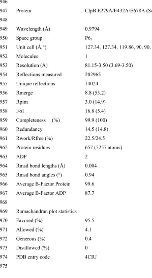
(residues 159 to 858; E432A mutation; Table 1). The subunit structure is very similar
131
to that of T. thermophilus ClpB (Figure 1-supplement 4). Since none of the available
132
crystal structure conformations fit in the EM map, domains were fitted as separate
133
rigid bodies connected at hinge regions (Figure 1C). For the AAA-1 ring, it was
134
possible to build a hexamer model based on the crystal structure of hexameric ClpC 19
135
(PDB code 3PXG), a homologue that also displays disaggregation activity in vitro 39.
136
The resulting ClpB AAA-1 hexamer model was automatically docked into the AAA-1
137
layer as a rigid body (Figure 1D). This strategy was chosen over the fitting of a single
138
subunit followed by hexamerisation because it is expected to provide a more accurate
picture of the subunit interface, which is difficult to determine at the resolution of our
140
EM map. However, this approach did not work for the AAA-2 layer since the ClpB
141
AAA-2 hexamer based on the ClpC crystal structure was not compatible with the
142
density. In this case a ~40° tilt of the monomers into the plane of the ring was
143
required to obtain the optimal fit, which resembles the pseudo-atomic model of the
144
homologue ClpA AAA-2 ring 37. Coordinates of the E. coli ClpB ND (residues 1 to
145
148) were obtained from the PDB (1KHY). A single N domain was fitted manually,
146
maintaining the connection to AAA-1, and then hexamerised by applying symmetry
147
in Chimera 58. 148
149
The AAA+ rings have a central opening of ~30 Å and therefore are not as compact as
150
in one of the previous models 20,21,40, but not as expanded as in the other 22. The
Arg-151
fingers are at the interface between subunits, available to catalyse hydrolysis as
152
expected from mutational studies 28,41,42. 153
154
The ClpB coiled-coil MDs were separately docked in the rod-shaped densities
155
surrounding the AAA-1 ring (Figure 3D), maintaining the connection to the AAA-1
156
small subdomain. The pseudo-atomic model of one ClpB subunit obtained from this
157
fitting differs from the crystal structure by rotations about the inter-domain hinge
158
points (Figure 1-supplement 4B).
159
160
In this position, the MD contacts the neighbouring AAA-1 via its motif 1, while
161
motif 2 makes intrasubunit AAA-1 interactions. This is in good agreement with recent
162
biochemical data showing protection of these two motifs upon ClpB oligomerisation
163
and formation of an intermolecular disulphide bond between E344C of AAA-1 and
164
L424C of a neighboring MD motif 1 33 (Figure 1E).
165
166
Moreover, intramolecular disulphide cross-links engineered between AAA-1 and
167
motif 2 residues in TthClpB, EcoClpB and yeast Hsp104 are also compatible with the
168
fitted structure (Figure 1E; K476C/E358C 33; G175C/R484C, H362C/Q473C E. coli
169
numbering 20; G175C/S499C 34; Hsp104 K358C/D484C 35). However, this
170
arrangement is not compatible with some of the engineered disulphide bonds
171
observed in yeast Hsp104 43 (D427C/E475C, D427C/E471C and E320C/N467C). 172
To investigate possible species-specific structural differences we reconstructed the
174
functional yeast homologue HAP (Hsp104 with the ClpA tripeptide for ClpP binding)
175
in complex with ClpP. Using ~10,000 particles and analyzing the data as described
176
above, an independent map of HAP was obtained at ~21 Å resolution (Figure 2A;
177
Figure 1-supplement 3). The three-layered structure is comparable to BAP and there
178
is density accounting for all domains including the MD, which surrounds the AAA-1
179
ring. The atomic coordinates of the ND and AAA+ rings obtained from the BAP
180
analysis were fitted as rigid bodies. In order to fit the density, the MD must adopt a
181
more horizontal orientation (Figure 2A). In summary, HAP shows overall the same
182
structural organisation as its bacterial homologue ClpB.
183
184
Cryo EM reconstructions of ClpB with and without ClpP support the negative
185
stain maps
186
Since negative stain EM of ClpB (BAP) in complex with ClpP gave a clear result
187
different from all previous cryo EM maps of ClpB and Hsp104, we collected cryo EM
188
data on BAP-ClpP as well as on ClpB alone. The same strategy of using only clearly
189
identifiable side views was applied. Complexes were imaged in the presence of
190
ATPγS and independent maps were obtained by de novo angular reconstitution in
191
each case (Figure 2B,C).
192
For cryo EM of the BAP-ClpP complex, we used a Trap (E279A/E678A) variant,
193
which can bind but not hydrolyse ATP due to mutations in both Walker B motifs. We
194
anticipated that the Trap construct would be more stable than the wild type, but data
195
collection was challenging because side views were not abundant. Eventually, ~4500
196
particles were collected and the same processing strategy was used as for the negative
197
stain data. This variant is more likely to trap non native substrates in its central
198
channel, and the extra density seen in this complex is likely to arise from denatured
199
protein, possibly ClpP, present in the sample (Figure 2B).
200
In the case of isolated ClpB, side views were sorted on the basis of multivariate
201
statistical analysis (MSA). Briefly, particles representing all views of ClpB were
202
picked, centered and classified by MSA. Only particles belonging to classes
203
representing side-views (80°<β<100°) were extracted and used for further processing.
204
Both maps show overall dimensions and density distributions comparable to the
205
negative stain structure of BAP in complex with ClpP, confirming that the ClpB
206
hexamer structure is not significantly altered either by negative staining or by binding
207
to ClpP. The atomic model derived by fitting the negative stain BAP-ClpP map could
be docked as a rigid body into both cryo EM reconstructions, but the MD assumes a
209
more horizontal orientation, similar to that in HAP (Figure 2).
210
211
ClpB activity mutants show altered MD orientations
212
E432A and Y503D are ClpB point mutations at opposite ends of the MD coiled-coil,
213
which result in repressed (E432A) and hyperactive (Y503D) states 30,33. Repressed
214
ClpB-E432A is deficient in DnaK interaction and cannot be activated by its Hsp70
215
partner. Hyperactive ClpB-Y503D shows high ATPase and substrate unfolding
216
activity even in the absence of Hsp70 30,33. We collected negative stain EM datasets of 217
the ClpP complexes of these variants and obtained 3D reconstructions of
BAP-218
E432A and BAP-Y503D at 18 Å and 20 Å, respectively (Figure 3; Figure
1-219
supplement 3). Starting models were independently (Figure 3-supplement 1)
220
generated by angular reconstitution and refined with 6-fold symmetry. Both mutants
221
assemble into three layers similar to the wild type and show high variability in the ND
222
ring. Atomic coordinates of the AAA+ rings can be fitted as described for the wild
223
type, using the ClpC hexamer as starting point. Some rearrangement was necessary to
224
fit the ND into density (Figure 3). The most notable difference between the two maps
225
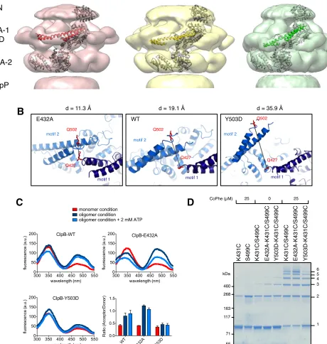
is a ~30º difference in orientation of the motif 1 blade of the MD and the loss of the
226
motif 2 density in the hyperactive state (Figure 4A,B). Another difference observed
227
upon alignment of the AAA-1 ring in the repressed, wild-type and hyperactive maps
228
is a ~15° rotation of the wild-type AAA-2 ring relative to the mutants.
229
230
In the BAP-E432A repressed mutant the MD is clearly visible around the AAA-1
231
ring. It has a horizontal orientation so that motif 2 forms a contact with motif 1 of the
232
neighbouring MD (Figure 3B; Figure 4B). This packing of the MD is supported by
233
biochemical data showing overprotection of motif 2 in this mutant 33. Moreover, a
234
very similar intersubunit motif 1-motif 2 contact is seen in all the ClpB crystal
235
structures, in which ClpB monomers are arranged in a spiral assembly (TthClpB,
236
1QVR 20; EcoClpB current study) (Figure 4-supplement 1).
237
238
In the Y503D hyperactive form there is density for motif 1 in a more tilted
239
orientation, but there is no density visible for motif 2, suggesting that this region
240
becomes either disordered or highly mobile (Figure 3C,D). This finding is in
241
accordance with H/D exchange data showing deprotection of this region in the
BAP-242
Y503D mutant 33. We conclude that, in the Y503D hyperactive form, the motif 1 arm
of the MD is tilted downwards so that it can no longer contact motif 2 of the adjacent
244
MD, and binds to a lower region of the neighbouring AAA-1 domain. Motif 2 is
245
mobile and solvent exposed (Figure 3C,D; Figure 4A,B).
246
247
Alignment of AAA-1 domains of wild type, repressed and hyperactive forms of
248
ClpB/BAP confirms that the MD rotates ~30° from a tilted orientation in the
249
hyperactive state, to a horizontal one in the repressed state, with the wild type
250
occupying an intermediate orientation (Figure 4B). In this movement motif 1 switches
251
from being protected against AAA-1 to engaging motif 2 in trans. These different
252
orientations of the MD have opposite consequences for DnaK binding. The horizontal
253
MD position, with motif 1 contacting motif 2 of the neighbouring subunit, is
254
incompatible with DnaK binding at the tip of motif 2 30,32. In contrast, tilting of the
255
MD exposes motif 2 for DnaK interaction.
256
257
To confirm the close proximity of motif 1 and motif 2 of neighbouring MDs we
258
performed fluorescence resonance energy transfer (FRET) experiments using Q427W
259
(motif 1) as FRET donor and IAEDANS labelled Q502C (motif 2) as FRET acceptor.
260
The FRET pair (Förster radius of 22 Å) has a distance of 19.1 Å in the wild type
261
model and was introduced into the tryptophan-free ClpB-W462Y/W543L variant. In
262
addition we coupled the FRET pair to E432A and Y503D mutations, to monitor the
263
consequences of repressed and hyperactive states on FRET efficiency. The distance
264
between the FRET partners is either reduced (11.3 Å) or increased (35.9 Å) in models
265
of the repressed and hyperactive variants, respectively (Figure 4B). All ClpB variants
266
were IAEDANS labeled with similar efficiencies (70-80%; Figure 4-supplement 2).
267
High IAEDANS fluorescence and thus FRET efficiency was observed for wild type
268
ClpB and E432A upon ClpB oligomerization. Increase in acceptor fluorescence in
269
general correlated with reduced tryptophan emission except for ClpB wild type upon
270
oligomerization. In contrast, IAEDANS fluorescence remained low under all
271
conditions tested when the FRET pair was linked to Y503D (Figure 4C).
272
273
Furthermore, to test for direct interaction between motif 1 and motif 2, we introduced
274
cysteine residues into motif 1 (K431C) and motif 2 (S499C) and analyzed whether
275
intermolecular crosslinks form under oxidizing conditions. Low Cu-Phenanthrolin
276
concentrations (25 μM) yielded a ladder of crosslink products from ClpB dimers to
277
hexamers. Although non-specific dimer formation was observed in single cysteine
variants, higher oligomers were only found in double cysteine variants (Figure 4D).
279
Introducing the activating (Y503D) mutation to ClpB-K431C/S499C decreased but
280
did not abolish crosslink formation, suggesting rapid fluctuation of MDs between
281
different conformations even in the hyperactive state (Figure 4D). These findings
282
support the interaction of MD motifs 1 and 2 from adjacent subunits as observed in
283
the EM reconstructions and strengthening or loosening of the contact in repressed and
284
hyperactive states, respectively.
285
286
Motif 1 is essential for keeping ClpB in the repressed state
287
Mutating MD motif 1 can impair Hsp70 interaction and consequently protein
288
disaggregation 30,33. On the other hand a ClpB motif 1 deletion variant retains
289
substantial disaggregation activity 30,44, providing no clarification of its role in protein
290
disaggregation. Our EM structural data indicate that motif 1 acts as a crucial
291
component of ClpB regulation by stabilizing MDs in a horizontal position through
292
interaction with an adjacent motif 2. Indeed, ClpB-ΔM1 (ΔE410-Y455) shows
293
hyperstimulation of ATPase activity in the presence of the substrate casein compared
294
to wild-type ClpB, resembling the deregulated ATPase activity of hyperactive ClpB
295
variants lacking motif 2 or the entire MD 33 (Figure 5A). The basal ATPase activity of 296
ClpB-ΔM1 was reduced in comparison to wild type due to partial oligomerization
297
defects, in agreement with earlier reports 33,44. ClpB-ΔM1 oligomerization defects
298
were more pronounced than in ClpB-ΔM1/M2, suggesting that motif 2 in absence of
299
motif 1 might impede hexamer formation.
300
Next we tested whether hyperstimulation of ATPase activity is linked to high
301
unfolding power. We employed BAP variants of the respective deletion constructs
302
and tested for unfolding and degradation of casein-YFP in presence of ClpP.
BAP-303
ΔM1 unfolds the YFP moiety of casein-YFP, an activity only observed for
304
hyperactive BAP variants but not wild type BAP 33 (Figure 5B). Loss of ClpB
305
regulation in ClpB-ΔM1 was also linked to severe toxicity upon expression in E. coli
306
ΔclpB mutant cells (Figure 5C). However, higher expression levels of ClpB-ΔM1
307
than of ClpB-Y503D are required to observe the same degree of toxicity probably
308
because deletion of motif 1 results in oligomerisation defects 33 (Figure 5C). In
309
conclusion ClpB-ΔM1 exhibits the three major characteristics of hyperactive ClpB
310
variants (high ATPase and unfolding activities linked to cellular toxicity),
311
demonstrating the crucial regulatory role for motif 1 in interacting with an adjacent
312
motif 2 to ensure tight activity control of ClpB.
Asymmetric reconstructions show variable orientations of the MD around the
314
ring
315
As mentioned above, we imposed 6-fold symmetry as a first approximation, to
316
simplify the alignment and reconstruction problem. Nevertheless, crystal structures of
317
the AAA+ protein ClpX show that the homo-hexameric assembly can be markedly
318
asymmetric 18,45,46. We therefore reanalysed our negative stain EM data without
319
imposing symmetry, in order to study the conformational variability within the
320
hexamer.
321
Using ~15,000, ~10,000 and ~9000 particles for wild-type BAP, BAP-E432A and
322
BAP-Y503D we obtained asymmetric maps at 21 Å, 21 Å and 25 Å resolution,
323
respectively (Figure 6A; Figure 1-supplement 3; Figure 6-supplement 1). Although
324
the numbers of particles were similar to those used for 6-fold analysis, the resolution
325
is only slightly worse and the maps are comparable in quality to the symmetrised
326
ones, consistent with the structure being asymmetric. The structure of the hyperactive
327
mutant is less defined and has lower resolution than the other two, particularly for the
328
MDs, probably owing to their higher mobility. Therefore, only wild-type BAP and
329
BAP-E432A were used for further analysis.
330
331
The asymmetric structures show similar density distributions in the AAA+ rings, even
332
though not all six subunits in a ring can be aligned simultaneously owing to 5°-15°
333
variations in rotational orientation. This rotational variability may explain the
334
apparent ~15° rotation of the AAA-2 wild-type ring relative to the mutants observed
335
in the symmetrised reconstructions. As expected from the eigenimage analysis, the
336
ND ring is not very well defined and the density does not account for all six NDs.
337
Therefore, we did not attempt any atomic structure fitting into this region. Docking of
338
the atomic structures of AAA-1, AAA-2 and MD was performed as follows.
339
340
Structures of AAA+ proteins crystallised in their hexameric assemblies show that the
341
large and small subdomains of the AAA fold assume a range of conformations that
342
can be clustered into open or closed forms 46,47 (Figure 6-supplement 2). The closed
343
conformation is ATP-binding competent and there are intermediate forms that might
344
also represent weak binding states. Using a gallery of available AAA+ crystal
345
structures we modelled open, closed and intermediate conformations of ClpB AAA-1
346
and AAA-2. We also created AAA+ dimers of adjacent ClpB subunits based on ClpX
347
pseudo-hexameric crystal structures 18,46 (Figure 6-supplement 2) that were fitted as
rigid bodies into the asymmetric reconstructions. Crystallographic dimers are likely to
349
provide more realistic models of subunit interfaces than can be deduced by fitting
350
individual subunits into low-resolution maps.
351
352
The AAA-2 ring density could be almost entirely interpreted using this approach and
353
the fit suggests that 3 to 5 subunits are sufficiently closed to bind ATP (Fig.
6-354
supplement 2). We analyzed nucleotide (ADP) binding to wild type ClpB by
355
isothermal calorimetry (ITC) revealing a binding stoichiometry of 7.5 +/- 0.1 ADP
356
per hexamer (Figure 6-supplement 3). ITC experiments using ClpB-K212A, which is
357
deficient in nucleotide binding in AAA-1, allowed us to determine a binding
358
stoichiometry of 3.7 +/- 0.3 ADP in AAA-2 of the mutant hexamer (Figure
6-359
supplement 3). The same ADP binding stoichiometry was also found for the repressed
360
and hyperactive variants (Figure 6-supplement 3). The deduced stoichiometries are in
361
good agreement with the distinct AAA-2 conformations observed in the asymmetric
362
EM reconstructions. Similar calculations have been reported for the AAA+ proteins
363
MCM, ClpX and HslU 48,49. The AAA-1 ring is more asymmetric than AAA-2 and it
364
is not easily interpretable by fitting crystallographic dimer models. There is sufficient
365
density to guide rigid body fitting of all AAA-1 domains, but this fitting does not
366
allow deductions of nucleotide occupancy (Figure 6-supplement 2).
367
368
Asymmetric reconstructions of both wild-type BAP and repressed mutant display
369
clear densities accounting for the MDs that lie outside the AAA-1 ring and assume
370
different orientations similar to those observed in the symmetrised maps of wild type
371
and mutants. In the wild-type asymmetric reconstruction, the MD orientation ranges
372
from horizontal as in the repressed state to highly tilted, similar to the hyperactive
373
state, passing through the intermediate wild type-like state (Figure 6B). In one
374
subunit, motif 1 contacts AAA-2 of the same subunit, suggesting a route of allosteric
375
communication between AAA-1 and AAA-2. Additionally, this contact is compatible
376
with recently reported engineered disulphide bonds between motif 1 residues and
377
AAA-2 in Hsp104 and ClpB 43. The tilted MDs are clustered together, consistent with
378
the release of motif 1-motif 2 contacts, freeing adjacent subunits.
379
380
In the asymmetric reconstruction of the repressed state, five MDs are found in the
381
horizontal orientation, followed by a sixth for which there is no clear density
382
(Figure 6C). In the wild type two MDs can make the motif 1 to motif 2 contact, while
in the repressed state up to five MDs are compatible with this contact (Figure 6B,C).
384
Conversely, in the wild type at least two MDs exist in a clearly activated state but in
385
the repressed state none of the MDs are visible in the tilted, hyperactive conformation.
386
As a consequence, the MD conformations in BAP-E432A do not support Hsp70
387
binding and ATPase activation, in agreement with previous findings 30,33. However,
388
ClpB wild type hexamers harbour two MDs that favour Hsp70 recruitment,
389
potentially priming the ClpB ring for further activation.
390
391
Discussion
392
The BAP construct of ClpB complexed with ClpP, combined with a conservative
393
approach of using only clearly identifiable side views and basing the analysis mainly
394
on negative stain images (with independent confirmation from cryo EM) enabled us
395
to unambiguously locate all subunit domains in the oligomer, including the coiled-coil
396
MD propeller. Although the quoted resolutions for some previous structures of ClpB
397
and Hsp104 were better, the globular shape, flexibility and asymmetry of these
398
hexamers reduce the reliability of orientation assignment. Moreover, previous
399
published structures of ClpB/Hsp104 were obtained by cryo EM only, which provides
400
a lower signal-to-noise ratio (SNR) than negative stain data.
401
402
Rigid body fitting of E. coli ClpB atomic coordinates (PDB code 4CIU) into our new
403
maps reveals that the MD is not projecting outwards from the hexamer 20,21,40 nor is
404
intercalated between subunits 22,23, but is instead lying on the surface of the ClpB
405
hexameric ring with a variable degree of tilt (Figure 4A; Figure 6B,C). A similar
406
position of the MD is seen for Hsp104, underlining the conserved activity and
407
mechanism of these disaggregases. This new MD arrangement is in excellent
408
agreement with recent, extensive biochemical analysis of the MD 33. It was proposed
409
that the MD works as a molecular toggle switching from a repressed state in which
410
both motif 1 and 2 ends of the propeller are protected, to an active state where motif 2
411
is deprotected, exposing the binding site for DnaK/Hsp70 30,32,33. Our EM
412
reconstructions of repressed, hyperactive and wild type ClpB reveal a lever-like
413
movement of the MD that switches from the repressed state with head-to-tail motif
1-414
motif 2 binding between adjacent subunits, to a mobile, activated state with motif 2
415
free and available for binding to DnaK (Figure 7A). The wild type conformation is
416
intermediate between these states and thus poised for switching.
417
Our data thus explain the critical role of motif 1, which regulates the accessibility of
419
motif 2 and consequently ClpB activity. Confirming its important regulatory role,
420
deletion of motif 1 results in hyperactive ATPase and unfolding activity of ClpB-∆M1
421
as well as cell toxicity (Figure 5), representing key characteristics of the hyperactive
422
state 33. Moreover, our maps suggest that motif 1, through its contacts with either the
423
adjacent motif 2 or AAA-1, plays a key role in direct communication between
424
neighbouring subunits, which must therefore act in a coordinated manner.
425
426
It has become clear that AAA+ proteins are highly dynamic molecular motors
427
unlikely to exist in a homogeneous structural state. Therefore we generated
428
asymmetric reconstructions of ClpB. Although the resolution of the asymmetric
429
structures is not sufficient to support a detailed mechanistic model, these
430
reconstructions provide the first visualization of the MD conformational flexibility
431
that was inferred from biochemical analysis 20,33,34. The asymmetric structures show 432
that the MD orientation varies around the ring occupying the repressed, wild type-like
433
and hyperactive positions described by the symmetrised averages. The variable tilts of
434
MDs observed around the ring suggest that 2 to 4 adjacent subunits are available for
435
DnaK binding in the wild-type versus only 1 in the repressed mutant (Figure 6B,C).
436
This is consistent with the estimated stoichiometry of 2-5 molecules of DnaK per
437
ClpB hexamer required for activation 30,43. It also suggests that at least 4 subunits
438
must have detached MDs to allow activity, perhaps through movements of the AAA+
439
domains, in agreement with the number of ClpX subunits that hydrolyze ATP in a
440
coordinated manner to unfold GFP in single molecule experiments 50. Moreover, we 441
calculated an ADP binding stoichiometry of 4 for both wild type and mutants
442
(Figure 6-supplement 3), which indicates that although ATP hydrolysis is strongly
443
affected, detachment of the MD does not change the nucleotide binding.
444
445
The cryo EM reconstruction of wild-type ClpB suggests that the various MD
446
conformations exist only transiently, poised between hyperactive and repressed states,
447
with the balance shifted slightly towards repressive motif 1-motif 2 contacts
448
(Figure 2). DnaK binding to an accessible motif 2 stabilizes the MD in a tilted
449
conformation, thereby in turn breaking the repressive contacts with MDs in
450
neighbouring subunits. Thus, an initial encounter of DnaK will facilitate DnaK
451
binding in the neighbouring MDs. In this model, breakage or formation of motif
1-452
motif 2 contacts provides a mechanistic basis for signalling DnaK binding or
dissociation in a wavelike manner around the ClpB ring. (Figure 7B). The model
454
predicts spatial proximity of multiple DnaK molecules, which is the case for
455
aggregated but not soluble DnaK substrates, directing ClpB activity specifically to
456
protein aggregates. Moreover, the activation of ClpB by DnaK binding, combined
457
with movements of the highly mobile N domain, might act to deliver the substrate to
458
the channel entrance, where it would be engaged for threading, unfolding and
459
consequent extraction from the aggregate.
460
461
462
Material and methods
463
Strains, plasmids and proteins
464
E. coli strains used were derivatives of MC4100. Mutant derivatives of ClpB/BAP
465
were generated by PCR mutagenesis and standard cloning techniques in pDS56 and
466
were verified by sequencing. Wild type and mutant ClpB were purified using Ni-IDA
467
(Macherey-Nagel) and size exclusion chromatography (Superdex S200, Amersham)
468
following standard protocols. Purifications of DnaK, DnaJ, GrpE, ClpP, Luciferase
469
and Casein-YFP were performed as described previously 33. Pyruvate kinase and α–
470
casein were purchased from Sigma. Protein concentrations were determined with the
471
Bio-Rad Bradford assay.
472
473
Electron microscopy of negatively stained and vitrified specimens
474
BAP(HAP)-ClpP complexes were formed in 20 mM Tris-HCl, pH 7.5, 20 mM KCl,
475
15 mM MgCl2, 1 mM DTT and 2 mM ATPγS. Proteins were applied to glow-476
discharged carbon coated grids (EM sciences), previously coated with 5 kDa
poly-477
lysine (Sigma-Aldrich) to positively charge the surface. Samples were stained with
478
2% uranyl acetate.
479
For cryo-EM imaging, BAPtrap-ClpP was applied to holey carbon grids coated with a
480
thin carbon film and pretreated with poly-lysine while ClpB specimens were loaded
481
onto lacey carbon grids. Cryo-EM specimens were vitrified in a Vitrobot (FEI).
482
Images were recorded on a 4k Gatan CCD camera at a magnification of 50,000× for
483
negatively stained specimens (pixel size 2.2 Å; underfocus range: 0.5-1.2 μm) and of
484
80,000× for cryo specimens (pixel size 1.4 Å; underfocus range: 1.5 µM-4 μm). All
485
data were collected on a Tecnai F20 FEG operated at 200 kV under low dose
486
conditions.
487
Single particle processing
489
The contrast transfer function (CTF) for each CCD frame was determined with
490
CTFFIND3 51 and corrected by phase flipping using Bshow1.6 52. Side views of
BAP-491
ClpP 2:2 complexes were manually picked using Boxer 53 and extracted into 256×256
492
boxes. The boxed particles were band-pass filtered between 300 and 10 Å for the
493
negative stain dataset and between 300 and 5 Å for the cryo dataset. They were then
494
normalized to the same mean and standard deviation. Particles were initially aligned
495
to the total sum of 10-20 vertically oriented particles using SPIDER 54. Individual 1:1
496
BAP-ClpP complexes were extracted with circular masks and classified by MSA in
497
IMAGIC-5 55 to remove images that did not represent BAP-ClpP, yielding 17470 498
particles of wild type BAP-ClpP, 12588 of BAP-E432A-ClpP, 9436 of
BAP-Y503D-499
ClpP and 12568 of HAP-ClpP. The BAPtrap-ClpP cryo dataset included 4592
500
particles. At this stage, particles were high-pass filtered to 160 Å and initial class
501
averages of 20-30 images each were obtained by MSA. All alignments of 1:1
BAP-502
ClpP complexes were done limiting the in-plane rotation to +/- 20°. Upon further
503
classification, 5-10 good classes were used to generate a starting model by angular
504
reconstitution 56. Alternative starting models were also created by applying 6 fold
505
symmetry to single classes and then using the resultant 3D map, generally composed
506
of three discs corresponding to the three layers of the molecule, to generate an anchor
507
set for Euler angle assignment. A low-resolution density map was independently
508
created for each dataset by angular reconstitution with 6-fold symmetry. Particle
509
orientations were refined in multiple cycles of AP SHC alignment in SPIDER, MSA
510
and angular reconstitution in IMAGIC and the resulting 3D reconstruction, filtered to
511
30 Å, was used as an initial model for projection matching in SPIDER. By applying a
512
rectangular mask, only the AAA+ layers were refined. After 8-10 cycles of projection
513
matching, using progressively smaller angular sampling steps (4° to 1°) and filtering
514
the 3D to the estimated resolution at each cycle, final structures were generated of the
515
whole complex and their resolution was estimated by Fourier shell correlation with a
516
0.5 correlation cutoff. Based on cross correlation coefficient, around 70-80% of each
517
dataset was included in the final reconstruction.
518
The differences between the mutant structures were tested by refining with
519
interchanged starting models. In both cases, the original mutant structure was
520
recovered despite the use of the other map as a starting model for projection matching
521
(Figure 3-supplement 1).
For cryo EM ClpB specimen, the views were randomly oriented and the initial
523
strategy was to extract clearly identifiable side views by MSA and classification. 7606
524
side views were used to generate a starting model by angular reconstitution, which
525
was refined to 29 Å resolution (Figure 1-supplement 3) by projection matching.
526
For the reconstructions without imposed symmetry, particle orientations were
527
determined using either the 6-fold symmetrized map filtered to 50 Å or a sphere
528
obtained from the average of all particles as a starting model. Subsequently, particle
529
orientations were refined with ~10 cycles of projection matching without imposed
530
symmetry. The particle orientations were well distributed around the BAP-ClpP
531
central axis (Figure 6-supplement 1). The resolution was estimated by Fourier shell
532
correlation (FSC) at 0.5 (Figure 1-supplement 3).
533
534
Atomic Structure Fitting
535
Docking was done using the crystal structures of E. coli ClpB-E432A (current study,
536
PDB code: 4CIU) and E. coli ClpB ND (PDB code 1KHY). A homology model of
537
Hsp104, obtained using Phyre2 57 was used for fitting into the HAP reconstruction.
538
A hexameric ClpB/Hsp104 AAA-1 ring was modelled on the ClpC hexamer crystal
539
structure (PDB code 3PXG) and was automatically fitted as a rigid body into the
540
symmetrised maps using the UCSF Chimera package 58. The N-terminal, the middle 541
and the AAA-2 domains were first fitted manually and then local fitting was
542
optimized in Chimera, followed by symmetrisation. In the asymmetric
543
reconstructions, dimers of adjacent ClpB subunits were modelled on crystal structures
544
of ClpX hexamers and fitted as rigid bodies in Chimera.
545
546
Biochemical assays
547
Steady-state ATP hydrolysis rates were determined in buffer A (50 mM Tris pH 7.5, 5
548
mM MgCl2, 20 mM KCl, 2 mM DTT) as described 34. ClpB disaggregation activities 549
were determined by following the refolding of aggregated firefly Luciferase
550
according to published protocols 34. Chaperones were used at the following
551
concentrations: 1 μM ClpB (wild type or derivatives), 1 μM DnaK, 0.2 μM DnaJ, 0.1
552
μM GrpE. Oligomerisation of ClpB variants was tested as described previously 28,34.
553
Site-specific labelling of ClpB using 1,5-IAEDANS,
5-([(2-554
iodoacetyl)amino]ethylamino)naphthalene-1-sulfonic acid (Invitrogen) was
555
performed according to the manufacturers’ instructions. The intrinsic tryptophan
556
fluorescence of ClpB variant harbouring a single tryptophan (ClpB*-Q427CW, 1 μM
each) was measured on a Perkin-Elmer LS50B spectrofluorimeter at 25°C in low salt
558
buffer A (50 mM Tris pH 7.5, 5 mM MgCl2, 20 mM KCl, 2 mM DTT). Tryptophan 559
and IAEDANS emission spectra of labeled ClpB variants were recorded in high and
560
low salt buffer A (supplemented with 400 mM KCl or 20 mM KCl, respectively) in
561
the absence or presence of 2 mM nucleotide between 300 and 550 nm at a fixed
562
excitation wavelength of 290 nm. The Förster radius of the Trp-IAEDANS FRET pair
563
was calculated as 22 Å 59.
564
For formation of disulfide bridges ClpB cysteine variants were first dialyzed to
565
remove DTT. Cysteine oxidation was achieved by adding 25 μM Cu-Phenanthroline
566
to 4 μM ClpB and incubating the mixture for 1 min at room temperature. Oxidation
567
and disulfide bond formation was stopped by addition of 50 mM iodoacetamide and
568
SDS-sample buffer containing 5 mM EDTA. Crosslink products were analyzed by a
569
non-reducing SDS gradient gel (3-8%).
570
Casein-YFP unfolding and degradation assays were carried out using 6 μM BAP
571
(wild type or variants), 9 μM ClpP and 0.5 μM YFP. Degradation of
Casein-572
YFP was determined by monitoring YFP fluorescence at 535 nm (excitation
573
wavelength 515 nm) at a Perkin-Elmer LS50B spectrofluorimeter.
574
575
Spot tests
576
E. coli cells harbouring plasmid-encoded clpB alleles were grown in the absence of
577
IPTG overnight at 30°C. Serial dilutions were prepared, spotted on LB-plates
578
containing different IPTG concentrations and incubated for 24 h at 37°C.
579
580
Isothermal titration calorimetry
581
ClpB wild type and variants were extensively dialyzed against low salt buffer A (50
582
mM Tris (pH 7.5), 25 mM KCl, 20 mM MgCl2, 5% glycerol). Isothermal titration 583
calorimetry (ITC) was performed using an ITC calorimeter (iTC200Microcalorimeter,
584
MicroCal). Consecutive injections of nucleotide into a 300 l cell containing ClpB
585
were performed after sample equilibration at 30°C. Integration and fitting of ITC data
586
were performed using ORIGIN software (GE). ClpB and ADP concentrations were
587
determined by UV absorbance at 280 nm.
588
589
Hydrogen-exchange experiments
590
HX-MS experiments were performed similar to those described earlier 33. Briefly,
591
ClpB (100 pmol), BAP (100 pmol) or BAP-ClpP complex (100 pmol and 200 pmol
respectively) were incubated for 3 min at 30°C in low salt buffer A (50 mM Tris, pH
593
7.5, 25 mM KCl, 20 mM MgCl2, 2 mM DTT) in presence of ATP or ATP S and 594
diluted 20-fold into D2O-based MDH buffer to initiate amide proton-deuteron 595
exchange. The exchange reaction was stopped after 1 min by the addition of 1 volume
596
of ice-cold quench buffer (0.4 M K-phosphate buffer, pH 2.2). Quenched samples
597
were immediately injected into the HPLC setup, with (peptide analysis) or without
598
(full length protein analysis) online peptic digest, and analysed on an electrospray
599
ionization quadrupole time-of-flight mass spectrometer (QSTAR Pulsar, Applied
600
Biosystems) as described in Rist et al, 200360. Analysis of deuteron incorporation into 601
peptide was performed by using AnalystQS software (Applied Biosystems/MDS
602
SCIEX).
603
604
Acknoledgments
605
We thank Elena Orlova for help in image processing, Christos Savva for EM
606
technical assistance, David Houldershaw for computing support and the Birkbeck EM
607
group for useful discussions. This work was funded by Wellcome Trust grants 089050
608
and 079605 to H. Saibil. E. Kummer was supported by the Hartmut
Hoffmann-609
Berling International Graduate School of Molecular and Cellular Biology. Y. Oguchi
610
was supported by a Humboldt fellowship.
611
612
Accession Numbers
613
The EM maps have been deposited in the 3D-EM database (www.emdatabank.org)
614
with accession codes EMD-2555 (BAP-E432A C6), EMD-2556 (BAP-E432A C1),
615
EMD-2557 (BAP wild type C6), and EMD-2558 (BAP wild type C1), EMD-2559
616
(BAP-Y503D C6), EMD-2560 (BAP-Y503D C1), EMD-2561 (HAP), EMD-2562
617
(BAPtrap cryo) and EMD-2563 (ClpB wild type cryo). The corresponding FSC
618
curves have also been deposited.
619
620
References
625
1. Doyle, S.M. & Wickner, S. Hsp104 and ClpB: protein disaggregating machines.
626
Trends in Biochemical Sciences34, 40-8 (2009). doi: 10.1016/j.tibs.2008.09.010. 627
2. Glover, J.R. & Lindquist, S. Hsp104, Hsp70, and Hsp40: a novel chaperone system
628
that rescues previously aggregated proteins. Cell 94, 73-82 (1998).
629
doi: 10.1016/S0092-8674(00)81223-4 630
3. Goloubinoff, P., Mogk, A., Zvi, A.P., Tomoyasu, T. & Bukau, B. Sequential
631
mechanism of solubilization and refolding of stable protein aggregates by a 632
bichaperone network. Proceedings of the National Academy of Sciences of the United
633
States of America96, 13732-7 (1999). doi: 10.1073/pnas.96.24.13732 634
4. Haslberger, T., Bukau, B. & Mogk, A. Towards a unifying mechanism for
635
ClpB/Hsp104-mediated protein disaggregation and prion propagation. Biochemistry
636
and cell biology = Biochimie et biologie cellulaire 88, 63-75 (2010).
637
doi: 10.1139/o09-118. 638
5. Motohashi, K., Watanabe, Y., Yohda, M. & Yoshida, M. Heat-inactivated proteins
639
are rescued by the DnaK.J-GrpE set and ClpB chaperones. Proceedings of the
640
National Academy of Sciences of the United States of America 96, 7184-9 (1999). 641
doi: 10.1073/pnas.96.13.7184 642
6. Zolkiewski, M. ClpB cooperates with DnaK, DnaJ, and GrpE in suppressing protein
643
aggregation. A novel multi-chaperone system from Escherichia coli. The Journal of
644
biological chemistry274, 28083-6 (1999). doi: 10.1074/jbc.274.40.28083 645
7. Mogk, A. et al. Identification of thermolabile Escherichia coli proteins: prevention
646
and reversion of aggregation by DnaK and ClpB. The EMBO journal 18, 6934-49
647
(1999). doi: 10.1093/emboj/18.24.6934 648
8. Parsell, D.A., Kowal, A.S., Singer, M.A. & Lindquist, S. Protein disaggregation
649
mediated by heat-shock protein Hsp104. Nature 372, 475-8 (1994).
650
doi:10.1038/372475a0 651
9. Haslberger, T. et al. Protein disaggregation by the AAA+ chaperone ClpB involves
652
partial threading of looped polypeptide segments. Nature structural & molecular
653
biology15, 641-50 (2008). doi: 10.1038/nsmb.1425. 654
10. Kim, Y.I., Burton, R.E., Burton, B.M., Sauer, R.T. & Baker, T.A. Dynamics of
655
substrate denaturation and translocation by the ClpXP degradation machine. 656
Molecular Cell5, 639-48 (2000). doi: 10.1016/S1097-2765(00)80243-9 657
11. Lum, R., Tkach, J.M., Vierling, E. & Glover, J.R. Evidence for an 658
unfolding/threading mechanism for protein disaggregation by Saccharomyces 659
cerevisiae Hsp104. The Journal of biological chemistry 279, 29139-46 (2004). ,
660
12. Tessarz, P., Mogk, A. & Bukau, B. Substrate threading through the central pore of the 662
Hsp104 chaperone as a common mechanism for protein disaggregation and prion 663
propagation. Molecular Microbiology 68, 87-97 (2008). doi:
10.1111/j.1365-664
2958.2008.06135.x. 665
13. Weber-Ban, E.U., Reid, B.G., Miranker, A.D. & Horwich, A.L. Global unfolding of a
666
substrate protein by the Hsp100 chaperone ClpA. Nature 401, 90-3 (1999).
667
doi:10.1038/43481 668
14. Weibezahn, J. et al. Thermotolerance requires refolding of aggregated proteins by
669
substrate translocation through the central pore of ClpB. Cell 119, 653-65 (2004).
670
doi: 10.1016/j.cell.2004.11.027 671
15. Erzberger, J.P. & Berger, J.M. Evolutionary relationships and structural mechanisms
672
of AAA+ proteins. Annual Review of Biophysics and Biomolecular Structure35,
93-673
114 (2006). doi: 10.1146/annurev.biophys.35.040405.101933 674
16. Ogura, T. & Wilkinson, A.J. AAA+ superfamily ATPases: common
structure--675
diverse function. Genes to cells : devoted to molecular & cellular mechanisms 6,
676
575-97 (2001). doi: 10.1046/j.1365-2443.2001.00447.x 677
17. Bochtler, M. et al. The structures of HsIU and the ATP-dependent protease
HsIU-678
HsIV. Nature403, 800-5 (2000). doi:10.1038/35001629
679
18. Glynn, S.E., Martin, A., Nager, A.R., Baker, T.A. & Sauer, R.T. Structures of
680
asymmetric ClpX hexamers reveal nucleotide-dependent motions in a AAA+ protein-681
unfolding machine. Cell139, 744-56 (2009). doi: 10.1016/j.cell.2009.09.034
682
19. Wang, F. et al. Structure and mechanism of the hexameric MecA-ClpC molecular
683
machine. Nature471, 331-5 (2011).doi: 10.1038/nature09780
684
20. Lee, S. et al. The structure of ClpB: a molecular chaperone that rescues proteins from
685
an aggregated state. Cell115, 229-40 (2003).doi: 10.1016/S0092-8674(03)00807-9
686
21. Lee, S., Sielaff, B., Lee, J. & Tsai, F.T. CryoEM structure of Hsp104 and its
687
mechanistic implication for protein disaggregation. Proceedings of the National
688
Academy of Sciences of the United States of America 107, 8135-40 (2010).
689
doi: 10.1073/pnas.1003572107. 690
22. Wendler, P. et al. Atypical AAA+ subunit packing creates an expanded cavity for
691
disaggregation by the protein-remodeling factor Hsp104. Cell131, 1366-77 (2007).
692
doi: 10.1016/j.cell.2007.10.047 693
23. Wendler, P. et al. Motor mechanism for protein threading through Hsp104. Molecular
694
Cell34, 81-92 (2009). doi: 10.1016/j.molcel.2009.02.026.
695
24. Gai, D., Zhao, R., Li, D., Finkielstein, C.V. & Chen, X.S. Mechanisms of
696
conformational change for a replicative hexameric helicase of SV40 large tumor 697
antigen. Cell119, 47-60 (2004). doi: 10.1016/j.cell.2004.09.017
25. Huyton, T. et al. The crystal structure of murine p97/VCP at 3.6A. Journal of
699
Structural Biology144, 337-48 (2003).doi: 10.1016/j.jsb.2003.10.007 700
26. Desantis, M.E. & Shorter, J. The elusive middle domain of Hsp104 and ClpB:
701
location and function. Biochimica et Biophysica Acta 1823, 29-39 (2012).
702
doi: 10.1016/j.bbamcr.2011.07.014. 703
27. Kedzierska, S., Akoev, V., Barnett, M.E. & Zolkiewski, M. Structure and function of
704
the middle domain of ClpB from Escherichia coli. Biochemistry42, 14242-8 (2003).
705
doi: 10.1021/bi035573d 706
28. Mogk, A. et al. Roles of individual domains and conserved motifs of the AAA+
707
chaperone ClpB in oligomerization, ATP hydrolysis, and chaperone activity. The
708
Journal of biological chemistry278, 17615-24 (2003). doi: 10.1074/jbc.M209686200 709
29. Miot, M. et al. Species-specific collaboration of heat shock proteins (Hsp) 70 and 100
710
in thermotolerance and protein disaggregation. Proceedings of the National Academy
711
of Sciences of the United States of America 108, 6915-20 (2011).
712
doi: 10.1073/pnas.1102828108. 713
30. Seyffer, F. et al. Hsp70 proteins bind Hsp100 regulatory M domains to activate
714
AAA+ disaggregase at aggregate surfaces. Nature structural & molecular biology19,
715
1347-55 (2012). doi: 10.1038/nsmb.2442. 716
31. Sielaff, B. & Tsai, F.T. The M-domain controls Hsp104 protein remodeling activity
717
in an Hsp70/Hsp40-dependent manner. Journal of Molecular Biology 402, 30-7
718
(2010). doi: 10.1016/j.jmb.2010.07.030. 719
32. Rosenzweig, R., Moradi, S., Zarrine-Afsar, A., Glover, J.R. & Kay, L.E. Unraveling
720
the mechanism of protein disaggregation through a ClpB-DnaK interaction. Science
721
339, 1080-3 (2013). doi: 10.1126/science.1233066.
722
33. Oguchi, Y. et al. A tightly regulated molecular toggle controls AAA+ disaggregase.
723
Nature structural & molecular biology19, 1338-46 (2012). doi: 10.1038/nsmb.2441. 724
34. Haslberger, T. et al. M domains couple the ClpB threading motor with the DnaK
725
chaperone activity. Molecular Cell 25, 247-60 (2007).
726
doi: 10.1016/j.molcel.2006.11.008 727
35. Lipinska, N. et al. Disruption of ionic interactions between the nucleotide binding
728
domain 1 (NBD1) and middle (M) domain in Hsp100 disaggregase unleashes toxic 729
hyperactivity and partial independence from Hsp70. The Journal of biological
730
chemistry288, 2857-69 (2013). doi: 10.1074/jbc.M112.387589. 731
36. Mizuno, S., Nakazaki, Y., Yoshida, M. & Watanabe, Y.H. Orientation of the
amino-732
terminal domain of ClpB affects the disaggregation of the protein. The FEBS journal
733
279, 1474-84 (2012). doi: 10.1111/j.1742-4658.2012.08540.x.
734
37. Effantin, G., Ishikawa, T., De Donatis, G.M., Maurizi, M.R. & Steven, A.C. Local
735
microscopy: functional connotations. Structure 18, 553-62 (2010). 737
doi: 10.1016/j.str.2010.02.016. 738
38. Parsell, D.A., Kowal, A.S. & Lindquist, S. Saccharomyces cerevisiae Hsp104 protein.
739
Purification and characterization of ATP-induced structural changes. The Journal of
740
biological chemistry269, 4480-7 (1994). 741
39. Schlothauer, T., Mogk, A., Dougan, D.A., Bukau, B. & Turgay, K. MecA, an
742
adaptor protein necessary for ClpC chaperone activity. Proceedings of the National
743
Academy of Sciences of the United States of America 100, 2306-11 (2003).
744
doi: 10.1073/pnas.0535717100 745
40. Lee, S., Choi, J.M. & Tsai, F.T. Visualizing the ATPase cycle in a protein
746
disaggregating machine: structural basis for substrate binding by ClpB. Molecular
747
Cell25, 261-71 (2007). doi: 10.1016/j.molcel.2007.01.002
748
41. Biter, A.B., Lee, J., Sung, N., Tsai, F.T. & Lee, S. Functional analysis of conserved
749
cis- and trans-elements in the Hsp104 protein disaggregating machine. Journal of
750
Structural Biology179, 172-80 (2012). doi: 10.1016/j.jsb.2012.05.007 751
42. Yamasaki, T., Nakazaki, Y., Yoshida, M. & Watanabe, Y.H. Roles of conserved
752
arginines in ATP-binding domains of AAA+ chaperone ClpB from Thermus 753
thermophilus. The FEBS journal 278, 2395-403 (2011). doi:
10.1111/j.1742-754
4658.2011.08167.x. 755
43. Desantis, M.E. et al. Conserved Distal Loop Residues in the Hsp104 and ClpB
756
Middle Domain Contact Nucleotide-binding Domain 2 and Enable Hsp70-dependent 757
Protein Disaggregation. The Journal of biological chemistry 289, 848-67 (2014).
758
doi: 10.1074/jbc.M113.520759. 759
44. del Castillo, U. et al. A quantitative analysis of the effect of nucleotides and the M
760
domain on the association equilibrium of ClpB. Biochemistry50, 1991-2003 (2011).
761
doi: 10.1021/bi101670s 762
45. Kon, T. et al. The 2.8 A crystal structure of the dynein motor domain. Nature 484,
763
345-50 (2012).doi: 10.1038/nature10955.
764
46. Stinson, B.M. et al. Nucleotide binding and conformational switching in the
765
hexameric ring of a AAA+ machine. Cell 153, 628-39 (2013).
766
doi: 10.1016/j.cell.2013.03.029 767
47. Cho, C. & Vale, R.D. The mechanism of dynein motility: insight from crystal
768
structures of the motor domain. Biochimica et Biophysica Acta1823, 182-91 (2012).
769
doi: 10.1016/j.bbamcr.2011.10.009. 770
48. Moreau, M.J., McGeoch, A.T., Lowe, A.R., Itzhaki, L.S. & Bell, S.D. ATPase site
771
architecture and helicase mechanism of an archaeal MCM. Molecular Cell28, 304-14
772
49. Yakamavich, J.A., Baker, T.A. & Sauer, R.T. Asymmetric nucleotide transactions of 774
the HslUV protease. Journal of Molecular Biology 380, 946-57 (2008).
775
doi: 10.1016/j.jmb.2008.05.070. 776
50. Sen, M. et al. The ClpXP protease unfolds substrates using a constant rate of pulling
777
but different gears. Cell155, 636-46 (2013). doi: 10.1016/j.cell.2013.09.022
778
51. Mindell, J.A. & Grigorieff, N. Accurate determination of local defocus and specimen
779
tilt in electron microscopy. Journal of Structural Biology 142, 334-347 (2003).
780
doi: 10.1016/S1047-8477(03)00069-8 781
52. Heymann, J.B. & Belnap, D.M. Bsoft: Image processing and molecular modeling for
782
electron microscopy. Journal of Structural Biology 157, 3-18 (2007).
783
doi: 10.1016/j.jsb.2006.06.006 784
53. Ludtke, S.J., Baldwin, P.R. & Chiu, W. EMAN: Semiautomated software for
high-785
resolution single-particle reconstructions. Journal of Structural Biology 128, 82-97
786
(1999). doi: 10.1006/jsbi.1999.4174 787
54. Frank, J. et al. SPIDER and WEB: Processing and visualization of images in 3D
788
electron microscopy and related fields. Journal of Structural Biology 116, 190-199
789
(1996). doi: 10.1006/jsbi.1996.0030 790
55. van Heel, M., Harauz, G., Orlova, E.V., Schmidt, R. & Schatz, M. A new generation
791
of the IMAGIC image processing system. Journal of Structural Biology116, 17-24
792
(1996). doi: 10.1006/jsbi.1996.0004 793
56. van Heel, M. et al. Single-particle electron cryo-microscopy: towards atomic
794
resolution. Quarterly Reviews of Biophysics33, 307-69 (2000).
795
57. Kelley, L.A. & Sternberg, M.J. Protein structure prediction on the Web: a case study
796
using the Phyre server. Nature protocols4, 363-71 (2009).doi: 10.1038/nprot.2009.2.
797
58. Pettersen, E.F. et al. UCSF Chimera--a visualization system for exploratory research
798
and analysis. Journal of computational chemistry 25, 1605-12 (2004).
799
doi: 10.1002/jcc.20084 800
59. Jeganathan, S., von Bergen, M., Brutlach, H., Steinhoff, H.J. & Mandelkow, E.
801
Global hairpin folding of tau in solution. Biochemistry 45, 2283-93 (2006).
802
doi: 10.1021/bi0521543 803
60. Rist, W., Jørgensen, T.J.D., Roepstorff, P., Bukau, B., & Mayer, M.P. Mapping
804
temperature-induced conformational changes in the Escherichia coli heat shock
805
transcription factor σ32 by amide hydrogen exchange. Journal of Biological
806
Chemistry