Augustin, H. and McGourty, K. and Steinert, J.R. and Cochemé, H.M. and
Adcott, J. and Cabecinha, M. and Vincent, A. and Halff, E.F. and Kittler,
J.T. and Boucrot, Emmanuel and Partridge, L. (2017) Myostatin-like proteins
regulate synaptic function and neuronal morphology. Development 144 (13),
pp. 2445-2455. ISSN 0950-1991.
Downloaded from:
Usage Guidelines:
Please refer to usage guidelines at
or alternatively
RESEARCH ARTICLE
Myostatin-like proteins regulate synaptic function and neuronal
morphology
Hrvoje Augustin1,2, Kieran McGourty3,*, Joern R. Steinert4,*, Helena M. Cochemé1,2,5,6, Jennifer Adcott1,2, Melissa Cabecinha1, Alec Vincent1, Els F. Halff7, Josef T. Kittler7, Emmanuel Boucrot3and Linda Partridge1,2,‡
ABSTRACT
Growth factors of the TGFβsuperfamily play key roles in regulating neuronal and muscle function. Myostatin (or GDF8) and GDF11 are potent negative regulators of skeletal muscle mass. However, expression of myostatin and its cognate receptors in other tissues, including brain and peripheral nerves, suggests a potential wider biological role. Here, we show that Myoglianin (MYO), theDrosophila
homolog of myostatin and GDF11, regulates not only body weight and muscle size, but also inhibits neuromuscular synapse strength and composition in a Smad2-dependent manner. Both myostatin and GDF11 affected synapse formation in isolated rat cortical neuron cultures, suggesting an effect on synaptogenesis beyond neuromuscular junctions. We also show that MYO actsin vivoto inhibit synaptic transmission between neurons in the escape response neural circuit of adult flies. Thus, these anti-myogenic proteins act as important inhibitors of synapse function and neuronal growth.
KEY WORDS:Drosophila, GDF11, Myoglianin, Myostatin, Muscle size, Synapse
INTRODUCTION
Organismal muscle mass is tightly regulated by positive and negative endocrine and autocrine/paracrine factors. Myostatin (also known as growth and differentiation factor 8 or GDF8), a member of
the transforming growth factor β(TGFβ) superfamily of secreted
differentiation and growth factors, is a potent inhibitor of skeletal
muscle mass in mammals. Myostatin (Mstn) gene mutations or
deletions cause hyperplastic and/or hypertrophic muscle growth in mice (McPherron et al., 1997) and a number of other species, including humans (Carnac et al., 2007), with consequent loss of muscle function (Gentry et al., 2011). Myostatin-like protein GDF11 (also known as BMP11) was also recently identified as a
circulating inhibitor of skeletal muscle regeneration in rodents and, potentially, humans (Egerman et al., 2015).
Both GDF8 and GDF11 bind to Activin-type receptor complexes, leading to the phosphorylation of intracellular Smad2/ 3 transcription factors, followed by their translocation to the nucleus (Oh et al., 2002; Rebbapragada et al., 2003). In addition to its action on muscles, GDF11 is a negative regulator of neuron number in the olfactory epithelium (Kawauchi et al., 2009; Wu et al., 2003), an inhibitor of neuronal precursors that give rise to olfactory receptors (Gokoffski et al., 2011) and an antagonist of neurogenesis during
retinal development (Kim et al., 2005).Mstntranscript was recently
detected in mouse brain (Lein et al., 2007) and myostatin receptors are expressed in several tissues, including brain and peripheral nerves. Apart from a study demonstrating an inhibitory effect of myostatin on
neuronal colony formationin vitro(Wu et al., 2003), the potential role
of myostatin in the nervous system remains unexplored despite its potential biological and therapeutic significance.
TheDrosophila myoglianin(myo) gene encodes the invertebrate Activin-type ligand with the highest amino acid sequence homology to myostatin and GDF11, both of which share 46% amino acid identity and >60% similarity with MYO (Lo and Frasch,
1999). Unlike the predominant Mstn expression in vertebrate
skeletal muscle (Lee, 2004),myois strongly expressed not only in
different muscle types throughout development but also in embryonic (Lo and Frasch, 1999) and larval brain glia (Awasaki
et al., 2011). Considering the strong expression of Gdf11 in the
mammalian nervous system during development and adulthood (Nakashima et al., 1999; Shi and Liu, 2011), it is tempting to think of Myoglianin as combining the functions of myostatin and GDF11 in flies.
In this study, we identified MYO as a strong inhibitor of synaptic function and composition at the larval NMJ, in addition to its role as an inhibitor of body weight and muscle size. These synaptic effects of MYO were mediated mainly by the transcription factor Smad2
(also known as Smox) and Shaggy, the Drosophila glycogen
synthase kinase 3 (GSK3) homolog. Myostatin could reverse the effect of MYO depletion on synaptic strength in larvae. Furthermore, myostatin and GDF11 inhibited neuronal growth and synapse specification in rat cortical neurons, indicating that they can
act directly on neurons that are not associated with muscle. Thein
vivorole of MYO in regulating neuronal function was confirmed in
a central, non-NMJ synapse in adult flies. Our findings show that MYO and its mammalian orthologs myostatin and GDF11 have previously unsuspected roles in the nervous system, acting as important inhibitors of synapse function and neuronal growth.
RESULTS
MYO inhibits NMJ synapse strength and composition
The larval body wall musculature of Drosophila is composed of
bilaterally symmetrical hemisegments, each consisting of 30 easily
Received 5 April 2017; Accepted 15 May 2017
1Institute of Healthy Ageing, and GEE, University College London, Darwin Building,
Gower Street, London WC1E 6BT, UK.2
Max Planck Institute for Biology of Ageing, Joseph-Stelzmann-Str. 9b, Cologne D-50931, Germany.3
Institute of Structural and Molecular Biology, University College London, Darwin Building Gower Street, London WC1E 6BT, UK.4
MRC Toxicology Unit, Hodgkin Building, University of Leicester, Lancaster Road, Leicester LE1 9HN, UK.5
MRC Clinical Sciences Centre, Du Cane Road, London W12 0NN, UK.6
Institute of Clinical Sciences, Imperial College London, ICTEM Building, Hammersmith Hospital Campus, Du Cane Road, London W12 0NN, UK.7Department of Neuroscience, Physiology and
Pharmacology, University College London, Gower Street, London WC1E 6BT, UK. *These authors contributed equally to this work
‡Author for correspondence ( [email protected])
H.A., 0000-0002-2041-3844; L.P., 0000-0001-9615-0094
This is an Open Access article distributed under the terms of the Creative Commons Attribution License (http://creativecommons.org/licenses/by/3.0), which permits unrestricted use, distribution and reproduction in any medium provided that the original work is properly attributed.
DEVEL
O
identifiable longitudinal and oblique multinucleated muscle cells/fibers. We focused on ventral longitudinal muscles 6 and 7 (Fig. S1A), which are innervated by two axons forming a single glutamatergic neuromuscular junction (Ruiz-Cañada and Budnik, 2006), a complex synapse composed of muscle, neuronal and glial cells.
We investigated the functional significance of the presence
of MYO in larval musculature (Awasaki et al., 2011)
electrophysiologically. We used microRNA (miRNAmyo) or
dsRNA (UAS-myoRNAi) to downregulate, and aUAS-myoglianin
(WT) construct (Awasaki et al., 2011) to enhancemyoexpression by
means of theMef2-GAL4muscle driver (Brand and Perrimon, 1993;
Ranganayakulu et al., 1995), resulting inmyoexpression changes in
larval muscle preparations (Fig. S1B). Currents resulting from the spontaneous release of presynaptic vesicles [miniature excitatory
junctional currents (mEJCs), or‘minis’] and evoked release [evoked
excitatory junctional currents (eEJCs)] represent two functional outputs at the neuromuscular synapse (Melom et al., 2013). Nerve-evoked postsynaptic currents, and the frequency of spontaneous
release, reflect presynaptic Ca2+-dependent vesicular release
(Peron et al., 2009), whereas mini amplitudes mainly reflect the postsynaptic sensitivity to transmitter, determined largely by the properties of glutamate receptors (DiAntonio et al., 1999). When eEJCs from muscle 6 were measured in the voltage-clamp mode (the
membrane potential was clamped to −60 mV), we observed that
experimentally reduced expression of myo in muscle increased
eEJC amplitude, whereas overexpression reduced it (Fig. 1A,B). Although the mean mEJC frequency and amplitude remained unchanged across genotypes (Fig. S1C,D), the amplitude distribution showed a significant shift towards larger synaptic
currents with myo knock-down in muscles (KS test, P<0.0001)
(Fig. 1C,D), indicating increased postsynaptic sensitivity to glutamate. These data thus revealed that muscle-derived MYO is a potent suppressor of synaptic transmission at the NMJ through impact on both presynaptic release and postsynaptic sensitivity. On the postsynaptic side of the excitatory larval NMJ, heterotetrameric ionotropic glutamate receptors (GluRs) comprise two functionally distinct subtypes: IIA, containing the GluRIIA subunit; and IIB, containing the GluRIIB subunit. Type IIA receptors generate larger synaptic currents and mediate functional strengthening of the NMJ (Petersen et al., 1997; Sigrist et al., 2002). Type IIB receptor subunits are characterized by faster desensitization kinetics and lower responsiveness to vesicularly released neurotransmitter (DiAntonio et al., 1999). Brp (Bruchpilot), a presynaptic marker, promotes active zone assembly and integrity, and vesicular neurotransmitter release (Kittel et al., 2006); the presence of Brp has been associated with presynaptic strengthening at larval NMJ (Weyhersmuller et al., 2011).
Prompted by our electrophysiological results, we measured the density of the GluRIIA receptor field and the number of Brp puncta in the NMJ boutons (each bouton contains multiple active zones)
(Fig. 1E-G). Although myo levels negatively correlated with
GluRIIA signal intensity (Fig. 1G), only myo downregulation
( positively) affected the total active zone number (Fig. S1E) and the number of Brp puncta normalized to the NMJ area (Fig. 1G). This
indicates that myo upregulation and silencing affect presynaptic
release through different mechanisms. To address the issue of potential off-target effects of the miRNA construct, we have confirmed our results by measuring the GluRIIA intensity in flies
expressing an anti-myoRNAi construct (Awasaki et al., 2011) in
[image:3.612.311.562.54.575.2]somatic muscles (Fig. S1F). MYO also negatively affects NMJ length and branching pattern (Fig. S1G), in line with increased
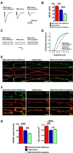
Fig. 1. MYO is a negative regulator of synaptic physiology and composition.(A) Representative samples of eEJCs recorded from muscle 6 in B. (B) Quantification of evoked EJCs from the larvae with reduced (Mef2-GAL4/UAS-miRNAmyo) or increased (Mef2-GAL4/UAS-myoglianin)
myoexpression in muscles. Control phenotype:+/Mef2/GAL4(n=5-9).
Representative traces (C) and cumulative frequency (CF) diagram (D) of
mEJC amplitudes from the larvae expressingmyotransgenes in muscle;
larger synaptic currents are indicated by a shift of the curve to the right
(n=6-12 animals,∼500-1200 events measured per genotype). (E,F)
Representative confocal images showing the 3rd instar larval NMJ 6/7 staining for GluRIIA (E) and Brp (F). Anti-HRP labels presynaptic
(motoneuronal) membrane. Scale bars: 20μm. (G) Left: quantification of
GluRIIA signal intensities in larvae expressing variousmyoconstructs in
larval muscles (n=10-18). Right: number of Brp puncta normalized to the
area of the 6/7 NMJ (n=12-15). Data are mean±s.e.m. ANOVA+Tukey’s
post-test: **P<0.01, ***P<0.001; n.s., not significant.
DEVEL
O
axonal branching in myostatin-null mice (Gay et al., 2012). The lack
of effect on mini amplitudes inmyooverexpressing animals, despite
the reduction in IIA staining, could be attributable either to a compensatory increase in the levels of other GluR subunits present at the NMJ or to GluRIIA epitope masking (Renden and Broadie,
2003). We observed no effect ofmyomanipulations on the levels of
IIB type synaptic receptors (Fig. S1H), indicating a receptor subtype-specific action of MYO. Together with our physiological data, these results demonstrate a significant inhibitory effect of muscle-derived MYO on the function and composition of the neuromuscular synapse.
Glia-expressedmyohas a modulatory role at the NMJ
We next examined whether MYO was produced in the larval
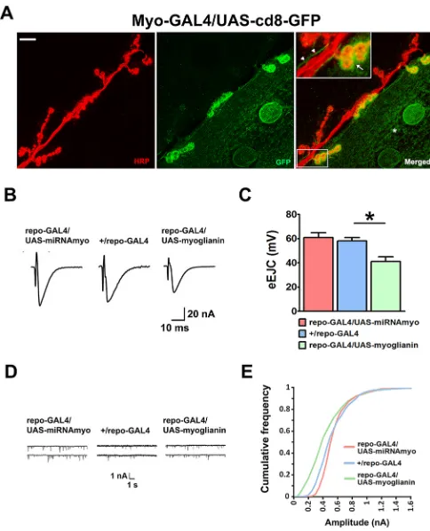
NMJ glia. We used aUAS-GFPconstruct driven by theMyo-GAL4
driver (Awasaki et al., 2011), and detected a strong GFP-positive signal around synaptic boutons and in the extramuscular tracts running in parallel with the motoneurons innervating muscles 6 and 7 (green signal in Fig. 2A). Although the increased GFP signal intensity around boutons likely stems from the elaborate infoldings of the muscle membrane ensheathing the boutons, known as the subsynaptic reticulum, the extramuscular tracts (Fig. 2A,
arrowheads) imply glial myo expression at the larval NMJ,
consistent with the previous detection of the myo transcript in
peripheral larval glia (Fuentes-Medel et al., 2012). The effect of
manipulation ofmyoexpression in glia on synaptic physiology was
less prominent than in muscle, probably because of the small size of the glial compartment at the NMJ in comparison with muscle, with only upregulation reducing the mean evoked response amplitude (Fig. 2B,C). We also observed a small, but significant (KS test,
P<0.0001), negative effect of glial myo on the distribution of
miniature amplitudes (Fig. 2D,E), with the‘mini frequency’and
mean ‘mini amplitude’ remaining unperturbed (Fig. S2A,B).
Knockdown of glial myo increased synaptic GluRIIA
fluorescence (Fig. S2C), consistent with the effect of myo
knockdown on the distribution of mini amplitudes (Fig. 2D,E);
we did not detect GluRIIA changes in myo-overexpressing
animals, possibly owing to relatively minor changes in receptor number and/or composition in these larvae (Fig. S2C). Type-IIB
receptor levels were unaffected by myoexpression (Fig. S2D),
and no significant effect ofmyodownregulation was seen on the
levels of type IIA receptors when myo was silenced in the
motoneurons innervating larval body-wall muscles (Fig. S2E), consistent with absence of MYO in this cell type. Together, these results imply a modulatory role for MYO of glial origin at the neuromuscular synapse.
MYO displays a myostatin-like effect on larval weight and muscle size
Having established a role for MYO at the NMJ, we next determined whether MYO resembles myostatin in its negative impact on body weight, and adult (McPherron et al., 1997) and embryonic (Manceau et al., 2008) muscle size. We first examined the effect of MYO on larval mass and muscle size. The wet weight of 3rd instar wandering larvae (72-96 h after hatching) was reduced
by experimentally increased expression ofmyo, and increased by
its knockdown, in larval muscle preparations (Fig. 3A).
Developmental progression (time to pupariation) was unaffected in these genotypes (Fig. S3A). Wet weight was also increased in
larvae expressing the previously usedmyoRNAi construct driven
by a different muscle driver (24B-GAL4), and decreased in animals
expressing an alternate UAS-myoglianin construct (see Materials
and Methods) (Fig. S3B). Interestingly, we observed a similar effect
on larval weight whenmyoconstructs were driven with the pan-glial
repodriver (Fig. 3B). Whereas miRNA againstmyoin motoneurons
(Fig. S3C) or fat body (Fig. S3D) had no effect on larval weight,
downregulation of myo in the midgut resulted in significantly
increased weight (Fig. S3D), suggesting a role for MYO outside the nervous system and muscle. Body wall muscles are the major constituent of the larval body in terms of size and mass (Bate et al.,
1999), and we therefore examined the effect ofmyoexpression on
the size of the larval body-wall muscles 6 and 7 (Fig. 3C). Similar to larval weight, the surface area of both muscles was reduced by
increasedmyoexpression, and increased by its knockdown in the
muscle (Fig. 3D and Fig. S3E). We observed no difference between
genotypes whenmyoexpression was manipulated in glia (Fig. 3E).
Larval crawling speed was also negatively correlated with myo
expression levels (Fig. 3F, Movies 1-6), showing that manipulations
of myo in muscle and glial cells have significant behavioral
consequences. Together, these data establish a role for muscle- and
glia-expressedmyoas a strong negative regulator of larval weight
and motility, and establish that muscle-derived MYO has a
myostatin-like function in regulating muscle size in Drosophila
[image:4.612.316.558.56.355.2]larvae.
Fig. 2. MYO is produced at the larval NMJ and is a modulator of its function.(A) Confocal images showing the NMJ expression of a GFP
construct underMyo-GAL4control. Anti-HRP (red) marks motoneurons
innervating the 6/7 NMJ; anti-GFP antibody (green) was used to enhance the GFP signal. Asterisk marks the GFP-positive area in the muscle. Arrow (inset) indicates strong GFP signal in the synaptic boutons, with the arrowheads indicating thread-like, GFP-labeled, extramuscular structures running
alongside neuronal projections. Scale bar: 20μm. (B-E) Physiological
measurements in larvae mis-expressingmyoin glia.
(B) Representative eEJCs traces. (C) Quantification of evoked EJCs
(n=5-9; ANOVA+Tukey’s post-test: *P<0.05). (D) Representative mEJC traces.
(E) Cumulative frequency diagram of mEJC amplitudes (n=6-12).
DEVEL
O
Downregulation ofmyopromotes signaling through GSK3/ Shaggy
We next identified potential intracellular mediators of reduced MYO and their relevance for MYO action on synaptic physiology. Akt plays an important role in modulating synaptic plasticity in
Drosophila (Guo and Zhong, 2006) and in mammals through
phosphorylation-induced inhibition of GSK3β (Peineau et al.,
2007). We therefore investigated how manipulations of myo
expression in muscles affected the levels of these signaling
proteins in larval body-wall musculature. Downregulation ofmyo
significantly increased the levels of active phosphorylated Akt (Fig. S4A,B), with total Akt levels remaining stable across genotypes
(Fig. S4C), whereas phosphorylated Akt was unaffected by myo
overexpression. Although muscle-specific silencing of myo
significantly increased the phosphorylation of GSK-3/Shaggy (Fig. S4A,D), with up-regulation again having no effect, the levels of p-S6K, a marker for mTOR activation, were unperturbed
bymyomanipulations (Fig. S4E). We next wanted to examine the
potential dependency ofmyodownregulation on GSK3/Shaggy and
Akt in regulating synaptic physiology. GeneticAktsuppression in
the muscle caused larval lethality in both control and ‘reduced
MYO’ background, precluding the investigation of genetic
interactions betweenmyoandAkt. RNAi-mediated downregulation
of GSK3/Shaggy (sggRNAi), however, completely abolished the
positive effect ofmyo silencing on the main electrophysiological
parameters: eEJC (Fig. S4F,G) and mEJC (KS test, P<0.0001)
(Fig. S4H,I). Overall, these results implicate Shaggy as an intracellular effector of MYO signaling at the larval NMJ synapse.
Smad2 mediates MYO signaling at the NMJ
The canonical model of TGFβsignaling inDrosophilaassumes two
possible intracellular mediators of MYO action: the transcription factors MAD and Smad2 (Van der Zee et al., 2008). Whereas the Activin-type ligands phosphorylate Smad2, BMP-like ligands in
[image:5.612.52.419.54.517.2]Drosophilawork through the transcription factor MAD (Fuentes-Medel et al., 2012; Peterson et al., 2012). If reduced MYO results in reduced MAD or Smad2 activity, then their forced activation should reverse the effects of MYO depletion. We expressed constitutively
Fig. 3. MYO negatively regulates larval weight and muscle size.(A) Larval weights
in animals with muscle-expressingmyo
constructs. (B) Wet weight in larvae with
glia-manipulatedmyoexpression:repo-GAL4/
UAS-miRNAmyo(silencing),repo-GAL4/ UAS-miRNAmyo(upregulation) and
+repo-GAL4(control).n=14-68 measurements
per genotype, three to five larvae per measurement. (C) Part of a single larval abdominal hemisegment containing
muscles 6 and 7. Scale bar: 40μm. (D,E)
Surface area of fibers 6 and 7 in indicated genotypes (n=5-11). (F,G) Crawling speed in
3rd instar larvae withmyolevels manipulated
in muscle (F) and glial (G) cells (n=15-51).
Data are mean±s.e.m. ANOVA+Tukey’s
post-test: *P<0.05, **P<0.01, ***P<0.001; n.s., not significant.
DEVEL
O
active forms of MAD or Smad2 in myo knockdown flies and measured evoked synaptic responses, the main readout for NMJ transmission strength. Whereas activated MAD had no effect on
evoked response inMef2-GAL4/UAS-miRNAmyolarvae, expression
of the constitutively active Smad2 fully reversed the amplitude of the responses (Fig. 4A,B). Activated Smad2 also completely (KS
test, P<0.0001) reversed the effect of suppressed myo on the
amplitude of spontaneous NMJ responses (Fig. 4C). Activated
MAD had a significant (KS test,P<0.019) effect on the distribution
of mEJCs (Fig. 4A,C), but was unable to fully reverse the phenotype inMef2-GAL4/UAS-miRNAmyoanimals. We observed no effect of Smad2 or MAD activation on larval weight (Fig. 4D), indicating that weight regulation by MYO requires alternative intracellular mediators. Smad2 is therefore a principal effector of MYO action on synaptic physiology in the larval NMJ.
Human myostatin reverses the effects ofmyosilencing on synaptic strength in developing larvae
Genetic manipulations of myo only imply, but do not prove, a
commensurate effect on the levels of MYO protein. We therefore conducted an experiment to establish whether human myostatin
protein could reverse the effects ofmyoknockdown. We injected
either human myostatin or control solution (BSA) into 2nd instar larvae 25-48 h after hatching; this juvenile stage is characterized by rapid tissue growth and peak larval protein synthesis rate (Church and Robertson, 1966). Importantly, both myostatin and MYO have
been shown to bind to theDrosophilaTGFβ(Wit/Babo) receptor
complex (Lee-Hoeflich et al., 2005). If the effect of reducedmyo
expression on larval weight and/or synaptic physiology is mediated via reduced MYO synthesis and secretion, then extracellular injection of myostatin should reverse these effects in 3rd instar
wandering stage larvae. Injected myostatin (∼50 pg/larva, see
Materials and Methods) completely reversed the elevated mean
eEJC response inMef2-GAL4/UAS-miRNAmyoanimals (Fig. 5A,B);
the postsynaptic density of type IIA glutamate receptors was also reduced (Fig. 5C,D) in these larvae, demonstrating the influence of myostatin on both synaptic compartments. The inability of injected myostatin to reverse the weight phenotype (Fig. 5E) could be due to an insufficiently high myostatin concentration acting on the somatic muscle tissue during larval growth. These results support the notion
that the positive effect ofmyosilencing on synaptic composition and
strength was due to reduced expression, synthesis and secretion of muscle-derived native MYO in developing larvae. They also suggest that myostatin might regulate synaptic function in the mammalian nervous system.
Myostatin and GDF11 negatively affect synapse formation and neuronal morphology
The impact ofmyomis-expression on synaptic composition at the
NMJ cannot be unambiguously attributed to a direct action on neurons. We therefore tested whether physiological levels (10 ng/ ml) (Chen et al., 2016; Lakshman et al., 2009; Schafer et al., 2016; Szulc et al., 2012) of mammalian MYO homologs myostatin and GDF11 could modulate synaptogenesis in isolated mammalian neurons. Consistent with its role in synaptic development and plasticity (Caraci et al., 2015; Zhang et al., 1997), addition of
TGFβ1 (5 ng/ml) (Czarkowska-Paczek et al., 2006; Ramesh et al.,
1990) onto primary cortical rat neurons increased neurite outgrowth, reduced excitatory synapse formation and increased inhibitory synapse formation (Fig. 6, Fig. S5). This effect was likely mediated
by Smad2/3 signaling, because inhibition of Alk5 (a TGFβ
receptor) with the small inhibitor A83-01 had the opposite effect,
whereas direct activation of Smad2/3 with alantolactone (bypassing
the TGFβ receptor) mimicked addition of TGFβ1 (Fig. 6C-E,
[image:6.612.322.542.51.533.2]Fig. S5). As expected from its inhibition of neurogenesis (Nakashima et al., 2001), supraphysiological levels of BMP2
Fig. 4. Smad2 mediates effects of MYO on synaptic function. (A) Representative traces of evoked (top) and spontaneous (bottom)
responses for indicated genotypes. (B) Activation of Smad2 in‘low myo’
background (Mef2-GAL4/UAS-miRNAmyo/Smad2↑) abolished the effect of
reducedmyoexpression on evoked response (n=8-10). (C) Cumulative
frequency graph showing the distribution of‘mini amplitudes’in various
mutants. Downregulation ofmyocaused a significant increase in the amplitude
of‘minis’(red line) that was completely abolished by simultaneous Smad2
activation (yellow line) (n=5-15).Mef2-GAL4/Smad2↑flies (gray line)
generated miniature amplitudes than were higher than in+/Mef2-GAL4
controls, and significantly lower than inMef2-GAL4/UAS-miRNAmyoanimals
(KS test,P<0.0001). (D) Wet weight measurements of 3rd instar larvae of
indicated genotypes (n=13-26). Data are mean±s.e.m. ANOVA+Tukey’s
post-test: *P<0.05, ***P<0.001; n.s., not significant.
DEVEL
O
(10 ng/ml) (Fei et al., 2013) had the opposite effect, with a reduction in neurite outgrowth, increased excitatory synapse formation and reduced inhibitory synapse formation (Fig. 6, Fig. S5). Surprisingly, addition of myostatin and GDF11 also reduced neurite outgrowth
(Fig. 6A-C), indicating that these two mammalian orthologs ofmyo
do act directly on neurons and limit their capacity to connect with distant cells. This effect appears to be conserved across species,
becausemyodownregulation in larval muscles leads to an increased
number of neuron-to-muscle connections at the larval NMJ (Yu
et al., 2013). Similar to TGFβ1, myostatin and GDF11 signal
through the Smad2/3 pathway (Oh et al., 2002; Rebbapragada et al., 2003). Interestingly, myostatin reduced inhibitory synapse formation, whereas GDF11 increased excitatory synapse formation (Fig. 6), both affecting mainly the levels of pre-synaptic markers (Fig. S5). Altogether, these findings show that myostatin and GDF11 act directly on neurons by inhibiting neurite growth and modulating synaptogenesis.
MYO inhibits a central synapse
To determine in vivo whether MYO controls synapse function
outside of the larval NMJ, we examined neurotransmission in the giant fiber system (GFS) of adult flies. This circuit mediates escape response by conveying visual and mechanosensory signals from the brain to the thoracic ganglia via two GF interneurons. The GFs activate the leg extensor muscle (TTM) via TTM motoneurons (TTMn) and electro-chemical GF-TTMn synapses; they also activate flight muscles (DLMs) by forming electro-chemical connections with the peripherally synapsing interneuron (PSI), which in turn chemically synapses onto DLM motoneurons (DLMs) (Allen et al., 2006) (Fig. 7A).
Midline glia have been shown to promote GF-TTMn synapse formation during pupal development via Netrin-Frazzled signaling, and TTMn dendrites appear to physically contact the midline glia during development (Orr et al., 2014). We used the midline
[image:7.612.48.358.57.534.2]glia-specificslit-GAL4 driver to manipulatemyoin these cells during
Fig. 5. Myostatin injections into developing larvae reverse the effect ofmyodownregulation in muscles. (A) Representative evoked response traces for indicated genotypes (+BSA or MST) for the quantification shown in
B. (B) Myostatin reverses the effect ofmyodownregulation
on the mean evoked EJCs in the +/Mef2-GAL4and
Mef2-GAL4/UAS-miRNAmyolarvae (n=5-9). Two-way ANOVA analysis: the treatment/genotype interaction is highly significant (P=0.0043). (C) Myostatin negatively regulates the abundance of type II NMJ glutamate receptors in 3rd
instar larvae with muscle-reducedmyoexpression.
Representative confocal images for
Mef2-GAL4/UAS-miRNAmyolarvae injected with BSA (left) or myostatin
(right). Scale bar: 30μm. (D) Quantification of synaptic
GluRIIA density in injectedMef2-GAL4/UAS-miRNAmyo
larvae (n=6 or 7). (E) Injection of myostatin (maroon bars)
into 2nd instar larvae does not reverse the effect ofmyo
downregulation in muscle (n=18-26) on larval weight. Two-way ANOVA analysis: the treatment/genotype interaction is
not significant. Data are mean±s.e.m. ANOVA+Tukey’s
post-test (A,E) or unpairedt-test (D): *P<0.05, **P<0.01,
***P<0.001.
DEVEL
O
pupal development, and examined the effect on the GFS function in young adult flies by measuring the latency between the stimulation of the GF cell bodies in the brain and TTM (or DLM) depolarization
(Fig. 7A). Silencing of myo had speeded up the transmission
through the TTM (Fig. 7B,C) but, as expected, not through the DLM (Fig. S6) branch of the circuit, resulting in a mean response
latency that is shorter than in the control genotype (+/slit-GAL4).
Overexpression of myo had the opposite effect, lengthening the
[image:8.612.126.494.52.526.2]muscle response time following brain stimulation (Fig. 7B,C). To assess a possible role of the NMJ between the TTMn and TTM, we stimulated the motoneuron directly by placing the stimulating electrodes in the thorax, thereby bypassing the GF axon (Fig. 7A).
Fig. 6. Myostatin and GDF11 modulate neurite outgrowth and synapse formation.(A) Images of rat brain isolated cortical neuron culture treated as indicated
with either DMSO (control), 5 ng/ml TGFβ1 (TGFβ), 10 ng/ml BMP2, 10 ng/ml myostatin (also called GDF8) or 10 ng/ml GDF11 for 5 days commencing from 6
DIV. Cultures were immunostained for excitatory pre- (vGLUT1, green) and post- (PSD95, red) synaptic density markers in addition to a neuronal marker (MAP2, blue). Higher magnification insets underneath correspond to boxed regions in the top row and arrowheads indicate synapses, as indicated by co-labeling with
vGLUT1 and PSD95 localized to neurites (MAP2). Scale bars: 15μm. (B) Images of rat brain cortical neuron culture treated as in A. Cultures were immunostained
for inhibitory pre- (VGAT, green) and post- (GPHN, red) synaptic density markers in addition to a neuronal marker (MAP2, blue). Higher magnification insets underneath correspond to boxed regions in the top row and arrowheads indicate synapses, as indicated by co-labeling with VGAT and GPHN localized to neurites
(MAP2). Scale bars: 15μm. (C) Microscopy image quantification of the median neurite area occupied per image normalized to control after indicated treatments in
A, in addition to a TGFβ1 signaling antagonist (TGFβinhib, 400 nM) and agonist (TGFβbypass, 400 nM) (n=3 independent experiments). (D) Microscopy image
quantification of the median synapse frequency per neurite area per image normalized to control after indicated treatments in B. Synapses are indicated by co-labeling with vGLUT1 and PSD95 localized to neurites (MAP2) (n=3 independent experiments). (E) Microscopy image quantification of the median synapse frequency per neurite area per image normalized to control after indicated treatments in B. Synapses are indicated by co-labeling with VGAT and GPHN localized
to neurites (MAP2) (n=3 independent experiments). Data are mean±s.e.m. ANOVA+Dunnett’s test: *P<0.05; **P<0.01; ***P<0.001; n.s., not significant.
DEVEL
O
The response latencies measured this way were normal (∼0.6 ms) (Tanouye and Wyman, 1980) and did not differ between the genotypes (Fig. 7D), implying no effect of MYO of midline glial origin on this NMJ. These data firmly implicate MYO in the
formation of functional GF-TTMn synapses during adult
development. Together, our results show that MYO is an in vivo
inhibitor of synaptic transmission between neurons.
DISCUSSION
Growth factors regulate many aspects of tissue development, growth and metabolism. Myostatin and GDF11 are highly homologous
members of the TGFβ superfamily of growth factors. Whereas
GDF11 plays a role in a variety of systems, the role of myostatin appears to be confined to skeletal and cardiac muscles (Huang et al., 2011; Lee, 2004).
MYO is a negative regulator of synaptic transmission, larval weight and muscle size
Despite the previously described roles of MYO in neural remodeling and synapse refinement (Awasaki et al., 2011; Yu et al., 2013), very little is known about the impact of MYO on synaptic physiology. We first established muscle-derived MYO as a negative regulator of both spontaneous and evoked response at the NMJ, demonstrating its role as a broad regulator of synaptic transmission. The highly coordinated apposition of active zones and glutamate receptors underlies their ability to regulate synaptic strength and plasticity of the larval NMJ (Marrus and DiAntonio,
2004). We show that muscle expression of myoinversely affects
the NMJ quantity of Brp and GluRIIA, which are crucial pre- and postsynaptic proteins, and determinants of evoked neurotransmitter
release and quantal size (i.e. postsynaptic sensitivity to presynaptically released transmitter), respectively (DiAntonio et al., 1999; Kittel et al., 2006). Although it is possible that MYO exerts its influence on synaptic strength through other mediators, GluRIIA and Brp are their likely downstream effectors. Our
electrophysiological results, obtained using theGAL4-UASsystem
for targeted manipulation of myo, differ from the ones obtained
recently using a genetic nullmyomutant showing slightly reduced
miniature amplitudes (Kim and O’Connor, 2014). The likely
explanation is that compensatory effects happen in other tissues in the tissue-specific knockdown animals that cannot occur in genetic nulls, especially for systemic type factors. The other possible explanation is differential cross-regulation between different (MYO-like) ligands in genetic null versus tissue knockdown animals. These results thus indicate the relevance of tissue specificity of MYO action,
and ofmyo expression levels, in regulating synaptic function, and
emphasize the need for caution when interpreting results from different types of gene manipulations.
We detected myo expression in the glial cells of the larval
neuromuscular junction. Although Drosophila NMJ contains at
least two subtypes of glia (Augustin et al., 2007),myoexpression
appears confined to the‘repo-positive’subtype both in the central
(Awasaki et al., 2011) and peripheral nervous system (this work). The dual muscle and glial presence makes MYO ideally positioned for regulating NMJ function. Owing to the small size of the compartment, however, glia-derived MYO likely has a modulatory role at the neuromuscular junction.
We have also found that muscle suppression of MYO, a
[image:9.612.48.390.55.389.2]Drosophila homolog of myostatin and GDF11, promotes increased larval weight and body-wall muscle size in developing
Fig. 7. MYO inhibits transmission in an adult synapse.(A) Schematic diagram of the fly giant fiber system (GFS) with the indicated positions of main electrode insertion sites for
electrophysiological measurements (upper stimulating electrode, stimulation of the GFS cell bodies; lower stimulating electrode, motoneuronal stimulation). PSI forms cholinergic synapses with five DLMns (only three shown). The NMJs between TTM and DLM motoneurons and their target muscles are chemical (glutamatergic).
(B) Representative traces showing latency periods (double-headed arrow) between the stimulation and TTM depolarization. (C) Quantification of response latencies in the TTM branch of the GFS circuit (n=7 or 8). (D) TTM responses following thoracic (NMJ) stimulations (n=6 or 7). Data are
mean±s.e.m. ANOVA+Tukey’s post-test: *P<0.05,
***P<0.001; n.s., not significant.
DEVEL
O
larvae, resembling the effect of Mstn knockdown in mammals.
Interestingly, pan-glial expression ofmyonegatively affected larval
wet weight, but not the size of somatic myofibers, suggesting previously unsuspected systemic roles for glial cells.
Smad2 is a downstream effector of MYO
We found that Smad2 is a mediator of MYO action on both evoked response and postsynaptic sensitivity, with MAD having a minor effect on the latter. Although MAD primarily functions as a cytoplasmic transducer of BMP signaling, it has been demonstrated that, under certain conditions, MAD can be phosphorylated in response to Activin pathway activation (Peterson et al., 2012).
We have detected elevated levels of phosphorylated Akt and GSK3/Shaggy in larval somatic muscles of animals with reduced
myoexpression in this tissue. In flies and mammals, the Akt-mTOR
axis promotes skeletal muscle growth (Piccirillo et al., 2014), and phosphorylation-induced inhibition of GSK3/Shaggy induces hypertrophy in skeletal myotube (Vyas et al., 2002). The effects
of attenuatedmyoexpression on larval tissue size, however, do not
appear to be mediated by Smad2 (or MAD) activation, as their
overexpression does not reverse the weight phenotype in‘lowmyo’
background. Indeed, ‘non-Smad’ signaling pathways have been
demonstrated for various TGFβ ligands in vertebrates and
Drosophila(Huang et al., 2011; Ng, 2008). In addition to its role as an inhibitor of the NMJ growth (Franco et al., 2004) and active
zone formation (Viquez et al., 2009) in developing Drosophila
larvae, GSK3β is also a crucial promoter of synaptic plasticity
(Nelson et al., 2013; Peineau et al., 2009, 2007), possibly through regulation of glutamate receptor function or trafficking (Bradley et al., 2012; Salcedo-Tello et al., 2011; Wei et al., 2010). Our work has revealed Shaggy as a mediator of reduced MYO action, and as a negative regulator of synaptic strength at the larval NMJ. Although MYO likely affects both sides of the synapse directly, an unlikely but possible scenario is that presynaptic motoneuron responds to a retrograde signal released from muscle/glial cells at the NMJ in response to an induction by MYO. An attractive hypothesis is that MYO negatively regulates presynaptic release directly, in conjunction with muscle-secreted Gbb, a positive regulator of neuromuscular synapse development and growth (McCabe et al., 2003). The effects of MYO could also be mediated through the transmembrane protein Plum, previously proposed to regulate connectivity at the larval NMJ by sequestrating MYO (Yu et al., 2013).
Myostatin negatively regulates synaptic function and neuronal morphology
We found that injections of myostatin into rapidly growing larvae
abolish the positive effect ofmyodownregulation on NMJ strength
and composition, and reverse the elevated muscle p-Akt levels. Furthermore, both myostatin and GDF11 suppressed the growth of neuronal processes and perturbed the formation of synapses in cultured brain neurons, suggesting a direct action on neurons and regulation of synaptogenesis beyond neuromuscular junctions. Recently, myostatin transcript and protein were detected in the mouse hippocampus and olfactory system neurons, respectively (Iwasaki et al., 2013; Lein et al., 2007), and myostatin type I (Alk4/5) and type II (ActIIB) receptors were found to be expressed in the mammalian nervous system (Böttner et al., 1996; Cameron et al., 1994; Rebbapragada et al., 2003). Our results therefore expand on these findings, suggesting functional relevance for myostatin in both peripheral and central nervous system, and beyond its action as a canonical regulator of skeletal muscle growth. These novel roles remain to be further explored.
MYO is a broad regulator of synaptic function in flies
We have expanded our analysis of the functional relevance of MYO in the nervous system by demonstrating its importance in a non-NMJ synapse. Specifically, MYO plays a role in the development of
a mixed electrochemical synapse in theDrosophilaescape response
pathway, likely by regulating the density of shakB-encoded gap
junctions at the GF-TTMn synapse (Blagburn et al., 1999). These findings implicate MYO as a broad negative regulator of neuronal function across the nervous system and developmental stages. Our
work thus reveals broad and novel roles for anti-myogenic TGFβ
superfamily of proteins in the nervous system and suggests new targets for interventions into synaptic function across species.
MATERIALS AND METHODS
Drosophilaexperiments
Fly stocks and husbandry
All stocks were maintained and all experiments were conducted at 25°C on a 12 h:12 h light:dark cycle at constant humidity using standard sugar/yeast/ agar (SYA) media (15 g/l agar, 50 g/l sugar, 100 g/l autolyzed yeast, 100 g/l nipagin and 3 ml/l propionic acid) (Bass et al., 2007). Second and 3rd instar larvae used in the experiments were selected based on morphological (larval spiracles and mouth-hook) and behavioral criteria. Flies were mated for 48 h before separating females from males.Drosophilastocks used in the paper are described in the supplementary Materials and Methods.
Larval NMJ electrophysiology
Recordings were performed as previously described (Robinson et al., 2014). TEVC recordings using sharp electrodes were made from ventral longitudinal muscle 6 in abdominal segments 2 and 3 of 3rd instar larvae.
GFS electrophysiology
Recordings from the giant fiber system were carried out as described previously (Allen et al., 1999; Augustin et al., 2011).
Larval microinjections
Second instar larvae were injected with myostatin or BSA using a microinjector, and successful delivery was visualized using blue food dye. For further details, see supplementary Materials and Methods.
Time to pupariation and weight measurements
Measuring the time to pupariation was carried out essentially as described recently (Johnson et al., 2013). For further details, see supplementary Materials and Methods.
Crawling speed
Larval motility was measured using a custom-made tracking and analysis software (S. Pletcher, University of Michigan, Ann Arbor, MI, USA). For further details, see supplementary Materials and Methods.
Statistical analyses
Most statistical analyses were performed using GraphPad Prism 5 software. A two-way ANOVA test was used to perform (age×genotype) interaction calculations. For other comparisons between two or more groups, a one-way ANOVA followed by a Tukey-Kramer or Dunnett’s (for cell culture experiments) post-hoc test was used. In all instances,P<0.05 is considered to be statistically significant (*P<0.05; **P<0.01; ***P<0.001). Values are reported as the mean±s.e.m. The Kolmogornov-Smirnov (KS) test was used to analyze the cumulative distribution of‘miniature amplitudes’.
Immunocytochemistry and confocal microscopy
Immunocytochemistry and confocal microscopy were performed as described previously (Augustin et al., 2007) using Zeiss 700 inverted confocal microscope. All neuromuscular junction (NMJ) images and analyses were from NMJs on larval ventral longitudinal muscles 6 and 7 (hemisegments A3-A4). Measurements of the density of postsynaptic
DEVEL
O
glutamate receptors were made using ImageJ by drawing a circle around quantifying mean postsynaptic immunofluorescence intensity relative to fluorescence in surrounding muscle tissue (Fsynapse−Fbackgroundmembrane).
Brp densities were calculated by counting the number of Brp puncta per NMJ and dividing by the area of the presynaptic motor neuron. For further details, see the supplementary Materials and Methods.
Western blots
Larval muscle preparations were dissected (six preparations per sample, three to five samples per genotype per experiment) in cold HL3 buffer and flash frozen prior to western blot analysis. For further details, see the supplementary Materials and Methods.
RNA extractions
RNA extractions were carried out using a modified Trizol-based protocol. For further details, see supplementary Materials and Methods.
cDNA synthesis using superscript system for RT-PCR
cDNA synthesis was carried out using standard molecular biology protocols. For further details, see supplementary Materials and Methods.
Cell culture experiments
Neuronal cell cultures were prepared and treated as outlined in more detail in supplementary Materials and Methods, after which cells were fixed in 4% paraformaldehyde (PFA), permeabilized with 0.1% Triton-PBS and labeled using DAPI, MAP2 and either vGLUT1 and PSD95 or anti-Gephyrin and anti-VGAT (for details of antibodies, see supplementary Materials and Methods). Images of labeled cells were acquired using a high-content analysis system (ImageXpress, Micro XLS, Molecular Devices). Image analysis was performed using a protocol established in CellProfiler image analysis software (Kamentsky et al., 2011) and is a variation on a protocol established previously (Nieland et al., 2014). A set of image analysis algorithms or‘pipeline’was constructed to measure the properties of interest within the cortical neuron culture labeled with either DAPI, MAP2, PSD95 and vGLUT1 or with DAPI, MAP2, anti-Gephyrin and anti-VGAT. Each image-set, corresponding to one field of view or site and comprising four fluorescently labeled channels, was analyzed independently using this pipeline. Nine sites per well were analyzed and repeated in triplicate experiments.
Statistical analyses
Results shown are mean normalized to GAPDH. One-way ANOVA and Dunnett’s test were performed using Prism 5 (GraphPad Software). Significance of mean comparison is annotated as follow: *P<0.05; **P<0.01; ***P<0.001.
Acknowledgements
We thank Michael O’Connor (University of Minnesota, USA) for myoglianin lines, useful discussions and comments on the manuscript; Takeshi Awasaki (Janelia Farm, USA and Kyorin University, Japan) for myoglianin miRNA lines; and the Bloomington Drosophila Stock Center for reagents.
Competing interests
The authors declare no competing or financial interests.
Author contributions
Conceptualization: H.A., E.B., L.P.; Methodology: H.A., K.M., J.R.S., H.M.C., J.A., E.F.H., J.T.K., E.B.; Software: H.A., K.M.; Validation: H.A., J.R.S.; Formal analysis: H.A., K.M., J.R.S., H.M.C.; Investigation: H.A., H.M.C., J.A., M.C., A.V., E.F.H., J.T.K., E.B., L.P.; Resources: K.M., J.R.S., E.B., L.P.; Writing - original draft: H.A., E.B., L.P.; Writing - review & editing: H.A., K.M., E.B., L.P.; Visualization: H.A., K.M.; Supervision: E.B., L.P.; Project administration: L.P.; Funding acquisition: E.B., L.P.
Funding
This work was funded by a Wellcome Trust Strategic Award to L.P., by the Max Planck Society and by a Biotechnology and Biological Sciences Research Council David Phillips fellowship (to E.B.). Deposited in PMC for immediate release.
Supplementary information
Supplementary information available online at
http://dev.biologists.org/lookup/doi/10.1242/dev.152975.supplemental
References
Allen, M. J., Shan, X., Caruccio, P., Froggett, S. J., Moffat, K. G. and Murphey, R. K.(1999). Targeted expression of truncated glued disrupts giant fiber synapse formation in Drosophila.J. Neurosci.19, 9374-9384.
Allen, M. J., Godenschwege, T. A., Tanouye, M. A. and Phelan, P.(2006). Making an escape: development and function of the Drosophila giant fibre system.Semin. Cell Dev. Biol.17, 31-41.
Augustin, H., Grosjean, Y., Chen, K., Sheng, Q. and Featherstone, D. E.(2007). Nonvesicular release of glutamate by glial xCT transporters suppresses glutamate receptor clustering in vivo.J. Neurosci.27, 111-123.
Augustin, H., Allen, M. J. and Partridge, L.(2011). Electrophysiological recordings from the giant fiber pathway of D. melanogaster.J Vis Exp, e2412.
Awasaki, T., Huang, Y., O’Connor, M. B. and Lee, T. (2011). Glia instruct developmental neuronal remodeling through TGF-beta signaling.Nat. Neurosci.
14, 821-823.
Bass, T. M., Grandison, R. C., Wong, R., Martinez, P., Partridge, L. and Piper, M. D. W.(2007). Optimization of dietary restriction protocols in Drosophila. J. Gerontol. A Biol. Sci. Med. Sci.62, 1071-1081.
Bate, M., Landgraf, M. and Ruiz Gomez Bate, M.(1999). Development of larval body wall muscles.Int. Rev. Neurobiol.43, 25-44.
Blagburn, J. M., Alexopoulos, H., Davies, J. A. and Bacon, J. P.(1999). Null mutation in shaking-B eliminates electrical, but not chemical, synapses in the Drosophila giant fiber system: a structural study.J. Comp. Neurol.404, 449-458.
Böttner, M., Unsicker, K. and Suter-Crazzolara, C.(1996). Expression of TGF-beta type II receptor mRNA in the CNS.Neuroreport7, 2903-2908.
Bradley, C. A., Peineau, S., Taghibiglou, C., Nicolas, C. S., Whitcomb, D. J., Bortolotto, Z. A., Kaang, B.-K., Cho, K., Wang, Y. T. and Collingridge, G. L.
(2012). A pivotal role of GSK-3 in synaptic plasticity.Front. Mol. Neurosci.5, 13.
Brand, A. H. and Perrimon, N.(1993). Targeted gene expression as a means of altering cell fates and generating dominant phenotypes.Development 118, 401-415.
Cameron, V. A., Nishimura, E., Mathews, L. S., Lewis, K. A., Sawchenko, P. E. and Vale, W. W. (1994). Hybridization histochemical localization of activin receptor subtypes in rat brain, pituitary, ovary, and testis.Endocrinology134, 799-808.
Caraci, F., Gulisano, W., Guida, C. A., Impellizzeri, A. A. R., Drago, F., Puzzo, D. and Palmeri, A.(2015). A key role for TGF-beta1 in hippocampal synaptic plasticity and memory.Sci. Rep.5, 11252.
Carnac, G., Vernus, B. and Bonnieu, A.(2007). Myostatin in the pathophysiology of skeletal muscle.Curr. Genomics8, 415-422.
Chen, Y., Guo, Q., Zhang, M., Song, S., Quan, T., Zhao, T., Li, H., Guo, L., Jiang, T. and Wang, G.(2016). Relationship of serum GDF11 levels with bone mineral density and bone turnover markers in postmenopausal Chinese women.Bone Res4, 16012.
Church, R. B. and Robertson, F. W.(1966). A biochemical study of the growth of Drosophila melanogaster.J. Exp. Zool.162, 337-351.
Czarkowska-Paczek, B., Bartlomiejczyk, I. and Przybylski, J.(2006). The serum levels of growth factors: PDGF, TGF-beta and VEGF are increased after strenuous physical exercise.J. Physiol. Pharmacol.57, 189-197.
DiAntonio, A., Petersen, S. A., Heckmann, M. and Goodman, C. S.(1999). Glutamate receptor expression regulates quantal size and quantal content at the Drosophila neuromuscular junction.J. Neurosci.19, 3023-3032.
Egerman, M. A., Cadena, S. M., Gilbert, J. A., Meyer, A., Nelson, H. N., Swalley, S. E., Mallozzi, C., Jacobi, C., Jennings, L. L., Clay, I. et al.(2015). GDF11 increases with age and inhibits skeletal muscle regeneration.Cell Metab.22, 164-174.
Fei, Z.-H., Yao, C.-Y., Yang, X.-L., Huang, X.-E. and Ma, S.-L.(2013). Serum BMP-2 up-regulation as an indicator of poor survival in advanced non-small cell lung cancer patients.Asian Pac. J. Cancer Prev.14, 5293-5299.
Franco, B., Bogdanik, L., Bobinnec, Y., Debec, A., Bockaert, J., Parmentier, M. L. and Grau, Y.(2004). Shaggy, the homolog of glycogen synthase kinase 3, controls neuromuscular junction growth in Drosophila. J. Neurosci. 24, 6573-6577.
Fuentes-Medel, Y., Ashley, J., Barria, R., Maloney, R., Freeman, M. and Budnik, V.(2012). Integration of a retrograde signal during synapse formation by glia-secreted TGF-beta ligand.Curr. Biol.22, 1831-1838.
Gay, S., Jublanc, E., Bonnieu, A. and Bacou, F.(2012). Myostatin deficiency is associated with an increase in number of total axons and motor axons innervating mouse tibialis anterior muscle.Muscle Nerve45, 698-704.
Gentry, B. A., Ferreira, J. A., Phillips, C. L. and Brown, M.(2011). Hindlimb skeletal muscle function in myostatin-deficient mice.Muscle Nerve43, 49-57.
Gokoffski, K. K., Wu, H.-H., Beites, C. L., Kim, J., Kim, E. J., Matzuk, M. M., Johnson, J. E., Lander, A. D. and Calof, A. L.(2011). Activin and GDF11 collaborate in feedback control of neuroepithelial stem cell proliferation and fate. Development138, 4131-4142.
Guo, H.-F. and Zhong, Y.(2006). Requirement of Akt to mediate long-term synaptic depression in Drosophila.J. Neurosci.26, 4004-4014.
Huang, Z., Chen, X. and Chen, D.(2011). Myostatin: a novel insight into its role in metabolism, signal pathways, and expression regulation. Cell. Signal. 23,
1441-1446.
DEVEL
O
Iwasaki, S., Miyake, M., Watanabe, H., Kitagawa, E., Watanabe, K., Ohwada, S., Kitazawa, H., Rose, M. T. and Aso, H.(2013). Expression of myostatin in neural cells of the olfactory system.Mol. Neurobiol.47, 1-8.
Johnson, T. K., Crossman, T., Foote, K. A., Henstridge, M. A., Saligari, M. J., Forbes Beadle, L., Herr, A., Whisstock, J. C. and Warr, C. G.(2013). Torso-like functions independently of Torso to regulate Drosophila growth and developmental timing.Proc. Natl. Acad. Sci. USA110, 14688-14692.
Kamentsky, L., Jones, T. R., Fraser, A., Bray, M.-A., Logan, D. J., Madden, K. L., Ljosa, V., Rueden, C., Eliceiri, K. W. and Carpenter, A. E.(2011). Improved structure, function and compatibility for CellProfiler: modular high-throughput image analysis software.Bioinformatics27, 1179-1180.
Kawauchi, S., Kim, J., Santos, R., Wu, H.-H., Lander, A. D. and Calof, A. L.
(2009). Foxg1 promotes olfactory neurogenesis by antagonizing Gdf11. Development136, 1453-1464.
Kim, M.-J. and O’Connor, M. B.(2014). Anterograde Activin signaling regulates postsynaptic membrane potential and GluRIIA/B abundance at the Drosophila neuromuscular junction.PLoS ONE9, e107443.
Kim, J., Wu, H. H., Lander, A. D., Lyons, K. M., Matzuk, M. M. and Calof, A. L.
(2005). GDF11 controls the timing of progenitor cell competence in developing retina.Science308, 1927-1930.
Kittel, R. J., Wichmann, C., Rasse, T. M., Fouquet, W., Schmidt, M., Schmid, A., Wagh, D. A., Pawlu, C., Kellner, R. R., Willig, K. I. et al.(2006). Bruchpilot promotes active zone assembly, Ca2+ channel clustering, and vesicle release. Science312, 1051-1054.
Lakshman, K. M., Bhasin, S., Corcoran, C., Collins-Racie, L. A., Tchistiakova, L., Forlow, S. B., St Ledger, K., Burczynski, M. E., Dorner, A. J. and Lavallie, E. R. (2009). Measurement of myostatin concentrations in human serum: circulating concentrations in young and older men and effects of testosterone administration.Mol. Cell. Endocrinol.302, 26-32.
Lee, S.-J.(2004). Regulation of muscle mass by myostatin.Annu. Rev. Cell Dev. Biol.20, 61-86.
Lee-Hoeflich, S. T., Zhao, X., Mehra, A. and Attisano, L.(2005). The Drosophila type II receptor, Wishful thinking, binds BMP and myoglianin to activate multiple TGFbeta family signaling pathways.FEBS Lett.579, 4615-4621.
Lein, E. S., Hawrylycz, M. J., Ao, N., Ayres, M., Bensinger, A., Bernard, A., Boe, A. F., Boguski, M. S., Brockway, K. S., Byrnes, E. J. et al.(2007). Genome-wide atlas of gene expression in the adult mouse brain.Nature445, 168-176.
Lo, P. C. H. and Frasch, M.(1999). Sequence and expression of myoglianin, a novel Drosophila gene of the TGF-beta superfamily.Mech. Dev.86, 171-175.
Manceau, M., Gros, J., Savage, K., Thome, V., McPherron, A., Paterson, B. and Marcelle, C.(2008). Myostatin promotes the terminal differentiation of embryonic muscle progenitors.Genes Dev.22, 668-681.
Marrus, S. B. and DiAntonio, A.(2004). Preferential localization of glutamate receptors opposite sites of high presynaptic release.Curr. Biol.14, 924-931.
McCabe, B. D., Marqués, G., Haghighi, A. P., Fetter, R. D., Crotty, M. L., Haerry, T. E., Goodman, C. S. and O’Connor, M. B.(2003). The BMP homolog Gbb provides a retrograde signal that regulates synaptic growth at the Drosophila neuromuscular junction.Neuron39, 241-254.
McPherron, A. C., Lawler, A. M. and Lee, S.-J.(1997). Regulation of skeletal muscle mass in mice by a new TGF-beta superfamily member.Nature387, 83-90.
Melom, J. E., Akbergenova, Y., Gavornik, J. P. and Littleton, J. T.(2013). Spontaneous and evoked release are independently regulated at individual active zones.J. Neurosci.33, 17253-17263.
Nakashima, M., Toyono, T., Akamine, A. and Joyner, A.(1999). Expression of growth/differentiation factor 11, a new member of the BMP/TGFbeta superfamily during mouse embryogenesis.Mech. Dev.80, 185-189.
Nakashima, K., Takizawa, T., Ochiai, W., Yanagisawa, M., Hisatsune, T., Nakafuku, M., Miyazono, K., Kishimoto, T., Kageyama, R. and Taga, T.(2001). BMP2-mediated alteration in the developmental pathway of fetal mouse brain cells from neurogenesis to astrocytogenesis. Proc. Natl. Acad. Sci. USA 98, 5868-5873.
Nelson, C. D., Kim, M. J., Hsin, H., Chen, Y. and Sheng, M. (2013). Phosphorylation of threonine-19 of 95 by GSK-3beta is required for PSD-95 mobilization and long-term depression.J. Neurosci.33, 12122-12135.
Ng, J.(2008). TGF-beta signals regulate axonal development through distinct Smad-independent mechanisms.Development135, 4025-4035.
Nieland, T. J. F., Logan, D. J., Saulnier, J., Lam, D., Johnson, C., Root, D. E., Carpenter, A. E. and Sabatini, B. L. (2014). High content image analysis identifies novel regulators of synaptogenesis in a high-throughput RNAi screen of primary neurons.PLoS ONE9, e91744.
Oh, S. P., Yeo, C. Y., Lee, Y., Schrewe, H., Whitman, M. and Li, E.(2002). Activin type IIA and IIB receptors mediate Gdf11 signaling in axial vertebral patterning. Genes Dev.16, 2749-2754.
Orr, B. O., Borgen, M. A., Caruccio, P. M. and Murphey, R. K.(2014). Netrin and Frazzled regulate presynaptic gap junctions at a Drosophila giant synapse. J. Neurosci.34, 5416-5430.
Peineau, S., Taghibiglou, C., Bradley, C., Wong, T. P., Liu, L., Lu, J., Lo, E., Wu, D., Saule, E., Bouschet, T. et al.(2007). LTP inhibits LTD in the hippocampus via regulation of GSK3beta.Neuron53, 703-717.
Peineau, S., Nicolas, C. S., Bortolotto, Z. A., Bhat, R. V., Ryves, W. J., Harwood, A. J., Dournaud, P., Fitzjohn, S. M. and Collingridge, G. L.(2009). A systematic investigation of the protein kinases involved in NMDA receptor-dependent LTD: evidence for a role of GSK-3 but not other serine/threonine kinases.Mol. Brain2, 22.
Peron, S., Zordan, M. A., Magnabosco, A., Reggiani, C. and Megighian, A.
(2009). From action potential to contraction: neural control and excitation-contraction coupling in larval muscles of Drosophila.Comp. Biochem. Physiol. A Mol. Integr. Physiol.154, 173-183.
Petersen, S. A., Fetter, R. D., Noordermeer, J. N., Goodman, C. S. and DiAntonio, A.(1997). Genetic analysis of glutamate receptors in Drosophila reveals a retrograde signal regulating presynaptic transmitter release.Neuron19, 1237-1248.
Peterson, A. J., Jensen, P. A., Shimell, M. J., Stefancsik, R., Wijayatonge, R., Herder, R., Raftery, L. A. and O’Connor, M. B.(2012). R-Smad competition controls activin receptor output in Drosophila.PLoS ONE7, e36548.
Piccirillo, R., Demontis, F., Perrimon, N. and Goldberg, A. L. (2014). Mechanisms of muscle growth and atrophy in mammals and Drosophila.Dev. Dyn.243, 201-215.
Ramesh, C., Pohl, R., Balon, R. and Yeragani, V. K.(1990). Effect of imipramine on liver function tests.Pharmacopsychiatry23, 56-57.
Ranganayakulu, G., Zhao, B., Dokidis, A., Molkentin, J. D., Olson, E. N. and Schulz, R. A.(1995). A series of mutations in the D-MEF2 transcription factor reveal multiple functions in larval and adult myogenesis in Drosophila.Dev. Biol.
171, 169-181.
Rebbapragada, A., Benchabane, H., Wrana, J. L., Celeste, A. J. and Attisano, L.
(2003). Myostatin signals through a transforming growth factor beta-like signaling pathway to block adipogenesis.Mol. Cell. Biol.23, 7230-7242.
Renden, R. B. and Broadie, K.(2003). Mutation and activation of Galpha s similarly alters pre- and postsynaptic mechanisms modulating neurotransmission. J. Neurophysiol.89, 2620-2638.
Robinson, S. W., Nugent, M. L., Dinsdale, D. and Steinert, J. R.(2014). Prion protein facilitates synaptic vesicle release by enhancing release probability.Hum. Mol. Genet.23, 4581-4596.
Ruiz-Cañada, C. and Budnik, V.(2006). Introduction on the use of the Drosophila embryonic/larval neuromuscular junction as a model system to study synapse development and function, and a brief summary of pathfinding and target recognition.Int. Rev. Neurobiol.75, 1-31.
Salcedo-Tello, P., Ortiz-Matamoros, A. and Arias, C.(2011). GSK3 function in the brain during development, neuronal plasticity, and neurodegeneration. Int. J. Alzheimers Dis.2011, 189728.
Schafer, M. J., Atkinson, E. J., Vanderboom, P. M., Kotajarvi, B., White, T. A., Moore, M. M., Bruce, C. J., Greason, K. L., Suri, R. M., Khosla, S. et al.(2016). Quantification of GDF11 and myostatin in human aging and cardiovascular disease.Cell Metab.23, 1207-1215.
Shi, Y. and Liu, J.-P.(2011). Gdf11 facilitates temporal progression of neurogenesis in the developing spinal cord.J. Neurosci.31, 883-893.
Sigrist, S. J., Thiel, P. R., Reiff, D. F. and Schuster, C. M.(2002). The postsynaptic glutamate receptor subunit DGluR-IIA mediates long-term plasticity in Drosophila. J. Neurosci.22, 7362-7372.
Szulc, P., Schoppet, M., Goettsch, C., Rauner, M., Dschietzig, T., Chapurlat, R. and Hofbauer, L. C.(2012). Endocrine and clinical correlates of myostatin serum concentration in men–the STRAMBO study.J. Clin. Endocrinol. Metab. 97, 3700-3708.
Tanouye, M. A. and Wyman, R. J.(1980). Motor outputs of giant nerve fiber in Drosophila.J. Neurophysiol.44, 405-421.
Van der Zee, M., da Fonseca, R. N. and Roth, S.(2008). TGFbeta signaling in Tribolium: vertebrate-like components in a beetle. Dev. Genes Evol. 218, 203-213.
Viquez, N. M., Fuger, P., Valakh, V., Daniels, R. W., Rasse, T. M. and DiAntonio, A.(2009). PP2A and GSK-3beta act antagonistically to regulate active zone development.J. Neurosci.29, 11484-11494.
Vyas, D. R., Spangenburg, E. E., Abraha, T. W., Childs, T. E. and Booth, F. W.
(2002). GSK-3beta negatively regulates skeletal myotube hypertrophy. Am. J. Physiol. Cell Physiol.283, C545-C551.
Wei, J., Liu, W. and Yan, Z.(2010). Regulation of AMPA receptor trafficking and function by glycogen synthase kinase 3. J. Biol. Chem. 285, 26369-26376.
Weyhersmuller, A., Hallermann, S., Wagner, N. and Eilers, J.(2011). Rapid active zone remodeling during synaptic plasticity.J. Neurosci.31, 6041-6052.
Wu, H.-H., Ivkovic, S., Murray, R. C., Jaramillo, S., Lyons, K. M., Johnson, J. E. and Calof, A. L.(2003). Autoregulation of neurogenesis by GDF11.Neuron37, 197-207.
Yu, X. M., Gutman, I., Mosca, T. J., Iram, T., Özkan, E., Garcia, K. C., Luo, L. and Schuldiner, O.(2013). Plum, an immunoglobulin superfamily protein, regulates axon pruning by facilitating TGF-beta signaling.Neuron78, 456-468.
Zhang, F., Endo, S., Cleary, L. J., Eskin, A. and Byrne, J. H.(1997). Role of transforming growth factor-beta in long-term synaptic facilitation in Aplysia. Science275, 1318-1320.