N A N O E X P R E S S
Open Access
Thermal diffusivity measurement of spherical
gold nanofluids of different sizes/concentrations
Gerardo A López-Muñoz
1,2*, José A Pescador-Rojas
3, Jaime Ortega-Lopez
4, Jaime Santoyo Salazar
3and J Abraham Balderas-López
2Abstract
In recent times, nanofluids have been studied by their thermal properties due to their variety of applications that range from photothermal therapy and radiofrequency hyperthermia (which have proven their potential use as coadjutants in these medical treatments for cancer diseases) to next-generation thermo-fluids. In this work, photoacoustic spectroscopy for a specific study of thermal diffusivity, as a function of particle size and
concentration, on colloidal water-based gold nanofluids is reported. Gold nanoparticles were synthetized in the presence of hydroquinone through a seed-mediated growth with homogenous sizes and shapes in a range of 16 to 125 nm. The optical response, size and morphology of these nanoparticles were characterized using
ultraviolet–visible spectroscopy and transmission electron microscopy, respectively. Thermal characterizations show a decrease in the thermal diffusivity ratio as the nanoparticle size is increased and an enhancement in thermal diffusivity ratio as nanoparticle concentration is added into the nanofluids. Compared with other techniques in the literature such as thermal lens and hot wire method, this photoacoustic technique shows an advantage in terms of precision, and with a small amount of sample required (500μl), this technique might be suitable for the thermal diffusivity measurement of nanofluids. It is also a promising alternative to classical techniques.
Keywords:Gold nanoparticles, Nanofluids, Photoacoustic, Thermal diffusivity
Background
Thermal properties of nanofluids, which are mixtures of nanomaterials suspended in an organic or inorganic base fluid, are especially interesting due to their variety of appli-cations that range from photothermal therapy and radiofre-quency hyperthermia (which have proven their potential use as coadjutants in these medical treatments for cancer diseases [1,2]) to next-generation thermo-fluids. Recently, it has been found that nanofluids exhibit higher thermal con-ductivity and thermal diffusivity than base fluids them-selves. Thermal research in nanofluids has been mainly focused in thermal conductivity measurements [3-5], but in recent years, other techniques have been developed for thermal diffusivity measurements in nanofluids, such as the hot-wire technique and the thermal lens spectrometry [6-8]. However, these methodologies are limited due to the
difficulty in achieving accurate results, the complexities in their theoretical model and the amount of sample required for measurements.
In this article, an aqueous synthesis of gold nanoparticles in the presence of hydroquinone through a seed-mediated growth is presented. Also, the study of thermal diffusivity in gold nanofluids as a function of particle size (16–125 nm) and concentration by photoacoustic spectroscopy opens new horizons for the thermal research area in nanofluids.
Methods
Hydroquinone synthesis of gold nanoparticles
Typical synthesis of gold nanoparticles by the chemical re-duction of gold chloride using sodium citrate can only pro-duce quality particles up to 50 nm in size; beyond which, they are poly-dispersed and non-spherical [9]. The reduc-tion of gold chloride onto nanoparticle seeds (gold nano-particles, <20 nm) together with hydroquinone as a selective reducing agent improves these obtained particles with homogenous sizes and shapes in a size range of 50 to 200 nm [10,11].
* Correspondence:[email protected]
1
Química Aromática SA, Río Grande S/N, Col. Santa Catarina Acolman, México State, CP 55875, Mexico
2
UPIBI-IPN, Av. Acueducto S/N, Col. Barrio la Laguna Ticomán, México City, CP 07340, Mexico
Full list of author information is available at the end of the article
Gold nanoparticle seeds were synthetized by sodium cit-rate reduction. A solution of gold chloride is brought to boil and immediately followed by the addition of a solution of sodium citrate. The solution is removed from heat once nanoparticle maturation is completed, as indicated by the colour transition.
Large-diameter gold nanoparticles were synthetized by hydroquinone method, using consistent concentrations of gold chloride, sodium citrate and hydroquinone but de-creasing the number of seeds, which results in the growth of larger nanoparticles. The addition of less seeds results in larger gold nanoparticle diameters. The different size nano-fluids were centrifuged at 6,000 rpm for 30 min and re-dispersed with high-performance liquid chromatography (HPLC) water for a final concentration of 0.1 mg/ml.
For concentrated nanofluids, stock solutions of gold nanoparticles synthetized by hydroquinone method were centrifuged at 6,000 rpm for 30 min for final concentrations of 1 mg/ml. The concentrated solutions were re-dispersed with HPLC water to obtain different gold nanoparticle concentrations.
All chemicals used were of analytical grade from Sigma-Aldrich Corporation (St. Louis, MO, USA) and were used as received; HPLC water was used in all the gold nanoparti-cles’synthesis.
Basic photoacoustic theoretical scheme
As shown elsewhere [12], the photoacoustic signal in trans-mission configuration, assuming sample thicknessLas the only variable (with fixed modulation frequency f) in the liquid’s sample thermally thick regime, can be expressed as follows:
δP¼EexpðσsLÞ; ð1Þ
whereEis a complex constant andσs= (1 +i)(πf/αs)1/2, with αs as the sample’s thermal diffusivity. The amplitude jδPj and phaseФ, of this equation can be written as follows:
δP
j j ¼j jE exp ffiffiffiffiffi
πf
αs s
L
!
; ð2Þ
Φ¼Φ0 ffiffiffiffiffiffi πf
αs s
L: ð3Þ
The signal phaseФ is a linear function of the sample thickness with the slope given by the following:
B¼ πf
as
1 2
; ð4Þ
from which the sample’s thermal diffusivity can be obtained.
Experimental setup
A transversal section of the photoacoustic experimental setup for thermal diffusivity measurements is shown in Figure 1. The photoacoustic cell consisted of a cylin-drical cavity made in a stainless steel body and
commu-nicated with a commercial electret microphone
(Panasonic, Kadoma, Osaka, Japan), model WM-61A. The resultant signals detected by the microphone were processed by a lock-in amplifier model SR830 (Stanford Research, Menlo Park, CA, USA) for amplification and de-modulation; the transistor-transistor logic output of the lock-in was used for the modulation control of a 660-nm laser diode system (Qioptiq Photonics Ltd. (formerly Point Source Ltd.), Hamble, Hampshire, UK) model IFLEX-2000 at a fix modulation frequency of 2 Hz.
The photoacoustic signal was recorded as a function of the sample thickness, taking 16 experimental points from a relative sample thicknessl0at 10-μm steps using a micro-linear actuator model T-NA08A50 (Zaber Tech-nologies, Inc., Vancouver, Canada). Linear fits were done for the photoacoustic phase to obtain parameter B, as described in the theoretical section, from which the sam-ple’s thermal diffusivity was obtained by means of the re-lation αs= 2π/B2. Measurements were performed at
[image:2.595.294.538.441.727.2]room temperature 25 ± 2°C.
Results and discussion Nanoparticle characterization
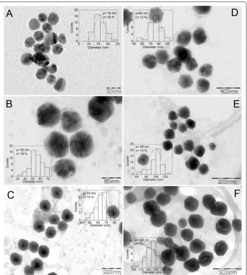
The particle size, shape/morphology and distribution of nanoparticles in the base medium were determined by transmission electron microscopy (TEM) with a JEOL
[image:3.595.58.540.165.701.2]JEM2010 equipment (JEOL Ltd., Akishima, Tokyo, Japan) at 200 kV and 106μA. Samples were prepared by adding 50 μl of the stock nanoparticle solution onto 200-mesh carbon-coated copper grids to obtain TEM images. In Figure 2, TEM images show a larger diameter
of the gold nanoparticles by decreasing the number of seeds, as it was expected. The dispersion of gold nano-particles has a median size distribution near 12% for all the samples, including large-diameter gold nanoparticles. This is an advantage if compared with the citrate method, in which it is only possible to produce mono-dispersed and spherical gold nanoparticles of up to 50 nm in diameter.
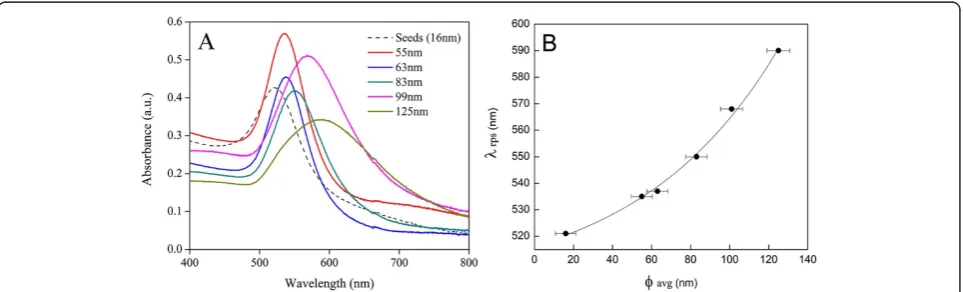
The absorption spectra of synthetized nanoparticles were measured as 1:2 dilutions (stock to H2O) using an ultraviolet–visible (UV–vis) spectrophotometer (8453 Agilent Technologies, Inc., Santa Clara, CA, USA). Figure 3A shows the UV–vis absorption spectra of the different size gold nanofluids studied. As expected, the increase in gold nanoparticle diameters resulted in a longer absorption λmax values (Figure 3B), according to
Mie’s theory [13,14].
Thermal diffusivity measurement
Thermal diffusivity values of 9.31 × 10⁻⁴ ± 0.04 × 10⁻⁴ cm2s−1 for glycerol (J.T. Baker Reagent Chemicals, Phillipsburg, NJ, USA) and values of 14.35 × 10⁻⁴± 0.04 × 10⁻⁴cm2s−1for HPLC grade water (Sigma-Aldrich Cor-poration) were obtained by the photoacoustic system as reference samples for system calibration with a coeffi-cient of variation near 2% to the reported values in the literature [15,16]. The resulting thermal diffusivity values for the different sizes and concentrations of gold nano-fluids are summarized in Tables 1 and 2, respectively; these are average values over five measurements of each sample taking the standard deviation in measurements as the uncertainty.
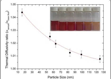
[image:4.595.304.538.122.221.2]Nanoparticle size
Figure 4 shows the thermal diffusivity values of gold colloid nanofluids as a function of nanoparticle size; it shows that if the nanoparticle concentration is con-stant (0.1 mg/ml), then the thermal diffusivity ratio
(αsample/αbase fluid) of nanofluids increases as the nano-particle diameter is reduced. This behaviour can be ex-plained as follows: as the particle size decreases, the effective surface area increases, and the Brownian mo-tion of nanoparticles enhances convective heat transfer mechanisms; thermal diffusivity and thermal conduc-tivity of nanofluids increase as a result. However, for large-diameter nanoparticles, the surface area and the Brownian motion both decrease, and as a consequence, there is no enhancement of thermal diffusivity and ther-mal conductivity by convection [3].
Concentration
Figure 5 shows the nanoparticle concentration de-pendency of the nanofluid thermal diffusivity for 55 and 83 nm size nanoparticles; as the nanoparticle concentration increases, the thermal diffusivity ratio (αsample/αbase fluid) of nanofluids increases; the results depict similar behaviours reported in the literature for metal nanofluids by thermal lens spectrometry, and photopyroelectric and the hot wire methods [7,8,17,18]. This dependency can be explained as follows: the dis-sociation of the surfactant (sodium citrate) may intensify the enhancement of thermal properties by inducing elec-trostatic repulsion among the suspended nanoparticles resulting in stabilized suspensions. Moreover, with the
Figure 3UV–vis absorption spectra and absorption shift of gold nanoparticles.(A) UV–vis absorption spectrum of the synthetized gold nanoparticles. (B) Shift in absorptionλrpsof gold nanoparticles with particle size.
Table 1 Thermal diffusivity of gold nanofluids for different nanoparticle diameters for constant nanoparticle concentration of 0.1 mg/ml
Particle size (nm) α(10−4cm2s−1) ± 0.04
16 14.98
55 14.71
63 14.63
83 14.53
99 14.51
[image:4.595.58.540.558.704.2]volumetric increase of nanoparticles, the specific heat of nanofluids decreases; as a consequence, the thermal dif-fusivity of the colloidal suspension increases [18]. The enhancement of convection and the decrease in specific heat increase the thermal diffusivity ratio of 55 nm con-centrated nanofluids compared with 83 nm concon-centrated nanofluids.
Conclusions
The present study investigates the effect of concentra-tion and size of gold nanoparticles on the thermal diffu-sivity of gold nanofluids prepared by hydroquinone method through a seed-mediated growth by photoacous-tic spectroscopy. The thermal diffusivity ratio changed inversely with the nanoparticle size; this effect was stud-ied in a size range of 16 to 125 nm for gold nanofluids. The thermal diffusivity ratio has been found to increase with concentration; this effect was studied in a range of 1 to 0.2 mg/ml for gold nanofluids. The experimental data for thermal diffusivity ratio as a function of
concentration and size for gold nanofluids depict similar behaviours in enhancement ratio compared to thermal conductivity reports and other techniques in the litera-ture such as thermal lens and hot wire method with high accuracy, and with a small amount of sample required (500 μl), photoacoustic spectroscopy might be suitable for thermal diffusivity measurement of nanofluids and being a promising alternative to classical techniques.
Abbreviations
HPLC: high-performance liquid chromatography; TEM: transmission electron microscopy; UV–vis: ultraviolet–visible.
Competing interests
The authors declare that they have no competing interests.
Authors’contributions
GALM and JAPR prepared all the samples, set up the photoacoustic method and measured and analysed the UV–vis and photoacoustic data. JSS measured and analysed the TEM data. JOL and JABL designed the experiments and wrote the manuscript. All authors read and approved the final manuscript.
Acknowledgements
The authors acknowledge Zaber Technologies, Inc. for providing a micro-linear stage, Química Aromática SA, COFAA-IPN and CONACyT for the partial support of this work.
Author details
1
Química Aromática SA, Río Grande S/N, Col. Santa Catarina Acolman, México State, CP 55875, Mexico.2UPIBI-IPN, Av. Acueducto S/N, Col. Barrio la Laguna
Ticomán, México City, CP 07340, Mexico.3Physics Department, CINVESTAV, Av. IPN 2508, Col. San Pedro Zacatenco, México City CP07360, Mexico.
4
Biotechnology Department, CINVESTAV, Av. IPN 2508, Col. San Pedro Zacatenco, México City, CP 07360, Mexico.
Received: 22 June 2012 Accepted: 24 July 2012 Published: 30 July 2012
References
[image:5.595.307.539.89.259.2]1. Moran CH, Wainerdi SM, Cherukuri TK, Kittrell C, Wiley BJ, Nicholas NW, Curley SA, Kanzius JS, Cherukuri P:Size-dependent joule heating of gold
Figure 4Thermal diffusivity decrease ratio with gold nanoparticle size.The nanoparticle concentration was 0.1 mg/ml. The inset shows (left to right) the different size nanofluids of 16 (seeds), 55, 63, 83, 99 and 125 nm.
[image:5.595.56.293.115.213.2]Figure 5Thermal diffusivity enhancement ratio with gold nanofluid concentration.The nanoparticle average diameters were 55 and 83 nm.
Table 2 Thermal diffusivity of gold nanofluids at different nanoparticle concentrations
Concentration α
55 nm (10−4cm2s−1
) ± 0.04
α83nm (10−4cm2s−1)
± 0.04
0.2 mg/ml 14.74 14.56
0.4 mg/ml 14.81 14.59
0.6 mg/ml 14.85 14.64
0.8 mg/ml 14.91 14.69
[image:5.595.56.291.517.683.2]nanoparticles using capacitively coupled radiofrequency fields.Nano Research2009,2:400.
2. Raoof M, Curley SA:Non-invasive radiofrequency-induced targeted hyperthermia for the treatment of hepatocellular carcinoma.Int J of Hepatology2011, doi:10.4061/2011/676957.
3. Paul G, Sarkar S, Pal T, Das PK, Manna I:Concentration and size dependence of nano-silver dispersed water based nanofluids.J Colloid Interface Sci2012,371:20.
4. Kleinstreuer C, Feng Y:Experimental and theoretical studies of nanofluid thermal conductivity enhancement: a review.Nanoscale Res Lett2011,
6:229.
5. Jang SP, Choi SU:Effects of various parameters on nanofluid thermal conductivity.J. of Heat Transfer2007,129:617.
6. Zamiri R, Azmi BZ, Shahriari E, Naghavi K, Saion E, Rizwan Z, Husin MS:
Thermal diffusivity measurement of silver nanofluid by thermal lens technique.J Laser Appl2011,23:042002.
7. Jiménez Pérez JL, Gutierrez Fuentes R, Sanchez Ramirez JF, Cruz-Orea A:
Study of gold nanoparticles effect on thermal diffusivity of nanofluids based on various solvents by using thermal lens spectroscopy.Eur. Phys. J. Special Topics2008,153:159.
8. Zhang X, Gu H, Fujii M:Study of the effect of volume fraction concentration and particle materials on thermal conductivity and thermal diffusivity of nanofluids.J Appl Phys2006,100:044325. 9. Frens G:Controlled nucleation for the regulation of the particle size in
monodisperse gold suspensions.Nature Phys. Sci1973,241:20. 10. Perrault SD, Chan WC:Synthesis and surface modification of highly
monodispersed, spherical gold nanoparticles of 50–200 nm.J Am Chem Soc2009,131:17042.
11. Brown KR, Walter DG, Natan MJ:Seeding of colloidal Au nanoparticle solutions. 2. Improved control of particle size and shape.Chem Mater
2000,12:306.
12. Balderas-López JA, Mandelis A:Novel transmission open photoacoustic cell configuration for thermal diffusivity measurements in liquids.Int J Thermophys2002,23:605.
13. Haiss W, Thanh NT, Aveyard J, Fernig DG:Determination of size and concentration of gold nanoparticles from UV–vis spectra.Anal Chem
2007,79:4215.
14. Amendola V, Meneghetti M:Size evaluation of gold nanoparticles by UV– vis spectroscopy.J Phys Chem,113(C2009):4277.
15. Balderas-López JA, Mandelis A, Garcia JA:Thermal-wave resonator cavity design and measurements of the thermal diffusivity of liquids.Rev Sci Instrum2000,71:2933.
16. Balderas-López JA, Mandelis A:Simple, accurate and precise measurements of thermal diffusivity in liquids using a thermal-wave cavity.Rev Sci Instrum2001,72:2649.
17. Dadarlat D, Neamtu C, Streza M, Turcu R, Craciunescu I, Bica D, Vekas L:
High accuracy photopyroelectric investigation of dynamic thermal parameters of Fe3O4and CoFe2O4magnetic nanofluids.J of Nanoparticle
Res.2008,10:1329.
18. Murshed SM, Leong KC, Yang C:Determination of the effective thermal diffusivity of nanofluids by the double hot-wire technique.J Phys D: Appl Phys2006,39:5316.
doi:10.1186/1556-276X-7-423
Cite this article as:López-Muñozet al.:Thermal diffusivity measurement of spherical gold nanofluids of different sizes/concentrations.Nanoscale Research Letters20127:423.
Submit your manuscript to a
journal and benefi t from:
7Convenient online submission 7Rigorous peer review
7Immediate publication on acceptance 7Open access: articles freely available online 7High visibility within the fi eld
7Retaining the copyright to your article