© 2017, IRJET | Impact Factor value: 5.181 | ISO 9001:2008 Certified Journal | Page 3593
Brain Computer Interface: A Review
Er. Tejinder Kaur
1, Prof. Birinder Singh
21
Student, Dept. of Computer Science and Engineering, Baba Banda Singh Bahadur Engineering College
2Professor, Dept. Of Computer Science and Engineering, Baba Banda Singh Bahadur Engineering College
Punjab, India
---***---Abstract -
Brain Computer Interface is a technique which allows people to interact with machines independent of muscles. Brain Computer Interface record the electrical signals from the brain and convert the recorded signals into corresponding control signals with the help of specialized computer. The main purpose of Brain Computer Interface is to enable the paralyzed people to communicate with their environment. BCI can be implemented in various ways like invasive, semi-invasive and non-invasive. The non-invasive way to implement Brain Computer-interface is known as Electroencephalogram(EEG). This paper gives a brief introduction about Brain Computer Interface, EEG and its applications.Key Words: Amyotrophic Lateral Sclerosis (ALS), Brain Computer Interface (BCI), Electrocorticogram (ECoG), Electroencephalogram (EEG), Magnetoencphalogram (MEG), Near Infrared Spectroscopy (NIRS), Functional Magnetic Resonance Imaging (fMRAI).
1.INTRODUCTION
There are some diseases which lead the patient in the locked in syndrome. In this condition, the person is cognitively intact but the body is paralyzed. Amyotrophic lateral sclerosis(ALS) is one example of such types of disease or a person can be paralyzed due to some accident like road accident, radiation accident etc. The main cause of ALS is unknown and this disease does not have any cure. Here the word paralyzed means that person has lost any voluntary control over the muscles, but his or her mind is as active as ours. The patient cannot move their legs, arms or faces. They totally depend upon the artificial respirator. In that state the patient can communicate effectively with their environment with the help of device that can read the electrical signals from the brain and can convert them into the corresponding control signals. Such devices are called Brain Computer Interface.
Back in 60s, controlling some electrical devices with the brain waves was considered as the pure science fiction. In 1929, the German Scientist Hans Berger recorded the brain activity from the human scalp, but at that time the technology required for measuring and processing the brain signals are too limited. The BCI is firstly portray by Dr. Grey Walter in 1964. He connected the electrodes directly to the motor areas of the patient. The patient was undergoing surgery for other reasons. Then the patient was asked to
press a button to advance a slide projector and Dr. Walter
was recording the brain activity correspondingly. He found
that he had to introduce a delay from the detection of the
brain activity until the slide projector advanced because the slide projector would otherwise advance before the patient
pressed the button. Unfortunaltely, Dr. Walter did not
publish their work beside of it he presented a talk about it to a group named the Oster Society in London[7].Later on the BCI research is developed for many more years.
The natural form of control or conversation needed nerves and movement of muscles. This mechanism begins with the user’s intent. A complex process is prompted by this intent which includes some of the brain areas. As the result of this process, some signal are generated from brain which
is used in BCI. A straightforward conversation link between a
human or animal brain and an exterior equipment is called Brain-Computer Interface. It is also known as Brain-Machine Interface or Direct Neural interface. Basically, Brain Computer Interface is an artificial system that bypasses the body’s normal efferent pathways, which are the neuromuscular output channels. BCI measures brain activity correlated with the user’s intent and converts the recorded brain activity into the corresponding control signals [4]. These converted signals are further used for BCI applications. The translation of electrical brain signals into the signals used by the BCI applications is done by the computer which usually involves signal processing and pattern recognition. Since the measured activity or signals emerge directly from the brain and not originates from the muscles or peripheral devices, the system is called a Brain– Computer Interface.
A BCI system must have some components. BCI must measure the signals directly from the brain in the invasive or non-invasive method. A BCI must give feedback to the user in the real-time scenario. Lastly, the BCI system must rely on the voluntary control. Although BCI is a communication tool but nowadays the BCI systems are gripping above a communication tool for the users who are not able to communicate otherwise. BCIs achieving consideration even from the healthy peoples and have few new goals like hands-free gaming or rehabilitation. There are some people that still do not have any knowledge about the Brain-Computer Interface.
© 2017, IRJET | Impact Factor value: 5.181 | ISO 9001:2008 Certified Journal | Page 3594 the EEG are due to the mental activity. These adjustments in
signals are detected by the BCI system and convert it into a control signal which can be used in many applications such as the motion of a wheel chair, video game etc. Electroencephalographic waveforms (Brain Waves) in which an Electroencephalographic (EEG) time series is presented and Neurophysiological model is discussed in an Air Force research program. These Neurophysiological model relate EEG waveshape with the response of neural populations[26]. The voluntary neural network hand movements can be detected by EEG signals [13]. The machines which are used in BCIs should be able to learn to discriminate between different patterns of brain activity carefully. Neuroprostheses is an example of such machine which does
not only receive output from the nervous system but also
provide input. The BCI user should able perform a different mental activity in order to produce distinct brain signals. The major phases of BCI are:
1. Data Acquisition
2. Signal Processing & Classification 3. Computer Interface
4. Application
1.1 Early History
In 1875, Richard Caton recorded EEG signals in animals from the cortical surface. Later on Hans Berger who is a German neuroscientist record the electrical signals from the human brain first time in 1929 using EEG [15]. Alpha Waves are recorded by Berger for the first time. E. Adrian and B. Matthew does interpretation of the potential waves of the brain with in the cortex region [11]. E. Adrian and K. Yamagiwa discovered the location of the EEG signals are varied on the head and the standardized position of electrodes over the head is defined [12]. R. Stanley, D. A. Daniel et al. [24] uses reciprocity theorem to determine sensitivity of EEG signals which helps to find the location and orientation of signal source in the Brain. Later on in 1970, EEG activities are further explore by the Defence Advanced Research Projects Agency of USA, before the EEG was only used for exploring the brain function and clinical diagnosis.
1.2 Advancement
D. K. Mejdi, C. Nidhal et al. [9] develop a drowsiness monitoring system which helps to reduce road accidents by estimating the drowsiness level of the driver by the means of EEG (electroencephalography) signals records. A.Wafa, A. dalal et al. [3] develop a BCI-based smart home system, which allow a person who suffer from quadriplegia to open/close doors using brain signals only. This further reduce their need for caregivers. Q. Shiyuan, L. Zhijun et al. [19] presents a teleoperation control for an exoskeleton robotic system which is based on the vision feedback and brain-machine interface. The proposed algorithm decode the EEG signals and then generates control signals for the exoskeleton robot using features extracted from neural activity. A. P. Z. Mohammad, P. Manoranjan proposed a
seizure prediction method based on a patient-specific approach by extracting undulated global and local features of preictal and interictal periods of EEG signals [2].
According to Ayurveda, Brahmi a kind of herb, used as the treatment of brain and nervous system disorders, such as epilepsy, neuropathic pain, and helping people to stay calm. W.Piyawan, S. Phakkharawat research to study the results of the consumption of the Brahmi extract whether it affected to the human brainwaves and spatial ability. The results were analyzed, after the eight weeks which shows that the volunteers who consumed the Brahmi every day, showed significantly and statistically improving scores of spatial ability test and their brainwaves had also changed. W.Piyawan, S. Phakkharawat et al. [31] makes conclusion, that the there will be the significant and statistical improvement of the spatial ability by consumption of the Brahmi extract of 300 mg per day for eight weeks.
2. TYPES OF BRAIN COMPUTER INTERFACE
The basic purpose of BCI is to catch the electrical signals that pass between neurons in the brain and convert these signals to the signal that can be sensed by external devices. BCI measures the activities of the brain, then do some computations on it and finally produce the control signals which mirror the user’s intent. To understand how BCI will work, one has to understand how the activities of the brain can be measured. There are several types of brain-computer interfaces which are listed below.
2.1 Invasive Brain Computer Interface
The Invasive Brain Computer Interface is mostly used to provide the functionality to paralyzed people. With the help of Invasive BCI one can restore the vision by connecting the brain with external cameras and also restore the use of limbs by using brain controlled robotic arms and legs. Invasive BCI requires surgery to implant the mandatory sensors. Invasive
BCI requires surgery to implant the mandatory sensors. The
method used in the invasive BCI is neurosurgery. The surgery comprises the opening of the skull through a surgical procedure and cutting the membranes which cover the brain. On the surface of the cortex, the electrodes are placed the signals from recorded from these electrodes are known as Intracortical signals.
© 2017, IRJET | Impact Factor value: 5.181 | ISO 9001:2008 Certified Journal | Page 3595 William inserted single array BCI containing 68 electrodes
into Jerry’s visual cortex and succeeded to restore his vision. Invasive BCI and Semi-invasive methods have some advantages over non-invasive methods. However, these advantages come with the serious drawback of requiring surgery. Financial Ethical and some other considerations make neurosurgery impractical except for some users who need a BCI to communicate. Even then, some of these users may prefer noninvasive BCI meets their needs. It is also unclear Intracortical recordings can provide stable and safe recording over years. Long-term stability may be especially problematic in the case of Intracortical recordings. The tissue reactions can be caused by the electrodes which are implanted into cortical tissue which can lead to deteriorating signal quality or even complete electrode failure. Invasive BCIs is difficult because of the cost and risk of neurosurgery.
2.2 Partially Brain Computer Interface
Partially invasive BCI is a type of BCI in which the devices are embedded inside the skull but rest outside the brain in-stead of the grey matter. The electrodes are embedded in a thin plastic pad that is placed above the cortex, beneath the dura mater. The signal strength of partially invasive BCI is bit weaker when it compares to invasive BCI. But still, it will generate better resolution signals than the non-invasive BCIs. The partially invasive BCIs have less exposure of scar tissue formation as compared to Invasive BCI.
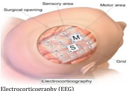
Partially Invasive BCI requires surgery to implant the mandatory sensors. This is a special type of surgery namely, the craniotomy. The surgery comprises the opening of the skull through a surgical procedure and cutting the membranes which cover the brain. On the surface of the cortex, the electrodes are placed, the signals from these electrodes are recorded. The technique which is used to record the electrical signals using invasive BCI is called
Electrocortiogram(ECoG). Electrocortiogram(ECoG) worn
the similar technology as electroencephalography, in the case of the non-invasive brain-computer interface.
Electrocorticogram(ECoG) technologies were first trade-in
humans in 2004.
[image:3.595.338.548.100.246.2]ECoG does not damage any neurons because no electrodes penetrate the brain. The ECoG records the integrated activity of a much larger number of neurons that are in the proximity of the ECoG electrodes. However, any
Fig -1: Electrocorticography (EEG)
invasive technique has better spatial resolution than the EEG.It is also unclear in the case of Partially-invasive brain computer interface whether it can provide safe and stable recording over years.
2.3 Non Invasive Brain Computer Interface
The third type of brain-computer interface is non-invasive brain-computer interface. It has the least signal clarity but it is recognized as very safest when compared to other types of BCI. The non-invasive BCIs device is able to restore the partial movement and give a patient the ability to move muscle implants. Most of the BCIs are dependent upon the electrical measures of the brain activities.
A Non-Invasive technique is one in which the electrodes are placed directly on the desired part of the brain. The devices or sensors are mounted on the headbands or caps which read brain signals. The non-invasive BCI approach record signals less effectively because electrodes are placed over the cap not on the desired part of the brain.
Electroencephalography (EEG) is very effective method of
recoding electrical signals from the scalp[5].
Electroencephalography(EEG) indicate that
electrical activity of the brain is recorded from the scalp with
the help of electrodes. It is capable of providing a good
temporal resolution. It is moveable, low-cost and easy to use [4]. The equipments used in Electroencephalography(EEG)
are lightweight, inexpensive.A non-invasive BCI based on
Electroencephalography(EEG) consist of a cap on which electrodes are placed, cables which broadcast the recorded signals from the brain to the signal amplifier, this bio-signal amplifier is a device which convert electrical brain signals from analog to digital format and a computer that operate on the data and controls and run the BCI applications.
© 2017, IRJET | Impact Factor value: 5.181 | ISO 9001:2008 Certified Journal | Page 3596 Fig -2: A Typical EEG based BCI
movements or eye blinks (Electrooculographic activity, EOG) and from muscles (Electromyographic activity, EMG) close to the recording sites. External electromagnetic sources such as the power line can also contaminate the EEG. Furthermore, although the EEG is not very technically demanding, the setup procedure can be cumbersome. To achieve adequate signal quality, the skin areas that are contacted by the electrodes have to be carefully prepared with special abrasive electrode gel. Because gel is required, these electrodes are also called wet electrodes.
The number of electrodes required by current BCI systems range from only a few to more than 100 electrodes. Most groups try to minimize the number of electrodes to reduce setup time and hassle. Since electrode gel can dry out and wearing the EEG cap with electrodes is not convenient or fashionable, the setting up procedure usually has to be repeated before each session of BCI use. From a practical viewpoint, this is one of largest drawbacks of EEG-based BCIs. A possible solution is a technology called dry electrodes [21]. Dry electrodes do not require skin preparation not electrode gel. This technology is currently being researched, but a practical solution that can provide signal quality comparable to wet electrodes is not in sight at the moment.
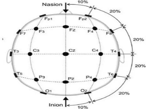
Electroencephalography(EEG) signals are collected by placing various channels over the head. To get the consistent recordings from the brain the electrodes are placed over the specific area of brain. Mostly scientist relay on the standard system to place the electrode accurately. This standard system is known as the 10-20 system and is widely used in the clinical EEG recordings, Electroencephalography and Brain computer interface research.
The name 10-20 system suggest the positions of the electrodes which are most commonly used while EEG recordings. These positions are 10,20,20,20,20 and 10% of
the total nasion and the inion distance. The buldge on the back of the skull and just above the neck is known as the inion. The intersection of the nasal and frontal bones at the bridge of the nose is known as the nasion. The other electrodes are placed over the same distance. The inter-electrode are placed over the equal distance from front to back and from left to right. The study shows that the most prominent regions in the brain are the regions where the variations in the electrical signals are occur. The positions where the variations occur are not fixed but they vary from person to person.
The label letters indicate the brain region where the electrodes are placed. O- occipital, P- parietal, T-temporal, C-central, F- frontal, Fp- pre-frontal. The electrode C3 record the motor movements from the right hand and the electrode C4 record the motor movement from the left hand. The electrode Cz record the motor movements from foots. But it is not possible to distinguished between the left and right feet. Because Electroencephalography(EEG) record the electrical signals from the scalp which have weak signal strength and also the origins of the left and right feet movements are very close to each other.
Fig -3: Standard 10-20 System
3. OTHER NEUROIMAGING APPROACHES IN BRAIN
COMPUTER INTERFACE
The brain computer interface need brain signals to perform any type of task. To get the signals from the brain BCIs rely on recording these electrical signals and then converting them into the corresponding commands. Two types of brain activities may be monitored:
i) Electrophysiological ii) Hemodynamic.
[image:4.595.315.549.384.559.2]© 2017, IRJET | Impact Factor value: 5.181 | ISO 9001:2008 Certified Journal | Page 3597 3.1 Electrophysiological
Electrophysiological activity is generated by electro-chemical transmitters exchanging information between the neurons. Ionic currents are generated by the neurons which flow across and within neuronal assemblies. With the help of dipole conducting current from a source to a sink through the dendritic trunk the large variety of current pathways can be simplified [5]. Electrophysiological activity can be measured by Electroencephalography(EEG), Magnetoencephalography(MEG), Intracortical recording, Electrocorticography(ECoG) and electrical signal acquisition in single neurons. Electroencephalography, Electrocorticography, Intracortical recording are already discussed above.
3.1.1 Magnetoencephalography(MEG)
Magnetoencephalography record the magnetic activities of the brain by means of magnetic induction. MEG is a non-invasive way to record the signals from the brain. It measures the intracellular currents flowing through dendrites of the neuron which produce magnetic fields. This magnetic field can be measure outside of the head [30]. Magnetic fields are detected by superconducting quantum interferences devices, which are extremely sensitive to magnetic disturbances produced by neural activity. The signals produced by MEG are identical to EEG signals. The magnetic fields are less distorted by the skull and scalp than electric fields which becomes advantage of MEG and it provides signals with higher spatiotemporal resolution than EEG. MEG has also been successfully used to localize active regions inside the brain. Although MEG have these types of advantages, In BCI system MSG is not often used because MEG is a expensive and bulky technology.
3.2 Hemodynamic
When the blood releases glucose at a greater rate than in the area of inactive neurons to active neurons this process is known as the hemodynamic response. The oxygen and glucose delivered through the blood stream results in a surplus of oxyhemoglobin in the veins of the active area, and there is a distinguishable change of the ratio of oxyhemoglobin to deoxyhemoglobin. Neuroimaging methods are used to quantified these changes such as functional magnetic resonance and near infrared spectroscopy[27]. These methods are categorized as indirect methods, as they measure the hemodynamic response, which is not directly related to neuronal activity.
3.2.1 Functional Magnetic Resonance Imaging
(fMRI)
Functional Magnetic Resonance Imaging (fMRI) detects the changes in local cerebral blood volume, oxygenation levels and cerebral blood flow during neural activation. It record the changes by the means of
electromagnetic fields. It is a non-invasive technique. fMRI can be performed using MRI scanners which apply electromagnetic fields of strength in the order of 3T or 7T. The main advantage of fMRI is high space resolution. Therefore fMRI have been applied for localizing active regions inside the brain [20]. But fMRI has a low temporal resolution, physiological delay from 3 to 6 seconds which is introduce by hemodynamic response [18] and is highly susceptible to head motion artifacts. It is typically used to measure the Blood Oxygen Level Dependent (BOLD) during neuronal activation . The data set acquired using fMRI techniques were processed offline and the results only became available after several hours or even days. Non-clinical fMRI applications are not expected because fMRI requires overly bulky and expensive hardware.
3.2.2 Near Infrared Spectroscopy (NIRS)
Near Infrared Spectroscopy (NIRS) method employs infrared light to acquired fluctuations in cerebral metabolism during neural activity. It is a non-invasive and an optical spectroscopy method. Infrared light penetrates the skull below its surface, to a depth of approximately 1–3 cm. where the intensity of the attenuated light allow salt-erations in oxy-hemoglobin and de-oxyhemoglobin concentrations to be measured. Due to shallow light penetration in the brain, this optical neuro-imaging technique is limited to the outer cortical layer.
NIRS system consists of a light source, a light detector, signal processing devices, a driving electronic device and a recording device. The light source is an infrared emitting diode (IRED) which is placed in direct contact with scalp. The light detector is a photodiode, which is placed right next to the light source.
The signal processing devices are filters and amplifiers which process the electrical signal and reduce the noise. The driving electronic device is an electronic circuit which controls the IRED in order to modulate the light. The recording device is a personal computer or any other device that digitalizes, stores, and displays the electrical signal. The performance of the NIRS and quality of signals can be effected by head motions or hair obstruction [29].
The nature of the hemodynamic response is the major limitations of NIRS, because within the certain number of seconds vascular changes occur, spatial resolution of NIRS is quite low. NIRS offers high portability, low cost, and an acceptable temporal resolution [22]. This neuroimaging modality can be a good alternative to EEG, as it do not require conductive gel or corrosive electrodes.
4. BRAIN WAVES
© 2017, IRJET | Impact Factor value: 5.181 | ISO 9001:2008 Certified Journal | Page 3598 are firstly discovered by Berger. These waves are associated
with the different functions which are performed by a human body [23]. Types of brain waves are listed below.
4.1 Infra-Low (<0.5 Hz)
Few studies have been done on Infra-Low brainwaves due their slow nature, which makes them difficult to detect and accurately measure. These waves are also known as Slow Cortical Potentials. Infra-Low brainwaves are thought to be the basic cortical rythms had a major role in networking function and brain timing. These waves have rang below 0.5 Hz.
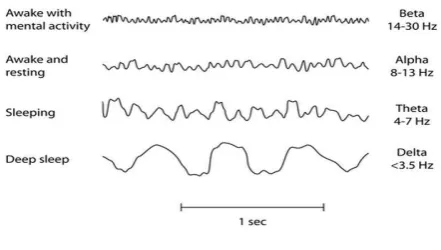
4.2 Delta Waves (0.5 to 4 Hz)
Another type of the brain waves is the delta waves which have the lowest frequencies. The delta waves are generated in the dreamless sleep or very deep sleep and in deepest meditation. The domain of the delta waves is theunconscious mind. These waves suspend external awareness therefore healing and regeneration process faster in this state. This describes why sleep is essential for human beings.
4.3 Theta Waves (4 to 8 Hz)
[image:6.595.60.281.562.687.2]Theta waves are associated with light sleep or deep meditation. These waves mostly occur in sleep but in deep meditation they are dominating. Therefore it is a border between unconscious and conscious state. Our senses are focused on signals originating from within and are withdrawn from original world in the theta waves. In these we are in dream, imagery, vivid, intuition that’s we hold history, nightmares, fear, trouble etc. Theta waves are calm and silent waves. The deep spirituality connection are comes from theta waves.
FIG -4: Brain Waves
4.4 Alpha Waves (8 to 13 Hz)
Alpha waves are the resting state of human brain. It is the power of now to be in the present. These waves helps in the overall coordination. Alpha waves are also known as the
gateways from the lower base of self-consciousness to the subconscious mind [9]. Alpha waves are dominant during the meditative states.
4.5 Beta Waves (14 to 30 Hz)
Beta waves are associated with fast activity, when humans are alert, engaged in problem solving, judgment, decision making, and engaged in focused mental activity, attentive. Beta waves are further divided into three bands which are i) Lo-Beta ranges from 12 to 15Hz. It can be thought of as musing or 'fast idle. ii) Beta which ranges from 15 to 22Hz and are generated when a person is actively figuring something out or high engagement. iii) Hi-Beta ranges from 22 to 38Hz. It is highly complex thought, integrating new experiences, high anxiety, or excitement. Beta waves are also associated with logical reasoning decision making.
4.6 Gamma Waves (14 to 30 Hz)
Gamma waves are associated with language processing, generation of ideas and memory processing. These waves are the fastest brain waves with high frequency. These brain waves are also responsible for the simultaneous processing of information from different brain areas. How gamma waves are generated remains a mystery because gamma waves have frequency above the frequency neuronal firing. Gamma waves have range from 38 to 42Hz.
5. APLLICATIONS OF BRAIN COMPUTER INTERFACE
5.1 Military
In an actual fight the experience of the soldier can’t be duplicated by machines. In that case soldier, while in simulation using BCI, the voluntary movements and decision of soldier can be used to guide robots/drones while the soldier sits safely playing simulation [1].
5.2 Medical Applications
BCI have variety of application in the medical field which can take advantage of brain signals in its associated phases including detecting, prevention, diagnosis, rehabilitation and restoration. Such as seizure predicting [2].
5.2.1 Prevention
© 2017, IRJET | Impact Factor value: 5.181 | ISO 9001:2008 Certified Journal | Page 3599 5.2.2 Detection and Diagnosis
BCI system has also contribute in the detecting and forecasting the health issues such as the abnormal Brain Structure(Brain Tumor), Sleep Disorder(Narcolepsy), Seizure Disorder(Epilepsy) and Brain Swelling(Encephalitis).EEG based BCI can be used to detect Breast Cancer, Dyslexia (a kind of brain disorder).
5.2.3 Rehabilitation and Restoration
When the brain cells are suddenly die because of the lack of oxygen that condition is known as the stroke due to which the patient can lose the ability to speak, or control over the limbs. Stroke may be fully recovered with the help of BCI [18]. Neuroprosthetic devices, which are used to regain the normal functionality for the patients who can not recover the mobility and communication like wheel chair, robotics prosthetic or an exoskeleton.
5.3 Smart Environment
K. K. Ang, C.Guan et al. [18] have proposed a Brain Computer Interface-based Smart Living Environment Auto-Adjustment Control System(BSLEACS), which is a cognitive control system that monitor the users mental state and accordingly adapts the surrounding component. Using audio-video environment the alcoholic drivers, who contributes to a road accident, could be characterized using EEG.
5.4 Neuromarketing and Advertisement
The EEG evaluation is also beneficial for the TV Advertisement, related to the both political fields and commercial fields.G. Vecchiato, F. Babiloni et al. [15] have consider the effect of cognitive function in the neuro-marketing field.
5.5 Self-Regulation and Education
In the sports competition an emotional intelligence based on EEG has been applied to control the stress. Functional Magnetic Resonance Imaging (fMRI) neurofeedback has been elaborated in self-regulations and skill learning [5].
5.6 Games and Entertainment
Now a days many researchers have the topic to combining the features of the exiting game with the brain controlling capabilities. Tan and Nijholt describe the brainball game which helps to drop the stress level [11].Halicopters are made to fly in any dimension either 2D or 3D. BCI provide a simplified driving simulator with mental-tasked condition for the verification of drivers identity. Cognitive Biometric or electrophysiology, which only modalities using bio-signals(such as brain signals) can be used to protect highly confidentional information.
6. CHALLENGES OF BRAIN COMPUTER INTERFACE
The development of the Brain Computer System rely on the accurate selection of the signals then the data should be acquire in the correct manner, feature extraction, translation algorithm, dependent/independent mode, output devices, synchronous/asynchronous mode, training user group, choice of application and protocols all these have their great effect on working of the BCI system [22]. The main challenges which are faced by BCI system are:
6.1 Information Rate
It depends upon the sensors, the more the sensors are used to acquire the brain signals the more will be the information rate and upon the channels bandwidth which is used to pass these signals to corresponding system/device.
6.2 Error Rate
Error in the BCI is defines as classification error occurred while allocating task to external device. If there is higher the error rate in the BCI system then there will be the more complication while interacting with environment.
6.3 Autonomous
BCI systems cannot be used autonomously by disabled people, the reasons behind it is that BCI systems require assistants to apply electrodes or signal-receiving devices [1].
6.4 Midas Touch Problem
For the BCI system it is very difficult to distinguish between the voluntary or in-voluntary acts of the person. Midas is such a type of problem in which the system wrongly interprets the fixations which may be caused due to the long processing time. As user can off the BCI system by sending input to it but can not turn it on back again.
6.5 Cognitive load
It is the load on the brain to control the complex task of the hybrid BCI system, which may further depend upon the critical system means the user has to control in how many dimension.
7. ETHICAL ISSUES
As the raise in the BCI, the user would be haunt with the privacy issue, it means that attackers would be able to hack targets brain.
REFERENCES
© 2017, IRJET | Impact Factor value: 5.181 | ISO 9001:2008 Certified Journal | Page 3600 Egyptian Informatics Journal, Vol. 16, pp. 213-230,
July,2015.
[2] A.P. Z. Mohammad, P. Manoranjan, “Seizure Prediction Using Undulated Global and Local Features”, Vol. 64, issue. 1, Pages: 208 – 217, 2017.
[3] A. Wafa, A. dalal, A. Amenah, “Smart home: toward daily use of BCI-based systems”, 2017 International Conference on Informatics, Health & Technology (ICIHT), pp.1-5, 2017.
[4] B. Benjamin, D. Guido, “The Berlin Brain–Computer Interface: EEG-Based Communication Without Subject Training”, IEEE transactions on neural system and rehabilitation engineering, vol. 14, no. 2, pp. 147-152, 2006.
[5] C. Babiloni, V. Pizzella, C. D. Gratta, A. Ferretti, G. L. Romani, “Fundamentals of Electroencefalography, Magnetoencefalography, and Functional Magnetic Resonance Imaging”, Brain Machine Interfaces for Space Applications,New York, Vol. 86, pp. 67–80, 2009. [6] C. T. Lin, B. S. Lin, F. C. Lin, C. J. Chang, “Brain computer
interface-based smart living environmental auto-adjustment control system in UpnP home networking”, IEEE Systems Journal, Vol. 8, Issue. 2, June 2014. [7] D.C. Dennett, Consciousness explained, Back Bay Books,
Lippincott Williams & Wilkins, 1992.
[8] D. Di, C. Zhihua, F. Ruifang, L. Guangyu, L. Tian, “ Study on human brain after consuming alcohol based on eeg signal. In: Computer Science and Information Technology (ICCSIT)”, 3rd IEEE International Conference, vol. 5, pp. 406–09, 2010.
[9] D. K. Mejdi, C. Nidhal, W. Ali, A. M. Adel, “Towards an automatic drowsiness detectionsystem by evaluating the alpha band of EEG signals”, Applied Machine Intelligence and Informatics (SAMI), pp. 000371 – 000376, 2017.
[10] D.S. Tan, A. Nijholt, “Brain-computer interfaces: applying our minds to human-computer interaction”, Springer, 2010.
[11] E. Adrian and B. Matthews, “The interpretation of potential waves in the cortex,” Journal of Physiology, vol. 81, pp. 440-471, 1934.
[12] E. Adrian and K. Yamagiwa, “The origin of the Berger rhythm,” Brain, vol. 58, p. 323–351, 1935.
[13] E. Abbas, G. Mahdi, “EEG Signals can be used to detect the voluntary hand neural network movements by using an enhanced resource-allocating”, Proceedings of the 23rd Annual EMBS International Conference, October 25-28, Istanbul, Turkey, pp. 721-724, 2001.
[14] G. Vecchiato, F. Babiloni, L. Astolfi , J. Toppi, P. Cherubino, J. Dai, W. Kong, D. Wei, “Enhance of theta eeg spectral activity related to the memorization of commercial advertisings in chinese and italian subjects”, Biomedical Engineering and Informatics (BMEI), 4th International Conference, vol. 3, pp. 1491–94, 2011.
[15] H. Berger, “Uber das Elecrtenkphalogramm des Menchen,” Arch Psychiat Nervernkr,vol. 87, pp. 527-570, 1929.
[16] J. R. Wolpaw, N. Birbaumer, D.J. McFarland, G. Pfurtscheller, and T.M. Vaughan, Braincomputer interfaces for communication and control. Clin Neurophysiol, 113, Jun., 767–791, 2002.
[17] K. K. Ang, C.Guan, K. C. Geok, C. Kuah, C. Wang, H. Zhang, “Clinical study of neurorehabilitation in stroke using eeg based motor imagery brain-computer interface with robotic feedback. In: Engineering in Medicine and Biology Society (EMBC)”, Annual International Conference of the IEEE, pp. 5549–52, 2010.
[18] N. Weiskopf, K. Mathiak, S. W. Bock, F. Scharnowski, R. Veit, W. Grodd, R. Goebel, N. Birbaumer, “Principles of a brain-computer interface (BCI) based on real-time functional magnetic resonance imaging (fMRI)”, IEEE Trans. Biomed. Eng., vol. 51, pp. 966–970, 2004. [19] Q. Shiyuan, L. Zhijun, H. Wei, Z. Longbin,Y.Chenguang, S.
Chun-Yi, “Brain–Machine Interface and Visual Compressive Sensing-Based Teleoperation Control of an Exoskeleton Robot”, IEEE Transactions on Fuzzy Systems, Vol. 25, issue. 1, pp. 58-69, 2017.
[20] R. C. deCharms, K. Christoff, G. H. Glover, J. M. Pauly, S. Whitfield, J. D. E. Gabrieli, “Learned regulation of spatially localized brain activation using real-time fMRI”, Neuroimage, vol. 21, pp. 436–443, 2004.
[21] R. Fazel-Rezai, B. Z. Allison, C. Guger, E. W. Sellers, S. C. Kleih, A. Kübler, “P300 brain computer interface: current challenges and emerging trends”, Frontiers in Neuroengineering, vol. 5, 2012.
[22] R. P. Kennan, S. G. Horovitz, A. Maki, Y. Yamashita, H. Koizumi, J. C. Gore, “Simultaneous recording of event-related auditory oddball response using transcranial near infrared optical topography and surface EEG”, Neuroimage, vol. 16, pp. 587–592,2002.
[23] R. Salmelin, M. Hámáaláinen, M. Kajola, R. Hari, “Functional segregation of movement-related rhythmic activity in the human brain”, Neuroimage 1995, vol. 2, pp. 237–243, 1995.
[24] R. Stanley, D. A. Daniel, “EEG Electrode Sensitivity-An Application of Reciprocity” IEEE Transactions on Biomedical Engineering, Vol. BME-16, No. 3, pp. 204-205, 1969.
[25] S. Baillet, J. C. Mosher, R. M. Leahy, “Electromagnetic brain mapping”, IEEE Signal Process, Vol. 18, pp. 14-30, 2001.
[26] S. Bernard, B. R. Neil, M. A. Miles, C. G. Edward, “A New Approach, to Signal Analysis in Electroencephalography” IRE Transactions on Medical Electronics, Vol. PGME-8, pp.24-30,1957.
© 2017, IRJET | Impact Factor value: 5.181 | ISO 9001:2008 Certified Journal | Page 3601 [28] S. M. Coyle, T. E. Ward, C. M. Markham, “Brain-computer
interface using a simplified functional near-infrared spectroscopy system”, J. Neural Eng. 2007, vol. 5, pp. 43- 50, 2007.
[29] S. Waldert, T. Pistohl, C. Braun, T. Ball, A. Aertsen, C. Mehring, “A review on directional information in neural signals for brain-machine interfaces”, J. Phisiol, Vol. 103, pp.244–254, 2009.
[30] W. Wang, G. P. Sudre, Y. Xu, R. E. Kass, J. L. Collinger, A. D. Degenhart, A. I. Bagic, D. J. Weber, “Decoding and cortical source localization for intended movement direction with MEG”, Neurophysiol, Vol. 104, pp. 2451–2461, 2010.
[31] W.Piyawan, S. Phakkharawat, “Effect of Brahmi extract on human brainwaves”, 2017 International Conference on Digital Arts, Media and Technology (ICDAMT), pp. 364 – 368,2017.
BIOGRAPHIES