Mutation in the TCR
aa
subunit constant gene
(TRAC) leads to a human immunodeficiency
disorder characterized by a lack of TCR
ab
ab
+
T
cells
Neil V. Morgan, … , Sophie Hambleton, Eamonn R. Maher
J Clin Invest.
2011;
121(2)
:695-702.
https://doi.org/10.1172/JCI41931
.
Inherited immunodeficiency disorders can be caused by mutations in any one of a large
number of genes involved in the function of immune cells. Here, we describe two families
with an autosomal recessive inherited immunodeficiency disorder characterized by
increased susceptibility to infection and autoimmunity. Genetic linkage studies mapped the
disorder to chromosomal region 14q11.2, and a homozygous guanine-to-adenine
substitution was identified at the last base of exon 3 immediately following the translational
termination codon in the TCR
a
subunit constant gene (
TRAC
). RT-PCR analysis in the two
affected individuals revealed impaired splicing of the mRNA, as exon 3 was lost from the
TRAC
transcript. The mutant TCR
a
chain protein was predicted to lack part of the
connecting peptide domain and all of the transmembrane and cytoplasmic domains, which
have a critical role in the regulation of the assembly and/or intracellular transport of TCR
complexes. We found that T cells from affected individuals were profoundly impaired for
surface expression of the TCR
ab
complex. We believe this to be the first report of a
disease-causing human
TRAC
mutation. Although the absence of TCR
ab
+T cells in the
affected individuals was associated with immune dysregulation and autoimmunity, they had
a surprising level of protection against infection.
Research Article
Immunology
Find the latest version:
Mutation in the TCR
α
subunit constant
gene (
TRAC
) leads to a human
immunodeficiency disorder characterized
by a lack of TCR
αβ
+
T cells
Neil V. Morgan,1 Sarah Goddard,2 Tony S. Cardno,3 David McDonald,4 Fatimah Rahman,1
Dawn Barge,5 Andrew Ciupek,3 Anna Straatman-Iwanowska,1 Shanaz Pasha,1
Mary Guckian,2 Graham Anderson,6 Aarnoud Huissoon,2 Andrew Cant,7
Warren P. Tate,3 Sophie Hambleton,4,7 and Eamonn R. Maher1,8
1Wellchild Paediatric Research Centre, Department of Medical and Molecular Genetics and Centre for Rare Diseases and Personalised Medicine,
University of Birmingham School of Medicine, Birmingham, United Kingdom. 2Regional Department of Immunology, Heartlands Hospital,
Birmingham, United Kingdom. 3Department of Biochemistry, University of Otago, Dunedin, New Zealand. 4Institute of Cellular Medicine,
Newcastle University, Newcastle upon Tyne, United Kingdom. 5Regional Immunology Laboratory, Royal Victoria Infirmary, Newcastle upon Tyne,
United Kingdom. 6MRC Centre for Immune Regulation, The Medical School, University of Birmingham, Birmingham, United Kingdom. 7Paediatric Immunology and Infectious Diseases Service, Great North Children’s Hospital, Newcastle upon Tyne, United Kingdom.
8West Midlands Regional Genetics Service, Birmingham Women’s Hospital, Birmingham, United Kingdom.
Inherited immunodeficiency disorders can be caused by mutations in any one of a large number of genes
involved in the function of immune cells. Here, we describe two families with an autosomal recessive inherited
immunodeficiency disorder characterized by increased susceptibility to infection and autoimmunity. Genetic
linkage studies mapped the disorder to chromosomal region 14q11.2, and a homozygous guanine-to-adenine
substitution was identified at the last base of exon 3 immediately following the translational termination
codon in the TCRα subunit constant gene (
TRAC
). RT-PCR analysis in the two affected individuals revealed
impaired splicing of the mRNA, as exon 3 was lost from the
TRAC
transcript. The mutant TCRα chain protein
was predicted to lack part of the connecting peptide domain and all of the transmembrane and cytoplasmic
domains, which have a critical role in the regulation of the assembly and/or intracellular transport of TCR
complexes. We found that T cells from affected individuals were profoundly impaired for surface expression
of the TCRαβ complex. We believe this to be the first report of a disease-causing human
TRAC
mutation.
Although the absence of TCRαβ
+T cells in the affected individuals was associated with immune dysregulation
and autoimmunity, they had a surprising level of protection against infection.
Introduction
Inherited immunodeficiencies have provided novel insights into T and B cell development and immune function. Failure of T cell development, of whatever cause, produces the clinical syndrome of SCID, characterized by profound susceptibility to opportunis-tic infection, failure to thrive, and death in infancy. In contrast, genetic disorders that impair T cell function may display a range of phenotypes; from SCID, through partial immunodeficiency with dysregulation and/or lymphoid neoplasia, to predominant autoimmunity (1, 2).
We investigated two consanguineous families with what we believe to be a novel immune dysregulatory disorder and per-formed gene mapping and identification studies, which led to the identification of a mutation in the TCRα subunit constant gene (TRAC).
Results
Clinical features . Two apparently unrelated children from consan-guineous families of Pakistani origin were noted to have clinical and immunophenotypic features in common. They presented at
the ages of 15 months (family 1 [F1]; II:3) and 6 months (family 2 [F2]; II:2) with features of combined immunodeficiency: recur-rent respiratory tract infection, otitis media, candidiasis, diarrhea, and failure to thrive. These infections (Table 1) responded well to conventional treatment but recurred frequently, until the institu-tion of continuous antibacterial and antifungal prophylaxis. One child (F1;II:3) showed clear predisposition to herpes viral infec-tion, experiencing an unusually chronic course of varicella at age 6 as well as chronic EBV and human herpesvirus 6 (HHV6) viremia. The same child showed evidence of ongoing infective damage to the respiratory tract and was treated with long-term oral antiviral therapy and intravenous immunoglobulin. In contrast, individual F2;II:2 had an uneventful episode of varicella at age 6 years and no evidence of chronic lung damage.
In addition to immunodeficiency, both children had evidence of immune dysregulation: child F1:II:3 showed hypereosinophil-ia, low-titer antinuclear antibodies (ANA), vitiligo, and alopecia areata, while child F2:II:2 had hypereosinophilia, eczema, auto-immune hemolytic anemia, antilymphocyte antibodies, anti-TTG antibodies, low-titer ANA, and pityriasis rubra pilaris. In contrast, humoral immunity against vaccine antigens appeared normal (Table 1). Both children developed moderate lymphadenopathy
and organomegaly (hepatomegaly in the case of F1;II:3 and hepa-Conflict of interest: The authors have declared that no conflict of interest exists.
research article
tosplenomegaly in F2;II:2). At age 2 years, weight gain faltered in individual F2;II:2, and imaging revealed thickened bowel loops and a mass at the porta hepatis. Histology of the liver showed patchy eosinophilia, with biliary fibrosis and no evidence of para-sites or fungi. It was felt that this may have been an autoimmune process, but infection such as cryptosporidium could not be excluded (feces were negative for cryptosporidium at this time). This child received a successful matched sibling bone marrow transplant age 7, while individual F1;II:3 underwent transplanta-tion at age 6 years, also from a matched sibling (both children
received reduced-intensity pretransplant conditioning with alem-tuzumab, fludarabine, and melphalan).
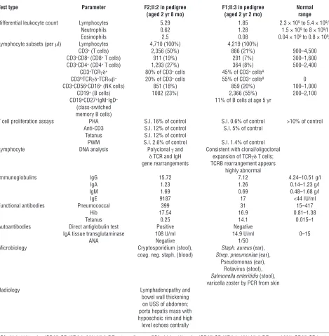
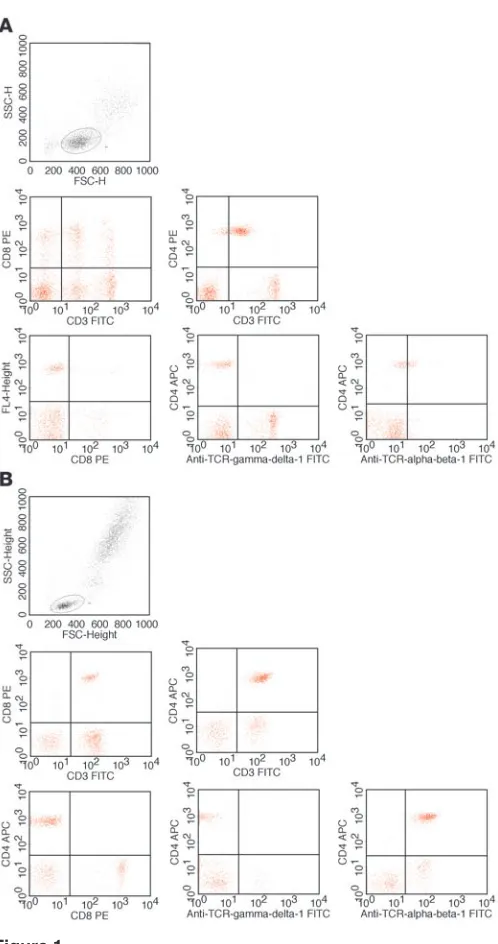
[image:3.585.57.526.112.590.2]Flow cytometry of peripheral blood lymphocytes. The flow cytometric peripheral blood lymphocyte profiles of the two affected individu-als were strikingly similar and unusual. Conventional CD3+ T cells were present but uniformly expressed TCRγδ, with or without CD8 (Table 1, Figure 1, and data not shown). Furthermore, an abnormal population of CD3lo cells was present, which expressed TCRαβ at extremely low levels. These CD3lo cells could be further subdi-vided on the basis of CD4 and CD8 expression; the majority were Table 1
Summary of laboratory and radiological investigations in two affected children with the immunodeficiency disorder
Test type Parameter F2;II:2 in pedigree F1;II:3 in pedigree Normal
(aged 2 yr 8 mo) (aged 2 yr 2 mo) range
Differential leukocyte count Lymphocytes 5.29 1.85 2.3 × 10⁹ to 5.4 × 10⁹/l
Neutrophils 0.62 1.28 1.5 × 10⁹ to 8 × 10⁹/l
Eosinophils 2.5 0.08 0.04 × 10⁹ to 0.8 × 10⁹/l
Lymphocyte subsets (per μl) Lymphocytes 4,710 (100%) 4,219 (100%)
CD3+ (T cells) 2,356 (50%) 886 (21%) 900–4,500
CD3+CD8+ (CD8+ T cells) 911 (19%) 291 (7%) 300–1,600
CD3+CD4+ (CD4+ T cells) 1,293 (27%) 364 (8%) 500–2,400
CD3+TCRγδ+ 80% of CD3+ cells 45% of CD3+ cellsA
CD3loTCRγδ–TCRαβ– 20% of CD3+ cells 55% of CD3+ cellsB 0
CD3–CD56+CD16+ (NK cells) 851 (18%) 859 (20%) 100–1,000
CD19+ (B cells) 1082 (23%) 2,366 (55%) 200–2,100
CD19+CD27+IgM–IgD– 11% of B cells at age 5 yr
(class-switched memory B cells)
T cell proliferation assays PHA S.I. 16% of control S.I. 0.6% of control >10% of control Anti-CD3 S.I. 12% of control S.I. 5% of control
Tetanus S.I. 12% of control
PWM S.I. 2.6% of control S.I. 1.4% of control Lymphocyte DNA analysis Polyclonal γ and Consistent with clonal/oligoclonal
δ TCR and IgH expansion of TCRγδ T cells; gene rearrangements TCRB rearrangement appears
highly abnormal
Immunoglobulins IgG 15.72 7.12 4.24–10.51 g/l IgA 1.23 1.26 0.14–1.23 g/l IgM 1.69 0.69 0.48–1.68 g/l IgE 9187 17 <44 IU/ml Functional antibodies Pneumococcal 399 31 15–417
Hib 17.54 16.9 0.81–1.38 Tetanus 0.25 14.1 0.015–1 Autoantibodies Direct antiglobulin test Positive Negative
IgA tissue transglutaminase 108 U/ml 14.9 U/ml 0–15 ANA Negative 1/50
Microbiology Cryptosporidium (stool), Staph. aureus (ear), coag. neg. staph. (blood) Strep. pneumoniae (ear),
Pseudomonas (ear), Rotavirus (stool),
Salmonella enteritidis (stool), varicella zoster by PCR from skin Radiology Lymphadenopathy and
bowel wall thickening on USS of abdomen; porta hepatis mass with hypoechoic rim and high level echoes centrally
AOf which 11% naive (CD27+CD45RA+), 26% HLA-DR+ at age 5 years. BOf which <1% naive (CD27+CD45RA+), 42% HLA-DR+, and 38% CD25+CD4+ at
age 5 years. coag. neg. staph., coagulase-negative staphylococcus; Hib, Haemophilus influenzae type B; S.I., stimulation index; Staph., Staphylococcus;
CD4+CD8–, while a smaller double-positive population decreased with age. Thymic emigrant equivalents (CD45RA+CD27+) were present within the CD3hi (TCRγδ+, 11%) but barely within the CD3lo (TCRγδ–, <1%) compartment. Proliferation of lymphocytes in response to phytohemagglutinin (PHA) and OKT3 was variably reduced compared with that in controls (Table 1). Despite these profound T cell abnormalities, class-switched memory B cells were present, immunoglobulin levels were within the normal range (apart from elevated IgE levels in F2;II:2), and vaccine responses were demonstrable. In keeping with these findings, the lymph node biopsy of individual F2;II:2 showed lymph node follicles and development of germinal centers (data not shown).
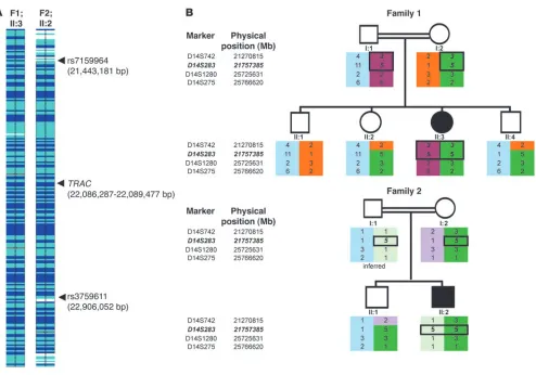
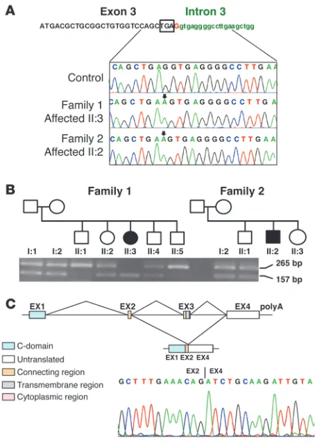
Molecular genetic analysis . In light of negative results of immu- nological and molecular investigations for known immunodefi-ciency disorders, we hypothesized that both children might have a novel autosomal recessive disorder and undertook genetic linkage studies using polymorphic microsatellite markers and genome-wide SNP genotyping using the Affymetrix 250K SNP microarray (Figure 2A). Linkage to the TRBC1 gene was excluded, and in both affected children the largest overlapping homozy-gous regions (1.46 Mb and 2.60 Mb) were at 14q11.2 (Figure 2A). The two children shared a common haplotype of 161 SNPs between rs7159964 (21,443,181 bp) and rs3759611 (22,906,052 bp) on chromosome 14. Genotyping of parents and siblings with microsatellite markers (D14S742, D14S283, D14S1280, and D14S275) confirmed linkage (Figure 2B). Mutation analysis of TRAC (chromosome 14:22,086,287–22,089,447 bp) was under-taken, revealing a homozygous G-to-A substitution (c.*1G>A) at the last nucleotide of exon 3 (immediately following the transla-tion termination codon) in both affected children (Figure 3A). Family studies confirmed that the sequence change segregated with disease status, and the c.*1G>A mutation was not detected in 384 ethnically matched control chromosomes.
In considering possible disease mechanisms, we first inves-tigated whether the TRAC c.*1G>A substitution might pro-mote a significant increase in readthrough of the termination codon, as previously shown for certain mammalian mRNAs (particularly at genetic recoding sites; refs. 3, 4). Thus, we compared the termination efficiency of the wild-type and mutant sequences (embedded in a typical +6 to +9 context sequence) by placing each between two luciferase reporter genes (RLuc and Luc+) and assaying the relative activities of
the two luciferases in human cultured cells. Although the mutant *A allele was associated with a significant increase in readthrough of the stop signal (compared with the wild-type *G), the absolute amount of readthrough product was relatively small, suggesting that this was unlikely to be the mechanism of disease in vivo (data not shown).
We next hypothesized that the c.*1G>A mutation would impair splicing of the TRAC transcript, since the mutation is within the consensus 5ʹ splice site. RT-PCR analysis of TRAC cDNA in members of the two affected families revealed exon skipping of the last coding exon (exon 3), resulting in an aberrant transcript joining exon 2 to the normally untranslated exon 4 (Figure 3, B and C) (testing for additional alternative patterns of TRAC splicing using primers anchored in exon 1 and intron 3 did not detect further transcripts). In the predicted transla-tion product, the 35 C-terminal amino acids are replaced by 56 amino acids encoded by exon 4 (p.Thr107LeufsX56). This results in partial loss of the connecting peptide domain and abolition of the transmembrane and cytoplasmic domains of the TCRα chain. Previously, deletion of as few as 9 amino acids from the C-terminus of the TCRα chain has been reported to impair assembly and/or intracellular transport of TCR-CD3 complexes (5, 6). Additionally a conserved motif (FETDxNLN), present in the α chain connecting peptide domain, was shown to be critical in controlling antigen responsiveness (7). Therefore, the c.*1G>A mutation–associated splicing defect would be expected to have a profound impact on TCRα chain structure and function.
Cellular localization and immunoblotting of TRAC mutant. When assessed by flow cytometry, surface staining for TCRαβ was strong- ly reduced in patient cells (Figure 1). To assess whether TCR poly-Figure 1
[image:4.585.46.295.76.548.2]research article
peptides were being retained within the cell, we prepared lysates of patient PBMCs and performed immunoblotting using antibodies against TCRα and TCRβ (Figure 4), normalizing for the propor-tion of TCRγδ– T cells. In contrast to normal control cells, patient cells showed undetectably low expression of either TCRα or TCRβ polypeptides, suggesting that the TRAC mutation not only resulted in TCRα deficiency but also reduced TCRβ expression.
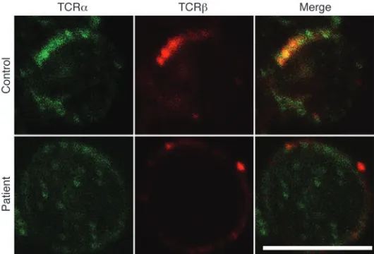
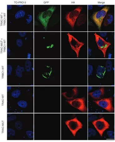
To investigate with greater sensitivity any effect on expres-sion and subcellular localization of the TCRαβ complex, we immunostained patient and control PBMCs with antibodies against T cell receptor α and β chains and observed localization using immunofluorescence microscopy. In control cells, colocal-ization of α and β chains within distinct areas of the cell mem-brane was readily apparent. Patient cells showed reduced levels of expression and no evidence of colocalization (Figure 5), suggest-ing that the mutant α chain fails to complex normally with the TCR β chain. Consistent with this and in contrast to wild-type TCRα, mutant HA-tagged TCRα, and GFP-tagged TCRβ failed to colocalize intracellularly in transfected HeLa cells (Figure 6). Surface expression in this system could not be assessed, owing to the lack of CD3 expression.
Discussion
[image:5.585.49.543.77.423.2]As well as displaying what we believe to be a novel immune dis-order, the two families harbor a most unusual mutation. To our knowledge, there are no previous reports of a mutation at the base following a termination codon (c.*1) in human disease. Prokary-otes and eukaryotes demonstrate highly significant deviations from the expected nucleotide distribution before and after the stop codon (3), and the mammalian termination signal decoding factor eRF1 absolutely requires a tetranucleotide as a minimum recognition element in vitro (8). Hence, nucleotide substitutions at the base following the termination codon might cause disease by impairing the efficiency of the “tetranucleotide stop signal” (3). However, when we modeled the effect in cultured cells, we found that although the c.*1G>A mutation did cause a small increase in readthrough, this was unlikely to be sufficient to cause a clinical phenotype, and the pathogenicity of the c.*1G>A mutation was due to abnormal splicing. Hence, the apparently unique nature of the c.*1 mutation in TRAC appears to be due to the unusual exon/intron structure of the gene. It is not known whether c.*1 mutations in other genes might cause human disease by causing a “leaky termination signal” alone, but such an effect may only be Figure 2
evident if the cells are sensitive to small changes in expression of the normal gene product or if the abnormal “readthrough protein” has a dominant effect on the function of the wild-type protein.
Most T cells express a heterodimeric T cell receptor consisting of β and α peptides in association with subunits of the CD3 com-plex (γ, δ, ε, ζ, and η chains). The antigen-binding region of the T cell receptor (the variable region) is assembled from an extensive repertoire of coding segments present in the TCR loci. During T cell development in the thymus, gene segments are rearranged to form a unique V exon in each thymocyte (9). The β chain gene is rearranged before TRAC, and, if productive, the TCRβ chain is initially expressed with preTα, an invariant chain. Functional signaling through this complex must occur for progression to
α chain rearrangement to take place (this is known as β selec-tion). This coincides with progression from the intermediate single-positive stage (CD3lo, CD4+, CD8– , in humans) to the dou-ble-positive stage (CD4+, CD8+). The αβ-expressing thymocyte then undergoes both positive selection (to ensure restriction to self-MHC) and negative selection to prevent recognition of self, called central tolerance (10). Defective expression of TCRα would be expected to impair αβT cell development beyond β selec-tion because of a failure to deliver positively selecting signals. Conversely, the development of γδT cells should not be directly affected, since TRAC rearrangement and TCRγδ expression are mutually exclusive outcomes.
Despite lacking a critical molecule for T cell function, the two children with a homozygous TRAC mutation did not succumb to infection and remained relatively healthy for some years on regular antibiotic prophylaxis. Furthermore, they were able to
produce class-switched antibodies and germinal centers and to mount antibody responses against both vaccine and auto-anti-gens. In this regard, they resemble the TCRα-knockout mouse model, in which γδT cells have been proposed to provide T cell help to B cells, including those responsible for autoantibody pro-duction (11, 12). Tcra–/–
mice possess normal numbers of dou-ble-positive and double-negative thymocytes (13), but generate few single-positive thymocytes, and most peripheral T cells bear TCRγδ . With increasing age, the mice develop an aberrant pop-ulation of peripheral T lymphocytes, but these are CD3+CD4+ TCRβ+/TCRα– cells that may express TCRβ homodimers (14) or pre-TCR (15). Their appearance is temporally and possibly caus- ally associated with the development of inflammatory bowel dis-ease (16, 17). Thus, in the Tcra–/– mouse as in our human patients,
the inability of single-positive thymocytes to express a mature
[image:6.585.50.277.81.402.2]αβTCR does not appear to prevent thymic egress absolutely but results in the development of T cells with an abnormal TCR phe-notype. It would have been of great interest to clarify what signals sustained the CD3lo population within our patients, since low-level signaling via the TCR has been proposed to be essential to T cell survival. Analysis of TCR gene rearrangement within sorted CD3lo cells might also have shed light on their developmental origins. As to their functional significance, we can unfortunately say little save to observe that other partial T cell deficiencies lead to a similar combination of reduced T cell effector function and T cell dysregulation (2).
Figure 3
Identification of a homozygous G-to-A substitution in the first base fol-lowing the termination codon (*1) in TRAC. (A) Wild-type and mutant
[image:6.585.352.482.522.656.2]TRAC sequence traces. The position of the *1G>A sequence change in the patients is indicated by the arrows. The boxed sequence shows the tetranucleotide translation termination signal. (B) RT-PCR analy-sis of TRAC showing skipping of exon 3, which segregates within the two families. (C) Schematic showing exon structure and domains of mutant and natively spliced mRNA and sequence trace from RT-PCR of TRAC, resulting in skipping of exon 3 (exon structure adapted from IMGT repertoire; http://www.imgt.org/textes/IMGTrepertoire/).
Figure 4
research article
Although the children have shown susceptibility to infection, neither opportunistic infections nor mycobacterial disease have been a feature. Cryptosporidium and salmonella have, however, been cultured during admissions to hospital with fever. Sus-ceptibility to these pathogens is associated with defects of the IFN-γ –IL-12/23 signaling pathway as well as T cell immunode-ficiencies including HLA class II deficiency (18). Infection by intracellular pathogens is also seen in the TRAC-deficient mouse model, with susceptibility to cryptosporidium, mycobacterium, and Leishmania (19–21). There is no evidence in the mouse model of increased risk of malignancy, although lymphadenopathy is a feature shared with our patients (22).
Finally we note that this is the second report of genetic involve-ment of the T cell receptor alpha locus (TRA@) in human disease, as polymorphic SNPs in TRA@ were recently found to be associ-ated with narcolepsy (the strongest association was with an SNP close to the junctional [J] segment gene [TRAJ10]) (23). The absence of this sleep disorder in children with TRAC mutations is consis-tent with the suggestion that the association between narcolepsy and TRA@ may be related to an autoimmune process involving destruction of hypocretin-producing neurons (24).
Methods
Patient ascertainment and sampling. This study was conducted according to the principles expressed in the Declaration of Helsinki. The study was approved by the South Birmingham Research Ethics Committee, United Kingdom. All participants provided informed consent for the collection of samples and subsequent analysis. DNA was extracted from whole blood and extracted (Gentra, Puregene DNA purification system) for all family members.
Gene mapping. A genome-wide linkage scan was carried out using the Affymetrix 250K SNP chip in the affected individual from each family with the immunodeficiency disorder (F1;II:3 and F2;II:2). This scan iden-tified a single region of homozygosity at chromosome 14q11.2 shared by the two affected individuals (Figure 2A). These homozygous regions were further analyzed by typing microsatellite markers (D14S742, D14S283, D14S1280, and D14S275) in all family members from whom DNA was available (Figure 2B).
Mutation analysis. We identified positional candidate genes using data from the National Center for Biotechnology Information (NCBI) and
University of California Santa Cruz human genome databases. Sequenc-ing was performed using standard methods on an ABI 3730 automated sequencer. For TRAC, we designed primer pairs using exon-primer (http:// ihg.helmholtzmuenchen.de/ihg/EasyExonPrimer.html) to amplify coding exons 1–3. The full coding region of human TRAC was PCR amplified from genomic DNA with the following primers: TRAC exon 1 (TRAC_Exon1_ FOR) 5ʹ-CAAAGAGGGAAATGAGATCATG-3ʹ and (TRAC_Exon1_ REV) 5ʹ-GGCCATTCCTGAAGCAAG-3ʹ; TRAC exon 2 (TRAC_Exon2_ FOR) 5ʹ-TGCCCAAGAACTAGGAGGTC-3ʹ and (TRAC_Exon2_REV) 5ʹ-GGTTATTGCGGGTTCATCAC-3ʹ; TRAC exon 3 (TRAC_Exon3_FOR) 5ʹ-GCTCTGCCTTGGGGAAAAC-3ʹ and (TRAC_Exon3_REV) 5ʹ -CTG-CAGGGAGGTTTGCTCTC-3ʹ. Genomic DNA from 192 ethnically matched controls was used to screen for the identified TRAC mutation.
RT-PCR. Total RNA was extracted from human lymphocytes using the RNeasy Mini Kit (QIAGEN). cDNA was synthesized using random prim-ers and AMV reverse transcriptase using the Promega reverse transcription system according to the manufacturer’s instructions (A3500, Promega). RT-PCR of TRAC was performed using the following primers: TRAC2F (5ʹ-GTTCCTGTGATGTCAAGCTGGTC-3ʹ) and TRACx4R2 (5ʹ -GGTAG-CAGCTTTCACCTCCTTG-3ʹ).
Plasmid constructs. Wild-type constructs of the full coding region of human TRAC and TRBC1 were PCR amplified from cDNA with the following primers: TRAC (TRAC_HA_FOR) 5ʹ -GCTAGTCGAC-TATATCCAGAACCCTGACCC-3ʹ and (TRAC_WT_REV) 5ʹ -CGATG-GTACCTCAGCTGGACCACAGCCGCA-3ʹ; TRBC1 (TRBC1_GFP_FOR) 5ʹ-GCTAGTCGACTTAGGACCTGAACAAGGTGTT-3ʹ and (TRBC1_WT_ REV) 5ʹ-CGATGGTACCACAGAAATCCTTTCTCTTGAC-3ʹ. The mutant
TRAC construct was PCR amplified from cDNA with the primers (TRAC_ HA_FOR) 5ʹ-GCTAGTCGACTATATCCAGAACCCTGACCC-3ʹ and (TRAC_MUT_REV) 5ʹ-CGATGGTACCCTGTTTCAAAGCTTTTCTC-3ʹ. Forward primers contained a SalI restriction site, and reverse primers contained a KpnI site to allow subcloning of the PCR fragments into the pEGFP-C2 and pCMV-HA vectors (BD Biosciences — Clontech). All plas-mid constructs were verified by sequencing.
Cell culture . HeLa cells were routinely maintained in DMEM (Sigma-Aldrich) supplemented with 10% fetal bovine serum at 37°C, 5% CO2.
[image:7.585.47.312.83.263.2]Cellular localization. PBMCs were obtained from patient F1;II:3 and control blood and stored frozen at –140°C in 10% DMSO, 90% fetal calf serum. After rapid thawing, cells were washed with PBS, fixed with 4% paraformaldehyde, and permeabilized with 0.1% Triton/PBS. Cells were blocked with 1% BSA and incubated with antibodies against TCRα (sc-9100) and TCRβ (sc-5277) (Santa Cruz Biotechnology Inc.). Cells were
Figure 5
TRAC mutation results in mislocalization of the TCRα and -β
washed and incubated with anti-rabbit Alexa Fluor 488 and anti-mouse Alexa Fluor 546 conjugates (Invitrogen). After washing, cells were mounted using ProLong Gold antifade and examined by confocal microscopy using a Leica SP2 confocal microscope.
HeLa cells were grown on coverslips in 24-well plates and transfected with 1 μg of plasmid DNA constructs (pCMV-HA, pCMV-HA.TRAC.WT, pCMV-HA.TRAC.MUT, pEGFP vector, and pEGFP.TRBC1.WT) using Lipo-fectamine 2000 (Invitrogen) according to the manufacturer’s instructions. After 24 hours, cells were washed with PBS, fixed in 4% paraformaldehyde, and permeabilized. The subcellular distribution of HA.TRAC was assessed by incubating cells with mouse monoclonal antibodies against HA (Sigma-Aldrich), detected using Alexa Fluor 546 conjugate (Invitrogen), and
nuclei were stained using TO-PRO-3 iodide (Invitrogen). Finally, cells were mounted using Citifluor (Citifluor Ltd.), and images were visualized using a Leica SP2 confocal microscope.
[image:8.585.110.478.80.533.2]Immunoblotting. Control and patient PBMCs and RPMI 8866 cells were lysed for 10 minutes on ice in lysis buffer (20 mM Tris-HCl, pH 7.4; 150 mM sodium chloride; 1% Triton-X 100; 5 mM EDTA; Halt protease inhibitors [Pierce]) before clarification at 13,000 g. Lysates were reduced (5 minutes at 70°C, LDS sample buffer and reducing agent [Invitrogen]) and alkylated (10 minutes at room temperature, 50 mM iodoacetamide) and electropho-resed at 25 mA/gel on duplicate 10% SDS polyacrylamide gels. Proteins were transferred to PVDF membrane at 30 mA/gel for 120 minutes. After blocking for 1 hour in 5% milk in TBS-0.5% Tween-20 (TBST), membranes were cut and
Figure 6
Cellular localization of TCRαβ complex in HeLa cells. HeLa cells grown on coverslips were cotransfected with pCMV.HA-TRAC and
pEGFP-TRBC1 (top row), pCMV.HA-TRACexon1–2 and pEGFP-TRBC1 (second row), pEGFP-TRBC1 (third row), pCMV.HA-TRAC (fourth row), and
pCMV.HA-TRACexon1–2 (fifth row). Cells were labeled with anti-HA antibody, followed by Alexa Fluor 546–conjugated secondary antibody, and
research article
probed with 0.4 μg/ml anti-TCRα (H-142, Santa Cruz Biotechnology Inc.), anti-TCRβ (G-11, Santa Cruz Biotechnology Inc.), or anti–β-actin (AC-74, Sigma-Aldrich) overnight at 4°C in 5% milk in TBST. After 4 washings of 5 minutes each in TBST, blots were incubated with the appropriate secondary antibody (10–20 ng/ml goat anti–mouse HRP or 20 ng/ml goat anti–rabbit HRP) for 1 hour. Blots were washed a further 4 times prior to incubation with chemiluminescent substrate (West Dura, Pierce) and exposure to film. Acknowledgments We thank the two families and our clinical and laboratory col-leagues for their help. We are grateful to P. Gissen for helpful advice. Financial support was provided by WellChild, the Well- come Trust, and Birmingham Children’s Hospital Research Foun-dation. W.P. Tate is supported by the Health Research Council of New Zealand and the Marsden Fund. Received for publication December 3, 2009, and accepted in revised form November 3, 2010. Address correspondence to: Eamonn R. Maher, Department of Medical and Molecular Genetics, University of Birmingham, Insti-tute of Biomedical Research, Edgbaston, Birmingham, B15 2TT, United Kingdom. Phone: 44.121.627.2741; Fax: 44.121.414.2538; E-mail: [email protected]. 1. Fischer A. Human primary immunodeficiency dis-eases. Immunity. 2007;27(6):835–845.
2. Liston A, Enders A, Siggs OM. Unravelling the association of partial T-cell immunodeficiency and immune dysregulation. Nat Rev Immunol. 2008;8(7):545–558.
3. McCaughan KK, Brown CM, Dalphin ME, Berry MJ, Tate WP. Translational termination efficiency in mammals is influenced by the base follow-ing the stop codon. Proc Natl Acad Sci U S A. 1995; 92(12):5431–5435.
4. Cridge AG, Major LL, Mahagaonkar AA, Poole ES, Isaksson LA, Tate WP. Comparison of char- acteristics and function of translation termina-tion signals between and within prokaryotic and eukaryotic organisms. Nucleic Acids Res. 2006; 34(7):1959–1973.
5. Shelton JG, Gülland S, Nicolson K, Kearse KP, Bäck- ström BT. Importance of the T cell receptor alpha-chain transmembrane distal region for assem-bly with cognate subunits. Mol Immunol. 2001; 38(4):259–265.
6. Bhatnagar A, et al. Mutational analysis of conserved amino acids in the T cell receptor alpha-chain trans- membrane region: a critical role of leucine 112 and phe-nylalanine 127 for assembly and surface expression.
Mol Immunol. 2003;39(15):953–963.
7. Bäckström BT, Milia E, Peter A, Jaureguiberry B, Baldari CT, Palmer E. A motif within the T cell receptor α chain constant region connecting peptide domain controls antigen responsiveness.
Immunity. 1996;5(5):437–447.
8. Poole ES, Major LL, Mannering SA, Tate WP. Trans-lational termination in Escherichia coli: three bases following the stop codon crosslink to release factor 2 and affect the decoding efficiency of UGA - contain-ing signals. Nucleic Acids Res. 1998;26(4):954–960. 9. Davis MM, Bjorkman PJ. T-cell antigen
recep-tor genes and T-cell recognition. Nature. 1988; 334(6181):395–402.
10. Anderson G, Moore NC, Owen JJ, Jenkinson EJ. Cellular interactions in thymocyte development.
Annu Rev Immunol. 1996;14:73–99.
11. Mombaerts P, et al. Mutations in T-cell antigen receptor genes alpha and beta block thymocyte development at different stages. Nature. 1992; 360(6401):225–231.
12. Wen L, et al. Immunoglobulin synthesis and generalized autoimmunity in mice congenitally deficient in alpha beta(+) T cells. Nature. 1994; 369(6482):654–658.
13. Eichelberger M, McMickle A, Blackman M, Mom- baerts P, Tonegawa S, Doherty PC. Functional analy-sis of the TCR alpha- beta+ cells that accumulate in the pneumonic lung of influenza virus-infected TCR-alpha–/– mice. J Immunol. 1995;154(4):1569–1576.
14. Takahashi I, Iijima H, Katashima R, Itakura M, Kiyono H. Clonal expansion of CD4+ TCRbetabeta+ T cells in TCR alpha-chain- deficient mice by gut-derived antigens. J Immunol. 1999;162(3):1843–1850. 15. Barber DF, Passoni L, Wen L, Geng L, Hayday AC.
The expression in vivo of a second isoform of pT alpha: implications for the mechanism of pT alpha action. J Immunol. 1998;161(1):11–16.
16. Wen L, et al. Germinal center formation, immuno- globulin class switching, and autoantibody produc-tion driven by “non alpha/beta” T cells. J Exp Med. 1996;183(5):2271–2282.
17. Takahashi I, Kiyono H, Hamada S. CD4+ T-cell population mediates development of inflammatory bowel disease in T-cell receptor alpha chain-deficient mice. Gastroenterology. 1997;112(6):1876–1886. 18. Casanova JL, Abel L. The human model: a genetic
dissection of immunity to infection in natural con-ditions. Nat Rev Immunol. 2004;4(1):55–66. 19. Waters WR, Harp JA. Cryptosporidium parvum
infection in T-cell receptor (TCR)-alpha- and TCR-delta-deficient mice. Infect Immun. 1996; 64(5):1854–1857.
20. Mogues T, Goodrich ME, Ryan L, LaCourse R, North RJ. The relative importance of T cell subsets in immunity and immunopathology of airborne Mycobacterium tuberculosis infection in mice.
J Exp Med. 2001;193(3):271–280.
21. Satoskar A, Okano M, David JR. Gamma delta T cells are not essential for control of cutaneous Leishmania major infection in genetically resistant C57BL/6 mice. J Infect Dis. 1997;176(6):1649–1652. 22. Viney JL, et al. Lymphocyte proliferation in mice con-genitally deficient in T-cell receptor alpha beta + cells.
Proc Natl Acad Sci U S A. 1994;91(25):11948–11952.
23. Hallmayer J, et al. Narcolepsy is strongly associ-ated with the T-cell receptor alpha locus. Nat Genet. 2009;41(6):708–711.