of long-term repopulating stem cells in
nonhuman primates
Brian C. Beard, … , Jennifer E. Adair, Hans-Peter Kiem
J Clin Invest.
2010;
120(7)
:2345-2354.
https://doi.org/10.1172/JCI40767
.
HSC transplantation using genetically modified autologous cells is a promising therapeutic
strategy for various genetic diseases, cancer, and HIV. However, for many of these
conditions, the current efficiency of gene transfer to HSCs is not sufficient for clinical use.
The ability to increase the percentage of gene-modified cells following transplantation is
critical to overcoming this obstacle. In vivo selection with mutant methylguanine
methyltransferase (MGMT
P140K) has been proposed to overcome low gene transfer
efficiency to HSCs. Previous studies have shown efficient in vivo selection in mice and
dogs but only transient selection in primates. Here, we report efficient and stable
MGMT
P140K-mediated multilineage selection in both macaque and baboon nonhuman
primate models. Treatment consisting of both O
6-benzylguanine (O
6BG) and
N
,
N¢
-bis(2-chloroethyl)-
N
-nitroso-urea (BCNU) stably increased the percentage of
transgene-expressing cells from a range of initial levels of engrafted genetically modified cells, with the
longest follow-up after drug treatment occurring over 2.2 years. Drug treatment was well
tolerated, and selection occurred in myeloid, lymphoid, and erythroid cells as well as
platelets. Retrovirus integration site analysis before and after drug treatments confirmed the
presence of multiple clones. These nonhuman primate studies closely model a clinical
setting and should have broad applications for HSC gene therapy targeting human
diseases of malignant, genetic, and infectious nature, including HIV.
Research Article
Genetics
Find the latest version:
http://jci.me/40767/pdf
Research article
Efficient and stable MGMT-mediated
selection of long-term repopulating
stem cells in nonhuman primates
Brian C. Beard,1 Grant D. Trobridge,1,2 Christina Ironside,1 Jeannine S. McCune,1,3 Jennifer E. Adair,1 and Hans-Peter Kiem1,2
1Clinical Research Division, Fred Hutchinson Cancer Research Center, Seattle, Washington, USA.
2Department of Medicine, Division of Hematology, and 3Department of Pharmacy, University of Washington, Seattle Washington, USA.
HSC transplantation using genetically modified autologous cells is a promising therapeutic strategy for
vari-ous genetic diseases, cancer, and HIV. However, for many of these conditions, the current efficiency of gene
transfer to HSCs is not sufficient for clinical use. The ability to increase the percentage of gene-modified
cells following transplantation is critical to overcoming this obstacle. In vivo selection with mutant
meth-ylguanine methyltransferase (MGMT
P140K) has been proposed to overcome low gene transfer efficiency to
HSCs. Previous studies have shown efficient in vivo selection in mice and dogs but only transient selection in
primates. Here, we report efficient and stable MGMT
P140K-mediated multilineage selection in both macaque
and baboon nonhuman primate models. Treatment consisting of both O
6-benzylguanine (O
6BG) and
N
,
N
′
-bis(2-chloroethyl)-
N
-nitroso-urea (BCNU) stably increased the percentage of transgene-expressing cells
from a range of initial levels of engrafted genetically modified cells, with the longest follow-up after drug
treatment occurring over 2.2 years. Drug treatment was well tolerated, and selection occurred in myeloid,
lymphoid, and erythroid cells as well as platelets. Retrovirus integration site analysis before and after drug
treatments confirmed the presence of multiple clones. These nonhuman primate studies closely model a
clini-cal setting and should have broad applications for HSC gene therapy targeting human diseases of malignant,
genetic, and infectious nature, including HIV.
Introduction
HSC transplantation using allogeneic cells has been a successful treatment modality for a variety of genetic diseases, because the graft stably produces phenotypically normal cells for the lifetime of the individual (1). However, many patients do not have suit-able donors for allogeneic transplantation. In addition, allogeneic transplantation is often associated with significant morbidity and mortality from graft-versus-host disease and infectious complica-tions due to immunosuppressive treatment (2). Thus, the infusion of genetically corrected autologous stem cells would circumvent these limitations of allogeneic transplantation (3). Furthermore, new treatment modalities using autologous HSCs harboring a potent anti-HIV transgene could be applied alone or in parallel with highly active antiretroviral treatment for HIV patients with advanced disease to limit disease progression. For all of these gene-modified autologous HSC strategies to be successful, high levels of genetically modified cells are required long term to provide a prolonged clinical benefit.
For treatment of hematopoietic diseases that affect a variety of lineages, such as erythrocytes (hemoglobinopathies and thalas-semia) and platelets (PLTs) (Wiskott-Aldrich syndrome), genetic modification at the HSC level is desirable to provide lifelong pro- duction of “corrected” hematopoietic lineages. A similar strat- egy of genetic modification of HSCs is favored to provide life-long “protected” macrophages and lymphocytes in HIV-infected patients. Successful HSC gene therapy has been unequivocally demonstrated for patients with X-linked SCID (SCID-X1) (4),
adenosine deaminase deficient SCID (5), X-linked chronic granu-lomatous disease (X-CGD) (6), and adrenoleukodystrophy (ALD) (7). Numerous advances in transduction protocols, including transduction on fibronectin fragment CH-296 (8), the use of mul-tiple cytokines (9, 10), and the use of different vector pseudotypes (11–13), have contributed to gene transfer levels of 20%–30% in clinically relevant nonhuman primates (14). Even with the gene marking levels (defined as the percentage or fraction of genetically modified cells) achieved clinically ranging from approximately 9%–15% in peripheral blood with reduced-intensity busulfan (6) and high-intensity cyclophosphamide and busulfan (7) and levels in the nonhuman primate ranging from 20%–30% in peripheral blood with total body irradiation (TBI) (14), for numerous clinical applications gene marking levels will still likely be below therapeu-tic thresholds (for review see ref. 15).
Without an intrinsic growth advantage provided by gene-modi-fied cells, such as that observed for SCID-X1, efficient and stable posttransplant in vivo selection will likely be required to increase the percentage of these cells into a therapeutic range for diseases such as thalassemia and other hemoglobinopathies. This approach has several therapeutic applications, such as using anti-HIV trans-genes to block infection or limit propagation of HIV or using drug resistance transgenes to limit myelosuppression from chemother- apy during cancer therapy to allow dose intensification. The sys-tem that currently meets the criteria for all of these applications is mutant methylguanine methyltransferase (MGMTP140K) (16–20),
because of stable multilineage in vivo selection that has been previ-ously described in mice (21) and dogs (22, 23). MGMTP140K
-medi- ated selection does not require chronic administration of pharma-cologic agents but instead can be achieved with limited treatments
Conflict of interest: The authors have declared that no conflict of interest exists.
using the wild-type MGMT inhibitor O6-benzylguanine (O6BG)
in combination with either N,N′-bis(2-chloroethyl)-N -nitroso-urea (BCNU) or temozolomide (TMZ). It is important to extend MGMTP140K
-mediated in vivo selection studies to nonhuman pri-mates and test durable sustained selection, because of the ability to directly translate transduction procedures (i.e., antibodies for HSC enrichment, growth factor cocktail, and vector pseudotype), similar dosing of drugs used for conditioning, and in vivo selec-tion to clinical applications. In addition, the nonhuman primate model is a powerful model for diseases such as HIV, which can be modeled using SHIV (24). With regard to safety, studies in the nonhuman primate are critical to assess the long-term implica-tions of cytotoxic drug treatment (BCNU and TMZ), for which the method of action is DNA damage, before proceeding to clinical applications. This long-term data will be especially important for application to nonmalignant disease, for which drug treatments with O6
BG and BCNU or TMZ would be new treatment modali- ties. Given these considerations, successful studies in the nonhu-man primate should readily translate to clinical applications.
Here, we tested MGMTP140K
-mediated in vivo selection and che- moprotection using gammaretrovirus- and HIV-derived self-inacti-vating (SIN) lentivirus-based vectors in baboons (Papio cynocephalus) and pigtailed macaques (Macaca nemestrina). We also extended these studies, using bicistronic and tricistronic vectors that allow for in vivo selection of gene-modified cells that also express anti-HIV transgenes, and also included reduced-intensity conditioning, using fractionated busulfan in the macaque. To accompany these efficacy studies and given the adverse events associated with gam- maretrovirus-based vectors in clinical trials for X-linked immuno-deficiency (SCID-X1) (25) and clonal expansion in X-CGD (6), we also carried out retrovirus integration site (RIS) mapping, before and after drug treatment, to monitor any early indication of clonal restriction that may lead to leukemic transformation. The combi-nation of these studies, along with long-term analysis following drug treatment, is critical to demonstrate the feasibility, safety and efficacy of MGMTP140K-mediated in vivo selection in an important
preclinical model. Our data supports the use and continued devel- opment of this strategy for chemoprotection during human can-cer therapy or to mediate in vivo selection for genetic or infectious diseases affecting the hematopoietic system.
Results
Efficient MGMTP140K-mediated in vivo selection and chemoprotection in baboons and macaques following transplantation with cells genetically modified with gammaretrovirus vectors. HSC gene therapy for a variety of severe hematopoietic genetic, infectious, and malignant diseases will likely require an efficient and robust in vivo selection strategy to attain therapeutic levels of gene marking (defined as the pres-ence of genetically modified cells). Here, we tested the MGMTP140K
-mediated in vivo selection in the well-established and clinically relevant baboon and macaque nonhuman primate models. We first evaluated gammaretrovirus vectors expressing MGMTP140K
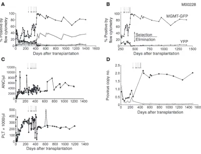
alone or with a fluorescent reporter EGFP and, in some cases, included cells genetically modified with enhanced yellow fluo- rescent protein (YFP) only as a control arm (26). Drug treat- ment was initiated after stable hematopoietic recovery and rela-tively stable gene marking, at least 90 days after transplantation (Supplemental Table 1; supplemental material available online with this article; doi:10.1172/JCI40767DS1). Initially, baboons M01044 and M00228 were given 3 cycles and 1 cycle, respectively,
of TMZ, with a single dose of O6BG before TMZ (Supplemental
Table 2). This resulted in transient in vivo selection (Figure 1, A and B, and Supplemental Figure 1B, dashed arrows) with no pro-nounced myelosuppression (Figure 1C). Both monkeys went on to receive TMZ dose escalation, adding a second dose of O6BG
(O6BG-2X) 7–8 hours after TMZ administration to maximize
inhibition of wild-type MGMT, further sensitizing unmodified cells (27) (Supplemental Table 2). This drug regimen strategy also resulted in transient in vivo selection (Figure 1, A and B, and Supplemental Figure 1B), with no pronounced myelosuppression (Figure 1C), even with TMZ doses of up to 1,100 mg/m2 (M01044) and
1,400 mg/m2 (M00228). As an alternative drug regimen for in
vivo selection, we next tested the combination of O6BG-2X and
BCNU, which we had previously shown to be very effective in the dog model, to facilitate MGMTP140K-mediated in vivo selection in
primates (23, 28). A drug treatment consisting of O6BG-2X and
BCNU resulted in substantial in vivo selection of MGMTP140K
gene-modified cells (Figure 1, A and B, and Supplemental Figure 1, see selection) and nearly a complete elimination of the YFP-only (unprotected) gene-modified cells (Figure 1, A and B, Supplemen-tal Figure 1, see elimination, and Supplemental Figure 2). Gene marking was monitored in baboons M01044 and M00228 by flow cytometry of the GFP- and YFP-positive cells (representative gene marking plot and flow cytometry in Supplemental Figure 3 of another baboon, M01277). To verify in vivo selection of the MGMTP140K-GFP cells and elimination of the YFP-only cells using
a DNA-based assay, we tracked cells using GFP-specific and YFP-specific real-time PCR (RT-PCR) primers and probes. As depicted in Figure 1D, following O6BG-2X and BCNU treatment, the trend
of MGMTP140K-GFP in vivo selection and YFP-only elimination
determined using RT-PCR followed the same trends determined using flow cytometry for the same samples in Figure 1, A and B.
We then wanted to extend the MGMTP140K-mediated in vivo
selection studies to the pigtail macaque (M. nemestrina), as this is a pertinent preclinical model for anti-HIV strategies (29). After sta-ble hematopoietic engraftment, we observed gene marking, with a provirus copy number of approximately 0.04, determined using RT-PCR, in total wbc of animal M02426. We then dose-escalated O6
BG-2X and BCNU treatments (Supplemental Table 2). Follow-ing 4 cycles of O6
BG-2X and BCNU treatments, gene marking sta- bilized at a provirus copy number of approximately 0.55 (Supple-mental Figure 4A). The increase in gene marking was verified with retrovirus-specific PCR of individual CFUs following the last cycle of drug treatment in which 62% of individual CFUs contained provirus (data not shown). Following the final dose of O6BG-2X
and BCNU, there was no pronounced neutropenia, while a control macaque with no MGMTP140K gene marking showed pronounced neutropenia at a lower dose of BCNU (Supplemental Figure 4B).
Gene-modified cells maintain multilineage hematopoietic repopulation potential following MGMTP140K-mediated in vivo selection . Many impor- tant hematopoietic and infectious diseases require stable multilin-eage in vivo selection, because more than one hematopoietic lineage is affected. To achieve stable multilineage selection, the strategy must select HSCs/early progenitors. To establish multilineage selection, we evaluated the level of gene marking in multiple hema-topoietic lineages (CD13+ granulocytes, CD13+ monocytes, CD20+
lymphocytes, and CD34+ BM cells) before and after MGMTP140K-
research article
forward- and side-scatter cell gates (Figure 1, A and B, and Sup-plemental Figures 1, 2, and 5). Further analysis of more distinct hematopoietic subsets showed the same trend as the overall popu-lation, with selection of all the MGMTP140K-GFP+ subsets and loss
of all unprotected YFP+ subsets (Figure 2). The increase in multiple
hematopoietic lineages was very stable, as subset analyses were car-ried out more than 1 year (379 days) after the last drug treatment (O6BG-2X and BCNU).
Efficient MGMTP140K-mediated in vivo selection and chemoprotection in macaques following transplantation with cells genetically modified with HIV-derived lentivirus vectors. Multiple retrovirus vectors have been used for stable gene transfer, including gammaretrovirus, HIV- and SIV-derived lentivirus, and foamy virus vectors (30). A vari-ety of factors contribute to the choice of one vector system over the others, but for clinical applications, the RIS profile may be the most important with regard to safety. Both in vitro (31, 32) and in vivo (33–35) RIS profile analyses suggest that lentivirus and foamy virus vectors have an improved safety profile, relative to gammaretrovirus vectors, based on integration in and around promoter regions, specifically those promoters driving proto-oncogene expression (33, 35). To this end, we initiated studies of MGMTP140K
-mediated in vivo selection using HIV-derived lentivi-rus backbones in pigtailed macaques.
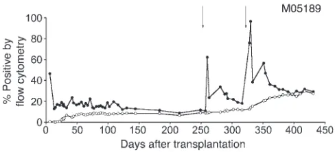
Hematopoietic recovery and long-term gene marking prior to MGMTP140K-mediated in vivo selection has been previously described
for monkeys J02043, J02370, and T04228 (14). Monkey M05189, which received cells transduced with a tricistronic lentivirus vec-tor containing an anti-HIV transgene (C46), MGMTP140K, and GFP,
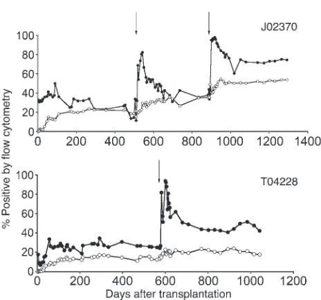
recovered complete blood counts, with normal engraftment kinetics (14) reaching an absolute neutrophil count (ANC) of more than 500 neutrophils per μ l by day 17 (24). Gene marking stabilized in granu-locytes and lymphocytes at 5.8% and 4.5%, respectively, by day 249 after transplantation and prior to initiating drug treatment on day 253 (Figure 3). Following each drug treatment, multilineage increas-es in gene marking were observed in monkeys with MGMTP140K
[image:4.585.93.493.80.388.2]and GFP (Figure 4) and a monkey with an anti-HIV transgene (C46) included in the expression cassette (Figure 3). The increase in gene marking was stable in monkey J02370 upon a follow-up of more than 14 months after the last drug treatment, which resulted in an increase in the gene marking level before drug treatment of 11.3% in granulocytes and 15.3% in lymphocytes to a gene marking level after drug treatment (2 cycles; Supplemental Table 2) of 76.9% in granu-locytes and 49.0% in lymphocytes (Figure 4). Furthermore, stable increases in gene marking were also observed in rbc and PLTs, with a gene marking level before drug treatment of 5.6% and 6.7% (14), respectively, and a gene marking level after drug treatment of 15.2%
Figure 1
Efficient MGMTP140K-mediated in vivo selection and chemoprotection in the baboon (M00228). (A) Gene marking in MGMTP140K-GFP granulocytes
(closed circles), MGMTP140K-GFP lymphocytes (open circles), YFP granulocytes (closed squares), and YFP lymphocytes (open squares), before
and after in vivo selection with either O6BG and TMZ or BCNU. (B) A truncated representation of the time following in vivo selection with O6BG and
TMZ or BCNU. Granulocytes are the only subset represented for clarity of selection (MGMTP140K-GFP [MGMT-GFP]) and elimination (YFP). (C)
Graphs are the corresponding neutrophil and PLT counts of the same baboon during the drug treatment cycles. (D) Provirus copy number data of MGMTP140K-GFP peripheral blood cells (closed circles) and YFP peripheral blood cells (open circles) of the flow cytometry data represented in A
and 64.0% (Figure 5), respectively. The EGFP signal is somewhat reduced in rbc, so the gene marking levels reported here are likely an underestimation. Finally, efficient MGMTP140K-mediated in vivo
selection was achieved using an HIV-derived lentivirus backbone, with an internal promoter of either elongation factor 1 α (EF1α) (J02043; data not shown) or spleen-focus forming virus (SFFV) (Figures 3 and 4) expressing MGMTP140K, confirming previous in
vitro selection data using similar HIV-derived lentivirus constructs and macaque CD34-selected cells (data not shown).
Efficient MGMTP140K-mediated in vivo selection in macaques following reduced-intensity busulfan conditioning. In the studies described above, we demonstrated efficient and stable MGMTP140K-mediated in vivo
selection and chemoprotection following TBI conditioning and transplantation. Since most of the current clinical gene therapy protocols for genetic diseases use a busulfan-based conditioning regimen, we tested targeted busulfan for pretransplant condi-tioning to provide sufficient myelosuppression and to facilitate engraftment of chemoprotected HSCs, while minimizing extrahe-matopoietic toxicity.
Based on previous results testing busulfan conditioning in control monkeys (Supplemental Table 3), we proceeded with a busulfan dose of 4 mg/kg on 2 consecutive days. This dose was well tolerated in the control monkey and resulted in significant neutropenia, indicating efficacy. We hypothesized that this level of myelosuppression would be sufficient to allow for engraftment of gene-modified cells. We also hypothesized that the length of neutropenia observed would be shortened as a result of the gene-modified cell infusion compared with the control animal that did not receive cells. Busulfan blood levels and clearance (Supplemen- tal Methods and Supplemental Table 3) were similar to data previ-ously reported in humans (36).
Following conditioning with busulfan and infusion of gene-modi-fied cells (approximately1.7 × 107 CD34-selected cells/kg), there was
moderate cytopenia, with an ANC of less than 500 neutrophils per
μl for 7 days, and thrombocytopenia, with a nadir of 18,000 PLTs per μl. Following stable hematopoietic recovery, we observed stable
gene marking, determined using RT-PCR, of 0.04 provirus cop-ies in peripheral blood mononuclear cells that, following a single cycle of O6BG-2X and BCNU treatment (Supplemental Table 2, see
K05079), rose to 0.16 (Figure 6) in animal K05079. Currently, gene marking has been stable for more than 9 months following drug treatment. Using this reduced-intensity conditioning approach, a high dose of gene-modified cells per kilogram for infusion (37), and posttransplant in vivo selection, we were able to achieve engraftment and selection of MGMTP140K gene-modified cells.
Polyclonal hematopoietic repopulation before and after in vivo selection. In the context of gene-modified in vivo selection following transplan-tation, either conferred intrinsically by a transgene (i.e., X-SCID) or conditionally during the presence of a biologic molecule or drug (i.e., MGMTP140K), it is important to assess the clonality of
hematopoiesis, because of the potential to restrict the clonal reper-toire as a result of in vivo selection. It is preferable to have multiple clones contributing to hematopoiesis following standard or gene-modified HSC transplantation to maintain sufficient long-term repopulating cells and clonal repertoire.
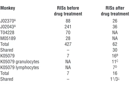
We have previously reported 235 unique lentivirus integration sites (RISs) from monkeys J02370 and J02043 prior to drug treat-ment (33). We have extended the analysis of monkeys J02370 and J02043 and included initial analysis of monkeys M05189, T04228, and K05079 prior to drug treatment and have identified 434 RISs that we believe to be unique (Table 1). Multiple RIS were identified in all monkeys prior to drug treatment; thus, we next wanted to deter- mine whether multiple clones contributed to hematopoiesis follow-ing drug treatment, as we have done before in the dog model (38).
In a limited RIS analysis of monkeys, J02370 (RIS analysis on day 883 after the initial transplant and 373 days after the first treat-ment with O6BG-2X and BCNU), J02043 (RIS analysis on day 1,129
after the initial transplant and 837 days after the second treatment with O6BG-2X and BCNU), and K05079 (RIS analysis on day 202
after the initial reduced-intensity transplant and 146 days after treatment with O6BG-2X and BCNU), we identified 78 sites that
[image:5.585.44.285.81.237.2]we believe to be unique. Of the 78 RISs identified in monkeys J02370, J02043, and K057079, 31 of the RISs were also identified in the initial RIS analysis prior to drug treatment (Table 1). It was clear, even in limited integration analysis following drug treatment, that multiple clones contributed to hematopoiesis in all monkeys analyzed. We wanted to extend this analysis, and considering the
Figure 2
Multilineage MGMTP140K-mediated in vivo selection. Bar graph
[image:5.585.298.542.557.668.2]repre-sentation of flow cytometry data from baboon M00228 gated on periph-eral blood cells positive for MGMT-GFP or YFP and a monoclonal anti-body for the indicated subset. White bars represent MGMT-GFP cells before drug treatment, and black bars represent MGMT-GFP cells after drug treatment (selection). Light gray bars represent YFP cells before drug treatment, and gray bars represent YFP cells after drug treatment (elimination).
Figure 3
Efficient MGMTP140K-mediated in vivo selection of cells harboring
anti-HIV transgenes in the macaque (M05189). Gene marking in granulocytes (closed circles) and lymphocytes (open circles), before and after in vivo selection of MGMTP140K-GFP genetically modified
cells in a macaque. Treatment with O6BG-2X and BCNU is denoted
research article
recurring theme between leukemic transformations in the SCID-X1 clinical trials has been provirus integration near the transcription start site (TSS) of proto-oncogenes, we focused on RIS proximity to proto-oncogene TSSs for safety evaluation. To accomplish this, we carried out integration site analysis both before and after drug treatment (Table 1 and Supplemental Tables 4–6) and mapped the closest proto-oncogene TSS relative to unique RIS and the percent-age of RISs within proto-oncogenes. We grouped unique RISs into 3 groups, before drug treatment, after drug treatment, and RISs found both before and after drug treatment (shared), and in all groups, the average distance to the closest proto-oncogene TSS was more than 1 Mb (Supplemental Tables 4–6). The percentages of RISs that mapped within proto-oncogenes from the same 3 groups were 5.2% (before drug treatment), 3.9% (after drug treatment), and 3.2% (shared). This suggests that, following MGMTP140K-mediated
in vivo selection, there is not a profound enrichment for RISs in or near proto-oncogene TSSs.
Discussion
Here, we show stable and efficient selection of hematopoietic repopulating cells in a highly relevant nonhuman primate model. We demonstrate stable multilineage selection and chemoprotec-tion in both the baboon (P. cynocephalus) and pigtailed macaque (M. nemestrina) after O6BG-2X and BCNU treatment, including
selection in PLTs and rbc, with up to 27 months of follow-up after selection. We also demonstrate efficient selection with an HIV- based lentivirus vector, following a reduced-intensity condition-ing regimen with single-agent busulfan. Before and after selection, hematopoiesis was polyclonal with both gamma and HIV-derived lentiviral vectors, as determined by linear amplification mediated– PCR (LAM-PCR) in all animals analyzed. Importantly, numerous clones were identified before and after selection, demonstrating that multiple MGMTP140K
gene-modified clones can engraft in mon- keys long-term (14, 26, 33), survive multiple rounds of in vivo selec-tion, and contribute to hematopoiesis following drug treatment. Most importantly, selection was well tolerated in all animals, with acceptable hematopoietic toxicity, transient liver transaminitis, and no pronounced extramedullary toxicity (23). Furthermore, normal hematopoiesis was confirmed before and after drug treatments by histopathology of bone marrow biopsies and bone marrow smears that demonstrated normal cellularity and no indication of malig-nant progression. These are important studies for the advancement of retrovirus-based gene therapy for clinical applications in which autologous gene-modified cells are appropriate, but in vivo selec-tion strategies will be required to elicit a therapeutic benefit or to enhance chemoprotection during cancer therapy.
For many single-gene hematopoietic diseases or infectious dis-eases, resolution of the disease will likely require high levels of gene marking (for review see ref. 15). For disorders affecting rbc (i.e., thalassemia and hemoglobinopathies) or PLTs (Wiskott-Aldrich syndrome), high engraftment levels are often required to help alleviate some of the indirect toxicities of the disease, such as iron overload and various organ pathology (39, 40). Also, high engraft-ment levels are likely required for applications using autologous cells genetically modified with anti-HIV transgenes to provide a significant portion of protected lymphocytes to limit HIV propa-gation. There are also technical issues that may reduce initial gene marking, resulting from relatively low viral titers and subsequently reduced gene transfer levels, including large complex expression cassettes (i.e., hemoglobinopathies), site-specific targeting vectors, and anti-HIV transgenes that target the HIV-derived lentivirus vec- tor system, thereby directly reducing titers. Therefore, these sce-narios will require a robust and stable in vivo selection strategy to increase gene marking to therapeutic thresholds and, ideally, a selection strategy that affects the level of HSCs, so the same system can be used in every situation instead of different systems for each application, depending on the hematopoietic lineage affected. Using both gammaretrovirus and HIV-derived lentivirus vectors, we demonstrate efficient selection of MGMTP140K gene-modified HSCs, based on long-term multilineage selection using the drug combination O6 BG-2X and BCNU, in agreement with our previ-Figure 4
Efficient MGMTP140K-mediated in vivo selection in macaques (J02370
and T04228). Gene marking in granulocytes (closed circles) and lym-phocytes (open circles), before and after in vivo selection of MGMTP140K-
GFP genetically modified cells in macaques. Treatment with O6BG-2X
[image:6.585.49.281.76.293.2]and BCNU is denoted by solid arrows.
Figure 5
[image:6.585.304.536.522.675.2]ous dog studies (23, 28). However, whereas in dogs, different drug doses are required, especially for BCNU, in monkeys, we used exactly what would be given and has been given to patients, sug-gesting our approach should readily translate to the clinic.
A recent report by Larochelle et al. suggested that only transient selection could be achieved with MGMTP140K gene-modified cells in the rhesus macaque, with pronounced pulmonary and gas-trointestinal (GI) toxicity (41), which contrasts with our studies, which resulted in substantial and stable increases in all hemato-poietic lineages analyzed, with acceptable extramedullary toxicity. The major disparities between the studies described by Larochelle et al., our previous reports employing the dog model (22, 23), and the studies described herein include the gene marking reported prior to initiating drug treatment, which, reported by Larochelle et al., was below 3% in the PB of all but one monkey and less than 10% in the remaining monkey, and the drug treatment regimens used for in vivo selection (41).
Low MGMTP140K gene marking levels (<3%) prior to drug treat-ment make in vivo selection difficult, because few gene-modified cells, contributing to hematopoietic recovery, following a pro-foundly myelosuppressive dose of cytotoxic drug could result in long durations of cytopenia, which in turn could allow for recovery of unmodified cells that were not killed following drug treatment, thereby diminishing the magnitude of in vivo selection. This is especially true if a suboptimal drug treatment regimen that does not afford an ideal selective advantage for MGMTP140K gene-modi-fied cells relative to unmodified cells is used for in vivo selection. Previously, we have shown that a fractionated regimen of O6BG and
TMZ over 3 days is not as effective as a single bolus of O6BG and
TMZ for in vivo selection in the dog (22). Larochelle et al. exclusive-ly used a 5-day fractionated regimen of O6
BG and TMZ that result-ed in only transient in vivo selection. This begs the question: what is the difference between bolus and fractionated regimens using TMZ? The relatively inefficient in vivo selection of MGMTP140K gene-modified cells, using a fractionated drug treatment regimen, is potentially due to the repair kinetics of a variety of DNA adducts fol-lowing O6BG and TMZ treatment (42–47). Importantly, in both cases
in which we efficiently increased relatively low levels of MGMTP140K gene marking in pigtailed macaques with a provirus copy number of approximately 0.04 (Figure 6), we used O6BG-2X and BCNU
without prior O6BG and TMZ treatment.
Similar to our previous studies in the dog (22), extending these nonhuman primate studies, we used a single bolus dose of O6BG
and TMZ initially in the baboon and then moved to a fractionated
dose of O6BG-2X before and after TMZ (27). Initially, we observed
transient selection of peripheral blood granulocytes, with no pro- nounced myelosuppression. This suggested that dosing was subop-timal for HSC selection; however, upon dose escalation of TMZ to levels far above what is dose limiting in humans (48), we continued to see transient granulocyte selection, without pronounced myelo-suppression. It should be noted that the lack of myelosuppression may have been misleading, as the MGMTP140K gene marking level at the time of O6BG and TMZ treatment would not have indicated
pronounced myelosuppression based on our previous dog studies (22). The YFP-only cells (unprotected) were essentially unaffected by the O6BG and TMZ treatments, suggesting that the delivery
of TMZ to the 2 baboons may not have been optimal, but with a single dose of O6BG-2X and BCNU, the YFP-only cells were almost
completely eliminated, while the MGMTP140K-GFP cells showed
robust and stable selection (26). This data, combined with previ-ous data in the nonhuman primate (41), suggest that MGMTP140K gene-modified cells are effective at preventing myelosuppression following O6BG-2X and TMZ treatment, but as of yet, an optimum
O6BG and TMZ drug regimen has not been established for in vivo
selection as previously demonstrated in the dog (22). Studies are currently ongoing to test new methods for efficient drug delivery using the new IV formulation of TMZ as well as using dacarbazine as an alternative drug that affects cell killing through the same active metabolite, MTIC (for review see ref. 49).
Regardless, unlike in the macaque studies reported by Laro-chelle et al. (41), both baboons were able to tolerate a single dose of O6BG-2X and BCNU following multiple rounds of O6BG
and TMZ. Although we have achieved promising results in both baboons and pigtailed macaques using MGMTP140K-mediated in
[image:7.585.56.265.80.175.2]vivo selection, with acceptable toxicity, we believe it is still crucial to minimize the number of drug treatment regimens, especially when applying this drug resistance strategy to increase gene-modi-fied cells to therapeutic levels for genetic or infectious diseases in which drug treatment is not the standard of care. However, this still does not address the disparity in the extramedullary toxicity seen in rhesus macaques (41). A patient population for which there
Figure 6
Efficient MGMTP140K-mediated in vivo selection in the macaque
fol-lowing reduced-intensity conditioning (K05079). Gene marking plotted is as provirus copy number from whole wbc, before and after in vivo selection of MGMTP140K-GFP genetically modified cells in a macaque.
Treatment with O6BG-2X and BCNU is denoted by a solid arrow.
Table 1
Source of integration sites analyzed
Monkey RISs before RISs after drug treatment drug treatment
J02370A 88 26
J02043A 241 36
T04228 70 NA M05189 28 NA Total 427 62 Shared – 30 K05079 7 16B
K05079 granulocytes NA 11C
K05079 lymphocytes NA 7D
Total 7 16 Shared – 1↑/3↓
To our knowledge, all RISs discussed in this table are unique. AWe
have previously reported 235 unique RISs (33). BRISs identified from
bulk DNA, lymphocyte-sorted DNA, and granulocyte-sorted DNA.
CRISs identified from granulocyte-sorted DNA. DRISs identified from
lymphocyte-sorted DNA. ↑Shared RIS found before and after treatment. ↓Shared RISs after treatment in sample from bulk DNA,
[image:7.585.309.532.532.678.2]research article
is extensive data with regard to the toxicity of O6BG, BCNU, and
TMZ includes patients with various types of brain tumors (for review see ref. 50). Although combinations of O6BG-BCNU and
O6BG-TMZ have been effective at reducing endogenous MGMT
activity in the tumor and at the same time sensitizing the previ-ously refractory tumor, O6
BG exacerbates the dose-limiting hema- topoietic toxicity of both BCNU and TMZ, while the extramedul-lary toxicity has been acceptable (51, 52). Extramedullary toxicity associated with BCNU is usually pulmonary toxicity caused by progressive fibrosis (for review see ref. 53). Although this is well documented, early onset/acute pulmonary toxicity associated with BCNU is rare and always associated with high-dose BCNU (54, 55). These findings would argue that the pronounced pulmonary and GI toxicity in the study by Larochelle et al. (41) more than likely associated with the heavy pretreatment of the monkeys with TBI and multiple rounds of 5-day fractionated O6BG-TMZ prior to
O6BG-BCNU and/or an unexpectedly high sensitivity of rhesus
macaques (Macaca mulatta) to BCNU.
Based on these findings, we believe that MGMTP140K strategies to
mediate in vivo selection for genetic and infectious diseases affect-ing the hematopoietic system or chemoprotection during cancer therapy are promising clinical approaches. We demonstrate effi-cient in vivo selection, beginning with gene marking levels as low as approximately 4%, and based on recent clinical studies (6, 7), it is reasonable to expect to routinely achieve this level of gene mark- ing using established, well-tolerated clinical conditioning regi-mens (reduced intensity and high intensity or myeloablative). The selection strategies, described here in nonhuman primates, using O6BG-2X and BCNU should be readily translatable to the clinic,
with respect to drug dosing relative to gene marking, but we also recognize that initial cell dose, conditioning regimen, gene transfer efficiency, and genetic background will all contribute to success-ful drug resistant clinical gene therapy applications. In particular, when comparing the data presented here, using reduced-intensity conditioning, to previous studies in nonhuman primates (37), the gene marking reported here prior to in vivo selection is generally higher and could be the result of an elevated dose of busulfan used for conditioning (8 mg/kg versus 4–6 mg/kg) and/or a higher cell dose per kilogram. Also, in comparison with recent successful clinical trials for CGD (6) and ALD (7), the cell dose in the monkey that received reduced-intensity conditioning in our study was also higher but generally was within 2–3 fold of the reported values. Although direct comparisons are difficult between the nonhu-man primate data and clinical data, gene-modified cell dose and an appropriate conditioning regimen are undoubtedly key com-ponents for successful gene therapy applications. With respect to the safety of the studies outlined here, our clonality studies, both before and after in vivo selection, show no obvious indication of progression toward monoclonal hematopoiesis. Although, with the limited RIS analysis, especially in the monkey with reduced-intensity conditioning, and fewer RISs identified, additional monitoring and analysis is critical to assess the long-term safety of the approach, to begin to address what impact the intensity of ablation for conditioning and in vivo selection have on engraft- ment and clonality of gene-modified cells. As with any experimen-tal treatment approach, safety is a primary concern. To this end, we have included studies with SIN HIV-derived lentivirus vectors that are widely considered to have a safer integration profile rela- tive to gammaretrovirus vectors and reduced-intensity condition-ing to minimize transplant-related toxicity. SIN lentiviral vectors
combined with a reduced-intensity conditioning regimen is a potentially promising approach for clinical studies and warrants further investigation. To continue to advance HSC gene therapy, continued research needs to focus on vector development, targeted integration approaches, and regulating transgene expression. For MGMTP140K strategies, additional work to define effective drug
regimens that allow for efficient in vivo selection, while minimiz-ing extramedullary toxicity, will be important.
Methods
Animal care and procedures. Healthy juvenile baboons (P. cynocephalus) and macaques (M. nemestrina) were housed at the University of Washington Regional Primate Research Center under conditions approved by the Ameri-can Association for the Accreditation of Laboratory Animal Care. Studies were conducted under protocols approved by the Institutional Review Board and Animal Care and Use Committees of the University of Washington.
Transplantation with gammaretrovirus-transduced baboon CD34-enriched cells. Transplantation procedures and protocols have been previously described, and TBI doses for the individual animals are listed in Supple-mental Table 1 (26, 56).
Transplantation with gammaretrovirus-transduced macaque CD34-enriched cells. Based on extensive comparisons among different vector backbones, incorporation of the SF91 design using the GRSΔ270 leader results in robust MGMTP140K expression in HSCs (57). This design has a number of
modifications in the 5′ untranslated leader sequences to remove all AUG codons and all viral coding sequences; thus, the probability of expression of unwanted peptides and the potential for homologous recombination is essentially eliminated (57–59). The backbone for the MGMTP140K
gam- maretrovirus construct (provided by D.B. Kohn, UCLA, Los Angeles, Cali-fornia, USA) was modified to eliminate all of the pol sequences from the 5′ -leader region. Four oligonucleotides were designed to amplify the delet-ed portion of the leader sequence in an asymmetric PCR reaction (SF-A, 5′ -TGTCTGCTGCAGCATCGTTCTGTGTTGTCTCTGTCTGACTGT-GTTTCTGTATTTGTCTGA-3′; SF-B, 5′ -TGGAGAGAGGGACTAT-TACTTAACTTTGTCGAGCTAATTTTCAGACAAATACAGAAACAC-3′; SF-C, 5′ -AGTAATAGTCCCTCTCTCCAAGCTCACTTACAGGCGGCC-GCCGCCATGGAAGCTTGGG-3′; and SF-D, 5′ -CCCAAGCTTCATG-GCGGCG-3′) (60). The modified leader sequence was cloned into the MND backbone using standard molecular biology techniques, yielding the construct pMND.GRS.P140K.
To create a stable producing line, Phoenix-GALV cells were plated at 1 × 10⁶
cells per well in 6-well tissue culture–treated cluster plates in DMEM sup-plemented with 10% fetal bovine serum and 1% penicillin/streptomycin. Approximately 24 hours after plating, cells were transfected in quadrupli-cate with the plasmid pMND.GRS.P140K and the blasticidin resistance plasmid pBS.br2, using standard calcium phosphate techniques. The gam-maretrovirus plasmid was in either a 20:1 or 100:1 molar excess to pBS. br2, to favor stable gammaretrovirus insertion in blasticidin resistant colo-nies. Three days after transfection, quadruplicate wells were trypsinized and combined into 3 different 10-cm plates, at increasing cell densities of 1 × 10⁴, 1 × 10⁵, and 1 × 10⁶ cells per plate. The following day, cells began
catalog MC-380; Kamiya Company). Clones that stained positive relative to control cells were thawed and tested again on HT1080 cells to deter-mine the highest titer line. The clone PhGALV-MND.GRS.P140K c22 was determined to be the highest titer line and was subsequently frozen down in several aliquots for master cell bank preparation.
Transplantation procedures and protocols have been previously described (26, 56), with the following modifications. First, CD34-selected cells were transduced as previously described (14), with a neat virus containing media (VCM) preload and 2 successive virus exposures with neat VCM at an MOI of 0.5 and 0.5. Second, in preparation for transplantation, the macaque received TBI, 900 cGy, administered from a linear accelerator at 7 cGy/min, as 2 equally divided doses 24 hours apart. After the irradiation, on the sec-ond day, approximately 2.5 × 10⁷ PhGALV-MND.GRS.P140K transduced
cells/kg were infused through an inserted central vein catheter.
Transplantation with HIV-derived lentivirus-transduced macaque CD34-enriched cells. Transplantation procedures and protocols have been previously described (14), with a few modifications explained below.
The vector developed for anti-HIV strategies (RSC-SC46-ires-MPGw2) was a SIN HIV-derived lentivirus vector based on the RRL system (61) (provided by Didier Trono, École Polytechnique Fédérale de Lausanne, Lausanne, Switzerland) with a backbone containing a central polypurine tract and a woodchuck posttranscriptional regulatory element. The internal promoters were a SFFV promoter expressing the membrane-anchored C-peptide (C46) derived from HIV-1 envelope glycoprotein gp41 (62) linked by an internal ribosome entry site (ires) to MGMTP140K
and a phosphoglycerate kinase pro-moter driving expression of EGFP for monkey M05189 (24). The vector was pseudotyped with VSV-G envelope, produced by transient transfection of 293T cells, and concentrated 100 fold as previously described (63).
The CD34-selected cells were transduced as previously described (26), with 2 successive virus exposures at an MOI of 5 and 4.3. In preparation for transplantation, the macaque received TBI, 900 cGy, administered from a linear accelerator at 7 cGy/min, as 2 equally divided doses 24 hours apart. After the irradiation, on the second day, approximately 2.3 × 10⁷ total
cells/kg or approximately 1.1 × 10⁷ RSC-SC46-ires-MPGw2–transduced
cells/kg (adjusted based on GFP+ gene marking, approximately 49.6%,
by flow cytometry of in vitro maintained cells) were infused through an inserted central vein catheter.
Reduced-intensity conditioning and transplantation with HIV-derived lenti-virus-transduced macaque CD34-enriched cells. Busulfan was administered to monkey K05079 intravenously over 2 hours (Supplemental Table 3). Gene-modified cells were infused approximately 24 hours after the second dose of busulfan, following our standard lentivirus transduction protocol (14). To avoid any potential immune response to MGMTP140K
gene-modi-fied cells, tacrolimus was administered orally twice a day (from day 2 to day 221) following transplantation, and the dose of tacrolimus was adjusted to between 10–15 ng/ml, based on serum concentrations.
Flow cytometric analysis of baboon and macaque hematopoietic cells. Leukocytes, isolated by ammonium chloride red cell lysis from heparinized peripheral blood (PBL) and bone marrow (BML) samples, drawn at multiple time points after transplantation, were analyzed for GFP/YFP-expression on a FACSVantage or FACSCalibur (Becton Dickinson). These PBLs and BMLs were identified out of at least 20,000 events (1 μg/ml propidium iodide, excluding forward and right-angle light scatter-gated). Flow cytometric data were analyzed by FlowJo version 8.5.2 to version 8.8.6 (Tree Star Inc.) or CELLQuest version 3.1f software (Becton Dickinson), with gating to exclude fewer than 0.1% control cells in the relevant region.
FACS sorting of hematopoietic subsets. Hemolyzed blood or bone marrow was used as starting material for wbc analysis. wbc were counted and labeled with antibodies specific to CD20 (clone 2H7, Becton Dickinson), CD13 (clone L138, Becton Dickinson), and CD34 (clone 563, Becton Dickinson)
in separate tubes. The cells were washed once before analysis on a FACSAria (Becton Dickinson). Cells were gated using propidium iodide exclusion (as described above); forward and right-angle light scatter was used to dis-tinguish between bulk lymphocytes, granulocytes, and monocytes. Cell lineage gates were further separated by 2-color flow cytometry, using PE-labeled antibodies to the surface markers for CD20+ lymphocytes, CD13+
monocytes or granulocytes, and CD34+ bone marrow cells.
Quantitative PCR (TaqMan). Gene marking was also analyzed by TaqMan 5′ nuclease quantitative RT-PCR assay. Sample DNA was analyzed in duplicate with a GFP-specific primer/probe combination (5′ -CTGCACCACCGGCAA-3′; 5′-GTAGCGGCTGAAGCACTG-3′ [Integrated DNA Technologies]; probe, 5′-FAM-CCACCCTGACCTACGGCCTG-TAMRA-3′ [Applied Bio-systems]), a YFP-specific primer/probe combination (5′ -CTGCACCACCG-GCAA-3′; 5′-GTAGCGGGCGAAGCACT-3′ [Integrated DNA Technologies]; probe, 5′-FAM-CCACCTTCGGCTACGGCCTG-TAMRA-3′ [Applied Biosys-tems]), or a lentivirus-specific primer/probe combination (5′ -TGAAAGC-GAAAGGGAAACCA-3′; 5′-CCGTGCGCGCTTCAG-3′ [Integrated DNA Technologies]; probe, 5′ -FAM-AGCTCTCTCGACGCAGGACTCGGC-TAMRA-3′ [Applied Biosystems]). In a separate reaction, a β -globin–spe-cific primer/probe combination (5′-CCTATCAGAAAGTGGTGGCTGG-3′; 5′-TTGGACAGCAAGAAAGTGAGCTT-3′ [Integrated DNA Technologies]; probe, 5′-FAM-TGGCTAATGCCCTGGCCCACAAGTA-TAMRA-3′ [Applied Biosystems]) was used to adjust for equal loading of genomic DNA per reac- tion. Standards consisted of dilutions of DNA extracted from cell lines trans- duced with a single copy of a GFP-, YFP-, or lentivirus-containing retrovi-rus vector and DNA from the same unmodified cell line. The approximate percentage of genetically modified cells can be calculated by making the assumption that each genetically modified clone we analyzed by PCR has a single copy, meaning that the fraction we report as 0.04 provirus copies can also be reported as a percentage, i.e., 4%. Reactions were run by means of the ABI Master Mix (Applied Biosystems) on the ABI Prism 7500 Sequence Detection System (Applied Biosystems), under the following thermal cycling conditions: 50°C for 2 minutes and 95°C for 10 minutes, followed by 40 cycles of 95°C for 15 seconds and 60°C for 1 minute.
Drug treatment with O6BG and TMZ or BCNU. Drug treatments were
always administered under general sedation. We dissolved 50 mg O6BG
(Sigma-Aldrich) in 30 ml of 40% polyethylene glycol in PBS, and the con-centration was adjusted to 1 mg/kg with prewarmed (37°C) PBS. The drug was further diluted in normal saline to a final volume of approximately 150–200 ml and was infused over 15–20 minutes. The dose of O6BG for
both baboons and macaques was 120 mg/m2. After initial drug treatment
regimens were tested in baboons M01044 and M00228 (see Supplemen-tal Table 2), a second identical dose of O6
BG was administered approxi-mately 7–8 hours after the end of TMZ or BCNU administration (27). TMZ capsules (Temodar, Schering-Plough) were crushed and mixed with cranberry juice (pH < 4) and administered via an oral gavage within 5 min-utes after completion of the O6BG infusion. BCNU (Bristol-Myers Squibb
and Sigma-Aldrich) was prepared as previously described (23) and admin-istered 45–60 minutes after the end of the first O6BG infusion. Whole
blood cell counts and blood chemistry values (including liver and kidney functions) were monitored on the day of drug treatment and twice a week thereafter until values returned to pretreatment levels.
LAM-PCR of RISs. Retroviral integration site analysis by LAM-PCR was performed on primate DNA isolated from PBLs. One hundred nano-grams of DNA served as template for LAM-PCR, which was performed as described previously (33) or with a modified approach that was described elsewhere (31, 64).