High-mobility group box 1 is involved in the
initial events of early loss of transplanted islets
in mice
Nobuhide Matsuoka, … , Masaru Taniguchi, Yohichi
Yasunami
J Clin Invest. 2010;
120(3)
:735-743.
https://doi.org/10.1172/JCI41360
.
Islet transplantation for the treatment of type 1 diabetes mellitus is limited in its clinical
application mainly due to early loss of the transplanted islets, resulting in low
transplantation efficiency. NKT cell–dependent IFN-
g
production by Gr-1
+CD11b
+cells is
essential for this loss, but the upstream events in the process remain undetermined. Here,
we have demonstrated that high-mobility group box 1 (HMGB1) plays a crucial role in the
initial events of early loss of transplanted islets in a mouse model of diabetes. Pancreatic
islets contained abundant HMGB1, which was released into the circulation soon after islet
transplantation into the liver. Treatment with an HMGB1-specific antibody prevented the
early islet graft loss and inhibited IFN-
g
production by NKT cells and Gr-1
+CD11b
+cells.
Moreover, mice lacking either of the known HMGB1 receptors TLR2 or receptor for
advanced glycation end products (RAGE), but not the known HMGB1 receptor TLR4, failed
to exhibit early islet graft loss. Mechanistically, HMGB1 stimulated hepatic mononuclear
cells (MNCs) in vivo and in vitro; in particular, it upregulated CD40 expression and
enhanced IL-12 production by DCs, leading to NKT cell activation and subsequent NKT
cell–dependent augmented IFN-
g
production by Gr-1
+CD11b
+cells. Thus, treatment with
either IL-12– or CD40L-specific antibody prevented the early islet graft loss. These findings
indicate that the HMGB1-mediated pathway eliciting early islet loss is a potential target […]
Research Article
Transplantation
Find the latest version:
Research article
High-mobility group box 1 is involved
in the initial events of early loss of
transplanted islets in mice
Nobuhide Matsuoka,1,2 Takeshi Itoh,1 Hiroshi Watarai,3 Etsuko Sekine-Kondo,3
Naoki Nagata,4 Kohji Okamoto,4 Toshiyuki Mera,1,2 Hiroshi Yamamoto,5
Shingo Yamada,6 Ikuro Maruyama,7 Masaru Taniguchi,3 and Yohichi Yasunami1
1Department of Regenerative Medicine and Transplantation and 2Department of Gastrointestinal Surgery, Faculty of Medicine, Fukuoka University, Japan. 3Laboratory for Immune Regulation, RIKEN Research Center for Allergy and Immunology, Yokohama, Japan. 4Department of Surgery 1, University of Occupational and Environmental Health, School of Medicine, Kitakyushu, Japan. 5Department of Biochemistry and Molecular Vascular Biology,
Kanazawa University Graduate School of Medical Science, Japan. 6Shino-Test Co., Sagamihara, Japan. 7Department of Laboratory and Vascular Medicine, Kagoshima University Graduate School of Medical and Dental Sciences, Japan.
Islet transplantation for the treatment of type 1 diabetes mellitus is limited in its clinical application mainly
due to early loss of the transplanted islets, resulting in low transplantation efficiency. NKT cell–dependent
IFN-
γ
production by Gr-1
+CD11b
+cells is essential for this loss, but the upstream events in the process remain
undetermined. Here, we have demonstrated that high-mobility group box 1 (HMGB1) plays a crucial role in
the initial events of early loss of transplanted islets in a mouse model of diabetes. Pancreatic islets contained
abundant HMGB1, which was released into the circulation soon after islet transplantation into the liver.
Treat-ment with an HMGB1-specific antibody prevented the early islet graft loss and inhibited IFN-
γ
production
by NKT cells and Gr-1
+CD11b
+cells. Moreover, mice lacking either of the known HMGB1 receptors TLR2 or
receptor for advanced glycation end products (RAGE), but not the known HMGB1 receptor TLR4, failed to
exhibit early islet graft loss. Mechanistically, HMGB1 stimulated hepatic mononuclear cells (MNCs) in vivo
and in vitro; in particular, it upregulated CD40 expression and enhanced IL-12 production by DCs, leading to
NKT cell activation and subsequent NKT cell–dependent augmented IFN-
γ
production by Gr-1
+CD11b
+cells.
Thus, treatment with either IL-12– or CD40L-specific antibody prevented the early islet graft loss. These
find-ings indicate that the HMGB1-mediated pathway eliciting early islet loss is a potential target for intervention
to improve the efficiency of islet transplantation.
Introduction
Pancreatic islet transplantation, although an attractive procedure for the treatment of type 1 diabetes mellitus, usually fails to achieve insulin independence of a diabetic recipient from a single donor due to early loss of transplanted islets and therefore requires sequential transplantations of islets with the use of 2–3 donors (1). Thus, the low efficiency of islet transplantation has been a major obstacle fac-ing islet transplantation and hampers its clinical application.
We have previously shown in mice that loss of transplanted islets soon after transplantation is caused by NKT cell–dependent IFN-γ production by Gr-1+CD11b+ cells and is successfully prevented by
treatment of NKT cells with repeated stimulation with their syn-thetic ligand, α-galactosylceramide (α-GalCer), to downregulate IFN-γ production of NKT cells, or by depletion of Gr-1+CD11b+ cells
with anti–Gr-1 antibody (2). However, precisely how it is involved in the upstream events in the activation of NKT cells and Gr-1+CD11b+
cells in the early loss of transplanted islets remains to be solved. High-mobility group box 1 (HMGB1) protein was initially found to be a DNA-binding protein present in almost all eukary-otic cells, where it stabilizes nucleosome formation and acts as a nuclear factor that enhances transcription (3, 4). Recently,
HMGB1 has been demonstrated to play crucial roles in response to tissue damage, indicating that HMGB1 is a prototype of the emerging damage-associated molecular pattern molecule (4, 5). HMGB1 is also known to be secreted by activated immune cells, including macrophages (6, 7), DCs (8), and NK cells (9) in response to infection and inflammatory stimuli. Once secreted, HMGB1 induces inflammatory responses by transduction of cel-lular signals through its receptors, such as TLR2, TLR4 (10–12), and receptor for advanced glycation end products (RAGE) (8, 13, 14). Moreover, HMGB1 levels are markedly increased during severe sepsis in humans and animals, and administration of neu- tralizing HMGB1-specific antibodies prevents lethality from sep-sis (6). Recent accumulating evidence now suggests that HMGB1 acquires or augments proinflammatory activity by binding to proinflammatory mediators such as LPS, IL-1 (14), and DNA (15–17). These observations indicate that HMGB1 is an essential mediator of organ damage; however, its precise role and mecha-nism remain unknown. Here, we investigate the mechanisms of action of HMGB1 in the early loss of transplanted islets.
Results
Involvement of HMGB1 in early loss of transplanted islets. It has previ-ously been shown that hyperglycemia of streptozotocin-induced (STZ-induced) diabetic recipient mice was ameliorated after trans-plantation of 400 syngenic islets in the liver but not of 200 islets (Figure 1A, no treatment), the number of islets isolated from a
Authorship note: Nobuhide Matsuoka, Takeshi Itoh, and Hiroshi Watarai contrib-uted equally to this work.
research article
single mouse pancreas (2). By using the diabetes model mice, we first investigated the effects of anti-HMGB1 antibody to examine whether HMGB1 is directly involved in early loss of transplanted islets. STZ-induced diabetic mice that received 200 islets together with anti-HMGB1 antibody once at the time of islet transplan-tation became normoglycemic, in contrast to mice treated with control chicken IgG (Figure 1A). The results demonstrated that
the anti-HMGB1 antibody ameliorates hyperglycemia of diabetic mice, indicating that the early loss of transplanted islets is pre-vented by anti-HMGB1. Thus, HMGB1 plays a crucial role in early loss of transplanted islets.
IFN-γ production of NKT cells and Gr-1+CD11b+ cells in the liver
[image:3.585.310.531.80.402.2] [image:3.585.58.279.81.538.2]receiv-ing islets is inhibited by anti-HMGB1 antibody. Next, we determined whether anti-HMGB1 antibody treatment has any effect on IFN-γ
Figure 1
Essential roles of HMGB1 in early loss of transplanted islets. (A) Nonfasting plasma glucose levels in STZ-induced diabetic mice received 200 syngeneic islets (top panel) and those treated with chicken anti-HMGB1 antibody or control chicken IgG. Individual lines represent glucose levels of each animal. (B) FACS profiles of liver MNCs from naive mice, STZ-induced diabetic mice that received 200 syngenic islets (Islet Tx), and islet transplanted mice treated with anti-HMGB1 antibody or with chicken IgG. NKT cells (top 2 rows) and Gr-1+CD11b+ cells (bottom 2 rows)
were analyzed for IFN-γ (second and fourth rows). The numbers in the figures represent the percentage of cells in the corresponding square areas. Representative data from 4 experiments are shown. (C) FACS profiles of NKT cells and Gr-1+CD11b+ cells after HMGB1 treatment. Liver
MNCs from wild-type or Jα18–/– mice treated with i.v. injection of saline or HMGB1 (100 μg/mouse) were isolated 2 hours after the injection and examined by flow cytometry for IFN-γ production by NKT cells and Gr-1+CD11b+ cells. The numbers in the figures represent the percentage of
research article
production by NKT cells and Gr-1+CD11b+ cells in the liver of
mice receiving islets, which are essential components of early loss of transplanted islets, as shown previously (2). For those purposes, mononuclear cells (MNCs) in the liver of recipient mice were isolat-ed at 6 hours after islet transplantation of 200 syngenic islets into the liver and examined by FACS as to IFN-γ production by NKT cells and Gr-1+CD11b+ cells in the liver. The results are in agreement
with the previous findings (2) that, within 6 hours after transplan-tation of syngeneic islets into the liver, NKT cells and Gr-1+CD11b+
cells accumulated into the liver with upregulated production of IFN-γ (Figure 1B). This upregulated production of IFN-γ after islet transplantation was inhibited by anti-HMGB1. Since the treatment with anti-HMGB1 antibody did not affect the number of infiltrated Gr-1+CD11b+ cells (Figure 1B), the recruitment of
Gr-1+CD11b+ cells was due not to HMGB1, but rather probably
to the events of transplantation itself. These findings suggest that HMGB1 is essentially involved in the activation of NKT cells and/or Gr-1+CD11b+ cells in the liver after islet transplantation.
NKT cell–dependent IFN-γ production by Gr-1+CD11b+ cells upon
stim-ulation with HMGB1. In order to confirm HMGB1-dependent IFN-γ production, we investigated whether HMGB1 has any stimulatory effects in vivo on NKT cells and/or Gr-1+CD11b+ cells in the liver
of mice (Figure 1C). For those purposes, HMGB1 was adminis-tered i.v. into naive wild-type and NKT cell–deficient Jα18–/– mice,
and their hepatic MNCs were isolated at 2 hours after the injec-tion and examined by flow cytometry. It was found that IFN-γ production was upregulated in NKT cells and Gr-1+CD11b+ cells
in the liver of wild-type mice treated with HMGB1 (Figure 1C). Importantly, the IFN-γ production by Gr-1+CD11b+ cells in the
liver of Jα18–/– mice treated with HMGB1 was not upregulated,
although accumulation of Gr-1+CD11b+ cells was similar to that
in wild-type mice (Figure 1C). These findings indicate that IFN-γ production by Gr-1+CD11b+ cells in the liver of mice treated with
HMGB1 is dependent on NKT cells.
Involvement of TLR2 and RAGE but not TLR4 in HMGB1-dependent early loss of transplanted islets. We further investigated whether HMGB1-dependent early loss of transplanted islets is dependent on TLR2, TLR4, and/or RAGE, which is known to be a potential receptor of HMGB1 (10–14). Isolated liver MNCs from wild-type mice induced augmented production of IL-12 and IFN-γ in response to HMGB1 in vitro (Figure 2A), which were greatly reduced in Tlr2–/– and Rage–/– liver
MNCs but not in Tlr4–/– liver MNCs, whose cytokine production levels
were equivalent to those of wild-type mice in response to HMGB1. To elucidate which receptor(s) for HMGB1 are actually involved in early loss of transplanted islets, STZ-induced diabetic Tlr2–/–,
Tlr4–/–, or Rage–/–
mice that received 200 syngenic islets were inves-tigated for glucose levels in the serum. Interestingly, all of Tlr2–/– or
Rage–/– mice (5 of 5) became normoglycemic, while Tlr4–/– mice
remained hyperglycemic after transplantation, indicating that TLR2 and RAGE, but not TLR4, play an essential role in the early loss of transplanted islets (Figure 2B).
Pancreatic islet cells are a major source of HMGB1, which mediates IFN-γ production by NKT cells and Gr-1+CD11b+ cells.To validate
the involvement of HMGB1 in early loss of transplanted islets, we carried out histological examination on islets before and after transplantation. HMGB1 was detected at a high level in cytoplasm as well as nucleus of transplanted islets as early as 3 hours after transplantation, while HMGB1 was stained only in the nucleus of islets in the naive pancreas and of isolated islets (Figure 3A). The results suggest that HMGB1 is localized in the nucleus of pancreatic islets, shuttled to cytoplasm, and possibly secreted into the circulation soon after transplantation.
Next, we examined the amounts of HMGB1 in isolated islets in comparison with those in other organs, including the thymus, lung, spleen, liver, and pancreas, as well as of FACS-sorted liver MNCs, including NK, NKT, T, B, Gr-1–CD11b+, and Gr-1+CD11b+
cells. Currently, there are no data available with respect to the
HMGB1 content in the different cell types, although tissue dis-Figure 2
HMGB1 receptors involved in early loss of transplanted islets. (A) In vitro cytokine production by liver MNCs. Liver MNCs (2 × 10⁶/well) isolated
[image:4.585.94.492.80.296.2]research article
tribution of HMGB1 has been reported previously (18). To our surprise, isolated islets contained high levels of HMGB1, which were 20 times more greater than in other organs or FACS-sorted cells tested (Figure 3B). The physiological roles of high concentra-tions of HMGB1 in islet cells as well as their etiology are a matter of interest and need to be clarified in future studies.
To investigate a direct relationship between HMGB1 and islet cell damage, we cultured isolated mouse islets in the absence or presence of cytotoxic proinflammatory cytokines, including IFN-γ, TNF-α, and IL-1β, which are known to induce islet cell death in vitro (19) with elevated concentrations of HMGB1 in the culture medium (20). IL-10 was used as a control. Islet cell death was assessed by fluores-cence microscopy with the use of the DNA-binding dye propidium iodide (PI) and Hoechst 33342 (HO 342) (19). PI, a highly polar dye that is impermeable to cells with preserved membranes, stains DNA
red when membranes are damaged. HO 342 freely passes the plasma membrane, readily enters cells with intact membranes, and stains DNA blue. Thus, the nuclei of dead cells stained red by PI, while those of intact cells stained blue without fragmentation and con- densation by HO 342. PI-positive islet cells were increased in num-ber with time in the presence of cytotoxic cytokines, while those in the absence of cytotoxic cytokines and in the presence of the control cytokine remained low in number (Figure 3C). In parallel, HMGB1 concentration in the islet cell culture medium increased with time in the presence of cytotoxic cytokines, while, in contrast, that in the absence of cytotoxic cytokines as well as in the presence of the con-trol cytokine remained low (Figure 3C).
[image:5.585.50.538.80.439.2]The above findings suggest that HMGB1 of transplanted islets may be released into the circulation of recipient mice in association with their damage soon after transplantation. In fact, the serum
Figure 3
research article
HMGB1 levels in the STZ-induced diabetic mice were elevated, with a peak at 24 hours, and returned to the preinjection levels by 72 hours after i.v. injection of STZ, while, after islet transplantation, HMGB1 peaked at 6 hours and returned to pretransplant levels by 7 days (Figure 3D). The findings suggest that the first peak of the serum HMGB1 elevation is due to islet cell damage caused by STZ injec-tion, which is a toxic agent to β cells of islets, while the second HMGB1 peak is due to the early loss of transplanted islets.
Cell types responsible for HMGB1-mediated cytokine production. We investigated the mechanisms of action of HMGB1 by measuring in vitro production of IFN-γ and IL-12 in the culture of isolated liver MNCs from wild-type and Jα18–/– mice in response to HMGB1,
since IFN-γ is critical in the early islet graft loss (2) and also because IL-12 is essential for IFN-γ production by NKT cells (21). Isolated liver MNCs from wild-type mice induced augmented production of IL-12 and IFN-γ in response to HMGB1 in vitro (Figure 4A). Importantly, the amount of IL-12 and IFN-γ produced by liver MNCs in NKT cell–deficient Jα18–/–mice treated with HMGB1
was greatly reduced (Figure 4A), indicating that NKT cells aug-ment HMGB1-dependent IL-12 and IFN-γ production.
We then investigated expression of Tlr2 and Rage by quantitative real-time PCR in each FACS-sorted cellular population from the liver, including NK1.1+CD3– NK, α-GalCer/CD1d dimer+ NKT,
CD3+ T, and CD19+ B cells; Gr-1+CD11b+CD11c– neutrophils
(Neu); and Gr-1–CD11b+ cells, which were further divided into
CD11c+F4/80– DCs and CD11c–F4/80+ monocytes/macrophages
(Mo/Mφ) (Supplemental Figure 1; supplemental material available online with this article; doi:10.1172/JCI41360DS1). Tlr2 and Rage were detected at high levels on Neu (Figure 4B). DCs also expressed modest levels of both Tlr2 and Rage. However, Mo/Mφ expressed modest levels of Tlr2 but low levels of Rage, while expression of either Tlr2 or Rage was barely detected in other cell populations (NK, NKT, T, and B cells) (Figure 4B).
[image:6.585.52.531.79.383.2]In order to dissect further the mechanisms of action of HMGB1, we investigated in vitro IL-12 and IFN-γ production in the culture of FACS-sorted individual cellular populations from liver MNCs in response to HMGB1. IL-12 was mainly produced from DCs rather than Neu or Mo/Mφ (Figure 4C). However, IFN-γ produc-tion was not detected in any individual cell population among all liver MNC subpopulations tested (Figure 4C).
Figure 4
NKT cell–dependent IL-12 and IFN-γ production by liver MNCs in response to HMGB1. (A) Liver MNCs (2 × 10⁶/well) isolated from wild-type
or Jα18–/– mice were cultured with the indicated doses of HMGB1 in vitro for 48 hours and measured for IL-12 and IFN-γ. Representative data from 2 experiments are shown. (B) PCR analysis on HMGB1 receptors. FACS-sorted liver MNCs (2 × 10³ for Tlr2, Rage, or Hprt) were
analyzed for mRNA levels by quantitative real-time PCR. Data were analyzed by the ΔΔCt method using the expression level in Mo/Mφ as nor-malized control. (C) Cytokine production in FACS-sorted liver MNCs upon stimulation with HMGB1. FACS-sorted cells were cultured in vitro (1 × 10⁵ cells/well) for 48 hours in the presence of HMGB1 (20 μg/ml). The amounts of IL-12 and IFN-γ were measured by CBA (n = 3). (D)
Cytokine production by DCs, Mo/Mφ, or Neu in the presence of NKT cells. FACS-sorted Gr-1–CD11b+CD11c+ DCs, Gr-1–CD11b+CD11c– Mo/Mφ,
and Gr-1+CD11b+CD11c– Neu (4 × 10⁴) were cocultured in vitro with NKT cells (2 × 10⁵) in the presence of HMGB1 (20 μg/ml) for 48 hours. The
amounts of IL-12 and IFN-γ were measured by CBA (n = 3). (E) Intracellular cytokine staining of liver MNCs after HMGB1 treatment. Liver MNCs (2 × 10⁶) were cultured with HMGB1 (20 μg/ml) for 24 hours, and the indicated cells were gated and analyzed for their production of IFN-γ by
research article
Since IL-12 was produced in vitro from DCs in response to HMGB1 (Figure 4C) and since NKT cell–dependent IFN-γ pro- duction by Neu is an essential component of early loss of trans-planted islets as shown previously (2), IL-12 and IFN-γ produc-tion of FACS-sorted DCs, Mo/Mφ, or Neu cocultured in the presence of NKT cells with addition of HMGB1 was examined. The production of IL-12 was greatly augmented in response to HMGB1, especially when DCs were cocultured with NKT cells (Figure 4D). The production of IFN-γ became evident in the culture medium of DCs cocultured with NKT cells in the pres-ence of HMGB1 (Figure 4D). The cell types responsible for the production of IFN-γ in response to HMGB1 in Figure 4D were NKT cells but not DCs, because intracellular cytokine staining revealed that NKT cells, but not DCs, produced IFN-γ (Figure 4E). It was also shown that Neu production of IFN-γ was augmented in the presence of NKT cells (Figure 4D).
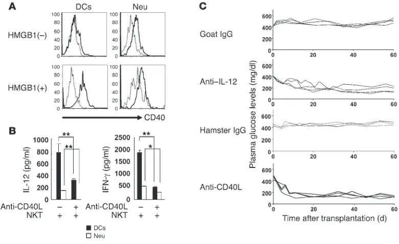
It is known that IFN-γ production by NKT cells is largely depen-dent on the interaction between CD40L expression on activated NKT cells and CD40 expression on DCs (22). Thus, we measured CD40 expression on DCs and Neu stimulated with HMGB1. CD40 surface expression was detected in both cell types in rest-ing conditions, while upregulation of CD40 was observed in DCs rather than Neu in HMGB1-treated conditions (Figure 5A). Fur-thermore, production of both IL-12 and IFN-γ mounted in vitro by HMGB1 stimulation was blocked by anti-CD40L antibody (Figure 5B), indicating that augmented IL-12 production from DCs and Neu and also IFN-γ production by NKT cells and Neu are triggered by CD40/CD40L interaction.
To confirm the data shown in Figure 5, A and B, we determined in vivo requirement of IL-12 and CD40-CD40L interaction in early loss of transplanted islets. Hyperglycemia of STZ diabetic mice receiving 200 syngenic islets in the liver was ameliorated by treatment with either anti–IL-12 or anti-CD40L antibody once at the time of islet transplantation, while that of mice treated with control antibody was not (Figure 5C). Together with the previous studies showing that the anti–IFN-γ treatment normalizes hyper-glycemia (22), the results indicate that IL-12 and CD40-CD40L interaction together with IFN-γ actually play a crucial role in vivo in early loss of transplanted islets.
Discussion
[image:7.585.98.488.82.321.2]Among the most important findings of the present study is that pancreatic islets contain abundant HMGB1 compared with other organs and individual cell populations in the liver, the site of islet transplantation. Immunohistochemical staining of the pancreas revealed that HMGB1 is mainly stained in the nucleus of islet cells but not in other cell types, while HMGB1 is detected in the circu-lation after islet cell damage. In fact, the plasma concentration of HMGB1 in wild-type mice was elevated and peaked at 24 hours after i.v. injection of STZ and returned to the preinjection level 72 hours after STZ injection. The plasma levels of HMGB1 in dia-betic recipient mice were elevated after islet transplantation with a peak at 6 hours and returned to pretransplant levels by 24 hours. These findings suggest that the first peak of the elevated HMGB1 levels is caused by destruction of islet cells by a toxic agent of STZ specific to β cells of islets and that the second peak in recipient
Figure 5
Involvement of CD40-CD40L interaction in production of IL-12 and IFN-γ in early loss of transplanted islets. (A) CD40 expression in DCs and Neu before or after treatment with HMGB1. Liver MNCs (2 × 10⁶) were treated without or with HMGB1 (20 μg/ml) for 24 hours and analyzed for CD40
expression (n = 3). (B) Requirement of CD40-CD40L interaction in the production of IL-12 and IFN-γ in the presence of NKT cells. DCs or Neu (4 × 10⁴) were cocultured in vitro with NKT cells (2 × 10⁵) in the presence of HMGB1 (20 μg/ml) for 48 hours with or without addition of anti-CD40L
research article
mice after islet transplantation is related to the damage of islet grafts soon after transplantation. Thus, combined with in vitro findings of elevated concentrations of HMGB1 in the culture medium of isolated islets in the presence of cytotoxic cytokines, the plasma levels of HMGB1 may reflect the degree of islet dam- age in the liver after transplantation. Furthermore, the treat-ment with anti-HMGB1 antibody delayed the onset of diabetes in NOD mice, suggesting that HMGB1 plays a significant role in disease progression (23). The above findings prompted us to determine whether HMGB1 is involved in the early loss of transplanted islets, which occurs within 6 hours after islet transplantation and is an event caused by inflam-matory cytokines, as we previously reported (2, 23). In fact, the hyperglycemia of islet-transplanted diabetic mice was ameliorated by treatment with anti-HMGB1 antibody, indicating that HMGB1 is essentially involved in the early loss of transplanted islets. Concerning the mechanisms of HMGB1-mediated early islet graft loss, 3 cell types, NKT cells, Gr-1–CD11b+CD11c+F4/80– DCs,and Gr-1+CD11b+ Neu, were found to be involved in the initial
phase of early loss of transplanted islets. Among these cell types, the primary cellular targets of HMGB1 does not seem to be NKT cells, since the receptors for HMGB1 (10–14) TLR2 and RAGE but not TLR4 (Figure 2B) are expressed on DCs, Mo/Mφ, and Neu, but not on NKT cells (Figure 4B). IL-12, which is essential for NKT cell–dependent production of IFN-γ, was mainly produced by DCs after HMGB1 stimulation (Figure 4C). Thus, it is likely that the first target for HMGB1 is DCs, which in turn activate NKT cells. Then, activated NKT cells themselves produce IFN-γ and also stimulate Neu to produce IFN-γ (Figure 4D), which is an essential component of HMGB1-mediated early loss of transplanted islets.
Thus, the present study unveils a role of DCs in HMGB1-dependent IFN-γ production by NKT cells. DCs stimulated with HMGB1 in vitro upregulate their CD40 expression and produce IL-12, which is markedly augmented in the presence of NKT cells, facilitating IFN-γ production by NKT cells and subsequently that of Neu. The requirement of CD40-CD40L interaction and IL-12 is confirmed by the fact that anti-CD40L and anti–IL-12 antibod- ies prevented early loss of transplanted islets, leading to amelio-ration of hyperglycemia of islet-transplanted diabetic recipient mice, while the corresponding control antibody did not. Thus, the uncovered pathways involved in the early loss of transplanted islets in the present study afford further new targets for interven-tion to improve the efficiency of islet transplantation. TLR2, TLR4, and RAGE as potential receptors for HMGB1 (10–14) are expressed mainly on DCs, Mo/Mφ , and Neu (Figure 4B). How-ever, in vitro and in vivo experiments revealed that TLR2 and RAGE but not TLR4 are involved in the early loss of transplanted islets (Figure 2B). It has been reported that HMGB1-mediated biological effects and usage of their receptors are different in the experimen-tal models. For example, TLR4, but not TLR2 or RAGE, has been shown to be an HMGB1 receptor in hepatic reperfusion injury (24). Similarly, HMGB1 signaling through TLR2 and TLR4 but not RAGE contributes to LPS-induced inflammation (11). In the case of SLE, HMGB1 present in DNA-containing immune com-plexes triggers activation of autoreactive B cells and plasmacytoid DCs through RAGE (17). These differences in HMGB1-mediated effects might be due to the presence, in different systems, of cell types with distinctly different HMGB1 receptor expression pro-files, and also due to the formation of complexes of HMGB1 with different molecules under varying disease conditions. Concerning the form of HMGB1, HMGB1 acquires and/or aug-ments inflammatory effects when it binds to other inflammatory molecules, such as IL-1β, the TLR4 ligand LPS, the TLR9 ligand CpG-ODN, or the TLR1-TLR2 ligand Pam3CSK (14–17). Recent studies on HMGB1-deficient mice also showed that HMGB pro- teins function as universal sentinels for nucleic acids (25). How-ever, in the present studies, it still remains unsolved what types of molecules interact with HMGB1 protein to mediate its function. Chen et al. (26) have reported that the direct effects of RAGE on conventional T cell functions resulted in the prolongation of syn-geneic and allogeneic islet graft transplanted in the subcapsular space of kidney, in that anti-CD3/CD28–induced T cell prolifera-tion, mixed lymphocyte reaction, and T cell production of IL-10, IL-5, and TNF-α but not IFN-γ were inhibited in RAGE-deficient mice and mice receiving RAGE inhibitor. Since no conventional T cells were involved in the early loss of islet transplanted in the liver, and also because IFN-γ, but not IL-10 nor IL-5, is a major player in the early islet loss, the mechanisms observed in the present studies are different from those described by Chen et al. (26). Concerning the potential sites for islet transplantation — includ-ing the liver, renal subcapsular space, omental pouch, abdominal cavity, intramuscular site, subcutaneum — the liver is currently the only site where insulin independence in patients with type 1 diabe-tes mellitus can be achieved with clinical islet transplantation, as reported by Shapiro et al. (1). Although we do not have any data on islet transplantation at non–NKT cell–dense sites, the NKT cell– mediated early loss of islets can occur at any tissue, as it has been demonstrated in the allogeneic heart transplantation model that NKT cells migrate immediately into non–NKT cell–dense trans- plantation sites, where CXCL16, the ligand for chemokine recep-tor CXCR6 selectively expressed on NKT cells, is expressed (27). Taken collectively, the findings in the present study shed light on the mechanisms involved in the early loss of transplanted islets as follows. First, islet cells themselves are a major source of HMGB1, which is released from transplanted islets. Since the plasma levels of HMGB1 reflect the degree of islet damage, HMGB1 could be a marker to predict rejection of transplanted islets. Second, HMGB1 stimulates production of inflammatory cytokines including IL-12 and IFN-γ in concert with DCs, NKT cells, and Neu in the liver receiving islets. Third, these inflammatory cytokines accelerated the injuries of transplanted islets. Thus, a vicious cycle harmful to transplanted islets is now unveiled. Therefore, each pathway involved in the early loss of transplanted islets revealed by the present study is a potential target for intervention to improve effi-ciency of islet transplantation. Methods
Mice. C57BL/6 mice were purchased from Charles River Japan Inc. or CLEA Japan Inc. Jα18-deficient mice were generated previously (28) and backcrossed more than10 times to C57BL/6 mice. Rage–/– mice (29) were described previously. Tlr2–/– and Tlr4–/– mice were provided by Shizuo Akira (Osaka University, Osaka, Japan). Mice were kept under specific patho-gen–free conditions and used at 8–16 weeks of age. All experiments were in accordance with protocols approved by the Animal Care and Use Commit-tee of Fukuoka University and RIKEN.
research article
Immunohistochemistry. The pancreas of naive mice, isolated islets, and the liver of transplant recipients were fixed in 10% formaldehyde solution, processed, and embedded in paraffin. The sections were stained immuno-histochemically with anti-mouse insulin antibody (Novocastra) and rabbit anti-bovine HMGB1 antibody (Shino-Test Co.) by a streptavidin-biotin-peroxidase complex method (33).
HMGB1 and cytokine measurement. HMGB1 levels in mouse serum and in the culture medium of isolated islets was measured with an ELISA kit (Shino-Test Co.) (34). IFN-γ concentrations in the culture supernatant of liver MNCs were determined by FACS with cytometric beads assay (CBA) (BD Biosciences). IL-12 concentration in the medium was mea-sured by ELISA (Endogen).
For measurement of tissue concentration of HMGB1, individual tis- sues (1–2 mg wet weight/organ), isolated islets (200 total), and FACS-sorted cells (2 × 10⁵ to 6 × 10⁵) of each population in the liver of mice
were sonicated in PBS. Then, the resulting tissues were treated as report-ed by Sanders (35) in which perchloric acid (HClO4) was added to the
homogenates with a concentration of 0.75 M. The content of HMGB1 in the solution was measured with ELISA after the adjustment of pH to 7.0 as well as the appropriate dilution with PBS containing 1% bovine calf serum. The sonicated tissues were also used for measuring DNA content with a Wako assay kit.
Reagents. Bovine HMGB1 was purchased from Shino-Test Co. Bovine HMGB1 was extracted from the bovine thymus and further purified by CM-Sephadex C25 ion column chromatography according to the method described by Sanders (35). The biological activity of purified HMGB1 was reported elsewhere (36). Anti-HMGB1 antibody was pur-chased from Shino-Test Co. This is a polyclonal antibody made by immunizing chicken with purified bovine HMGB1, and the neutral-izing effect of the anti-HMGB1 antibody was reported previously (37, 38). Goat anti-mouse IL-12 antibodies and rabbit anti-mouse CD40L antibody were purchased from BD Biosciences and Sigma-Aldrich, respectively. Recombinant mouse IFN-γ, TNF-α, IL-1β, and IL-10 were purchased from Sigma-Aldrich.
Flow cytometry. Antibodies used for flow cytometric analysis were as fol-lows: anti-mouse FcRγII/III (2.4G2), FITC- or Pacific blue–conjugated anti-CD3ε (145-2C11), FITC- or PerCP-Cy5.5-conjugated anti-CD11b (M1/70), allophycocyanin-conjugated (APC-conjugated) anti–IFN-γ (XMG1.2), peridinin–chlorophyll protein– (PerCP-) or FITC-conjugated anti-Gr-1 (RB6-8C5), PE-conjugated NK1.1 (PK136), PE-Cy7–conjugat-ed CD19 (1D3), biotinylated anti-CD11c (N418), APC-Cy7–conjugated avidin, APC-conjugated anti-F4/80 (BM8) (BD Biosciences or eBiosci-ence). PE- or APC-labeled α-GalCer–loaded CD1d dimer was prepared as described (39). Intracellular cytokine staining was performed as previ-ously described (39). Flow cytometry was performed using a FACSCalibur and FACSAria (BD) with FlowJo software (Tree Star). The purity of sorted cells was usually greater than 99%.
Cell preparation and culture. Liver MNCs were prepared as described pre-viously (40). For in vitro culture, liver MNCs and those of FACS-sorted cells were cultured in RPMI medium (Sigma-Aldrich) supplemented with 5% fetal bovine serum (Biosource) and 100 μg/ml kanamycin (Meiji Seika) and isolated islets in DMEM medium (Nissui) supplemented with 2% BSA (Sigma-Aldrich) and 100 μg/ml kanamysin in a CO2 incubator (95% air
plus 5% CO2) at 24°C or 37°C.
Quantitative real-time PCR. Total RNA was isolated from FACS-purified cell populations using TRIzol reagent (Invitrogen). cDNA was prepared by Superscript III RNase H–
Reverse Transcriptase with random hexam-ers (Invitrogen). Quantitative real-time PCR was performed with SYBR GreenER qPCR SuperMix (Invitrogen) for ABI PRISM 7900HT (Applied Biosystems). Total mRNA from cells (2 × 10³) was used as templates to
analyze expression levels of Tlr2, Rage, or Hprt. Gene-specific primer sequences were as follows: Tlr2-fw, GGGGCTTCACTTCTCTGCTT, Tlr2-rv,
AGCATCCTCTGAGATTTGACG; Rage-fw, 5′
-GTGTCGGGCAAC-TAACAGG-3′, Rage-rv, 5′-CTGGCTTCCCAGGAATCTG-3′; Hprt-fw, 5′ -TCCTCCTCAGACCGCTTTT-3′, Hprt-rv, 5′ -CCTGGTTCATCATCGCTA-ATC-3′. Quantitative analysis was performed by the ΔΔCt method by using Hprt as an internal control.
Statistics. The statistical significance of differences was determined by 1-tailed Student’s t test. Values were expressed as mean ± SD from inde-pendent experiments. Any difference with a P value less than 0.05 was considered significant. Acknowledgments We are grateful to Peter Burrows for helpful comments and con- structive criticisms in the preparation of the manuscript. Techni-cal support by Yuko Ueda (Department of Surgery 1, University of Occupational and Environmental Health, School of Medicine) is greatly appreciated. This work was supported by a grant from the Global FU Program of Fukuoka University (Y. Yasunami), a Health Science Research Grant from the Ministry of Health, Labor and Welfare, Japan (Y. Yasunami), and the Shimura Memo-rial Foundation (Y. Yasunami). Received for publication October 6, 2009, and accepted in revised form December 9, 2009. Address correspondence to: Masaru Taniguchi, Laboratory for Immune Regulation, RIKEN Research Center for Allergy and Immunology, Yokohama, Kanagawa 230-0045, Japan. Phone: 81.45.503.7001; Fax: 81.45.503.7003; E-mail: [email protected]. Or to: Yohichi Yasunami, Department of Regenerative Medicine and Transplantation, Faculty of Medicine Fukuoka University, Fukuoka, Fukuoka 814-0180, Japan. Phone: 81.92.801.1011 ext. 3630; Fax: 81.92.801.1019; E-mail: [email protected]. 1. Shapiro AM, et al. Islet transplantation in seven patients with type 1 diabetes mellitus using a glucocorticoid-free immunosuppressive regimen.
N Engl J Med. 2000;343(4):230–238.
2. Yasunami Y, et al. Vα14 NKT cell-triggered IFN-γ production by Gr-1+CD11b+ cells mediates early
graft loss of syngeneic transplanted islets. J Exp Med. 2005;202(7):913–918.
3. Muller S, Ronfani L, Bianchi ME. Regulated expres-sion and subcellular localization of HMGB1, a chromatin protein with a cytokine function. J Inter-nal Med. 2004;255(3):332–343.
4. Seong SY, Matzinger P. Hydrophobicity: an ancient damage-associated molecular pattern that initi-ates innate immune responses. Nat Rev Immunol. 2004;4(6):469–478.
5. Lotze MT, Tracey KJ. High-mobility group box 1 protein (HMGB1): nuclear weapon in the immune arsenal. Nat Rev Immunol. 2005;5(4):331–342. 6. Wang H, et al. HMG-1 as a late mediator of endotoxin
lethality in mice. Science. 1999;285(5425):248–251. 7. Gardella S, et al. The nuclear protein HMGB1
is secreted by monocytes via a non-classical, vesicle-mediated secretory pathway. EMBO Rep. 2002;3(10):995–1001.
8. Dumitriu IE, et al. Release of high mobility group box 1 by dendritic cells controls T cell activation via the receptor for advanced glycation end products.
J Immunol. 2005;174(12):7506–7515.
9. Semino C, Angelini G, Poggi A, Rubartelli A. NK/ iDC interaction results in IL-18 secretion by DCs at the synaptic cleft followed by NK cell activation and release of the DC maturation factor HMGB1. Blood. 2005;106(2):609–616. 10. Park JS, et al. Involvement of Toll-like receptors 2 and 4 in cellular activation by high mobility group box 1 protein. J Biol Chem. 2004;279(9):7370–7377. 11. Park JS, et al. High mobility group box 1 protein
interacts with multiple Toll-like receptors. Am J Physiol Cell Physiol. 2006;290(3):C917–C924.
12. Scaffidi P, Misteli T, Bianchi ME. Release of chro-matin protein HMGB1 by necrotic cells triggers inflammation. Nature. 2002;418(6894):191–195.
research article
1995;270(43):25752–25761.
14. Kokkola R, et al. RAGE is the major receptor for the proinflammatory activity of HMGB1 in rodent macrophages. Scand J Immunol. 2005;61(1):1–9.
15. Sha Y, Zmijewski J, Xu Z, Abraham E. HMGB1 devel-ops enhanced proinflammatory activity by binding to cytokines. J Immunol. 2008;180(4):2531–2537.
16. Tian J, et al. Toll-like receptor 9-dependent acti-vation by DNA-containing immune complexes is mediated by HMGB1 and RAGE. Nat Immunol. 2007;8(5):487–496.
17. Urbonaviciute V, et al. Induction of inflammatory and immune responses by HMGB1-nucleosome complexes: implications for the pathogenesis of SLE.
J Exp Med. 2008;205(13):3007–3018.
18. Mosevitsky MI, Novitskaya VA, Logannsen MG, Zebezhinsky MA. Tissue specificity of nucleo-cyto- plasmic distribution of HMGl and HMG2 pro-teins and their probable functions. Eur J Biochem. 1989;185(2):303–310.
19. Saldeen J. Cytokines induce both necrosis and apoptosis via a common Bcl-2-inhibitable path-way in rat insulin-producing cells. Endocrinology. 2000;141(6):2003–2010.
20. Steer SA, Scarim AL, Chambers KT, Corbett JA. Interleukin-1 stimulates β cell necrosis and release of the immunological adjuvant HMGB1.
PloS Med. 2006;3(2):e17. doi:10.1371/journal. pmed.0030017.
21. Tomura M, et al. A novel function of Vα14+CD4+
NKT cells: stimulation of IL-12 production by anti-gen-presenting cells in the innate immune system.
J Immunol. 1999;163(1):93–101.
22. Brigl M, Bry L, Kent SC, Gumperz JE, Brenner MB. Mechanism of CD1d-restricted natural killer T cell
activation during microbial infection. Nat Immunol. 2003;4(12):1230–1237.
23. Satoh M, et al. Successful islet transplantation to two recipients from a single donor by targeting pro-inflammatory cytokines in mice. Transplantation. 2007;83(8):1085–1092.
24. Tsung A, et al. HMGB1 release induced by liver ischemia involves Toll-like receptor 4-dependent reactive oxygen species production and calcium-mediated signaling. J Exp Med. 2007;204(12):2913–2923.
25. Yanai H, et al. HMGB proteins function as universal sentinels for nucleic-acid-mediated innate immune responses. Nature. 2009;462(7269):99–103.
26. Chen Y, et al. RAGE ligation affects T cell activa-tion and controls T cell differentiation. J Immunol. 2008;181(6):4272–4278.
27. Jiang X, et al. Cutting edge: critical role of CXCL16/ CXCR6 in NKT cell trafficking in allograft tolerance.
J Immunol. 2005;175(4):2051–2055.
28. Cui J, et al. Requirement for Vα14 NKT cells in IL-12-mediated rejection of tumors. Science. 1997;278(5343):1623–1626.
29. Myint KM, et al. Effects of RAGE gene disruption and administration of low–molecular weight heparin.
Diabetes. 2006;55(9):2510–2522.
30. Sutton R, Peters M, McShane P, Gray DW, Mor- ris PJ. Isolation of rat pancreatic islets by duc-tal injection of collagenase. Transplantation. 1986;42(6):689–691.
31. Okeda T, Ono J, Takaki R, Todo S. Simple meth-od for the collection of pancreatic islets by the use of Ficoll-Conray gradient. Endocrinol Jpn. 1979;26(4):495–499.
32. Kemp CB, Knight MJ, Scharp DW, Lacy PE, Ball-inger WF. Transplantation of isolated pancreatic islets into the portal vein of diabetic rats. Nature. 1973;244(5410):447.
33. Shibao K, et al. Enhanced coexpression of YB-1 and DNA topoisomerase IIα genes in human colorectal carcinomas. Int J Cancer. 1999;83(6):732–737. 34. Yamada S, Yakabe K, Ishii J, Imaizumi H, Maruyama
I. New high mobility group box 1 assay system. Clin Chim Acta. 2006;372(1–2):173–178.
35. Sanders O. A method for the fractionation of the high-mobility-group non-histon chromo-somal proteins. Biochem Biophys Res Commun. 1977;78(3):1034–1042.
36. Ito T, et al. High-mobility group box 1 protein pro-motes development of microvascular thrombosis in rats. J Thromb Haemost. 2007;5(1):109–116. 37. Abeyama K, et al. The N-terminal domain of
thrombomodulin sequesters high-mobility group-B1 protein, a novel antiinflammatory mechanism.
J Clin Invest. 2005;115(5):1267–1274.
38. Ueno H, et al. Contributions of high mobil- ity group box protein in experimental and clini-cal acute lung injury. Am J Respir Crit Care Med. 2004;170(12):1310–1316.
39. Watarai H, Nakagawa R, Omori-Miyake M, Dasht- soodol N, Taniguchi M. Methods for detection, iso-lation and culture of mouse and human invariant NKT cells. Nat Protoc. 2008;3(1):70–78.
40. Ohtsuka K, et al. Expansion of intermediate T cell receptor cells expressing IL-2Rα-β+, CD8α+β+, and
lymphocyte function-associated antigen-1+ in the
liver in association with intrahepatic islet xenograft rejection from rat to mouse: prevention of rejection with anti-IL-2Rβ monoclonal antibody treatment.