Usability engineering in the design and evaluation of a
functional electrical stimulation system for upper limb
rehabilitation.
CHRISTINE SMITH
School of Health Sciences,
University of Salford, Salford, UK
Table of Contents
ACKNOWLEDGEMENTS ... 15
GLOSSARY OF TERMS ... 16
ABSTRACT ... 17
1 CHAPTER ONE:INTRODUCTION ... 19
1.1 Overview of the thesis ... 19
1.1.1 Chapter Two ... 19
1.1.2 Chapter Three... 20
1.1.3 Chapter Four... 20
1.1.4 Chapter Five ... 20
1.1.5 Chapter Six ... 20
1.1.6 Chapter Seven ... 21
1.1.7 Chapter Eight ... 21
2 CHAPTER 2:LITERATURE REVIEW ... 22
2.1 The upper limb following stroke ... 22
2.1.1 Incidence and prevalence of upper limb impairments and functional limitations after stroke ... 22
2.1.2 Impact on quality of life ... 23
2.1.3 The recovery process following a stroke ... 24
2.2 Basic science studies ... 25
2.2.1 Timing of interventions ... 25
2.2.2 Intensity and scheduling of practice ... 26
2.2.3 Content and progression of training ... 29
2.2.4 Feedback on performance ... 31
3
2.3 Functional Electrical Stimulation (FES) ... 35
2.3.1 Basic science. What is FES? ... 35
2.3.2 A review of the efficacy of upper limb FES assisted practice ... 39
2.4 FES-systems for upper limb rehabilitation ... 43
2.4.1 Review of current commercial & research systems, including limitations on functionality ... 43
2.4.2 Limitations with existing FES systems ... 46
2.5 Use of health technology within clinical practice... 46
2.6 Adoption of rehabilitation technology ... 48
2.6.1 Perceived barriers to adoption and use in practice ... 48
2.7 Usability ... 49
2.7.1 Usability and usability engineering ... 49
2.7.2 Usability evaluation methods ... 50
2.8 A literature review of studies of ANRT that have reported on usability evaluation ... 51
2.8.1 Types and numbers of users. ... 53
2.8.2 Usability methods and tools ... 54
2.8.3 Exploitation of usability analysis to inform the design of an ANRT……… ... 57
2.8.4 Setup time for ANRT ... 57
2.9 Chapter summary and thesis aims ... 59
3 CHAPTER 3: RESEARCH THAT LED TO INCEPTION OF THE CURRENT PROJECT ………..62
3.1 Healthy Aims and the Clinical Setup Tool (CST) ... 62
3.2 The NEAT LO30 Project ... 64
3.3 The UL FES Rehab Tool ... 65
4
3.3.2 The hardware and programming environment ... 66
4 CHAPTER 4: USER INVOLVEMENT IN THE EARLY STAGES OF THE DESIGN PROCESS: IMPLEMENTATION AND ASSESSMENT OF ITS IMPACT ON THE DESIGN OF THE SOFTWARE AND GRAPHICAL USER INTERFACE (GUI). ... 69
4.1 Introduction ... 69
4.2 Phase One study ... 72
4.2.1 Methods ... 72
4.2.2 Advisory group participants ... 73
4.2.3 Data analysis ... 74
4.3 Results ... 75
4.4 Discussion of findings from the therapist advisory group meetings ... 79
4.4.1 Inputs to the design requirements... 80
4.4.2 Design Requirements ... 82
4.4.3 External Factors Affecting Adoption ... 84
4.5 Conclusion ... 84
5 CHAPTER FIVE: APPLICATION OF A USABILITY ENGINEERING APPROACH TO THE DESIGN OF THE GRAPHICAL USER INTERFACE (GUI): PHASES TWO AND THREE ... 86
5.1 Current status of the UL FES Rehab Tool ... 86
5.2 Chapter aims ... 87
5.3 Protocol for the phase two usability evaluation: software design refinement ... 87
5.3.1 Protocol aims ... 87
5.3.2 User selection and justification ... 88
5
5.3.4 Methods and procedure for evaluating usability of GUI version 1.0
………..89
5.3.5 Data analysis ... 91
5.4 Results ... 96
5.4.1 Results from stage one of the data analysis ... 96
5.4.2 The type of issue and the frequency ... 96
5.4.3 Post-test questionnaires ... 97
5.4.4 Triangulation ... 99
5.4.5 Stage two data analysis results. ... 99
5.4.6 Refined list prioritised using rating system ... 99
5.4.7 Summary of findings for version 1.0 of the GUI and usability problems that were addressed ... 101
5.4.8 Stage specific changes to the GUI ... 103
5.4.9 Rationale for progressing to next stage of testing ... 104
5.5 Phase 3: Usability engineering during the rapid prototyping phase of the full system, including hardware and development from v2.0 to v3.0 of the software. ... 104
5.5.1 Overview of the challenge and solution to state machine functioning ………104
5.5.2 Methods - Description of staged approach (healthy followed by stroke patients) ... 105
5.5.3 Usability problems identified during phase three, rapid prototyping and implemented solutions ... 106
5.6 Discussion of results, challenges and next steps... 110
5.6.1 A critical review and discussion of the usability testing in relation to the literature ... 110
5.6.2 Next steps following phase 3, the iterative design process ... 112
6
6 CHAPTER 6: DEVELOPMENT OF A TOOL TO PREDICT SETUP TIME ... 114
6.1 Introduction ... 114
6.2 Model development ... 115
6.2.1 Justification of the factors likely to influence setup time ... 115
6.2.2 Upper limb impairment ... 116
6.2.3 Task complexity ... 117
6.2.4 Components of the model that needed to be developed ... 118
6.3 Model implementation ... 123
6.3.1 Participant selection ... 124
6.3.2 Method ... 125
6.4 Results ... 127
6.4.1 Participant characteristics ... 127
6.4.2 Setup times ... 128
6.4.3 Relationship between task complexity and setup times ... 129
6.4.4 Relationship between the level of patients’ upper limb impairment and setup times. ... 130
6.4.5 A linear regression analysis for upper limb impairment and setup times……….130
6.4.6 Multiple regression analysis to predict setup time ... 131
6.5 Discussion ... 133
6.5.1 Participants’ level of upper limb impairment ... 133
6.5.2 The model ... 134
6.6 Limitations & conclusions ... 135
6.6.1 Limitations ... 135
7 7 CHAPTER 7: USABILITY AND FEASIBILITY TESTING OF THE FINAL PROTOTYPE UPPER LIMB FES REHAB TOOL, IN TWO SUB-ACUTE STROKE REHABILITATION
CENTRES. ... 137
7.1 Introduction ... 137
7.2 Summative usability evaluation of Advanced Neurological Rehabilitation Technologies in a sub-acute clinical setting ... 139
7.2.1 Studies of usability from the patients’ viewpoint only ... 139
7.2.2 Studies of usability from both patients’ and therapists viewpoint 141 7.2.3 Conclusion ... 144
7.3 Study protocol ... 145
7.3.1 Aims ... 145
7.3.2 Ethical approval ... 145
7.3.3 Identification and description of clinical sites ... 145
7.3.4 Therapist and Rehabilitation Assistant recruitment ... 146
7.3.5 Training for Therapists and Rehabilitation Assistants ... 146
7.3.6 Characterisation of Therapists and Rehabilitation Assistants ... 147
7.3.7 Patient recruitment ... 147
7.3.8 Characterisation of patients ... 149
7.3.9 Data capture ... 149
7.3.10 Procedure used during setup and practice ... 150
7.3.11 Post-session feedback ... 152
7.4 Results ... 153
7.4.1 Participant characteristics ... 153
7.4.2 Extent to which the FES Rehab Tool was used by therapists and possible explanatory factors ... 158
7.4.3 Functionality... 164
8
7.4.5 Post study data ... 172
7.5 Discussion ... 177
7.5.1 Recruitment ... 177
7.5.2 Functionality... 178
7.5.3 Usability ... 179
7.5.4 Setup time ... 182
7.6 Limitations and future work ... 184
7.7 Conclusions ... 185
8 CHAPTER 8.0: SUMMARY OF THE THESIS AND FUTURE WORK ... 187
8.1 Discussion ... 187
8.1.1 Introduction ... 187
8.2 Review of the thesis ... 187
8.2.1 Usability methods: what worked and what didn’t ... 187
8.2.2 The impact of the usability engineering approach on the final system design ... 191
8.2.3 Setup time ... 192
8.2.4 Education and training to facilitate uptake of rehabilitation technologies ... 193
8.3 Limitations ... 194
8.4 Conclusions ... 195
8.4.1 Novelty contributions ... 195
9 APPENDICES... 196
9
Tables
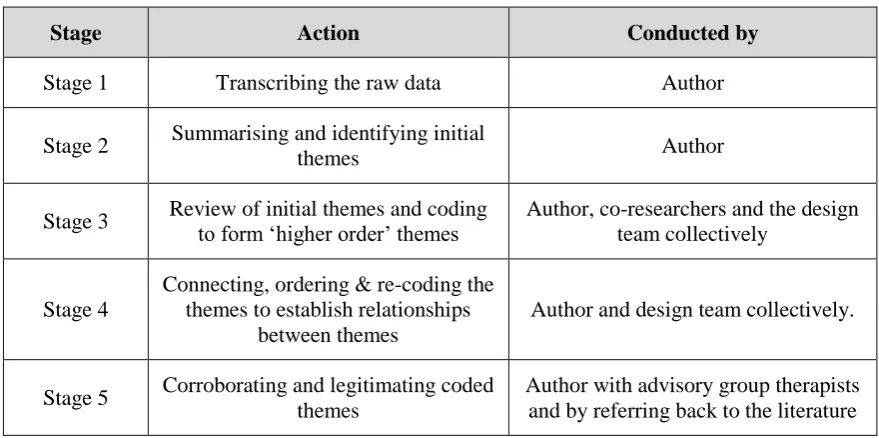
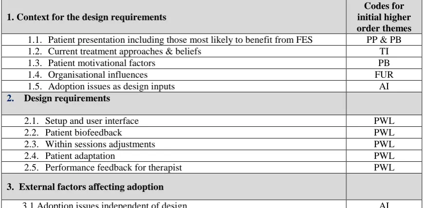
Table 2.1: Studies from the literature review that included Health Care Professions as part of the design and usability process. ... 54 Table 4.1: Table displaying the participant ID, designation, novice (N) or expert FES user (E) and meeting number attended ... 74 Table 4.2: Stages of the data analysis process. ... 75 Table 4.3: Results from stage 2 of the analysis - an advisory group trigger question with a summary of the responses. ... 75 Table 4.4: Stage 3 initial ‘higher order’ themes with coding ... 76 Table 4.5: Stage 4 initial ‘higher order’ themes mapped on to design process themes of the UL FES Rehab Tool. ... 77 Table 4.6: Summary of tasks, FES parameters and practice schedules for each category of patient, taken from the third advisory group meeting. ... 78 Table 4.7: UL FES Rehab Tool therapists’ design requirements in rank order of importance .
10 Table 6.4: Participant characteristics: impairment, function and Mini Mental
scores for the lab based testing...127 Table 6.5: Impairment level and setup times per participant and functional task.. ... 128 Table 7.1: Therapist & RA grade, level of experience in clinical practice, pre-study experience with FES and amount & type of computer use. ... 154 Table 7.2: Number and gender of patient’s screened and recruited at both centres. ... 155 Table 7.3: Reasons for exclusion and number of patients excluded at each centre ... 156 Table 7.4: Time since stroke, age (years), side affected by stroke, hand dominance, gender and Fugl-Meyer UE score per patient... 157 Table 7.5: Frequency of use of the UL FES Rehab Tool for patients and therapists / RA .... 158 Table 7.6: Individual therapists’ usefulness (U) and ease of use (EoU) TAM scores, in rank order of highest to lowest scores. ... 161 Table 7.7: Type of tasks, patients’ Fugl-Meyer UE scores, number of successful and unsuccessful UL FES assisted repetitions and task completion scores. ... 165 Table 7.8: Reasons for non-completion of upper limb reaching repetitions ... 166 Table 7.9: displays the FES Task Attainment Scale scores for each session.……….168 Table 7.10: Number of times assistance required during the setup process for each therapist and RA across FES sessions……… ... 169 Table 7.11 Total setup times (min) for the FES Rehab Tool. ... 170 Table 7.12: Individual therapists and RA’s pre and post usefulness (U) and ease of use ‘EoU’ scores, difference in pre and post ‘U’ and ‘EoU’ and total pre and post-study TAM scores, in rank order of highest to lowest scores. ... 173 Table 7.13: Total and median ease of setup scores taken from the post-session therapist
11
Figures
Figure 2.1: Interaction between growth control mechanisms and environmental stimuli during
physiological plasticity and repair. . ... 29
Figure 2.2: The graph shows the mean length of stay in hospital from 2001 to 2013-14.. ... 33
Figure 2.3: Total hours of therapy provided for all patients across the year against the target number of hours. ... 34
Figure 2.4: Propagation of an electrical impulse along the axon of a motor neuron... 36
Figure 2.5: Depolarisation of a motor neuron and generation of an action potential ... 36
Figure 2.6: Diagram to show the application of surface electrodes………..37
Figure 2.7: Typical waveform for FES . ... 37
Figure 2.8: Muscle fibre recruitment order with corresponding pulse widths ... 38
Figure 2.9: Stimulation frequency showing recruitment of different motor units over time resulting in a physiological tetanic state ... 38
Figure 2.10: Diagram to demonstrate the theory proposed by Rushton (2003). (a) The proposed normal physiology (b) a lesion in the system (c) the system following NMES intervention. ... 39
Figure 2.11: The Bioness H200®hand rehabilitation wireless system ... 44
Figure 2.12: The Stiwell med4 EMG-triggered FES system... 44
Figure 2.13: NeuroMoveTM NM900 ... 45
Figure 2.14: The MyoTrac Infiniti, produced by Saebo, USA. ... 45
Figure 2.15: Key components of a usability engineering process . ... 50
Figure 3.1: The 2 channel stimulator used with the CST. ... 62
Figure 3.2: The CST software interface. ... 63
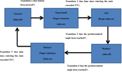
Figure 3.3: Example FSM for drinking from a glass.. ... 65
12
Figure 5.1: An example question from the post-test questionnaire. ... 90
Figure 5.2: An example of the in-test usability notes taken from the observers’ paper based data collection form……….. .. 92
Figure 5.3: Flow diagram showing the various components of the stage one usability analysis ... 93
Figure 5.4: Flow diagram showing the various components of the stage two usability analysis. ... 94
Figure 5.5: Structure of the usability problem categories ... 95
Figure 5.6: Results from stage 1 of the usability problem analysis ... 96
Figure 5.7: Stage 2 data analysis resulting in n= 23 changes made to the GUI ... 99
Figure 5.8: State machine diagram for sweeping coins into contralateral hand. ... 106
Figure 5.9: Screen shot of stage 3 GUI, v2.0 before changes. ... 107
Figure 5.10: Screen shot of stage 3 GUI , v3.0 following changes. ... 108
Figure 6.1: An example of the task, sweeping coins into contralateral hand. ... 119
Figure 6.2: The inter-relationship between upper limb impairment, task complexity and additional factors when predicting setup time and task selection. ... 122
Figure 6.3: Scatterplot of task complexity scores for Research Physiotherapist 1 (author) & Research Physiotherapist 2……….123
Figure 6.4: Scatterplot of task complexity against setup times ... 129
Figure 6.5: A scatterplot of participants’ upper limb impairment scores plotted against setup times………130
Figure 6.6: Predicted setup times plotted against measured setup times ... 133
Figure 7.1: The final prototype UL FES Rehab Tool – software and hardware components……….. ... 138
Figure 7.2: Patient attempting the ‘reach for coins’ task (a) with and (b) without the UL FES Rehab Tool. ... 150
Figure 7.3: Total post training confidence scores, per therapist & RA ... 160
13 Figure 7.5: A scatter plot of frequency of use of the UL FES Rehab Tool plotted against
the post training confidence scores ………162
Figure 7.6: A scatter plot of frequency of use of the UL FES Rehab Tool (normalised for no. of patients recruited at each centre) plotted against post training ‘Usefulness’ (U) TAM scores. ... 163
Figure 7.7: A scatter plot of frequency of use of the UL FES Rehab Tool (normalised for no. of patients recruited at each centre) plotted against post training ‘Ease of Use’ (EoU) TAM scores………...163
Figure 7.8: Scatter plot of Fugl-Meyer UE scores against % task completion rates……….166
Figure 7.9: Scatterplot of task complexity against setup times ... 171
Figure 7.10: Scatterplot of upper limb impairment against setup times. ... 172
Figure 7.11: SUMI Scale profiles ... 176
Figure 8.1: UL FES Rehab Tool development lifecycle, inputs and outputs and the associated usability evaluation methods for phases 1-5... 188
Appendices
Appendix 1: Full list of studies included in the usability literature review 194Appendix 2: NEAT LO30 funding documentation 208
Appendix 3: Phase two usability testing information sheet and consent form 210 Appendix 4: Phase two usability testing task sheets 213 Appendix 5: Phase 2, usability testing observation tool 216
14 Appendix 11: Phase four LREC and University of Salford ethical approval letters 236 Appendix 12: Phase five, proof of concept clinical trial NHS NRES, SRFT R & D and
University of Salford approval letters. 238
Appendix 13: Technology Acceptance Measure (TAM) questionnaire 241
Appendix 14: FES task attainment scale 247
Appendix 15: Phase five usability testing, post-test debrief form 248 Appendix 16: Software Usability Measurement Inventory (SUMI) 250 Appendix 17: Phase five, post-training confidence questionnaire 254 Appendix 18: Phase five usability testing, post-session debrief data 257
Appendix 19: SPSS tables from chapters 6 & 7 262
15
Acknowledgements
I would like to express my deepest appreciation to my PhD supervisory team, Professor Kenney, Professor Howard and Professor Hardiker who all contributed at some point to the completion of this thesis. An extra special ‘thank you’ to Professor Kenney, whose patience and guidance shaped the thesis into a coherent document. My gratitude also to Dr Sun whose thesis ran in parallel with mine and who came to the rescue when it felt like the software would never reach completion. In addition, a huge thank you to other members of the FES Rehabilitation Technologies research team, namely Karen Waring, Helen Luckie and Dr Williamson with whom it is a privilege and pleasure to work alongside.
My gratitude to the National Institute for Health Research who funded the NEAT LO30 project, and to all the therapists and patients who so willingly gave their time to take part in the study.
16
Glossary of terms
ANRT Advanced Neurological Rehabilitation Technologies
ATRAS Assistive Technologies for Rehabilitation of the Arm following Stroke project CIMT Constraint Induced Movement Therapy
FES Functional Electrical Stimulation GBS Guillain-Barré Syndrome
HCP Health Care Professionals
ITQ Immersive Tendencies Questionnaire MS Multiple Sclerosis
NIHR National Institute for Health Research NHS National Health Service
OT Occupational Therapy
P & C Patients and Carers
PT Physiotherapy
SALT Speech and Language Therapy SCI Spinal Cord Injury
SFQ Short Feedback Questionnaire
SUMI Software Usability Measurement Inventory SUS System Usability Scale
17
Abstract
Chronic physical impairment of the hemiplegic upper limb (UL) is seen in an estimated 50-70% of stroke patients, who place a high priority on regaining upper limb function. Current therapy is insufficiently intensive, often not task-oriented and hence poorly aligned with the evidence base. Functional electrical stimulation (FES) has the potential to not only increase the intensity of task-focused therapy, but also provide certain unique features, notably direct excitation of lower motor neurons. However, current FES systems are limited in their functionality and/or difficult to use. Systems are also poorly aligned to therapists’ ways of working and uptake remains limited. To address these problems, a novel FES technology (UL FES Rehab Tool) has been developed. The control system design is reported in Sun, (2014). The aims of my thesis were to: 1) design a Graphical User Interface (GUI) that would enable therapists to quickly and easily set up an individually tailored library of FES tasks for each patient; 2) evaluate the usability and functionality of the UL FES Rehab Tool (software and hardware) in both laboratory (lab) and clinical settings.
An iterative, mixed methods, five-phase usability engineering approach was used to design and evaluate the UL FES Rehab Tool. Phases one to three incorporated identification of therapists’ requirements, a user ‘assisted walkthrough’ of the software with expert and novice FES users and ‘rapid prototyping’ of the full system, using healthy participants. Further usability testing of the software & hardware was conducted in phase four with 1 physiotherapist and 6 patients, (total of 24 visits), in the chronic stage post-stroke. The work demonstrated in detail, for the first time, the impact of therapist involvement in the design of novel rehabilitation technology.
To address therapists’ focus on setup time, using the phase four data set, a novel model to predict setup time was devised. This model was able to explain 51% of the variance in setup time based on two parameters, task complexity and patient impairment.
18 Meyer scores 8–65). The usability methods effectively captured objective and subjective feedback from therapists and patients. However the previous setup time model was unable to predict setup time, suggesting other factors were important in a clinical setting. Although participant numbers were low, the results suggested therapists’ predisposition to using technology and post-training confidence in using the technology may influence their willingness to engage with novel rehabilitation technologies.
19
1
Chapter One: Introduction
1.1 Overview of the thesis
Rehabilitation technologies are showing promise as interventions to promote recovery of the hemiplegic upper limb post stroke. Functional Electrical Stimulation (FES) is one of the technologies that offers the potential to support the user in varied and challenging functional task practice. Current FES devices are limited in their functionality and hence more sophisticated devices are needed. The usability of devices in challenging clinical environments such as the acute setting, are likely to influence usage (Hochstenbach-Walen & Seelen, 2012), and hence great care is needed when designing more sophisticated rehabilitation devices to ensure usability. This study outlines a usability engineering approach to the design of a new FES system, the FES Rehab Tool, and the usability evaluation from the early design stages through to the proof of concept clinical trial, in two sub-acute stroke units.
The aim of chapter one is to outline the overall structure of the thesis chapter by chapter along with the accompanying rationale for each.
1.1.1 Chapter Two
20 Rehabilitation Technologies (ANRT) that have reported on usability evaluation is included, to provide context for subsequent aspects of the thesis.
The aims and objectives that informed the thesis are then stated.
1.1.2 Chapter Three
This chapter provides an overview of the work that led to the thesis. It outlines the early work on an accelerometer controlled upper limb FES system, the Clinical Setup Tool (CST) that was the forerunner to the UL FES Rehab Tool. In order to allow the reader to better understand the UL FES Rehab Tool and the systems that it was based on, the concept of finite state-machine control is introduced. Finally, the NEAT LO30 project that much of the thesis work contributed to is described. The NEAT LO30 project was supported both by the author’s work and that of a fellow PhD student, Mingxu Sun. The role of each of the authors in these complimentary pieces of work is also explained.
1.1.3 Chapter Four
This chapter describes a phased usability engineering approach to the design and evaluation of an UL FES Rehab Tool. It first outlines each of the phases of the design and usability evaluation. The authors’ approach to gaining therapists’ views from the advisory and focus group meetings is described and the findings presented.
1.1.4 Chapter Five
Phases two and three of the usability evaluation process are presented along with the findings from each of the phases. The chapter highlights the limited evidence base demonstrating the impact of usability engineering on ANRT design. Specifically the chapter demonstrates the impact of user involvement on the early design work on the GUI aspects of the UL FES Rehab Tool.
1.1.5 Chapter Six
21 patients’ level of impairment and task complexity. Data from six participants in the chronic stage of stroke were used to create the model and the model evaluation is presented. The relationship between impairment, task complexity and setup time is discussed along with the limitations of this approach.
1.1.6 Chapter Seven
Chapter seven presents the findings from the final proof of concept study, in which therapists set up and used the UL FES Rehab Tool in a clinical setting. The usability and feasibility of version 3 of the UL FES Rehab Tool when used in two sub-acute stroke units is presented and discussed. The methods adopted, including the use of a technology acceptance measure and the therapists training, are discussed. The findings are presented and discussed along with the challenges and limitations.
1.1.7 Chapter Eight
22
2
Chapter 2: Literature review
2.1 The upper limb following stroke
2.1.1 Incidence and prevalence of upper limb impairments and functional limitations after stroke
There are approximately 152,000 strokes in the United Kingdom (UK) every year, with the incidence predicted to increase in the coming years (Truelsen et al., 2006). Approximately one third of people who experience a stroke die as a direct result, leaving around 1.1 million stroke survivors living in the UK (Townsend et al., 2012). The total cost of stroke to the UK economy is estimated to be between £3.7 billion and £8 billion per year (DoH, 2010)
A stroke occurs when the blood supply to the brain is disrupted leading to death of nervous tissue. Eighty percent of strokes are caused by an occlusion in a cerebral artery, such as those caused by an embolus, resulting in an ischaemic stroke. The other main pathological cause of stroke (15%) is due to haemorrhage of a cerebral artery. The remaining five percent of strokes are classified as a subarachnoid haemorrhage (Lindley, 2008).
23 decreased movement smoothness (Alt Murphy, Willén, & Sunnerhagen, 2011), lower peak velocity, increased variability and timing of peak velocities and larger end point errors (van Vliet, Pelton, Hollands, Carey, & Wing, 2013). Poor inter-joint and intermuscular co-ordination are thought to be partially responsible for these deficits (van Kordelaar, van Wegen, & Kwakkel, 2012).
Stroke patients have been found to demonstrate a significant amount of non-use of their affected upper limb during unimanual and bimanual activities (Michielsen, Selles, Stam, Ribbers, & Bussmann, 2012). In the early stages following stroke, the patients’ ability to use their hemiplegic upper limb for functional activities is often severely impaired. This inability to use the upper limb can quickly lead to a phenomenon known as ‘learned non-use’ (Taub, Uswatte, Mark, & Morris, 2006). Factors such as recovery of the hand (Lin, Huang, Hsieh, & Wu, 2009) and hand dominance (Darling et al., 2013) are thought to influence functional recovery. However, further studies are required to fully understand the complex relationship between motor recovery and actual amount of use in people with chronic stroke.
2.1.2 Impact on quality of life
24 health-related quality of life as long as five years post stroke (Paul et al., 2005). Indeed, following a stroke, patients with severe upper limb dysfunction are approximately twice as likely to be admitted to institutionalized care (Hunter & Crome, 2002).
2.1.3 The recovery process following a stroke
The CNS has a capacity to reorganise in response to injury, pathology or behavioural demands placed on it (Xerri, 2012; Cramer et al., 2011; Pascual-Leone, Amedi, Fregni, & Merabet, 2005). This reorganisation occurs as a result of neuroplasticity of the neuromuscular system, which if influenced early after stroke, can have a positive effect on recovery of the upper limb. However, if this reorganisation is left to its own devices, it can be detrimental to recovery. Neuroplasticity can be defined as…….
“the ability of the nervous system to respond to intrinsic and extrinsic stimuli by reorganizing its structure, function and connections; can be described at many levels, from molecular to cellular to systems to behaviour; and can occur during development, in response to the environment, in support of learning, in response to disease, or in relation to therapy” (Cohen et al., 1997, pg.180).
25 dendritic branching in the relevant parts of the cortex and a resumption of the same kinematics of the movement as those utilised prior to the stroke. A ‘compensatory response’ is where neuroplastic changes may still take place as a result of re-learning, however the kinematics of the movement are different to those used pre-stroke (Metz, Antonow-Schlorke, & Witte, 2005). For example when stroke patients are attempting to reach for an object, they may employ compensatory forward flexion of the trunk in order to accommodate for a lack of shoulder flexion and elbow extension. Further studies are required before the relationship between ‘true recovery’ and ‘compensation’ can be fully understood. Section 2.2 outlines the factors that may influence recovery.
2.2 Basic science studies
In this section, the evidence from animal studies, together with recent clinical trials is introduced, pointing to the key features of effective therapy interventions.
2.2.1 Timing of interventions
26 place after 30 days. However, the mechanisms for these changes are less well documented. In addition, humans may continue to make considerable motor gains based on increases in strength or endurance of muscles, both of which are likely to impact on functional activity. In summary, more information is required regarding the variability in post-stroke injury and time-dependent neural activity before the optimum timing for rehabilitation interventions can be confirmed.
A systematic review of Randomized Controlled Trials (RCTs) that used assistive technologies (AT) for rehabilitation of the upper limb with stroke patients was undertaken by Farmer et al. (2014). The data was used to assess the effect size of the intervention across all dimensions of the ICF framework. A moderate benefit was found for AT when compared with usual care or in addition to usual care. There was a greater effect size for patients in the acute phase i.e. up to 6 weeks post stroke. There were two exceptions to this finding; 1) Neuromuscular Electrical Stimulation (NMES) to the shoulder (Church et al., 2006) and 2) Constraint induced Movement Therapy (CIMT) earlier than 6 weeks post-stroke (Dromerick, Lang, & Birkenmeier, 2009). Constraint-Induced Movement Therapy (CIMT) is a form of treatment for the hemiplegic upper limb that consists of constraint of the unaffected upper limb, simultaneous with intensive (up to 6 hours) and progressive task-based training of the hemiplegic limb. This intensive practice is often referred to as ‘shaping’. Wolf et al. (2010) delivered 2 weeks of CIMT to stroke patients randomized into either an early (3-9 months, n=106) or late (15-21 months, n=86) intervention group. Assessors who were blinded to the group allocation administered the Wolf Motor Function Test, Motor Activity Log (primary measures) and the Stroke Impact Scale (secondary measure) pre-intervention then at 2 weeks, 4, 8 and 12 months post intervention. Although both groups demonstrated significant improvements in upper limb recovery across all outcome measures, the early CIMT group showed the greatest relative improvement. This study reinforces the view that interventions targeted early after stroke are likely to advantageous.
2.2.2 Intensity and scheduling of practice
27 derived neurotrophic factor (BDNF), which has been found to improve sensorimotor recovery following ischemia. The rats were exposed to either an enriched or a non-enriched rehabilitation environment in order to examine the impact of varied reaching intensities and durations post brain lesion. The enriched rehabilitation environment typically contained multi-level cages with tubes, toys and ramps. Reaching interventions of 4 to 6 hours per day, 5 days per week for 8 weeks, with on average 300 reaching repetitions completed per session (MacLellan, 2011). The non-enriched environment was standard rat caging. Only rats in the enriched rehabilitation environment that reached a critical threshold of reaching activity (approximately 300) demonstrated recovery. Some rats engaged in the reaching activity but failed to reach the critical threshold of repetitions resulting in no recovery. Interestingly, the rats who did achieve recovery did not further benefit from the enriched rehabilitation environment when they were exposed to additional doses of the intervention. This study is important not only because it is the first to demonstrate that there appears to be a critical threshold of number of repetitions below which recovery will not occur, but also because it supports the use of task-specific interventions. One study by Birkenmeier, Prager, and Lang (2010) demonstrated that it is possible to achieve over 300 repetitions of an upper limb task within a 1 hour therapy session (3 tasks x 100 repetitions).
28 stroke has been poorly documented, and as such further work is needed to determine the optimum dose for upper limb rehabilitation.
29
2.2.3 Content and progression of training
Although the dose for therapeutic interventions is important when promoting upper limb recovery, without due consideration for the content and progression of training the picture is incomplete. The content and approach to therapy has been a matter of debate for many years (Wang, Chen, Chen, & Yang, 2005), with various authors proposing a particular approach e.g. Bobath, Orthopaedic, Motor re-learning amongst others. A recent Cochrane Review has found there to be no evidence of superiority of one method or approach over any other (Pollock, Baer, et al., 2014).
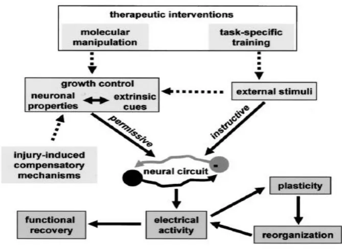
Animal studies have demonstrated that training needs to be task specific and challenging if it is to drive recovery (Nudo & Milliken, 1996). Rossi, Gianola, and Corvetti (2007) (Figure 2.1), have described the neurobiological changes that occur as a result of brain reorganisation following injury, and advocate task-specific training as a means of providing the most suitable form of extrinsic stimuli.
Figure 2.1: Interaction between growth control mechanisms and environmental stimuli during physiological plasticity and repair. Reproduced with permission of Rossi et al., (2007).
[image:29.595.125.460.353.597.2]30 result of the evidence from animal and human studies, task-oriented or task-specific training has been proposed as a fundamental ingredient when designing training schedules to promote skill reacquisition (French et al., 2010). A recent study has extended this concept by demonstrating that re-learned skills only generalise to similar movement sequences that occur in the same workspace, with similar joint co-ordinations as those learned during training (Panarese, Colombo, Sterpi, Pisano, & Micera, 2012). In addition, practice schedules need to progressively challenge the learner if motor learning is to take place. Guadagnoli and Lee (2004) describe this as creating ‘optimal challenge points’, which considers the level of skill the leaner has achieved, the task difficulty (including the environment that the task is performed in), and the amount of information available, as important variables. These variables have been taken into account when designing ‘iterative learning’ systems, and have been incorporated into new UL FES technologies that are currently under development (Meadmore et al., 2014).
In recent years as the evidence for a focus on reducing spasticity has waned, Progressive Resistance Training (PRT) has increased in popularity. Weakness is a dominant feature in post stroke hemiplegia, as a result of changes at a neural (supraspinal) and muscular level (Patten, Lexell, & Brown, 2004). Although a sufficiently large body of evidence remains to be collected, PRT appears to offer some merit (Porter, 2000; Hurley & Roth, 2000). However, PRT may not be beneficial for all severities of stroke patients. PRT has been shown to be most beneficial for patients with mild to moderate levels of impairment where voluntary effort can be initiated (Winstein et al., 2004; Thielman & Gentile, 2002). The transfer of improvements in strength to functional improvement remains to be fully examined.
31
2.2.4 Feedback on performance
One of the fundamental requirements to enhance motor relearning is provision of information or feedback, either during or following task performance. Feedback can be classified into two categories: ‘intrinsic’ feedback which is provided by the body’s own sensory-perceptual information, via internal sensory processes that occur as a result of movement e.g visual or proprioceptive feedback, and occur during performance of a task. ‘Extrinsic’ or ‘augmented’ feedback: usually arises from an external environmental source (Subramanian, Massie, Malcolm, & Levin, 2010). As intrinsic feedback is often disrupted following a stroke, provision of extrinsic feedback is crucial to supplement this deficit. Extrinsic feedback can be given verbally, manually or by using visual means such as a visual display, demonstration or video. Extrinsic feedback can be further divided into ‘knowledge of results’ (KR) and ‘knowledge of performance’ (KP). KR is “externally presented information about the outcome of performing a skill or about achieving the goal of the performance” (Magill, 2003). KP is “information about the movement characteristic that led to achievement of the goal”(Magill, 2003). For example, a patient may be instructed to straighten their elbow to more effectively reach a target object. Studies in healthy participants (Wulf & McConnel, 2002) and in stroke patients (Durham et al., 2013) have found KR to be more beneficial. Importantly for therapy that uses functional tasks, additional extrinsic feedback can be redundant if the outcome of the performance is inherent in the task (Platz et al., 2001; Beukers, Magill, & Hall, 1992), and might even be detrimental.
Timing of feedback is also important for the retention of information. Feedback can be provided concurrently (at the same time as the task is being performed) or terminally (after performance is complete). Terminal feedback can be further sub-divided into a number of sub-categories including bandwidth feedback (provided at intervals throughout training) and average feedback. When considering the timing of feedback, the patients’ stage of learning also needs to be taken into account (Mount et al., 2007; Ezekiel, Lehto, Marley, Wishart, & Lee, 2001). Wherever possible, patients should be encouraged to solve the motor problem (Mulder & Hochstenbach, 2003).
32 It is therefore not surprising that a review conducted by van Vliet and Wolf (2006) on provision of extrinsic feedback for motor relearning following stroke, concluded that although there are clear benefits for provision of feedback to enhance motor learning, further studies were required before it was possible to determine the most suitable type, frequency and attentional focus for each possible patient presentation.
In summary, from a review of basic science studies in animals and humans, there are varying amounts of evidence to support upper limb training schedules being:
1) Timely in that they should commence early post stroke, when neuronal processes can cope with external influences; 2) sufficiently intensive to support those individuals who have the potential to reach activity dependent recovery thresholds. This has been postulated to be in the region of 300 repetitions per training session. However, attention to the scheduling of practice is required so as to align to the patients’ needs; 3) the content of training should be functionally oriented, such as the approached used within task-oriented training. Training that aims to address ‘weakness’ rather than ‘spasticity’ appears to have clear benefits. However, movement sequences and tasks need to be progressive and optimally adapted to meet both the patients’ capability and the environment in which the skill is to be used; 4) And finally, due consideration needs to be given to the type, frequency and timing of feedback, adapted to align with the stage of learning and the patient’s presentation.
Section 2.2.5 reviews current therapy provision in order to allow comparison with the evidence presented in section 2.2.4.
33 Programme (SSNAP, April 2013-March 2014) (The Intercollegiate Stroke Working Party, 2014), found that out of the 85% of patients that required physiotherapy, they only received 32 minutes of therapy in just over half of their in-patient stay. Bearing in mind that these figures are based on therapy for restoration of mobility as well as upper limb function, and that previous studies have identified that therapy tends to focus on mobility rather than treatment of the upper limb (Cott, 2004), it is reasonable to assume that therapy for the upper limb falls significantly short of what is required to promote recovery (Rudd, Jenkinson, Grant, & Hoffman, 2009). In addition, the length of time that patients remain in hospital following a stroke has significantly decreased over the last decade (Figure 2.2).
Figure 2.2: the graph shows the mean length of stay in hospital from 2001 to 2013-14, showing that mean length of stay has decreased significantly. The top line depicts those patients who were discharged alive and the bottom line represents all patients (The Intercollegiate Stroke Working Party, 2014).
[image:33.595.160.497.289.498.2]34 Figure 2.3: Total hours of therapy provided for all patients across the year against the target number of hours (The Intercollegiate Stroke Working Party, 2014).
Figures taken from the same audit report showed that the total hours of therapy fall below target numbers (Figure 2.3), and on discharge 37% of patients required assistance with activities of daily living (ADL) (The Intercollegiate Stroke Working Party, 2014). As upper limb function is crucial to carrying out ADL, this most probably demonstrates that restoration of upper limb function had not occurred, and corroborates findings from previous studies (Kong, Chua, & Lee, 2011; DoH, 2010), that promoting upper limb recovery remains a significant challenge.
35 impaired patients (21%) and Functional Electrical Stimulation (FES) for those who were moderately (36%), or severely (18%) impaired.
It can therefore be concluded from the review of current practice, that it appears to be poorly aligned with the evidence base and indeed the recommended clinical guidelines. It is clear that existing rehabilitation practice alone will not address the increase in demand for rehabilitation and that new ways of tackling this growing problem are urgently required. Numerous studies have demonstrated the potential for technology to assist with the timing, intensity, and content of therapy, particularly for the upper limb (Demain et al., 2013; Hochstenbach-Waelen & Seelen, 2012; Rosser et al., 2011; Timmermans, Seelen, Willmann, & Kingma, 2009). These interventions have the potential to free up valuable therapist time and provide a situation where patients can access rehabilitation interventions in order to practise functional movements at their own pace. However, it is important to stress that rehabilitation technologies need to be seen as an adjunct to the therapeutic process, rather than one that replaces it. Both patients and practitioners need to be persuaded that the evidence base for their implementation is strong, and that it provides an additional dimension to the ‘toolbox’ of practitioners. Section 2.5 examines this issue in more depth.
The next section introduces the reader to FES, which is central to this thesis.
2.3 Functional Electrical Stimulation (FES)
2.3.1 Basic science. What is FES?
Before explaining electrical stimulation, a brief introduction to recruitment of muscle fibres is provided. Muscle activity is controlled by the Central Nervous System (CNS). Communication between the CNS and the muscles occur via motor neurons. A motor unit is a single motor neuron and its associated muscles fibres. Once a motor neuron is activated, all the muscle fibres it supplies are activated (Bear, Connors, & Paradiso, 1996).
36 is altered or ‘depolarised’. Depolarisation changes the permeability of the cell membrane causing diffusion of Na+ into the axon. The charge inside the cell becomes more positive, causing an ‘action potential’. Once this has been achieved the membrane becomes less permeable to Na+ and begins to favour K+ once again. K+ leave the axon until the resting potential is achieved once again. The membrane is then ‘re-polarised’ (RSC, 2004). This process of depolarisation and re-polarisation continues at a local level to allow transmission of the nerve impulse along the motor neuron. In a myelinated neuron, the electrical impulse jump from one ‘node of Ranvier’ to the next, thereby speeding up conduction (Figure 2.4).
Figure 2.4: Propagation of an electrical impulse along the axon of a motor neuron
"Action Potential" by Laurentaylorj - Own work. Licensed under CC BY-SA 3.0 via Wikimedia Commons -
https://commons.wikimedia.org/wiki/File:Action_Potential.gif#/media/File:Action_Potential.gif
FES is the use of Neuromuscular Electrical Stimulation (NMES) to activate paralysed muscles in a precise sequence and magnitude, resulting in the accomplishment of functional tasks. Lower motor neuron electrical stimulation alters the electrical field surrounding a nerve’s axon, and if the field reaches sufficiently high level, action potentials are induced (Figure 2.5).
Figure 2.5: Depolarisation of a motor neuron and generation of an action potential
37 updated by en:User:Diberri, converted to SVG by tiZom - Own work. Licensed under CC BY-SA 3.0 via Wikimedia Commons.
http://commons.wikimedia.org/wiki/File:Action_potential.svg#mediaviewer/File:Action_potential.sv
The most common method of applying stimulation is via surface electrodes as in Figure 2.6. However, FES has also been administered using percutaneous or implanted systems (Peckham & Knutson, 2005).
Figure 2.6: Diagram to show the application of surface electrodes. Reproduced and adapted from:
http://spectrum.ieee.org/automaton/robotics/medical-robots/robot-controls-human-arm
Stimulation protocols in FES typically utilise a current-controlled delivery method, whereby biphasic charge balanced pulsing is used to prevent the build-up of unwanted chemical substances. The first phase (stimulation phase) elicits the action potential. The second phase (reversal phase) reverses the electrochemical processes that occur during the stimulation phase. The polarity of a bi-phasic pulse can either be cathode-first or anode-first. Figure 2.7 illustrates a cathode-first example as this can affect the threshold of activation. Peripheral, cutaneous stimulation usually employs a cathode-first method due to the lower activation threshold (Merrill, Bickson, & Jefferys, 2005).
Figure 2.7: Typical waveform for FES (Merrill et al., 2005). The narrow pulse width is indicated PW. The interpulse interval (IPI) is the time between pulses.
38 In conventional electrical stimulation waveforms, the largest diameter nerve fibres are initially recruited (type IIb, fast twitch, depicted in red), followed by type IIa (green) and type 1 (slow twitch, fatigue resistant, blue) (Figure 2.8). This is the reverse order to that occurring under normal physiological conditions (Merrill et al., 2005).
Figure 2.8: Muscle fibre recruitment order with corresponding pulse widths (Hamouda, 2014)
The recruitment of different motor units over time produces a physiological tetanic state (Figure 2.9).
Figure 2.9: Stimulation frequency showing recruitment of different motor units over time resulting in a physiological tetanic state (Baker, Wederich, McNeal, Newsam, & Waters, 2000).
39 A unique feature of FES is that is activates motor nerve fibres both orthrodromically and antidromically. The antidromic impulse is postulated to have an effect on the plastic adaptations occurring at the anterior horn cell when performed in conjunction with voluntary effort via a damaged pyramidal motor system (Rushton, 2003) (Figure 2.10).
Figure 2.10: Diagram to demonstrate the theory proposed by Rushton (2003). (a) The proposed normal physiology (b) a lesion in the system (c) the system following NMES intervention. Diagram from Rushton (2003).
2.3.2 A review of the efficacy of upper limb FES assisted practice
[image:39.595.126.497.213.530.2]40 (de Kroon, van de Lee, IJzerman, & Lankhorst, 2002). TES has been aimed at reducing impairment, for example by increasing muscle strength or range of movement. Although it is useful for reducing impairment, there is limited evidence to support its impact on activity or function (Chae et al., 1998).
Another category of electrical stimulation is Functional Electrical Stimulation (FES). FES is a means of stimulating muscle in order to achieve functional tasks (de Kroon et al., 2002). FES can be used either on its own, or in combination with an orthosis to act primarily as an assistive device by enabling completion of everyday tasks (neuroprosthesis), for instance in patients with spinal cord injury, and/or as a training modality with the aim of promoting recovery of function. The latter approach is the one most commonly used with stroke patients. In a recent systematic review and meta-analysis by (Howlett, Lannin, Ada, & McKinstry, 2015), subgroup analysis from 8 studies (Page, Levin, Hermann, Dunning, & Levine, 2012; Faisal & Priyabanani Neha Om, 2012; Tarkka, Pitkanen, Popovic, Vanninen, & Kononen, 2011; Hara, Ogawa, Tsujiuchi, & Muraoka, 2008; Daly et al., 2005; Mann, Burridge, Malone, & Strike, 2005; Popovic, Popovic, Sinkjaer, Stefanovic, & Schwirtlich, 2004; Popovic, Popovic, Sinkjaer, Stefanovic, & Schwirtlich, 2003) (181 participants) found that FES had a large effect on upper limb activity (SMD 0.69, 95% CI 0.33 to 1.05) compared with a control group. However the control groups were generally traditional therapy, which was often not defined. In contrast to these findings, a recent Cochrane Systematic Review (Pollock, Farmer, et al., 2014) found there to be insufficient robust evidence from RCTs to support the use of FES as an intervention for upper limb recovery. However, the small number of participants in studies, the heterogeneity of studies and often insufficient control of the effect of duration of interventions across groups, makes it difficult to draw firm conclusions.
41 Thrasher, Zivanovic, McIlroy, & Popovic, 2008). Even when severely affected acute stroke patients, with minimal movement in their affected arm, undertake intensive functional task practice, using therapist-triggered FES, significantly improved clinical outcomes can be achieved (Thrasher et al., 2008). The following section reviews the evidence from studies where systems were patient-controlled.
In recent years there have been a number of reviews of FES for the upper limb. Bolton (2004) carried out a meta-analysis on EMG triggered neuromuscular stimulation on stroke motor recovery for the arm and hand (up to 2003). Only 5 studies were deemed to be sufficiently robust to be included in the analysis (Cauraugh & Kim, 2002; Cauraugh, Light, Kim, Thigpen, & Behrman, 2000; Francisco, Chae, & Chawla, 1998; Hummelsheim, Amberger, & Maurtiz, 1996; Kraft, Fitts, & Hammond, 1992), and of these only 1 study (Francisco, Chae, Chawla, & al., 1998) could be categorized as being in the acute phase post stroke (just 4 patients in the intervention group). Nevertheless, they found an overall beneficial effect on hand / arm function for acute / sub-acute (16%) and chronic stages (84%) of stroke. There was a significant mean effect size of 0.82 and a 95% confidence interval of 0.10-1.55. Some caution should be exerted when considering this review as non-randomised studies were included.
42 stimulation over other treatment modalities, such as standard physiotherapy. However, only 10 studies focused on electrical stimulation for the upper limb. Of the 10 studies, 6 were EMG triggered systems, predominantly for the wrist and hand in order to facilitate hand opening (Kimberley et al., 2004; Cauraugh & Kim, 2003; Cauraugh & Kim, 2002; Cauraugh et al., 2000; Francisco, Chae, Chawla, et al., 1998; Heckmann et al., 1997). The remaining studies were either not triggered (Linn, Granat, & Lees, 1999; Chae et al., 1998; King, 1996) or were triggered manually (Popovic et al., 2003). The heterogeneity of studies in these reviews makes it difficult to establish the efficacy of functional electrical stimulation, and impedes the generalisation of results. In spite of the promising basic science studies reviewed in section 2.2 the application to human studies with FES requires further research. A recent review by Quandt & Hummel, (2014) concisely summarises the position to date regarding the efficacy of FES; treatment doses, optimal stimulation parameters, timing of interventions and the level of severity of stroke patients likely to benefit from FES remains inconclusive.
There appears to be a growing evidence base for the use of voluntary movement (patient triggered) triggered FES, in particular movement that is triggered via accelerometers or electro-goniometers. This method harnesses the benefits of combining the patient’s voluntary effort with that of FES. A review by Popović et al. (2009) concluded that integration of electrical stimulation in combination with exercise-active movement enhanced motor re-learning following central nervous system damage. They also suggested that the therapeutic effects are likely to be more effective when treatment is applied in the acute, rather than the chronic phase of stroke. This seems to be in keeping with the basic science studies.
43 significantly improved from pre to post-intervention, alongside a reduction in arm support for unassisted FES performance.
This evidence suggests that functional improvements from FES may result from its use in supporting voluntary-triggered, task-focused practice. Clearly more evidence from larger, well designed multi-centre RCTs need to be undertaken before any firm conclusions can be drawn. However, larger studies can only be carried out if movement controlled FES devices that are sufficiently robust and flexible, in order to treat a wide range of patients, are in existence. Commercially available devices where stimulation is patient-controlled may be limited. A review of FES devices that are available commercially is reviewed in section 2.4.
2.4 FES-systems for upper limb rehabilitation
2.4.1 Review of current commercial & research systems, including limitations on functionality
Due to the growing promise of FES a number of commercial devices have made their way to market. The four most readily available FES devices will be reviewed in relation to their relative merits and shortcomings.
Bioness H200® wireless hand rehabilitation system (Figure 2.11)
44 Figure 2.11: The Bioness H200®hand rehabilitation wireless system
Otto Bock Stiwell Med4 system
The Stiwell med4 system (Figure 2.12) comprises of up to 4 channels of electrical stimulation combined with 2 EMG channels. The system is able to facilitate the achievement of more complex movement sequences involving multiple joints. The EMG channels allow the patient to initiate the triggering of stimulation by using muscle activity, and provides biofeedback on movement activity, including any compensatory muscle activity. The system has an integrated GUI and does not require PC support. Research studies involving patients who have used the system have commenced (Kwakkel et al., 2008; Rakos, Hahn, Uher, & Edenhofer, 2007) and although final results are yet to be released, there appear to be some promising results.
45
Zynex NeuroMoveTM NM 900 (Biomation, USA).
Figure 2.13: NeuroMoveTM NM900
The ‘NeuroMove’ (formerly the AM800) (Figure 2.13) is a surface EMG triggered neuromuscular electrical stimulation device. It measures peak values in the EMG signal to detect when a patient is attempting to move. It is marketed for use in stroke or spinal cord injury patients. It is recommended for use of no more than 30 minutes at a time. However, only one stimulation channel is available, greatly limiting its functionality.
[image:45.595.221.380.117.234.2]MyoTrac Infiniti
Figure 2.14: The MyoTrac Infiniti, produced by Saebo, USA.
46
2.4.2 Limitations with existing FES systems
Section 2.4.1 highlighted that a remaining problem with the majority of FES systems is that the triggering of the burst of stimulation often has to be triggered manually by the therapist carrying out the treatment session or is not controlled by movement of the limb, but instead by EMG. Devices that are triggered by therapists are not a practical solution if patients are to perform highly intensive practice. Although EMG triggered systems hold some promise, detecting the patients’ muscle activity amongst machine generated stimulation activity can be problematic. In addition, more severe patients can have minimal upper limb muscle activity for effective triggering of stimulation. By contrast, devices that where stimulation is triggered directly from other types of sensors e.g. accelerometers have the potential advantage of not requiring constant therapist support for their use. Accelerometer-triggered FES devices use the change in limb or hand-located accelerometer signal that results from voluntary movement to initiate or terminate stimulation. Although studies that have used accelerometer triggered stimulation show significant promise (Mann et al., 2011), the devices used to deliver the stimulation can be difficult and time consuming to set-up. Complex upper limb tasks usually require multiple stimulation channels which compounds the problem of long set-up times. In addition, such devices have often required specialist engineering support to setup (Tresadern, Thies, Kenney, & Howard, 2008). These issues, among others may limit the future uptake of FES devices into healthcare settings and provides the rationale for the development of a new advanced UL FES Rehabilitation Tool (UL FES Rehab Tool).
2.5 Use of health technology within clinical practice
47 technologies highlighted as the primary growth area (Cavuoto, Cornett, Grill, & Pope, 2009). The NHS Next Stage Review interim report highlighted the importance of technology in the NHS (DoH., 2007), and the availability of funding (for example from the Preventative Technology Grant, (http://www.ict-ageing.eu/?page_id=1617). In spite of this promise, uptake remains relatively low.
Therapists’ uptake of even very simple to use technology has traditionally been poor. From a survey of physiotherapists in the Republic of Ireland, even exercise equipment was not widely used, in spite of its wide spread availability, with less than 50% of therapists allowing patients to exercise on their own (Coote & Stokes, 2003). The focus of this thesis is on more sophisticated technologies than those addressed by Coote. In this thesis, a new term, advanced neurological rehabilitation technologies (ANRT) has been used, defined as software-controlled, electrical, mechanical or electro-mechanical devices or immersive multimedia designed to encourage sensory-motor recovery post neurological injury. This definition includes robotics, virtual reality and functional electrical stimulation (FES) systems.
Burridge and Hughes (2010) conducted a review of the most commonly used ANRT’s in clinical practice. They concluded that in spite of some of the technology having a growing body of evidence to support its use e.g. CIMT, uptake remained poor. This was in stark contrast to other less evidenced technologies such as the Saeboflex dynamic hand orthosis, where the company reported sales to 55 out of 320 NHS Acute and Primary Care Trusts. In 2010 O2developed and launched the “Wii fit’ for use by people with stroke as part of their global e-health strategy (EHI, 2010). Interestingly, in 2013 O2 decided to withdraw its health products due to poor uptake of the devices. A subsequent UK wide survey of stroke teams carried out as part of the Assistive Technologies for Rehabilitation of the Arm following Stroke (ATRAS) project, (McHugh et al., 2013) confirmed these findings in that assistive or rehabilitation technologies1 were not widely used. CIMT was the most widely used for mildly impaired patients and electrical stimulation for moderate and severe patients. A recent large survey of Health Care Professionals (HCP) (n=292) and Patients and Carers (P & C) (n=123) by Hughes, Burridge, Holtum Demain, et al. (2014) examined the translation of evidence-based assistive technologies into clinical practice. It reported
48 that 41% of health care professions (HCP) and 64% of patients and carers (P & C) had never used assistive technologies. FES was the most used by both HCP (34%) and P & C (47%) whilst robotics and biofeedback were the least used. Uptake of rehabilitation devices remains low for a variety of reasons, which will be discussed in section 2.6.
2.6 Adoption of rehabilitation technology
Successful adoption or uptake of ANRT into clinical practice is dependent upon a multitude of factors. The views of policy makers, service providers and service users is critical to adoption (Demain et al., 2013). The following section will examine the main barriers to adoption and use, by key stakeholders.
2.6.1 Perceived barriers to adoption and use in practice
The barriers to adoption of ANRT are complex and include user acceptance (clinicians and patients), cost, availability and flexibility of devices, insufficient robust evidence to convince health care commissioners of the value of rehabilitation devices, and often an insufficiently large effect size to drive organisational change (Cheeran, Cohen, & Dobkin, 2009). Although ANRT appear to offer promise, before this potential can be realised the barriers to uptake need to be addressed. Even when considering technology where efficacy has been demonstrated to some extent e.g. CIMT, robotics and virtual reality, uptake remains low (Arya, Pandian, Verma, & Garg, 2011; Brewer, McDowell, & Worthen-Chaudhari, 2007; Lum, Reinkensmeyer, Mahoney, Rymer, & Burgar, 2002).
49 and hardware requirements were extensive and highlighted that setup needed to be quick and easy and for the technology to be user-friendly. Therapists stressed the importance of short setup times, stating that therapy sessions were usually only 30 minutes, consequently lengthy setup times would severely hamper adoption. This reinforces the need to align new rehabilitation technology to the practices of therapists, and the needs of patients.
Even for FES devices, such as for foot-drop where NICE guidelines recommend its use, device reliability, inability to use in certain contexts e.g. near water, difficulty donning and doffing and allergic reaction to the electrodes have been cited by patients as barriers to uptake (Bulley, Shiels, Wilkie, & Salisbury, 2011). In the survey by Hughes, Burridge, Holtum Demain, et al. (2014), the top five factors that influence adoption were reported as: an evidence-base supporting use, ease of setup, safety, comfort and durability. The results from this survey and from Hochstenbach-Waelen and Seelen (2012) clearly highlight the importance of usability of ANRT to adoption and use within clinical practice.
2.7 Usability
2.7.1 Usability and usability engineering
In this thesis usability is defined in accordance with the International Standards Organisation (ISO) 9421-11 standard, part 11, as “the effectiveness, efficiency, and satisfaction with which specified users achieve specified goals in particular environments” (ISO, 1997, pg. 1). Users in the context of the thesis are either professional users, i.e. therapists, or end-users (patients).
50 Figure 2.15: Key components of a usability engineering process adapted from Shah, Robinson, and AlShawi (2009).
2.7.2 Usability evaluation methods
A systematic approach to the development of ANRT requires robust usability methods. Methods used mainly in the early stage of the usability engineering process to gather user requirements are termed “inquiry methods” and include focus groups, interviews and surveys; inspection methods lend themselves to the development stage at which point prototypes are available. Usability testing methods are most appropriately used towards the final stages of the design process, when summative feedback from users is required (Robinson et al., 2005). Section 2.8 reviews the literature to identify ANRT that have reported on usability evaluation. In order to guide the reader, some of the more frequently used methods are outlined below.