INTRODUCTION
The skin is the largest organ in mammals. Skin has many im-portant roles, including protection, maintenance of homeo-stasis, thermoregulation, vitamin D synthesis, and sensory perception. Damage to the skin by trauma or surgery activates a series of interdependent and sequential physiological events involving inflammation, cell proliferation, matrix deposition, and remodeling for tissue repair. Excessive collagen production and deposition during wound healing cause abnormal dermal proliferation and the formation of hypertrophic scars. Hyper-trophic scars often present with redness and itching as firm, rubbery lesions or shiny, fibrous nodules on the surface of the skin.1,2
Various mechanisms have been proposed to explain the
Effect of Relaxin Expression from an Alginate
Gel-Encapsulated Adenovirus on Scar Remodeling
in a Pig Model
In Sik Yun
1, Eunhye Kang
1, Hyo Min Ahn
2, Yong Oock Kim
1, Dong Kyun Rah
1,
Tai Suk Roh
1, Won Jai Lee
1, and Chae-Ok Yun
2,3,41Institute for Human Tissue Restoration, Department of Plastic & Reconstructive Surgery, Yonsei University College of Medicine, Seoul; 2Department of Bioengineering, College of Engineering, Hanyang University, Seoul;
3Institute of Nano Science and Technology (INST), Hanyang University, Seoul; 4GeneMedicine Co., Ltd., Seoul, Korea.
Purpose: Relaxin (RLX) is a transforming growth factor-β1 (TGF-β1) antagonist that is believed to function as a potent collagen re-arranger and a major suppressor of extracellular matrix components. Adenoviruses (Ads) are accepted vectors for cancer gene therapy. However, repeated treatments of Ad are limited by short-term biological activity in vivo. The efficacy of sustained RLX ex-pression to scar remodeling was assessed using an injectable alginate gel-matrix system.
Materials and Methods: Pig scar tissue was treated with relaxin-expressing Ad loaded in alginate gel (gel/Ad-RLX). Surface areas, color, and pliability of scars were compared, and various factors influencing scar formation and collagen arrangement were ana-lyzed.
Results: Gel/Ad-RLX decreased scar size, color index, and pliability. Immunohistochemistry showed decreased levels of major extracellular matrix proteins in the gel/Ad-RLX-treated group. Furthermore, treatment with gel/Ad-RLX reduced expression of tis-sue inhibitor of metalloproteinase-1 and alpha-smooth muscle actin and markedly increased expression of matrix metalloprotein-ase-1 in pig scar tissues. Gel/Ad-RLX also significantly downregulated TGF-β1 and upregulated TGF-β3 mRNAs in pig scar tissues.
Conclusion: These results support a prominent role for RLX in scar remodeling and suggest that gel/Ad-RLX may have therapeu-tic effects on scar formation.
Key Words: Gene therapy, scar remodeling, relaxin, adenovirus, alginate gel, pig scar model
pISSN: 0513-5796 · eISSN: 1976-2437
Received: November 22, 2018 Revised: June 25, 2019
Accepted: July 23, 2019
Co-corresponding authors: Chae-Ok Yun, PhD, Department of Bioengineering, College of Engineering, Hanyang University, 222 Wangsimni-ro, Seongdong-gu, Seoul 04763, Korea.
Tel: 82-2-2220-0491, Fax: 82-2-2220-4850, E-mail: [email protected] and Won Jai Lee, MD, PhD, Institute for Human Tissue Restoration, Department of Plas-tic & Reconstructive Surgery, Yonsei University College of Medicine, 50-1 Yonsei-ro, Seodaemun-gu, Seoul 03722, Korea.
Tel: 82-2-2228-2210, Fax: 82-2-393-6947, E-mail: [email protected]
•The authors have no potential conflicts of interest to disclose.
© Copyright: Yonsei University College of Medicine 2019
This is an Open Access article distributed under the terms of the Creative Com-mons Attribution Non-Commercial License (https://creativecomCom-mons.org/licenses/ by-nc/4.0) which permits unrestricted non-commercial use, distribution, and repro-duction in any medium, provided the original work is properly cited.
pathogenesis of scars.3 However, treatment is difficult due in
part to the lack of an accurate, practical, reproducible, and economical animal model with which to systematically study scar remodeling. Animal models are important for studying the complex interactions that occur in living tissue during wound healing without the limitations of in vitro techniques. Currently, many researchers use rodent models to study wound healing due to low costs, small animal size, and relative ease of handling and care. However, hypertrophic scars do not de-velop in these species. In addition, the dermal composition of rodents does not resemble that of humans. In contrast, por-cine skin is a good model for studying human dermal repair due to a number of similarities with human skin. Porcine and human skin has remarkably similar anatomies and physiolo-gies, similar thicknesses of dermal and epidermal layers, and the presence of similar dermal appendages.4-7 In addition,
ju-venile domestic pigs can be used instead of adult swine to ad-dress difficult handling and housing issues. Although juvenile animals tend to heal quicker than their older counterparts, the healing process takes a few months in juvenile pigs. Thus, sufficient time is available to assess the physiology and cellu-lar biology of the healing process in a juvenile porcine model.8
The 6-kDa polypeptide hormone relaxin (RLX) is a member of the insulin family of peptides. RLX is primarily produced by the ovaries and/or placenta during pregnancy in mammals. RLX has multiple pregnancy-related functions, including con-nective tissue remodeling in the reproductive tract and cervix during pregnancy and softening of the cervix and vagina at the time of delivery.9 Recent studies have demonstrated that RLX
also has biological effects on the brain, heart, kidneys, lungs, and connective tissues independent of its roles in reproduc-tion.10-12 These findings suggest that RLX may have
therapeu-tic applications for non-reproductive conditions, such as fibro-sis. RLX may reduce fibrosis by suppressing collagen synthesis and increasing collagenase activity with subsequent collagen degradation.13-15
During the past 20 years, the field of virus-mediated gene therapy has grown tremendously. Adenoviruses (Ads)-based vectors, in particular, have attracted attention as gene-delivery vehicles due to their high titer capability and transduction effi-ciency in both dividing and non-dividing cells.16,17 In this study,
we introduced a RLX-expressing Ad vector (dE1-RGD/LacZ/ RLX) into scar tissue on the skin of a pig model. A previous in-vestigation from our lab demonstrated that RLX-expressing Ad has potential therapeutic effects on keloid and hypertro-phic scars due to the reversal of pathological fibrosis.18-20
How-ever, a replication-incompetent Ad that expressed relaxin (Ad-RLX) vector system was limited by immunogenicity, local inflammation, a short half-life, enzymatic inactivation, and tran-sient effects, which resulted in short-term expression of RLX.21
To address this problem, we tested a matrix-based depot sys-tem with a biodegradable alginate gel, which is a natural poly-mer frequently used in biomedical applications. Alginate gel
can be used as a depot system for sustained release of Ad and maintenance of Ad viral activity due to its biocompatible envi-ronment.22,23 Thus, the use of alginate gel should ultimately
in-crease therapeutic efficacy of RLX.
MATERIALS AND METHODS
Scar formation
The scar model was created with three Yorkshire pigs (XP Bio, Anseong, Korea; four months of age, 40 kg). Anesthesia was in-duced in each pig via intramuscular injection of Zoletil (5 mg/ kg, Virbac, Carros, France) and Rompun (2 mg/kg, Bayer, Seoul, Korea). Hair was then completely removed from the backs and bellies of each pig. Inhalation anesthesia was achieved with isoflurane (IsoFlo, Abbott Laboratories, Abbott Park, IL, USA). After anesthetizing the pigs, 36 full-thickness skin defects (3×3 cm2) were created symmetrically on the back of each
York-shire pig with 18 wounds on each side of the midline. Each wound reached the depth of the muscle fascia to mimic scar formation. The distance of each wound was maintained 5 cm in the horizontal direction and 3 cm in the vertical direction to control the interval of each scar (Supplementary Fig. 1, only online). Intravenous antibiotics and wound dressings with TegaDerm® (3M, St. Paul, MN, USA) were administered
post-operatively for 5 days, after which open dressings were main-tained. Scar tissue, which was distinct in appearance from that of the peripheral normal tissue, formed at 50 days after creating the initial skin defect (Supplementary Fig. 2, only on-line).7 The Animal Care and Experiment Committee of Yonsei
University approved the experimental protocol (2011-0181).
Preparation of Ad and alginate gel
A replication-incompetent Ad that expressed relaxin (Ad-RLX) and control Ad (Ad-LacZ)18 was used in this study. The
propa-gation, purification, and titration of Ad were performed as previously described.24,25 An alginate (alginic acid sodium salt,
Sigma, St. Louis, MO, USA) solution of 5 wt.% was prepared with 0.5 g of alginate and 0.09 g of NaCl (Sigma) in 10 mL of phosphate-buffered saline. The alginate solution was stirred for 24 h at room temperature and gelated at various concentra-tions of CaCl2 (Sigma), ranging from 20 to 200 mM, to
deter-mine the optimal conditions for Ad transduction.
Injection of Ad into the pig scar model
gel encapsulating dE1-RGD/LacZ/RLX in each group respec-tively. Injections were performed with a 27-gauge needle and 1-mL syringe, and directly into the intradermal layers of the scar regions.
Therapeutic evaluation of scar area, color index, and pliability
The surface areas of scars were imaged with a digital camera. The images of the surface areas of the scars were measured with a ruler and compared to a 1-cm2 standard. Measurements
were assessed with Image J software (National Institutes of Health, Bethesda, MD, USA). The color of the scar was quanti-tatively analyzed with a spectrophotometer (CM-700D; Konica Minolta, Inc., Tokyo, Japan). The color of each scar was exam-ined with melanin (the degree of scar darkness) and erythema (the degree of scar redness) indices.26 Pliability was measured
with a durometer (H1000 Mini-Dial, RexGauge Co., Buffalo Grove, IL, USA). Each value was measured more than three times, and the average value was calculated. All of the evalua-tions were performed every 10 days until the 50th day after vi-rus injection by the same individual who was blinded to the experimental group. Scar area, color index, and pliability were measured by an independent blinded physician.
Immunofluorescence microscopy
Specimens were collected at 10 days after the administration of the Ad virus and were embedded in paraffin blocks. Paraf-fin-embedded tissues were sectioned, deparaffinized, hydrat-ed, and treated with blocking buffer (20% normal goat serum, S-100, Vector Lab Inc., Burlingame, CA, USA) for one hour. Af-ter removing the blocking buffer, sections were immersed in primary antibody (anti-relaxin, Santa Cruz Biotechnology, Dal-las, TX, USA) overnight at 4°C and then incubated in second-ary antibody (DyLight594, Vector Lab Inc.) at room temperature for 2 hours. The nucleus was stained with mounting solution containing DAPI. The degree of RLX expression in the tissue was evaluated by confocal microscopy (LSM700, Carl Zeiss, Oberkochen, Germany).
Arrangement of collagen fibers and mast cell count Picrosirius red (Sigma-Aldrich Co., St. Louis, MO, USA) and toluidine blue (Sigma-Aldrich Co.) staining was performed on specimens 50 days after virus injection. Full thickness skin tis-sues, 1×1 cm, were obtained from the center areas of scars and embedded in paraffin: we obtained 5 µm sections. Using the stained tissues, the number of mast cells (toluidine blue staining) and the collagen arrangement (picrosirius red stain-ing) were compared using optical microscopy (BX51, Olym-pus, Tokyo, Japan). The number of mast cells was counted in five different areas randomly chosen for each slide, and count-ing was performed by magnifycount-ing images under ×100 magnifi-cation.
Immunohistochemistry
Scar tissue sections were incubated at 4°C overnight with one of the following primary antibodies: mouse anti-collagen type-I (ab6308; Abcam, Ltd., Cambridge, UK), mouse anti-col-lagen type-III (C7805; Sigma-Aldrich Co.), mouse anti-elastin (E4013; Sigma-Aldrich Co.), mouse anti-fibronectin (sc-52331; Santa Cruz Biotechnology), rabbit anti-transforming growth factor-β1 (anti-TGF-β1) (ab9758; Abcam, Ltd.), or mouse anti-natural killer-1.1 (NK-1.1) antibody (108701; Biolegend, San Diego, CA, USA). Tissues were then incubated at room tem-perature for 20 min with the DAKO EnvisionTM Kit (DAKO,
Glostrup, Denmark) as a secondary antibody. Expression lev-els of type-I and type-III collagens, elastin, fibronectin, TGF-β1, and NK-1.1 were semi-quantitatively analyzed with MetaMorph®image analysis software (Universal Image Corp.,
Buckinghamshire, UK). Results are expressed as the mean op-tical density of six different digital images. Immunohisto-chemistry was performed by the same individual who was blinded to the experimental group, and the tissue sections for analysis were obtained from the center areas of scar tissue.
Real-time reverse transcriptase-polymerase chain reaction for expression of TGF-β1 and TGF-β3 mRNAs Scar tissues were treated with gel, gel/Ad-LacZ, or gel/Ad-RLX at 5×10⁷ PFU of Ad. On the injection date and 50 days
post-treatment, cDNA was synthesized with AccuPowerTM RT
Pre-Mix (Bioneer, Daejeon, Korea) after extracting total RNA with TRIzol reagent (Invitrogen, Carlsbad, CA, USA). PCR was per-formed in a 20-μL reaction volume containing the synthesized cDNA and 2× Taq Premix (Solgent, Daejeon, Korea). Thermal cycling was performed initially for 5 min at 94°C, followed by 30 cycles of 30 sec at 94°C, 30 sec at 58°C, and 30 sec at 72°C, and finally 5 min at 72°C. After electrophoresis on a 1% agarose gel, each band was evaluated by densitometry (Image J, Na-tional Institutes of Health). The standard value (β-actin) was converted to 1, and analyses were performed by obtaining the ratio with the other measured values [TGF-β1 (Ss03382325, ThermoFisher Scientific, MA, USA) and TGF-β3 (Ss03394349, ThermoFisher Scientific)]. PCR was performed three times for each mRNA in each tissue, and the average value was calcu-lated.
Statistical analysis
RESULTS
Relaxin-expressing Ad decreases scar size, color index, and pliability in pig scar tissue
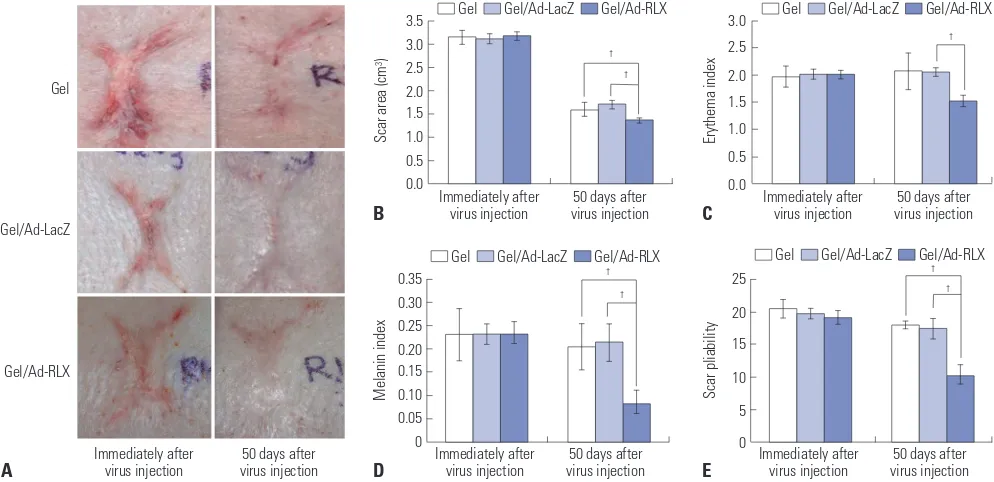
Scars with full thickness were generated on the backs of York-shire pigs. An RLX-expressing Ad vector entrapped in alginate gel was evaluated for effects on scar remodeling. The sizes of the initial scars were 3.15±0.15 cm2, 3.12±0.11 cm2, and 3.18±
0.08 cm2 in the gel, gel/Ad-LacZ, and gel/Ad-RLX groups,
re-spectively. Alginate gel (gel), alginate gel encapsulating RGD/LacZ (gel/Ad-LacZ), or alginate gel encapsulating dE1-RGD/LacZ/RLX (gel/Ad-RLX), respectively, was injected into the scar tissue. The sizes of the scars decreased to 1.61±0.15 cm2, 1.70±0.10 cm2, and 1.37±0.05 cm2 in each group,
respec-tively, by 50 days after treatment (Fig. 1A and B, Supplementa-ry Fig. 3, only online). These results indicated that RLX expres-sion from Ad (gel/Ad-RLX) reduces the size of scars compared to those of the control groups (gel or gel/Ad-LacZ) (p<0.05; Fig. 1B).
To further examine the effect of RLX on scar remodeling, color changes and pliability were investigated by spectropho-tometry in conjunction with erythema and melanin indices and a durometer. The erythema index values of the initial scars were 1.97±0.20, 2.01±0.10, and 2.01±0.09 in the gel, gel/Ad-LacZ, and gel/Ad-RLX groups, respectively. Erythema was significantly reduced (1.52±0.15; p<0.05) in the gel/Ad-RLX group at 50 days after treatment, compared to those in the gel (2.07±0.35) and gel/Ad-LacZ (2.05±0.08) groups (Fig. 1C). The initial melanin index values were 0.23±0.06, 0.23±0.02, and
0.23±0.03 in the gel, gel/Ad-LacZ, and gel/Ad-RLX groups, re-spectively (Fig. 1D). This index was also significantly (p<0.05) decreased to 0.08±0.02 in the gel/Ad-RLX group, compared to 0.21±0.05 and 0.21±0.04 in the gel and gel/Ad-LacZ control groups, respectively, at 50 days after treatment. Similarly, pli-ability was significantly (p<0.05) reduced in the gel/Ad-RLX group, compared to the gel and gel/Ad-LacZ control groups (Fig. 1E). The initial scars had pliability measurements of 20.50±1.42, 19.73±0.88, and 19.08±1.06 in the gel, gel/Ad-LacZ, and gel/ Ad-RLX groups, respectively. Pliability was reduced to 18.00± 0.58, 17.46±1.62, and 11.15±1.72 in the gel, gel/Ad-LacZ, and gel/Ad-RLX groups, respectively, by 50 days after treatment. Taken together, these data suggested that RLX expression from gel/Ad-RLX promotes the remodeling of scar tissue.
Relaxin-expressing Ad induces collagen
rearrangement and decreases expression of major ECM components in scar tissue
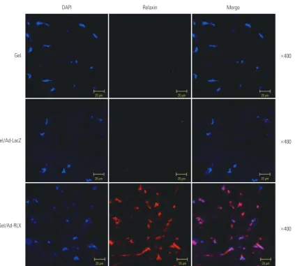
We examined RLX expression by immunofluorescence stain-ing in pig scar tissues transduced with gel, gel/Ad-LacZ, or gel/Ad-RLX. The gel/Ad-RLX-treated groups showed mark-edly increased immunoreactivity for RLX, compared to the control groups (gel or gel/Ad-LacZ). These results confirmed an association between increased RLX expression and reduced scar tissue formation in tissues transduced with the gel/Ad-RLX depot system (Fig. 2).
To evaluate the effects of RLX-expressing Ad on collagen-fi-ber arrangement, tissues were stained with picrosirius red, which binds specifically to collagen fibrils of various
diame-Fig. 1. Relaxin (RLX) expression reduces the size, color index, and pliability of pig scars. (A) Scars were created on the backs of pigs. The area of scars decreased, and the color improved at 50 days after the injection of relaxin-expressing Ad loaded in alginate gel (gel/Ad-RLX). (B) Scar size, (C) erythema index values, (D) melanin index values, and (E) pliability were significantly reduced (†p<0.05) compared to those of control groups [gel or LacZ-expressing
3.5 3.0 2.5 2.0 1.5 1.0 0.5 0.0
3.0 2.5 2.0 1.5 1.0 0.5 0.0
0.35 0.30 0.25 0.20 0.15 0.10 0.05 0
25
20
15
10
5
0 Immediately after
virus injection Immediately after virus injection
Immediately after virus injection Immediately after
virus injection Immediately after virus injection
50 days after
virus injection virus injection50 days after
50 days after virus injection 50 days after
virus injection virus injection50 days after
Scar area (cm
3)
Erythema index
Melanin index Scar pliability
B C
D E
†
†
†
† †
† †
Gel
Gel/Ad-LacZ
Gel/Ad-RLX
A
Gel Gel/Ad-LacZ Gel/Ad-RLX
Gel Gel/Ad-LacZ Gel/Ad-RLX
Gel Gel/Ad-LacZ Gel/Ad-RLX
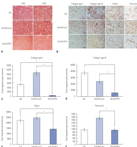
[image:4.595.56.553.454.695.2]ters. The gel/Ad-RLX-treated groups had closely packed colla-gen fibers and formed bundles of collacolla-gen (Fig. 3A), com-pared to those in the control groups (gel or gel/Ad-LacZ). These data suggested that RLX induces collagen rearrangement to resemble that of mature, bundle-shaped collagen fibers.
We next examined the effects of RLX overexpression on the major extracellular matrix (ECM) components of scar tissues. Immunohistochemical staining of scar sections revealed sig-nificant reductions in type-I collagen, type-III collagen, elastin, and fibronectin in the gel/Ad-RLX-treated group, compared with those in the gel/Ad-LacZ group (p<0.01) (Fig. 3B-F). These data strongly suggested that expression levels of the major ECM components were significantly decreased by RLX over-expression in pig scar tissues. Moreover, these results implied that RLX can play a prominent role in ECM remodeling during the development of scar tissue.
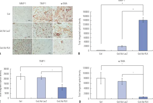
Relaxin-expressing Ad increases MMP-1 and decreas-es TIMP-1 and α-SMA expression in pig scar tissue Matrix metalloproteinase-1 (MMP-1), tissue inhibitor of me-talloproteinase-1 (TIMP-1), and alpha-smooth muscle actin (α-SMA) are important markers of the effects of TGF-β on wound repair. Therefore, we performed immunohistochemistry to examine expression of MMP-1, TIMP-1, and α-SMA. MMP-1 expression levels were significantly increased by 7.0-fold in pig scar tissues treated with RLX versus that of the gel/Ad-LacZ-treated tissues (p<0.01) (Fig. 4). In contrast, TIMP-1 and
α-SMA expression levels were significantly decreased by 1.9-and 9.9-fold, respectively, in pig scar tissues treated with gel/ Ad-RLX transduction (p<0.05). These results suggested that RLX upregulates MMP-1 and downregulates TIMP-1 and
[image:5.595.83.507.67.448.2]α-SMA, which are major players in collagen breakdown. Fig. 2. Immunofluorescence staining to evaluate the effects of relaxin on scar reduction in pig scar tissues. Increased expression of relaxin was de-tected after treatment with relaxin-expressing Ad loaded in alginate gel (gel/Ad-RLX). In contrast, control groups [gel or LacZ-expressing Ad loaded in alginate gel (gel/Ad-LacZ)] showed lower levels of relaxin. Nuclei were visualized by DAPI (4',6-diamidino-2-phenylindole) staining; original magni-fication, ×400; scale bar=20 µm.
Gel
Gel/Ad-LacZ
Gel/Ad-RLX
×400
×400
×400
DAPI Relaxin Merge
20 μm 20 μm 20 μm
20 μm 20 μm 20 μm
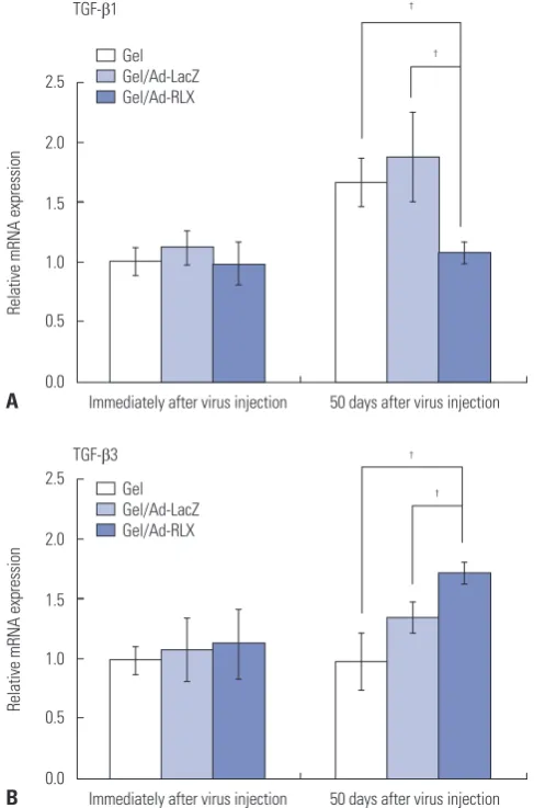
Relaxin-expressing Ad downregulates TGF-β1 and upregulates TGF-β3 expression in pig scar tissue To examine the mechanism by which RLX-expressing Ad sup-presses expression of the major ECM components, TGF-β1
β
gel/Ad-RLX group showed decreased TGF-β1 mRNA expres-sion and increased TGF-β3 mRNA expression in scar tissues, compared with those in the gel or gel/Ad-LacZ group (Fig. 5) (p<0.05), suggesting that the reduced expression of ECM
com-14000
12000 10000 8000 6000
4000 2000
0
50000
40000
30000
20000
10000
0
Gel Gel/Ad-LacZ Gel/Ad-RLX
Collagen type-I Collagen type-III
Total integrated optical density Total integrated optical density
C A
D Gel
Gel/Ad-LacZ
Gel/Ad-RLX
Gel
Gel/Ad-LacZ
Gel/Ad-RLX
×100 ×200
×400
Collagen type-I Collagen type-III Elastin Fibronectin
Gel Gel/Ad-LacZ Gel/Ad-RLX
*
* *
*
30000
25000
20000
15000
10000
5000
0
2000 1800 1600 1400 1200 1000 800 600 400 200 0
Gel Gel/Ad-LacZ Gel/Ad-RLX
Elastin Fibronectin
Total integrated optical density Total integrated optical density
E F Gel Gel/Ad-LacZ Gel/Ad-RLX
*
* *
[image:6.595.53.533.64.579.2]B
expression of TGF-β1 and increased expression of TGF-β3. We also checked TGF-β1 expression via immunohistochem-ical staining of the scar sections. The results revealed signifi-cant reductions in TGF-β1 in the gel/Ad-RLX-treated group, compared to the gel/Ad-LacZ group (p<0.05) (Supplementary Fig. 4, only online).
Relaxin-expressing Ad decreases mast cell counts and NK-1.1 expression in pig scar tissue
Mast cells are reported to be involved in the proliferation and contraction of fibroblasts, and the synthesis of ECM. They also play a key role in scar formation. The amount of mast cells was increased by four times in hypertrophic scars than in normal skin.27-29 To examine the local inflammatory effects of
RLX-ex-pressing Ad, mast cell counts and NK-1.1 immunohistochem-istry were assessed. Immediately after scar formation, the mean numbers of mast cells were 6.20±1.30, 7.00±0.80, and 6.30± 0.70 in the gel, gel/Ad-LacZ, and gel/Ad-RLX groups, respec-tively, with no significant differences. Fifty days after injection of the virus, the mean numbers of observed mast cells were 6.80±
0.42 in the gel group and 7.09±0.68 in the gel/Ad-LacZ group. A significant decrease was seen in the mean number of mast cells in the gel/Ad-RLX group (4.89±0.40; p<0.01; Supplemen-tary Fig. 5, only online). NK-1.1 expression was significantly lower in the gel/Ad-LacZ and gel/Ad-RLX-treated groups, compared to the gel group (p<0.05) (Supplementary Fig. 6, only online).
DISCUSSION
Scars, which are caused by collagen deposition, confer a large number of functional problems. Nowadays, many patients want to minimize scar formation, and many treatment modalities, such as scar revision, laser therapy, and medications, have been applied to prevent scar formation. However, the suc-cessful treatment of scars has not been fully resolved.30-32
This study was performed by adopting a pig scar model used our previous study. The aim of this study was to verify the anti-fibrotic effect of RLX on scars. Recent studies supported our
30000
25000
20000 15000
10000
5000 0
120000
100000
80000
60000
40000
20000
0 180000 160000 140000 120000 100000 80000 60000 40000 20000 0
Gel Gel/Ad-LacZ Gel/Ad-RLX Gel Gel/Ad-LacZ Gel/Ad-RLX
Gel Gel/Ad-LacZ Gel/Ad-RLX
Total integrated optical density Total integrated optical density
Total integrated optical density
C D
B A
Gel
Gel/Ad-LacZ
Gel/Ad-RLX
† †
*
*
†
†
MMP-1 TIMP-1 α-SMA
TIMP-1 α-SMA
[image:7.595.45.540.76.410.2]MMP-1
data and suggested that RLX exerts alternative biological effects mediated directly through TGF-β-dependent signaling.14,18,19,30-32
These effects elicit downregulation of TGF-β1 and upregula-tion of TGF-β3 to inhibit myofibroblast accumulation, colla-gen synthesis/secretion, and ultimately, fibrosis progression in scar tissue. Other studies have suggested that TGF-β1 is a potent fibrogenic growth factor, whereas TGF-β3 reduces scar tissue by decreasing synthesis and increasing degradation of type-I collagen.33 TGF-β plays a key role in the
pathophysiolo-gy of fibroplasia by decreasing the expression of MMPs and in-creasing the expression of TIMPs.19
Furthermore, TGF-β induces α-SMA expression, implying that α-SMA expression is a marker of TGF-β activity.32-37 Thus,
the increased expression of MMP-1 and decreased levels of TIMP and α-SMA suggest that dE1-RGD/LacZ/RLX plays a
prominent role in ECM remodeling in hypertrophic scars. MMP-1 promotes degradation and ECM remodeling, whereas TIMP-1 decreases the activities of plasminogen activators, such as MMPs, and shifts the ECM equilibrium toward degra-dation. Thus, increased MMP-1 production and decreased TIMP-1 expression should reduce the amount of abnormal or unfolded collagen during wound healing. In addition, reduced levels of α-SMA may be a positive indicator of TGF-β activity, which was decreased in the gel/Ad-RLX group in this study. Taken together, these studies suggest that dE1-RGD/LacZ/ RLX is a potential therapeutic intervention for hypertrophic scars.
Ad has been used extensively as a gene-delivery system in experimental models of cancer and cardiovascular diseases.38-40
However, one of the chief concerns with translating these ex-perimental results to the clinical setting is the large and re-peated dose that is usually required to generate a modest clin-ical effect or to achieve the desired therapeutic concentration at the target. To address this concern, we utilized an Arg-Gly-Asp-modified (RGD) replication-incompetent Ad vector as a strategy to allow direct delivery of the RLX gene to wounds. Previous studies from our lab showed that RGD-modified Ad markedly increases gene transfer efficiency in primary keloid fibroblasts. Thus, we believe that RGD-modified Ad is a highly efficient gene-transfer vehicle that does not adversely affect the replication potential of target cells.14,17
To further advance this potential therapy for hypertrophic scars, we generated and characterized an alginate gel-matrix system to entrap RLX-expressing Ad as a delivery vehicle.22,23
Previous studies have indicated that the biological activity of Ad loaded in alginate gel is prolonged, compared with that of naked Ad, over an extended period of time. Cells that were transduced with gel-released Ad expressed green fluorescence protein at a 13-fold greater intensity at seven days post-incuba-tion compared to that of naked Ad. Because long-term trans-duction is needed for in vivo applications, the Ad/alginate gel may be very useful as a depot system. The Ad/alginate gel acts as a reservoir that releases Ad in a sustained manner and maintains the biological activity of Ad. Alginate gel has also been shown to limit the mobility of Ad by entrapping Ad with-in its scaffold, further mwith-inimizwith-ing the spread of the replica-tion-incompetent Ad outside of the scar tissue. These results show that sustained and controlled delivery of Ad in alginate gel markedly augments therapeutic effects, prolongs mainte-nance of Ad activity, and provides specificity for local delivery of replication-incompetent Ad at high concentrations. These advantages of the Ad/alginate system will allow for potent lo-cal viral therapy to a target site.
In conclusion, we provide strong evidence that RLX pro-motes the remodeling of scar tissue. Moreover, sustained and controlled delivery of Ad in alginate gel markedly augments the therapeutic effects of RLX. Our data support further evalu-ation of gel/Ad-RLX as a novel gene-therapy system for
treat-2.5
2.0
1.5
1.0
0.5
0.0
2.5
2.0
1.5
1.0
0.5
0.0
Immediately after virus injection
Immediately after virus injection TGF-β1
TGF-β3
50 days after virus injection
50 days after virus injection
Relative mRNA expression
Relative mRNA expression
A
B
†
† †
† Gel
Gel/Ad-LacZ Gel/Ad-RLX
[image:8.595.54.298.73.442.2]Gel Gel/Ad-LacZ Gel/Ad-RLX
ing human scars.
ACKNOWLEDGEMENTS
This work was supported by a faculty research grant of Yonsei University College of Medicine (6-2012-0037, Dr. In Sik Yun), a grant from Hanyang University of Korea (HY-2011-G-2011 00000001880; Dr. Chae-Ok Yun) and the National Research Foundation of Korea (NRF-2018R1D1A1B07042537, Dr. In Sik Yun; 2016M3A9B5942352, Dr. Chae-Ok Yun). Hyo Min Ahn is a graduate student sponsored by the Brain Korea 21 plus pro-gram, Hanyang University, Seoul, South Korea (22A2013001 1095).
AUTHOR CONTRIBUTIONS
Conceptualization: Won Jai Lee and Chae-Ok Yun. Data curation: Eunhye Kang. Formal analysis: Hyo Min Ahn. Funding acquisition: Chae-Ok Yun. Investigation: In Sik Yun. Project administration: Yong Oock Kim. Supervision: Dong Kyun Rah and Chae-Ok Yun. Valida-tion: Won Jai Lee. Writing—original draft: In Sik Yun. Writing—review & editing: Tai Suk Roh.
ORCID iDs
In Sik Yun https://orcid.org/0000-0003-1103-7047
Eunhye Kang https://orcid.org/0000-0002-8094-0726
Hyo Min Ahn https://orcid.org/0000-0002-1268-8637
Yong Oock Kim https://orcid.org/0000-0002-3756-4809
Tai Suk Roh https://orcid.org/0000-0001-8681-159X
Won Jai Lee https://orcid.org/0000-0003-3056-0503
Chae-Ok Yun https://orcid.org/0000-0002-9466-4531
REFERENCES
1. Desmoulière A, Darby IA, Gabbiani G. Normal and pathologic soft tissue remodeling: role of the myofibroblast, with special em-phasis on liver and kidney fibrosis. Lab Invest 2003;83:1689-707. 2. Le AD, Brown JJ. Wound healing: repair biology and wound and
scar treatment. In: Bagheri SC, Bell RB, Khan HA, editors. Current therapy in oral and maxillofacial surgery. 1st ed. Philadelphia: Saunders; 2012. p.6-10.
3. Bae SH, Bae YC. Analysis of frequency of use of different scar as-sessment scales based on the scar condition and treatment meth-od. Arch Plast Surg 2014;41:111-5.
4. Corr DT, Gallant-Behm CL, Shrive NG, Hart DA. Biomechanical behavior of scar tissue and uninjured skin in a porcine model. Wound Repair Regen 2009;17:250-9.
5. Wang XQ, Kravchuk O, Liu PY, Kempf M, Boogaard CV, Lau P, et al. The evaluation of a clinical scar scale for porcine burn scars. Burns 2009;35:538-46.
6. Wang XQ, Liu PY, Kempf M, Cuttle L, Chang AH, Wong M, et al. Burn healing is dependent on burn site: a quantitative analysis from a porcine burn model. Burns 2009;35:264-9.
7. Yun IS, Jeon YR, Lee WJ, Lee JW, Rah DK, Tark KC, et al. Effect of human adipose derived stem cells on scar formation and remod-eling in a pig model: a pilot study. Dermatol Surg 2012;38:1678-88. 8. Zhao S, Lee HY, Sherwood OD. Porcine and human relaxin bioac-tivity: bioactivities of porcine relaxin and human relaxin do not
differ in mice and rats. Ann N Y Acad Sci 2005;1041:126-31. 9. Samuel CS, Hewitson TD, Unemori EN, Tang ML. Drugs of the
fu-ture: the hormone relaxin. Cell Mol Life Sci 2007;64:1539-57. 10. Sherwood OD. Relaxin's physiological roles and other diverse
ac-tions. Endocr Rev 2004;25:205-34.
11. Samuel CS, Hewitson TD. Relaxin in cardiovascular and renal disease. Kidney Int 2006;69:1498-502.
12. Dschietzig T, Bartsch C, Baumann G, Stangl K. Relaxin-a pleiotro-pic hormone and its emerging role for experimental and clinical therapeutics. Pharmacol Ther 2006;112:38-56.
13. Unemori EN, Erikson ME, Rocco SE, Sutherland KM, Parsell DA, Mak J, et al. Relaxin stimulates expression of vascular endothelial growth factor in normal human endometrial cells in vitro and is as-sociated with menometrorrhagia in women. Hum Reprod 1999;14: 800-6.
14. Kim JH, Lee YS, Kim H, Huang JH, Yoon AR, Yun CO. Relaxin ex-pression from tumor-targeting adenoviruses and its intratumoral spread, apoptosis induction, and efficacy. J Natl Cancer Inst 2006; 98:1482-93.
15. Unemori EN, Lewis M, Constant J, Arnold G, Grove BH, Normand J, et al. Relaxin induces vascular endothelial growth factor expres-sion and angiogenesis selectively at wound sites. Wound Repair Regen 2000;8:361-70.
16. Chartier C, Degryse E, Gantzer M, Dieterle A, Pavirani A, Mehtali M. Efficient generation of recombinant adenovirus vectors by ho-mologous recombination in Escherichia coli. J Virol 1996;70: 4805-10.
17. Kim J, Cho JY, Kim JH, Jung KC, Yun CO. Evaluation of E1B gene-attenuated replicating adenoviruses for cancer gene therapy. Can-cer Gene Ther 2002;9:725-36.
18. Lee WJ, Choi IK, Lee JH, Lee JS, Kim YO, Rah DK, et al. Relaxin-expressing adenovirus decreases collagen synthesis and up-regu-lates matrix metalloproteinase expression in keloid fibroblasts: in vitro experiments. Plast Reconstr Surg 2012;130:407e-17e. 19. Lee WJ, Kim YO, Choi IK, Rah DK, Yun CO. Adenovirus-relaxin
gene therapy for keloids: implication for reversing pathological fi-brosis. Br J Dermatol 2011;165:673-7.
20. Lee WJ, Yun CO, Yun IS, Kim YO, Choi IK, Yun TJ, et al. Augmen-tation of rat skin flap viability by relaxin-expressing adenovirus. Wound Repair Regen 2011;19:709-17.
21. Ahi YS, Bangari DS, Mittal SK. Adenoviral vector immunity: its im-plications and circumvention strategies. Curr Gene Ther 2011;11: 307-20.
22. Choi JW, Kang E, Kwon OJ, Yun TJ, Park HK, Kim PH, et al. Local sustained delivery of oncolytic adenovirus with injectable alginate gel for cancer virotherapy. Gene Ther 2013;20:880-92.
23. Park H, Kim PH, Hwang T, Kwon OJ, Park TJ, Choi SW, et al. Fabri-cation of cross-linked alginate beads using electrospraying for ad-enovirus delivery. Int J Pharm 2012;427:417-25.
24. Choi KJ, Zhang SN, Choi IK, Kim JS, Yun CO. Strengthening of an-titumor immune memory and prevention of thymic atrophy me-diated by adenovirus expressing IL-12 and GM-CSF. Gene Ther 2012;19:711-23.
25. Zhang SN, Choi IK, Huang JH, Yoo JY, Choi KJ, Yun CO. Optimiz-ing DC vaccination by combination with oncolytic adenovirus co-expressing IL-12 and GM-CSF. Mol Ther 2011;19:1558-68. 26. Yun IS, Lee WJ, Rah DK, Kim YO, Park BY. Skin color analysis using
a spectrophotometer in Asians. Skin Res Technol 2010;16:311-5. 27. Kitamura Y, Oboki K, Ito A. Molecular mechanisms of mast cell
development. Immunol Allergy Clin North Am 2006;26:387-405. 28. Sur R, Cavender D, Malaviya R. Different approaches to study mast
cell functions. Int Immunopharmacol 2007;7:555-67.
cell activation. Immunol Rev 2009;228:149-69.
30. Park BY, Shin IS, Yun IS. Dovetail scar revision. Dermatol Surg 2012;38:1716-21.
31. Kim SG, Kim EY, Kim YJ, Lee SI. The efficacy and safety of ablative fractional resurfacing using a 2,940-Nm Er:YAG laser for traumatic scars in the early posttraumatic period. Arch Plast Surg 2012;39: 232-7.
32. Greenhalgh DG. Consequences of excessive scar formation: deal-ing with the problem and aimdeal-ing for the future. Wound Repair Re-gen 2007;15 Suppl 1:S2-5.
33. Shah M, Foreman DM, Ferguson MW. Neutralisation of TGF-beta 1 and TGF-beta 2 or exogenous addition of TGF-beta 3 to cutane-ous rat wounds reduces scarring. J Cell Sci 1995;108(Pt 3):985-1002.
34. Wilgus TA, Vodovotz Y, Vittadini E, Clubbs EA, Oberyszyn TM. Reduction of scar formation in full-thickness wounds with topical celecoxib treatment. Wound Repair Regen 2003;11:25-34. 35. Liu W, Chua C, Wu X, Wang D, Ying D, Cui L, et al. Inhibiting scar
formation in rat wounds by adenovirus-mediated overexpression
of truncated TGF-beta receptor II. Plast Reconstr Surg 2005;115: 860-70.
36. Gallant CL, Olson ME, Hart DA. Molecular, histologic, and gross phenotype of skin wound healing in red Duroc pigs reveals an abnormal healing phenotype of hypercontracted, hyperpigment-ed scarring. Wound Repair Regen 2004;12:305-19.
37. Margulis A, Nocka KH, Wood NL, Wolf SF, Goldman SJ, Kasaian MT. MMP dependence of fibroblast contraction and collagen pro-duction induced by human mast cell activation in a three-dimen-sional collagen lattice. Am J Physiol Lung Cell Mol Physiol 2009; 296:L236-47.
38. Samuel CS, Cendrawan S, Gao XM, Ming Z, Zhao C, Kiriazis H, et al. Relaxin remodels fibrotic healing following myocardial infarc-tion. Lab Invest 2011;91:675-90.
39. Stewart DR. Scar prevention and cosmetically enhanced wound healing using relaxin. Ann N Y Acad Sci 2009;1160:336-41. 40. Du XJ, Xu Q, Lekgabe E, Gao XM, Kiriazis H, Moore XL, et al.