Bcl-2 and Bcl-X
L
serve an anti-inflammatory
function in endothelial cells through inhibition
of NF-
kk
B
A.Z. Badrichani, … , F.H. Bach, C. Ferran
J Clin Invest.
1999;
103(4)
:543-553.
https://doi.org/10.1172/JCI2517
.
To maintain the integrity of the vascular barrier, endothelial cells (EC) are resistant to cell
death. The molecular basis of this resistance may be explained by the function of
antiapoptotic genes such as
bcl
family members. Overexpression of Bcl-2 or Bcl-X
Lprotects
EC from tumor necrosis factor (TNF)–mediated apoptosis. In addition, Bcl-2 or Bcl-X
Linhibits activation of NF-
k
B and thus upregulation of proinflammatory genes. Bcl-2–
mediated inhibition of NF-
k
B in EC occurs upstream of I
k
B
a
degradation without affecting
p65-mediated transactivation. Overexpression of
bcl
genes in EC does not affect other
transcription factors. Using deletion mutants of Bcl-2, the NF-
k
B inhibitory function of Bcl-2
was mapped to
bcl
homology domains BH2 and BH4, whereas all BH domains were
required for the antiapoptotic function. These data suggest that Bcl-2 and Bcl-X
Lbelong to a
cytoprotective response that counteracts proapoptotic and proinflammatory insults and
restores the physiological anti-inflammatory phenotype to the EC. By inhibiting NF-
k
B
without sensitizing the cells (as with I
k
B
a
) to TNF-mediated apoptosis, Bcl-2 and Bcl-X
Lare
prime candidates for genetic engineering of EC in pathological conditions where EC loss
and unfettered activation are undesirable.
Article
Find the latest version:
Introduction
Under physiological conditions, vascular endothelium is a multifunctional anticoagulant and anti-inflammatory barrier (1, 2). The endothelial interface can dynamically modify its phenotype (EC activation), to evoke an inflam-matory environment in response to a variety of patho-physiological stimuli, including endotoxin, proinflam-matory cytokines, and immunological insults such as those associated with graft rejection and autoimmunity (2–4). Acquisition by EC of this activated phenotype has been thoroughly analyzed in the literature and implicates the de novo expression of genes such as those encoding for adhesion molecules (E-selectin), chemokines (inter-leukin-8 [IL-8]), and procoagulant factors (tissue factor [TF]). The induction of most of these genes is regulated by a key transcription factor: NF-κB (5, 6). In most cir-cumstances, EC resist the damaging potential associated with this proinflammatory environment and revert to their original quiescent phenotype. More recently, analy-sis of gene expression in EC of long-term surviving ham-ster to rat heart xenograft revealed the potential of EC to acquire a novel phenotype by expressing high levels of antiapoptotic proteins, namely the Zn finger protein A20 and the antiapoptotic bclmembers Bcl-2 and Bcl-XL(7).
Expression of these genes correlated with the absence of inflammation, thrombosis, and endothelial cell death, features seen in rejecting xenografts that lack the expres-sion of A20, Bcl-2, and Bcl-XLin their EC (7).
The Zn finger protein A20 was originally identified as a tumor necrosis factor (TNF)–inducible gene in human umbilical vein endothelial cells (HUVEC) (8, 9). We
recently demonstrated that A20 has a dual function in EC: protection from apoptosis and downregulation of EC activation through inhibition of the transcription factor NF-κB (10, 11). However, both expression and function of Bcl-2 and Bcl-XL in EC remain poorly
defined (12, 13). Bcl-2 and Bcl-XLare prototypic
cell-death regulators whose function is modulated by com-plex homo- and heterodimerizations with their proapop-totic homologues such as Bax and/or with other nonrelated molecules such as Raf-1 kinase (14–18). Both these proteins confer to cells resistance to a variety of proapoptotic stimuli, including hypoxia, radiation, growth factor withdrawal, and others (19), but their effect upon TNF-mediated apoptosis is still controver-sial (20, 21). TNF is associated with most proinflamma-tory conditions and is a potent activator of EC both in vivo and in vitro(22, 23). In this work, we studied the function of Bcl-2 and Bcl-XLin EC. Our results
demon-strate that expression of Bcl-2 or Bcl-XLin EC
signifi-cantly protects the cells from TNF-mediated apoptosis after sensitization with cycloheximide (CHX). In addi-tion, we show that Bcl-2 and Bcl-XLare able to
downreg-ulate EC activation independently of the agonist tested through specific inhibition of the transcription factor NF-κB. Inhibition occurs at a level upstream of IκBα degradation and involves stabilization of a slower migrating band that might represent a hyperphospho-rylated form of IκBα. Bcl-2 and Bcl-XLshare the same
function(s) in EC with the nonrelated antiapoptotic molecule A20. Their coexpression in EC of long-term surviving xenografts defines a novel protected
pheno-Bcl-2 and Bcl-X
Lserve an anti-inflammatory function
in endothelial cells through inhibition of NF-κB
A.Z. Badrichani,
1D.M. Stroka,
1G. Bilbao,
2D.T. Curiel,
2F.H. Bach,
1and C. Ferran
11Immunobiology Research Center, Department of Surgery, Beth Israel-Deaconess Medical Center, Harvard Medical School,
Boston, Massachusetts 02215, USA
2Gene Therapy Program, University of Alabama at Birmingham, Birmingham, Alabama 35294, USA
Address correspondence to: Christiane Ferran, Immunobiology Research Center, Beth Israel-Deaconess Medical Center, 99 Brooklyn Avenue, Room 370, Boston Massachusetts 02215, USA. Phone: (617) 632-0840; Fax: (617) 632-0880; E-mail: [email protected]
Received for publication December 15, 1997, and accepted in revised form January 4, 1999.
To maintain the integrity of the vascular barrier, endothelial cells (EC) are resistant to cell death. The molecular basis of this resistance may be explained by the function of antiapoptotic genes such as bcl
family members. Overexpression of Bcl-2 or Bcl-XLprotects EC from tumor necrosis factor
(TNF)–medi-ated apoptosis. In addition, Bcl-2 or Bcl-XLinhibits activation of NF-κB and thus upregulation of
proin-flammatory genes. Bcl-2–mediated inhibition of NF-κB in EC occurs upstream of IκBαdegradation without affecting p65-mediated transactivation. Overexpression of bclgenes in EC does not affect other transcription factors. Using deletion mutants of Bcl-2, the NF-κB inhibitory function of Bcl-2 was mapped to bclhomology domains BH2 and BH4, whereas all BH domains were required for the anti-apoptotic function. These data suggest that Bcl-2 and Bcl-XLbelong to a cytoprotective response that
counteracts proapoptotic and proinflammatory insults and restores the physiological anti-inflamma-tory phenotype to the EC. By inhibiting NF-κB without sensitizing the cells (as with IκBα) to TNF-medi-ated apoptosis, Bcl-2 and Bcl-XL are prime candidates for genetic engineering of EC in pathological
con-ditions where EC loss and unfettered activation are undesirable.
type of EC, whereby these molecules sum their potentials to protect the cell from death and from the untoward effect of EC activation (7). We suggest that the dual func-tion in EC of Bcl-2 and Bcl-XL(i.e., antiapoptotic and
anti-inflammatory, through inhibition of NF-κB) qual-ifies their cytoprotective role.
Methods
Cell culture and treatment. Bovine aortic endothelial cells (BAEC) were isolated and cultured in DMEM supplemented with L
-glu-tamine (2 mM), penicillin G (100 U/ml), and FCS (10%). Prima-ry cultures of BAEC were used between the fourth and the fifth passage. HUVEC were isolated and cultured as described (24).The 293 human embryonic kidney cell line was obtained from American Type Culture Collection (Rockville, Maryland, USA) and cultured in 10% FCS-supplemented DMEM. All cells were grown in culture at 37°C in a 5% humid CO2atmosphere. EC were stimulated with either 100 ng/ml of LPS from Escherichia coli0B55 (Sigma Chemical Co., St. Louis, Missouri, USA), 100 U/ml of recombinant human TNF (kind gift of Sandoz Phar-maceuticals, East Hanover, New Jersey, USA), or 5 ×10⁻⁸M of PMA (Sigma Pharmaceuticals, St. Louis, Missouri, USA).
Lipofection protocol. BAEC (3 ×10⁵per well) were plated in a 6-well plate and transfected when they reached 70% confluence. A total of 1.6 µg of DNA per well (test plasmids and reporter constructs) was added to 8 µg of Lipofectamine (GIBCO BRL, Grand Island, New York, USA), incubated at room temperature for 30 min, and then added to the cells in triplicate. In all exper-iments, 0.3 µg of the β-galactosidase (β-gal) reporter was used with 0.7 µg of the expression plasmids or the pAC control, and 0.6 µg of the E-selectin, IL-8, IκBα(ECI-6), or NF-κB-luciferase (luc) reporters. For the apoptosis experiments, the newly devel-oped Lipofectamine Plus Reagent (GIBCO BRL) was used according to the manufacturer’s instructions to achieve a high-er phigh-ercentage of transfection. In exphigh-eriments involving the induction of the IκBαreporter by the p65 (RelA) expression vector, 40 ng of p65 was used. For the HIV-wt-chlorampheni-col acetyltransferase (HIV-CAT) and HIV∆κB-CAT reporters experiments, 0.5 µg of the expression plasmids (mBcl-2,
mBcl-XL,or pAC) were transfected with 0.3 µg of the c-Tat expression plasmid and 0.6 µg of the HIV-CAT, or the HIV∆−
κB-CAT reporter along with 0.2 µg of the β-gal reporter. In all cotransfection experiments, FCS was added to the medium 5 h after transfection to achieve a final concentration of 10%. Forty-eight hours after transfection, the cells were stim-ulated with either human recombinant TNF (100U/ml), LPS (100 ng/ml), or PMA (5 ×10⁻⁸ M), harvested 7 h later, and assayed for β-gal, luciferase, and CAT.
Human embryonic kidney 293 cells were transfected using the Calcium-Phosphate method. Then 1 µg of the expression plasmids (mBcl-2, mBcl-XL,or pAC), 0.7 µg of IκBαreporter, and 40 ng of p65 (RelA) were added per well.
β-gal, luciferase, and CAT assays. Cellular extracts were assayed for β-gal activity per the Galacto-Light protocol (Tropix Inc., Bedford, Massachusetts, USA). Luciferase activity was assayed by adding 10 µl of cellular extract to 90 µl of a solution con-taining 24 mM glyclglycine (pH 7.8), 2 mM ATP (pH 7.5), and 10 mM MgSO4. Samples were read on the Microlumat LB 96P luminometer (EG&G Berthold, Wildbad, Germany) using an injection mix consisting of 24 mM glyclglycine and 0.1 mM luciferin (Sigma Chemical Co.).
Luciferase activity was normalized for β-gal by using the for-mula: luciferase activity / β-gal activity ×1,000. Normalized luciferase activity is given in relative light units (RLU). Signifi-cance was determined by the Student’s ttest. The CAT assay was performed for the HIV-wt reporter by means of a standard method using a Promega Kit (Promega Corp., Madison,
Wis-consin, USA) according to the manufacturer’s recommenda-tion. A portion of the xylene phase was mixed with scintillation liquid and counted in a scintillation counter (1900 TR; Packard Instrument Co., Downers Grove, Illinois, USA).
Reporter constructs. E-selectin reporter. The reporter construct used was described previously (25). Briefly, it represents bp –1286 to +484 of the porcine E-selectin promoter. This region includes the first complete intron and exon, as well as the beginning of the sec-ond exon up to the ATG site. The promoter was cloned into the pMAMneo-luc plasmid vector by replacing the mmTV promoter (CLONTECH Laboratories Inc., Palo Alto, California, USA).
IL-8 reporter. A gift from E. Hofer (VIRCC, Vienna, Austria) represents the human IL-8 promoter linked to the luciferase gene (p-UBT luc).
IκBα (ECI-6) reporter. The construction of this reporter has been described previously (26). It represents a 600-bp fragment of the porcine ECI-6/IκBαpromoter ligated into the luciferase expression vector p-UBT (p-UBT-luc), with the creation of an additional HindIII site.
NF-κB reporter. This reporter is a kind gift from A. Palmetshofer (Beth Israel Deaconess Medical Center, Boston, Massachusetts, USA). It consists of four copies of NF-κB elements taken from the porcine E-selectin promoter inserted upstream of a TK minimal promoter driving a luciferase gene. The vector backbone is a Blue-script KS+ plasmid (Stratagene, La Jolla, California, USA).
RSVβ-gal reporter.The full-length E. coliβ-gal gene (CLON-TECH Laboratories Inc.) was inserted into the pRc/RSV vec-tor (Invitrogen Corp., San Diego, California, USA) at the NotI site. This reporter is not inducible upon stimulation by the agonists used in this paper. Thus, its activity was used to cor-rect for transfection efficiency.
Expression plasmids. The murine Bcl-2 and Bcl-XLcDNA are a kind gift of T. Behrens (University of Minnesota, Minneapolis, Minnesota, USA). The full-length human Bcl-2 is a kind gift of G. Nünez (University of Michigan, Ann Arbor, Michigan, USA), and the deleted forms (∆1, ∆2, ∆3, ∆4, ∆6, ∆8, ∆12) are a kind gift of T.G. Parslow (University of California, San Francisco, San Francisco, California, USA). These cDNA were subcloned in the pAC expression vector. The pAC 8.8-kb plasmid vector contains a CMV promoter, a pUC19 polylinker site, and a SV40 splice/polyA site (a kind gift of R. Gerard, University of Texas Southwestern, Dallas, Texas, USA).
p65. The p65 expression plasmid is a kind gift of J. Anrather (Beth Israel Deaconess Medical Center, Boston, Massachusetts, USA) and represents the human RelA (from amino acid 2 to 551) fused to a NH2-terminal c-mycTag and cloned into the pcDNA3 expression plasmid (Invitrogen Corp.) at the Hin dI-II/XbaI polycloning sites.
anti–mouse and human Bcl-2 polyclonal antibodies (Santa Cruz Biotechnology Inc., Santa Cruz, California, USA). Furthermore, the presence of the expression cassette in the viral genome was con-firmed by DNA sequencing. The rAd.β-gal used as a control aden-ovirus is a kind gift of R. Gerard (University of Texas Southwest-ern). Production of rAd was done in the embryonic kidney 293 cell line. Recombinant adenoviruses were subsequently purified by two consecutive cesium chloride centrifugation and titered by limiting dilution on 293 cells.
Cells extracts. Transfected BAEC or HUVEC infected with rAd were harvested in PBS 48 h after transfection or infection and lysed in Ripa buffer (10 mM Tris [pH 7.5], 150 mM NaCl, 1% [vol/vol] Triton X-100, 0.5 µg/ml each of aprotinin, leupeptin, and antipain, 1 µg/ml of pepstatin, and 0.5 mM PMSF) for 20 min at 4°C. Cellular debris were pelleted by microcentrifugation for 20 min at 4°C, and supernatants were recovered and kept at –80°C until assayed. The protein concentration of these cell extracts was evaluated by the Lowry assay (Bio-Rad DC Protein Assay; Bio-Rad Laboratories Inc., Hercules, California, USA)
Immunoblots. Proteins (15–20 µg) were resolved on a reducing 12% SDS-polyacrylamide gel and transferred onto Immobilon-P transfer membranes (Millipore Corp., Bedford, Massachusetts, USA) at 0.8 mA/cm2. Membranes were preblocked at room tem-perature for 1 h in 5% (wt/vol) BLOTTO nonfat dry milk in 0.1% (vol/vol) Tween-20 PBS. Membranes were then labeled for 1 h with a first specific antibody. After four 10-min washings in PBS containing 0.1% Tween-20, membranes were incubated for 1 h at room temperature with secondary donkey anti–rabbit IgG anti-body conjugated to horseradish peroxidase (1:3,000 dilution) (Pierce Chemical Co., Rockford, Illinois, USA). Detection was then performed by enhanced chemiluminescence (ECL) using a commercially available kit (Amersham Corp., Arlington Heights, Illinois, USA) according to the manufacturer’s instructions.
Murine Bcl-2 (mBcl-2) expression was detected using a 1:500 dilution of a polyclonal rabbit antibody (N-19) directed against the amino acids 4–21 of the murine or human Bcl-2. Human wild-type Bcl-2 and Bcl-2 deletion proteins were detected using a 1:200 mouse monoclonal IgG antibody against Bcl-2 (DAKO Corp., Carpinteria, California, USA) or using the N-19 anti-body. Bcl-XLexpression was detected with a 1:500 dilution of a polyclonal rabbit antibody raised against the human and mouse Bcl-XS/L. IκBαexpression was detected using a rabbit polyclonal antibody (C-21; dilution 1:2,000). All these anti-bodies were purchased from Santa Cruz Biotechnology.
Flow cytometric analysis of E-selectin and vascular cell adhesion mol-ecule-1 expression.HUVEC were cultured to 90% confluence in 6-well plates and infected at a moiety of infection (MOI) of 100 with either the rAd.hBcl-2, the rAd.β-gal, or noninfected. Thir-ty-six to 48 h after infection, HUVEC were treated with 200 U/ml of TNF for 4 h (E-selectin expression) and 8 h (vascular cell adhesion molecule-1 [VCAM-1] expression), after which cells were harvested and labeled for surface expression of both adhe-sion molecules. Approximately 105cells were incubated with 40
µl (1 µg/ml) of mouse anti–human monoclonal antibodies to either E-selectin (CD62E) or VCAM-1 (CD106) (R&D Systems Inc., Minneapolis, Minnesota, USA) or an isotype-matched con-trol monoclonal antibody (IgG1) for 20 min on ice. Cells were then washed and incubated with 40 µl of FITC-conjugated goat anti–mouse IgG (1:300) for 20 min on ice. Surface antigen expression was analyzed with a FACScan bench-top model (Bec-ton Dickinson Immunocytometry Systems, San Jose, California, USA) using Cellquest acquisition and analysis software (Becton Dickinson Immunocytometry Systems). Data were collected from viable cells only as determined by forward- and side-scat-tered light properties and propidium iodide staining.
Evaluation of apoptosis in a transient-transfection assay. BAEC grown in 6-well plates were cotransfected using Lipofectamine Plus (GIBCO BRL) with 0.5 µg of the reporter plasmid
CMV-βgal together with 1 µg of the expression plasmids (mBcl-2, mBcl-XL, hBcl-2, and the different hBcl-2 mutants, or the pAC control plasmids). Twenty-four hours after transfection, cells were treated with 2 µg/ml of CHX (Sigma Chemical Co.), or with CHX followed 30 min later by 200 U/ml of TNF. After 12 h incubation with the respective treatments, the cells were fixed with 0.05% glutaraldehyde and stained with 5-bromo-4-chloro-3-indolyl-β-D-galactopyranoside for 4 h. BAEC were then visu-alized by phase-contrast microscopy, and the number of living β-gal–positive cells were evaluated in random 10 high-power fields per well (minimal number of 100 blue cells). Viable or apoptotic cells were distinguished based on morphological alter-ations of adherent cells undergoing apoptosis, including becom-ing rounded, condensed, and detached from the dish (31). The number of blue cells in CHX-treated BAEC and for each given expression plasmid was considered as corresponding to 100% of survival; the percentage of survival in the CHX-TNF–treated wells was calculated relative to that number as described else-where (32). CHX alone does not induce apoptosis in BAEC but sensitizes these cells to TNF-mediated apoptosis.
Results
Expression of Bcl-2 or Bcl-XLin BAEC protects CHX-sensitized
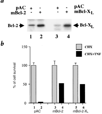
cells from TNF-mediated apoptosis. Expression of murine Bcl-2 and Bcl-XL after Lipofectamine transfection in BAEC
was confirmed by Western blot analysis (Fig. 1a). BAEC transfected with the empty expression plasmid show lit-tle expression of Bcl-2 or Bcl-XL(lanes 1and 3). In
con-trast, BAEC transfected with the Bcl-2 or Bcl-XL
expres-sion plasmids show high levels of expresexpres-sion (lanes 2and 4). We then tested whether expression of Bcl-2 or Bcl-XL
protected CHX-sensitized EC from undergoing TNF-mediated apoptosis.
As described in Methods, BAEC were cotransfected with mBcl-2, mBcl-XL, or pAC expression plasmids
together with the CMV-βgal plasmid. After 12 hours of CHX and TNF treatment, the percentage of cell survival was decreased to 3 ± 1% in pAC-transfected cells (Fig. 1b, lane 2vs. lane 1). Overexpression of mBcl-2 or mBcl-XL
leads to significant protection: the percentage of cell sur-vival reaches 52 ± 2% and 50 ± 4% in mBcl-2– or mBcl-XL–expressing cells (Fig. 1b, lane 4vs. lanes 3and 6
vs. lane 5). These results demonstrate that the antiapop-totic genes bcl-2and bcl-XLcan also interrupt
TNF-medi-ated apoptotic signaling in EC, consistent with those obtained in EC using another bclfamily member, A1 (33). Expression of Bcl-2 or Bcl-XLinhibits EC activation.The effect
of overexpression of murine Bcl-2 or Bcl-XLon the
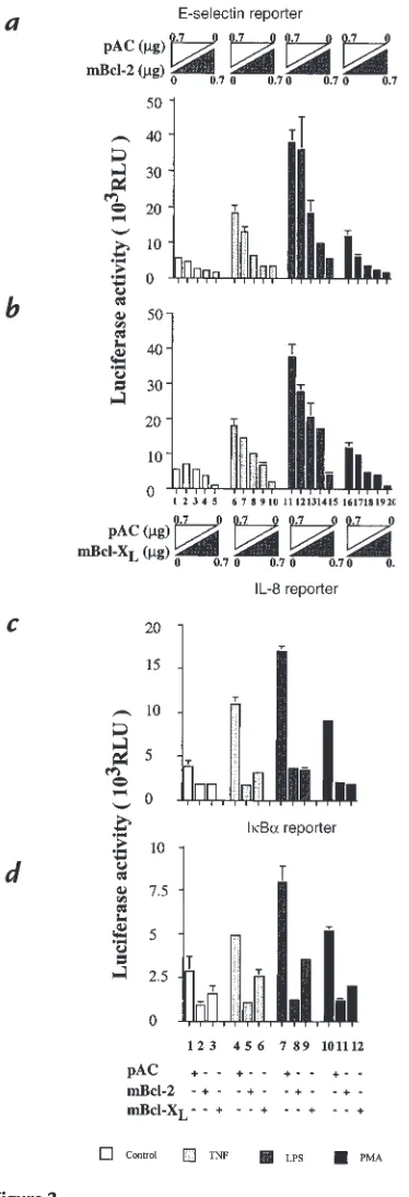
upreg-ulation of proinflammatory genes, which constitute a major part of EC activation, was first tested on the inducibility of an E-selectin reporter. E-selectin is a cell-specific marker of EC activation (34, 35). BAEC were cotransfected with the porcine E-selectin reporter con-struct, along with the expression plasmids encoding for mBcl-2, mBcl-XL, or with the control empty plasmid.
Bcl-2 or Bcl-XLexpression inhibits the induction of the
E-selectin reporter in a dose-dependent manner (Fig. 2, a and b, lanes 6–10). Stimulation of the E-selectin reporter with 100 U/ml of TNF leads to a threefold induction of the corrected luciferase activity. Transfection of BAEC with mBcl-2 or mBcl-XLexpression plasmids in amounts
ranging from 0.25 µg to 0.7 µg/5 ×105 BAEC leads to a
inhi-bition is significant at doses of 0.5 µg and higher for mBcl-2 (Fig. 2a, lanes 8and 9vs. lane 6; P < 0.03, P = 0.02, respectively) and at 0.6 µg for Bcl-XL(Fig. 2b, lane 9vs.
lane 6; P = 0.008). Inhibition is complete at 0.7 µg for either Bcl-2 or Bcl-XLcompared with the basal levels
detected in the nonstimulated cells (Fig. 2, aand b, lane 10vs. lane 1). These results demonstrate that expression of Bcl-2 and Bcl-XL in EC not only protects from
TNF-mediated apoptosis but also downregulates TNF-mediated EC activation as evaluated by analysis of the E-selectin reporter activity.
The inhibitory effect of the bclgenes was associated not only with activation of EC by TNF but also by LPS and PMA. Stimulation by LPS (100ng/ml) or PMA (5.10–8M) of
pAC-transfected BAEC leads to seven- and twofold induc-tion of the E-selectin reporter activity, respectively (Fig. 2, a and b, lanes 11and 16vs. lane 1). Similar to the observation with TNF, expression of either Bcl-2 or Bcl-XLinhibits
E-selectin reporter activity in a dose-dependent manner (Fig. 2, aand b,lanes 12–15, 17–20). This inhibition is com-plete when 0.7 µg of the given expression plasmid was used. The inhibitory effect of Bcl-2 and Bcl-XL is also seen with
other proinflammatory genes that are upregulated with EC activation. Reporters comprising the promoters of porcine IL-8 and IκBα(ECI-6) linked to the luciferin gene were tested in cotransfection experiments along with 0.7
µg of mBcl-2 or mBcl-XLexpression plasmids (previously
established as the optimal inhibitory amount). Expression of Bcl-2 or Bcl-XL inhibited the activity of the two
reporters after stimulation with either TNF, LPS, or PMA (Fig. 2, cand d). The luciferase activity of the IL-8 reporter, when cotransfected with pAC alone, increases 1.7-, 2.8-, and 1.8-fold after stimulation with TNF, LPS, or PMA, respectively (Fig. 2c, lane 1vs. lanes 4, 7, and 10). Expres-sion of Bcl-2 or Bcl-XL completely inhibited the
inducibil-ity of the IL-8 reporter by TNF, LPS, and PMA (lanes 5, 6, 8, 9, 11, and 12vs. lane 1).The same results were obtained when the porcine IκBαreporter is cotransfected along
with Bcl-2 or Bcl-XL(Fig. 2d). Induction of the IκBα
reporter activity after stimulation with TNF, LPS, and PMA reached 1.7-, 2.8- and 1.8-fold, respectively. Overex-pression of Bcl-2 or Bcl-XLled to significant inhibition of
reporter activity after TNF (lanes 5and 6; P < 0.03), LPS (lanes 8and 9; P < 0.04), or PMA stimulation (lanes 11and 12; P = 0.0002). Taken together, these results indicate that both Bcl-2 and Bcl-XLserve a new function in the EC (i.e.,
inhibition of EC activation) and that this function is inde-pendent of the agents tested.
Bcl-2 or Bcl-XLinhibits the activation of NF-κB without
affect-ing p65-mediated transactivation. We and others have shown that the induction of many proinflammatory genes that are upregulated upon EC activation is transcriptionally regulated and depends on NF-κB (3, 5, 26, 36). Our results demonstrating that the induction of 3 NF-κ B-dependent genes (E-selectin, IL-8, and IκBα)is inhibited by the expression of Bcl-2 or Bcl-XLprompted us to
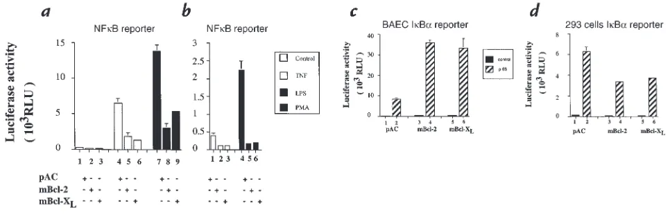
investigate whether inhibition relates to blockade of acti-vation of NF-κB. BAEC were cotransfected with a reporter construct dependent only on NF-κB for its acti-vation and mBcl-2, mBcl-XLexpression plasmids, or the
empty vector, pAC (0.7 µg). In pAC-transfected BAEC, the induction of the NF-κB reporter reached 24- and 52-fold after TNF and LPS stimulation, respectively (Fig. 3a, lanes 4and 7vs. lane 1). In Bcl-2–transfected BAEC, the induction of the NF-κB reporter activity decreased by 71.3% after TNF (Fig. 3a,lane 5vs. lane 4; P = 0.006) and by 78.3% after LPS stimulation (Fig. 3a, lane 8vs. lane 7; P < 0.001). Similarly, Bcl-XLexpression in BAEC inhibits
the inducibility of the NF-κB reporter by 79.5% after TNF (Fig. 3a, lane 6; P = 0.01) and by 61% after LPS stim-ulation (Fig. 3a,lane 9; P < 0.001). In addition, expression of Bcl-2 or Bcl-XLabrogates the 5.6-fold induction of the
NF-κB reporter after stimulation with PMA (Fig. 3b, lanes 5and 6vs. lane 4; P = 0.001).
[image:5.612.327.535.57.289.2]A recent report by Grimm et al. (37) demonstrated that expression of Bcl-2 in 293 cells inhibits NF-κB activation. Figure 1
Expression of Bcl-2 or Bcl-XLin BAEC after Lipofectamine-mediated
transfection inhibits TNF-induced apoptosis in CHX-sensitized EC. (a) Immunoblot detection of Bcl-2 (lanes 1and 2) and Bcl-XL(lanes 3and 4) in BAEC-transfected cells using polyclonal anti–Bcl-2 and anti–Bcl-XL
antibodies. Arrows indicate that transfected BAEC express high levels of Bcl-2 or Bcl-XLas opposed to control nontransfected cells. (b) BAEC were
cotransfected with a CMVβ-gal reporter (0.5 µg) and 1 µg of pAC (lanes
1and 2), mBcl-2 (lanes 3and 4), or mBcl-XL (lanes 5and 6). CHX was
added to transfected cells (all lanes) that were subsequently stimulated (lanes 2, 4, and 6) or not (lanes 1, 3, and 5) with TNF for 12 h . The per-cent cell survival was calculated as described in Methods. Expression of Bcl-2 or Bcl-XLrescues CHX-sensitized EC from TNF-mediated
These authors showed that overexpression of Bcl-2 in 293 cells did not prevent IκBαdegradation but rather down-modulated the transactivating potential of nuclear p65. To test whether Bcl-2 and Bcl-XLwould also affect
p65-medi-ated transactivation in EC, we cotransfected BAEC with the Bcl-2 or Bcl-XLexpression plasmids, or the empty vector
pAC together with the p65-dependent IκBα reporter. Expression of this reporter was then induced by cotrans-fection with a p65 expression plasmid. The IκBαreporter activity in pAC-transfected cells was increased 53-fold (Fig. 3c, lane 2). This induction was not inhibited by expression of Bcl-2 or Bcl-XL,which showed 68- and 74-fold induction,
respectively (Fig. 3c, lanes 4and 6). Thus, the inhibitory effect of Bcl-2 or Bcl-XLexpression upon p65-mediated
transactivation does not occur in EC as opposed to 293 cells, a result that we reproduced in our system (Fig. 3d). The difference could be explained by our use of primary cell cultures as opposed to the 293 cell line or could indi-cate cell-type specific function of the bclgenes.
Bcl-2 inhibits NF-κB activation at a level upstream of IκBα degradation.To determine the level at which Bcl-2 expres-sion acts upon activation of NF-κB, a recombinant repli-cation deficient human Bcl-2 adenovirus was generated as described. HUVEC were either noninfected or infected with rAd.hBcl-2 or rAd.β-gal at a MOI of 100, which usu-ally leads to >90% of expression of the transgene in HUVEC (38). Expression of the transgene (human Bcl-2) was evaluated by Western blot analysis of total cell extracts recovered 48 hours after infection (Fig. 4a, lane 3). In a first set of experiments, we confirmed that Bcl-2 retained its ability to inhibit EC activation in human cells. HUVEC were either noninfected or infected with rAd.hBcl-2 or rAd.β-gal. Forty-eight hours after infection, cells were treated with 200 U/ml of TNF, and both E-selectin and VCAM-1 surface expression was evaluated at four and eight hours after treatment, respectively. FACS analysis shows that overexpression of Bcl-2 significantly decreased TNF-mediated upregulation of E-selectin (Fig. 4b) and almost totally abrogated VCAM-1 upregulation (Fig. 4c). In addition, the impact of Bcl-2 upon IκBα degradation after TNF was also evaluated. Again, nonin-fected, rAd.Bcl-2, or rAd.β-gal–infected HUVEC were treated with 200 U/ml of TNF, and cell extracts were recovered 15 minutes and two hours after TNF stimula-tion. These extracts were evaluated by Western blot analy-sis for IκBαexpression. We demonstrate that overexpres-sion of Bcl-2 in EC inhibits IκBαdegradation by mainly stabilizing the slower migrating form of IκBα(Fig. 4d, lane 5vs. lanes 2and 8). The modest decrease of IκBαthat follows TNF treatment in Bcl-2–expressing HUVEC might relate to the small percentage of cells not express-ing the transgene. This data establishes that Bcl-2 inhibits NF-κB activation upstream of IκBαdegradation.
Expression of Bcl-2 or Bcl-XLin EC does not affect the
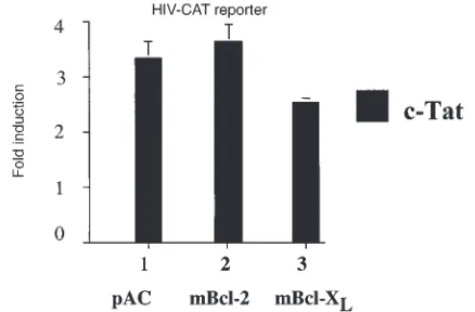
tran-scription factor Sp1.To rule out nonspecific or toxic effects of Bcl-2 or Bcl-XLexpression upon the whole
transcrip-tional machinery, NF-κB–independent induction of genes was examined. BAEC are cotransfected with the Bcl-2, Bcl-XL,or pAC expression plasmids together with
[image:6.612.308.495.50.597.2]HIV-CAT or HIV ∆κB−CAT reporters. Induction of the HIV-CAT reporter by the viral protein c-Tat is dependent upon the Sp1 binding sites of the HIV-LTR promoter and
Figure 2
Expression of Bcl-2 or Bcl-XLinhibits the induction by TNF, LPS, and PMA
of (aand b) E-selectin, (c) IL-8, and (d) IκBαreporters. E-selectin reporter induction by TNF, LPS, and PMA is inhibited by (a) Bcl-2 and (b) Bcl-XL
in a dose-dependent manner. Both mBcl-2 and mBcl-XL were titrated (0,
0.25, 0.5, 0.6, 0.7 µg) with pAC to equal 0.7 µg. The graphs shown are representative of four experiments. Significant inhibition is achieved at doses of 0.5 µg and higher. For (c) IL-8 and (d) IκBαreporters, 0.7 µg of either Bcl-2, Bcl-XL, or pAC expression plasmids was used for BAEC
trans-fection. The graphs shown are representative of four and three experi-ments, respectively. For all the reporters studies, BAEC were treated with TNF at a concentration of 100 U/ml, LPS at a concentration of 100 ng/ml, or PMA at a concentration of 5 ×10⁻⁸M. Results are given in RLU.
represents a means of gene induction independent of
NF-κB (39). Bcl-2 or Bcl-XLexpression does not affect the
threefold induction of this reporter stimulated by 0.3 µg of a c-Tat expression plasmid (Fig. 5). To further confirm that the presence in the wild-type HIV reporter of two κB binding sites did not interfere with its induction by c-Tat, a similar experiment was performed with the HIV ∆κ B-CAT reporter that is deleted from its κB binding sites. The results obtained were similar to those achieved using the wild-type reporter (data not shown), establishing that the transactivation property of the Sp1 transcription fac-tor is not altered by Bcl-2 or Bcl-XLexpression.
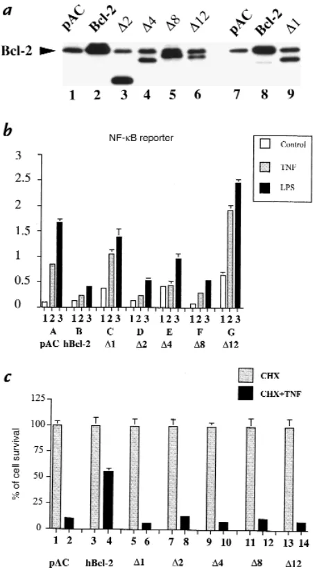
The bclhomology domains BH2 and BH4 are required for the inhibitory effect of Bcl-2 upon NF-κB activation in EC, whereas all BH domains are necessary for the anti-apoptotic effect of Bcl-2. To test whether the inhibitory effect of Bcl-2 upon NF-κB activation is associated with any of the described bcl homology domains, several human Bcl-2 deletion mutants were coexpressed togeth-er with the NF-κB reporter. Deletion of BH1, BH2, BH3, BH4, and the recently described negative regulatory domain (NRD) were included (Table 1). Expression of
each of the mutants after transfection of BAEC was con-firmed by Western blot analysis (Fig. 6a). The ability of these mutants to inhibit NF-κB activation was evaluat-ed after stimulation with TNF and LPS. Bcl-2 mutants lacking BH4 (∆1) or BH2 (∆12) were no longer able to inhibit NF-κB activation (Fig. 6b, lanes C2–3and G2–3 vs. lane B2–3). Additionally, expression of ∆1 (lane C1) or
∆12 (lane G1) leads to a significant increase in the basal luciferase activity of the NF-κB reporter compared with control (lane A1). Although still able to inhibit TNF- and LPS-mediated NF-κB activation, deletion of the BH3 domain (∆4) led to significantly less inhibition than that achieved by wild-type Bcl-2 (lanes E2–3vs. lanes B2–3). In contrast, deletion of BH1 (∆8) or the newly described NRD (∆2) did not affect the ability of Bcl-2 to inhibit EC activation upon TNF and LPS stimulation (Fig. 6b, lanes F2–3 and D2–3vs. lanes B2–3).The impact of the differ-ent Bcl mutants upon TNF-mediated apoptosis was also evaluated as described previously. Results demonstrate that all BH domains, as well as the NRD, are required to maintain the antiapoptotic function of Bcl-2 (Fig. 6c). The percentage of surviving EC after CHX and TNF treatment was comparable between the control pAC and all the Bcl-2 mutant–transfected cells (6%–13% of the transfected cells). In contrast, 55% cell survival was achieved in cells expressing the full-length hBcl-2 (Fig. 6c). Taken together, these results indicate the absence of a complete overlap between the Bcl-2 domains required for protection from apoptosis and those necessary for inhibition of NF-κB activation.
Discussion
Programmed cell death or apoptosis of EC can occur in vivo and has recently been involved in the pathogenesis of certain pathological conditions, including vasculitis, atherosclerosis, and graft rejection (7, 40–42). Different Figure 3
(aand b) Bcl-2 or Bcl-XLexpression in BAEC prevents the induction of an NF-κB reporter after TNF, LPS, and PMA stimulation without interfering with
(c) p65-mediated transactivation, in contrast with (d) 293 cells. (aand b) BAEC were cotransfected with 0.7 µg of pAC (a; lanes 1, 4, and 7), Bcl-2 (a; lanes 2, 5, and 8), or mBcl-XL(a; lanes 3, 6, and 9) expression plasmids along with 0.6 µg of the NF-κB reporter and 0.3 µg of β-gal reporter. Cells were
stimulated with either 100 U/ml TNF (a; lanes 4–6), 100 ng/ml LPS (a; lanes 7–9), or 5 ×10⁻⁸PMA (b; lanes 4–6). The graphs are representative of at
least three experiments performed. Results are given in RLU. Error bars are ± SE. (c) BAEC were cotransfected with 0.3 µg β-gal, 0.6 µg IκBαreporter, 40 ng p65 (RelA), and 0.7 µg of either pAC (lanes 1and 2), mBcl-2 (lanes 3and 4), or mBcl-XL (lanes 5and 6) expression plasmids. Overexpression of Bcl-2
or Bcl-XLdoes not inhibit the induction of IκBαby p65 (lanes 4and 6vs. lane 2). (d) 293 cells were transfected with the same plasmids with the
excep-tion of higher concentraexcep-tions of the expression plasmids (1 µg). Results show that expression of Bcl-2 or Bcl-XLinhibits the ability of p65 (RelA) to induce
[image:7.612.58.541.52.207.2]an IκBα reporter. Data shown are representative of three experiments performed. Results are expressed in RLU. Error bars represent ± SE.
Table 1
Bcl-2 deletion mutants
Proteins Amino acids deleted Domains deleted
wt-Bcl-2A 0 0
∆1 6–31 BH4
∆2 30–79 NRD
∆4 50–101 BH3
∆8 138–151 BH1
∆12 188–203 BH2
BH, bclhomology domain; NRD, negative regulatory domain. AAmino acids from
stimuli can trigger apoptosis in cultured EC, including growth factor deprivation, hemorrhagic snake venom, inhibition of anchorage-dependent cell spreading, and proinflammatory molecules,i.e., LPS and TNF (43–47).
TNF is a potent proinflammatory cytokine always detected at sites of inflammation. One of the functions of TNF is to trigger a pathway leading to programmed cell death. This function has been mapped to a specif-ic cytoplasmspecif-ic domain within the TNF-R type I protein (p55 TNF-R) that associates with a spectrum of newly defined molecules, including TRADD, FADD/MORT1, and MACH/Flice-1; the latter links TNF stimulus to death effectors (48–50). Although TNF-RI is expressed on EC and plays a crucial role in the acquisition by EC of a proinflammatory phenotype by activating NF-κB, these cells are highly resistant to TNF-mediated cell death (51). This resistance can be modulated by the addition of RNA or protein synthesis inhibitors such as CHX (52). We demonstrate that two antiapoptotic genes, bcl-2 and bcl-XL, extensively studied in other cell
types, also interrupt the cell-death pathway initiated by TNF in CHX-sensitized EC. Although Bcl-2 and Bcl-XL
were described for their ability to counteract a number of proapoptotic programs, including protection of murine aortic endothelial cells from growth factor withdrawal (53), their effect upon TNF-mediated apop-tosis is still questioned (14) and has not yet been eval-uated in EC. Our data concur with recent results show-ing that A1, a bcl-2 homologue, protects EC from TNF-mediated apoptosis (33).
In addition, we demonstrate that the function of Bcl-2 and Bcl-XLin EC is not limited to their antiapoptotic
[image:8.612.305.521.52.747.2]potential. Our data establishes that both Bcl-2 and Bcl-XL
Figure 4
are involved in a complex regulatory network that serves to downregulate EC activation and its associated gene upregulation. We show that expression of Bcl-2 or Bcl-XL
in BAEC inhibits the activation of reporter constructs, including promoters of E-selectin, IL-8, and IκBαused as readouts for EC activation. Although inducibility of these three markers is highly dependent upon activation of NF-κB, additional transcription factors can interfere with their upregulation; e.g., c-juntogether with activating transcription factor (ATF-2) and cyclic AMP–related ele-ment binding (CREB) can synergize with NF-κB for the upregulation of E-selectin and IL-8, respectively (54–56).
By showing that the expression of Bcl-2 or Bcl-XLinhibits
the upregulation of the activity of a reporter solely dependent upon NF-κB for its induction, we demon-strate that blockade of EC activation by Bcl-2 and Bcl-XL
relates to the inhibition of the transcription factor NF-κB. The downregulatory effect of Bcl-2 and Bcl-XL
upon EC activation is not limited to stimulation with TNF but applied to all stimuli tested. This agonist-inde-pendent inhibitory effect upon activation of NF-κB establishes the broad inhibitory potential of the bclgenes in EC. The novel dual role that we describe for bcl-2 and bcl-XLin EC (i.e., protection from apoptosis and blockade
of activation through inhibition of NF-κB) parallels our recent findings with the non–bcl-related antiapoptotic A20 protein (10, 11), suggesting that in EC, cell-death reg-ulators interfere with NF-κB activation.
We further show in this paper that the inhibitory effect of Bcl-2 upon NF-κB activation relates to stabilization of IκBα, mainly of a slower migrating form. This slower migrating form of IκBαis already seen before the addition of TNF in Bcl-2–expressing EC and is no longer degraded upon TNF stimulation. The identity of this IκBαband, which might constitute a hyperphosphorylated form of the protein, will help to give insights into the precise effect of Bcl-2 expression on IκBα. Experiments are currently
designed to check whether this form corresponds to a hyperphosphorylated form of IκBαand to map these phosphorylation sites. This effect of the bclgenes (i.e., inhi-bition of NF-κB activation in EC at a level upstream of IκBαdegradation) is demonstrated for the first time and confirms the data of Lin et al. (57), showing that Bcl-2 expression inhibits simian virus–induced NF-κB activa-tion by inhibiting translocaactiva-tion of NF-κB to the nucleus. Our data, however, contrasts with other reported bcl -medi-ated inhibitory mechanisms upon NF-κB activation, i.e., inhibition of p65-mediated transactivation (37) or decrease of nuclear levels of the transactivator RelA (p65)/p50 heterodimers to the advantage of the transin-hibitor p50/p50 homodimers (58). The situation is even more complex than in other cell types: Bcl-2 or Bcl-XL
expression either has no effect on NF-κB (L929, MCF-7 breast carcinoma cell line, and Jurkat T cells) (59–62) or restores the transactivating potential of NF-κB, as in HeLa cells activated by Fas (63). Our data are the first reported in primary cells. This novel NF-κB inhibitory function of the bclgenes could relate either to differences between pri-mary cells and tumor cell lines or alternatively could qual-ify as cell type–specific function of the bclgenes.
The molecular basis of the inhibitory effect of Bcl-2 or Bcl-XLupon NF-κB activation is still not defined.
How-ever, two of the already established functions of these bcl genes could explain the inhibitory effect: their protease inhibitor and antioxidant potentials (64, 65). Proteolysis of IκBαin the proteasome that follows its phosphoryla-tion and ubiquitinaphosphoryla-tion is an obligatory step for activa-tion of NF-κB (66–68). White and Gilmore (69) have recently identified an interleukin-1β converting enzyme (ICE)-like consensus site within the IκBαprotein and sug-gested that an ICE-like protease activity could be one of the pathways involved in the proteolysis of IκBα. Because Bcl-2 and Bcl-XLexert their antiapoptotic function by
inhibiting proteolytic cleavage of the ICE/ced-3 cysteine proteases (65), it is tempting to speculate that they would also inhibit an ICE-like protease involved in the proteoly-sis of IκBαin EC, which would explain the accumulation of an eventually hyperphosphorylated form of IκBα. An alternative mechanism relies on the described antioxidant function of Bcl-2 (64). We and others have shown that antioxidants, mainly belonging to the thiol group, are potent inhibitor of NF-κB activation and that this inhi-bition, like the one achieved by Bcl-2, occurs at an early step upstream of IκBαdegradation (36, 70).
[image:9.612.68.287.52.196.2]The bclgenes could also indirectly interrupt the path-way leading to activation of NF-κB by interfering with key signal transducers. Bcl-2 has been shown to interact physically with molecules such as p21Ras, p23Rras, cal-cineurin, and Raf-1 kinase (71–74). Interaction between Bcl-2 and Raf-1 targets this kinase to the mitochondrial membrane and enhances the antiapoptotic effect of 2. This interaction requires the BH4 domain of Bcl-2 (75). Raf-1 kinase is an early mediator involved in mul-tiple signaling pathways (TNF, LPS) acting at a level downstream of Ras and upstream of mitogen-activated kinase (MAPK) (76, 77). Cross-talk between MAPK and NF-κB signaling pathways is still debated; however, Raf-1 kinase interaction with Bcl-2 could be of some rel-evance in the pathway leading to IκBα phosphorylation Figure 5
Overexpression of Bcl-2 and Bcl-XLdoes not affect the transactivation
properties of Sp1. BAEC were cotransfected with mBcl-2, mBcl-XL, or
pAC together with an HIV-CAT reporter. Induction of this reporter was achieved by cotransfecting the viral protein c-Tat, which requires the Sp1 binding sites of the HIV-CAT reporter. Results show no difference in the c-Tat–mediated induction of the HIV-CAT whether the cells were trans-fected with pAC (lane 1), Bcl-2 (lane 2), or Bcl-XL (lane 3). Data shown
and degradation (78, 79). We demonstrate that deletion of the BH4 domain of Bcl-2 abrogates its inhibitory effect upon activation of NF-κB, which argues for a potential involvement of Raf-1 kinase in Bcl-2–mediat-ed inhibition of NF-κB activation. In addition to these hypotheses, our findings showing that expression of Bcl-2 in HUVEC stabilizes a lower (potentially hyperphos-phorylated) form of IκBαmight yet relate to a novel function of Bcl-2 that can modify IκBα. For instance, Bcl-2–mediated modification of IκBαcould prevent its phosphorylation at the specific serine residues, alter ade-quate ubiquitination, or enhance its resistance to degra-dation. These hypotheses are currently being tested.
Our data also show that Bcl-2 or Bcl-XLspecifically
interrupts activation of NF-κB without affecting another tested transcription factor, i.e., Sp1. We demonstrate that the c-Tat–driven Sp1 HIV reporter is not altered by Bcl-2 or Bcl-XLexpression in EC (39). Recent data from Linette
et al. (61) showed that in T cells, Bcl-2 expression specifi-cally impairs the transactivation properties of the nuclear factor of activated T cells (NFAT) without affecting other transcription factors, i.e., AP-1, NF-κB, and OCT-1. These data suggest that Bcl-2 and Bcl-XLhave different
tran-scriptional targets depending on the cell type studied. Bcl-2 family members can either promote or protect from cell death. A feature of these proteins is their ability to homo- or heterodimerize through the conserved Bcl-2 homology domains BH1, BH2, BH3, and BH4 (14, 17, 80, 81). Heterodimerization of Bcl-2 or Bcl-XLwith their
proapoptotic partners Bax, Bak, and Bcl-XSseems to
determine the life–death decision of a cell, although some controversies remain (82–84). Our experiments probe whether interaction with Bax is equally as important for the inhibitory effect of Bcl-2 upon NF-κB activation as for regulation of apoptosis. Mutagenesis studies showed that both BH1 and BH2 are required for Bcl-2 to het-erodimerize with Bax (85). Our results indicate that in EC, a Bcl-2 mutant lacking the BH1 domain is still able to inhibit NF-κB activation, whereas deletion of the BH2 domain abrogates this function. This result indicates that interaction between Bcl-2 and Bax is either not required for Bcl-2 function in EC or that only BH2 is necessary in EC to interact with Bax. Having mapped the inhibitory function of Bcl-2 on NF-κB activation to the BH4 and BH2 domains of the molecule, we are currently investi-gating which proteins in EC interact with these domains to sustain this novel bclfunction.
In contrast with the specific mapping of the Bcl-2 inhibitory effect upon NF-κB activation to BH2 and BH4, all BH domains, as well as the NRD of Bcl-2, were required for protection from apoptosis. This result indi-cates that the Bcl-2 motifs required for the anti-inflam-matory function (BH2, BH4) are also necessary for its antiapoptotic function, but not vice versa; indeed, cer-tain motifs are indispensable for the antiapoptotic func-tion (BH1, BH3, NRD) but have no impact upon the anti-inflammatory function. Our data does not rule out that a unique cellular target interacting with BH2 and BH4 could account for both functions of Bcl-2; this query is being investigated.
[image:10.612.308.535.53.471.2]From a basic point of view, we demonstrate that Bcl-2 and Bcl-XLplay a major protective role in EC by blocking
Figure 6
(a) Structure/function relationships of Bcl-2 deletion mutants. Expression of Bcl-2 deletion mutants in BAEC. (b) The Bcl homology domains BH4 and BH2 are required for the inhibitory effect of Bcl-2 upon NF-κB activa-tion after TNF and LPS stimulaactiva-tion, whereas (c) all BH domains and the NRD are required for the antiapoptotic function of Bcl-2. (a) Immunoblot detection of human wild-type and deletion mutants of Bcl-2 (lanes 2–6) in BAEC-transfected cells, using a polyclonal anti–Bcl-2 antibody. Mutant ∆1 is not recognized by this antibody and was detected using a monoclonal anti–Bcl-2 antibody (lane 9). (b) BAEC were cotransfected with 0.3 µg of
β-gal, 0.6 µg of NF-κB reporter along with 0.7 µg of pAC (lanes A1–3), human Bcl-2 (lanes B1–3), ∆1 (lanes C1–3), ∆2 (lanes D1–3), ∆4 (lanes
E1–3), ∆8 (lanes F1–3), and ∆12 (lanes G1–3). Cells were stimulated with 100 U/ml TNF (lanes 2) or 100 ng/ml LPS (lanes 3) Overexpression of ∆2,
∆4, or ∆8 inhibits the induction by TNF or LPS of a NF-κB reporter, in con-trast to ∆1 or ∆12. Graph shown is representative of four experiments. Results are expressed in RLU. Error bars represent ± SE. (c) BAEC were cotransfected with a CMVβ-gal reporter (0.5 µg) and 1 µg of pAC (lanes 1
TNF-mediated apoptosis in addition to inhibiting EC activation through blockade of NF-κB activation. The mechanism by which inhibition of NF-κB is accom-plished (i.e., stabilization of IκBα)is novel.
From a therapeutic standpoint, blockade of NF-κB has been suggested as a means of preventing proinflammatory consequences of EC activation implicated in different pathologies, including allograft and xenograft rejection (4, 86). Bcl-2 and Bcl-XLrepresent prime candidates for
genet-ic engineering of EC to achieve this purpose. In support of this approach is our data showing that expression of Bcl-2 and Bcl-XLin EC of long-term surviving xenografts is
asso-ciated with the absence of apoptosis, inflammation, and atherosclerosis, whereas they are not expressed in EC of rejecting xenografts (7). Although indirect, these results fur-ther argue for their protective and broad anti-inflammato-ry potential in vivo as well as establish the safety of their use.
Acknowledgments
We are grateful to Timothy Behrens, Gabriel Nunez, Tristram G. Parslow, and J. Reed for providing us with the murine Bcl-2 and Bcl-XL, the human Bcl-2, and the deletion mutants expres-sion plasmids. We acknowledge Joseph Anrather for providing us with the p65 expression plasmid and for valuable advice. We also thank Maria-Beatriz Arvelo and E. Czismadia for help with the virus purification and skilled technical assistance in cell cul-ture. We are also grateful to Shane T. Grey for criticism and advice.
1. Pober, J.S., et al. 1990. The potential roles of vascular endothelium in immune reactions [review]. Hum. Immunol. 28:258–262.
2. Cotran, R.S., and Pober, J.S. 1990. Cytokine-endothelial interactions in inflammation, immunity, and vascular injury. J. Am. Soc. Nephrol.
1:225–235.
3. Bach, F.H., et al. 1994. Endothelial cell activation and thromboregula-tion during xenograft rejecthromboregula-tion [review]. Immunol. Rev.141:5–30. 4. Gimbrone, M.A.J. 1995. Vascular endothelium: an integrator of
patho-physiologic stimuli in atherosclerosis [review]. Am. J. Cardiol.
75:67B–70B.
5. Read, M.A., Whitley, M.Z., Williams, A.J., and Collins, T. 1994. NF-κB and IκBα — an inducible regulatory system in endothelial activation. J. Exp. Med. 179:503–512.
6. Collins, T., et al. 1995. Transcriptional regulation of endothelial cell adhesion molecules — NF-κB and cytokine-inducible enhancers. FASEB J. 9:899–909.
7. Bach, F.H., et al. 1997. Accommodation of vascularized xenografts: expression of “protective genes” by donor endothelial cells in a host Th2 cytokine environment. Nat. Med. 3:196–204.
8. Opipari, A.J., Boguski, M.S., and Dixit, V.M. 1990. The A20 cDNA induced by tumor necrosis factor alpha encodes a novel type of zinc fin-ger protein. J. Biol. Chem. 265:14705–14708.
9. Opipari, A.J., Hu, H.M., Yabkowitz, R., and Dixit, V.M. 1992. The A20 zinc finger protein protects cells from TNF cytotoxicity. J. Biol. Chem.
267:12424–12427.
10. Ferran, C., and Stroka, D.M. 1997. Adenovirus-mediated gene transfer of A20 renders endothelial cells resistant to activation: a means of eval-uating the role of EC activation in xenograft rejection. Transplant Proc.
29:879–880.
11. Cooper, J.T., et al. 1996. A20 blocks endothelial cell activation through a NF-κB-dependent mechanism. J. Biol. Chem. 271:18068–18073. 12. LeBrun, D.P., Warnke, R.A., and Cleary, M.L. 1993. Expression of bcl-2
in fetal tissues suggests a role in morphogenesis. Am. J. Pathol.
142:743–753.
13. Lu, Q.L., Poulsom, R., Wong, L., and Hanby, A.M. 1993. Bcl-2 expression in adult and embryonic non-haematopoietic tissues. J. Pathol. 169:431–437.
14. Reed, J.C. 1994. Bcl-2 and the regulation of programmed cell death. J. Cell Biol. 124:1–6.
15. Boise, L.H., et al. 1993. bcl-x, a bcl-2-related gene that functions as a dom-inant regulator of apoptotic cell death. Cell. 74:597–608.
16. Oltvai, Z.N., Milliman, C.L., and Korsmeyer, S.J. 1993. Bcl-2 het-erodimerizes in vivo with a conserved homolog, Bax, that accelerates pro-grammed cell death. Cell. 74:609–619.
17. Sedlak, T.W., et al. 1995. Multiple Bcl-2 family members demonstrate
selective dimerizations with Bax. Proc. Natl. Acad. Sci. USA.92:7834–7838. 18. Wang, H.G., Takayama, S., Rapp, U.R., and Reed, J.C. 1996. Bcl-2 inter-acting protein, BAG-1, binds to and activates the kinase Raf-1. Proc. Natl. Acad. Sci. USA. 93:7063–7068.
19. White, E. 1996. Life, death, and the pursuit of apoptosis [review]. Genes Dev. 10:1–15.
20. Vanhaesebroek, B., et al. 1993. Effect of bcl-2 proto-oncogene on cellular sensitivity to tumor necrosis factor-mediated cytotoxicity. Oncogene. 8:1075–1081.
21. Hennet, T., Bertoni, G., Richter, C., and Peterhans, E. 1993. Expression of BCL-2 protein enhances the survival of mouse fibrosarcoid cells in tumor necrosis factor-mediated cytotoxicity. Cancer Res.53:1456–1460. 22. Pober, J.S., et al. 1986. Two distinct monokines, interleukin 1 and tumor necrosis factor, each independently induce biosynthesis and transient expression of the same antigen on the surface of cultured human vas-cular endothelial cells. J. Immunol.136:1680–1687.
23. Cotran, R.S., Gimbrone, M.A., Jr., Bevilacqua, M.P., Mendrick, D.L., and Pober, J.S. 1986. Induction and detection of a human endothelial acti-vation antigen in vivo. J. Exp. Med.164:661–666.
24. Gimbrone, M.J., Cotran, R.S., and Folkman, J. 1974. Human vascular endothelial cells in culture. Growth and DNA synthesis. J. Cell Biol.
60:673–684.
25. Brostjan, C., et al. 1996. Deletion analysis of the porcine E-selectin pro-moter. Transplant Proc.28:649–651.
26. de Martin, R., et al. 1993. Cytokine-inducible expression in endothelial cells of an IκBα-like gene is regulated by NF-κB. EMBO J. 12:2773–2779. 27. Bilbao, G., et al. 1999. Genetic cytoprotection of human endothelial cells during the preservation time with an adenoviral vector encoding the anti-apoptotic human Bcl-2 gene. Transplant Proc.In press.
28. Graham, F.L., and Prevec, L. 1991. Manipulation of adenovirus vectors. In Methods in molecular biology: gene transfer and expression protocols. 7th ed. E.J. Murray, editor. Press Inc. Clifton, NJ. 109–128.
29. McGrory, W.J., Bautista, D.S., and Graham, F.L. 1988. A simple tech-nique for the rescue of early region I mutations into infectious human adenovirus type 5. Virology. 163:614–617.
30. Bett, A.J., Prevec, L., and Graham, F.L. 1993. Packaging capacity and sta-bility of human adenovirus type 5 vectors. J. Virol. 67:5911–5921. 31. Kerr, J.F., Wyllie, A.H., and Currie, A.R. 1972. Apoptosis: a basic
biolog-ical phenomenon with wide-ranging implications in tissue kinetics [review]. Br. J. Cancer. 26:239–257.
32. McCarthy, J.V., Ni, J., and Dixit, V.M. 1998. RIP2 is a novel NF-κ B-acti-vating and cell death-inducing kinase. J. Biol. Chem.273:16968–16975. 33. Karsan, A., Yee, E., and Harlan, J.M. 1996. Endothelial cell death induced by tumor necrosis factor a is inhibited by the Bcl-2 family member A1. J. Biol. Chem.271:27201–27204.
34. Pober, J.S., and Cotran, R.S. 1990. Cytokines and endothelial cell biolo-gy. Physiol. Rev. 70:427–451.
35. Collins, T., Palmer, H.J., Whitley, M.Z., Neish, A.S., and Williams, A.J. 1993. A common theme in endothelial cell activation: insights from the structural analysis of the genes for E-selectin and VCAM-1. Trends Card. Med. 3:92–97.
36. Ferran, C., et al. 1995. Inhibition of NF-κB by pyrrolidine dithiocarba-mate blocks endothelial cell activation. Biochem. Biophys. Res. Commun.
214:212–223.
37. Grimm, S., Bauer, M.K.A., Bauerle, P.A., and Schultze-Osthoff, K. 1996. Bcl-2 down-regulates the activity of transcription factor NF-κB induced upon apoptosis. J. Cell. Biol.134:13–23.
38. Wrighton, C.J., et al. 1996. Inhibition of endothelial cell activation by ade-novirus-mediated expression of IκBα, an inhibitor of the transcription factor NF-κB. J. Exp. Med. 183:1013–1022.
39. Zimmermann, K., et al. 1991. trans-activation of the HIV-1 LTR by the HIV-1 Tat and HTLV-I Tax proteins is mediated by different cis-acting sequences. Virology. 182:874–878.
40. Collins, T. 1993. Biology of disease – endothelial nuclear factor-κB and the initiation of the atherosclerotic lesion. Lab. Invest. 68:499–508. 41. Laine, J., Etelamaki, P., Holmberg, C., and Dunkel, L. 1997. Apoptotic cell
death in human chronic allograft rejection. Transplantation. 63:101–105. 42. Thompson, C. 1995. Apoptosis in the pathogenesis and treatment of
dis-ease. Science. 267:1456–1462.
43. Abello, P.A., Fidler, S.A., Bulkley, G.B., and Buchman, T.G. 1994. Antiox-idants modulate induction of programmed endothelial cell death (apop-tosis) by endotoxin. Arch. Surg. 129:134–140.
44. Buchman, T.G., Abello, P.A., Smith, E.H., and Bulkley, G.B. 1993. Induc-tion of heat shock response leads to apoptosis in endothelial cells previ-ously exposed to endotoxin. Am. J. Physiol.265:165–170.
45. Araki, S., Ishida, T., Yamamoto, T., Kaji, K., and Hayashi, H. 1993. Induc-tion of apoptosis by hemorrhagic snake venom in vascular endothelial cells. Biochem. Biophys. Res. Commun. 190:148–153.
46. Re, F., et al. 1994. Inhibition of anchorage-dependent cell spreading trig-gers apoptosis in cultured human endothelial cells. J. Cell Biol.
127:537–546.
Tumor necrosis factor induces apoptosis (programmed cell death) in normal endothelial cells in vitro. Am. J. Pathol. 138:447–453.
48. Chinnayian, A.M., O’Rourke, K., Tewari, M., and Dixit, V. 1995. FADD, a novel death domain-containing protein interacts with the death domain of Fas and initiates apoptosis. Cell. 81:505–512.
49. Hsu, H., Xiong, J., and Goeddel, D.V. 1995. The TNF-receptor 1-associ-ated protein TRADD signals cell death and NF-κB activation. Cell. 81:495–504.
50. Hsu, H., Shu, H.B., Pan, M.G., and Goeddel, D.V. 1996. TRADD-TRAF2 and TRADD-FADD interactions define two distinct TNF receptor 1 sig-nal transduction pathways. Cell. 84:299–308.
51. Pohlman, T.H., and Harlan, J.M. 1989. Human endothelial cell response to lipopolysaccharide, interleukin-1, and tumor necrosis factor is regu-lated by protein synthesis. Cell. Immunol.119:41–52.
52. Polunovsky, V.A., Wendt, C.H., Ingbar, D.H., Peterson, M.S., and Bitter-man, P.B. 1994. Induction of endothelial cell apoptosis by TNF-α: mod-ulation by inhibitors of protein synthesis. Exp. Cell Res.214:584–594. 53. Kondo, S., et al. bcl-2 gene prevents apoptosis of basic fibroblast growth factor-deprived murine aortic endothelial cells. Exp. Cell Res.213:428–32. 54. Kaszubska, W., et al. 1993. Cyclic AMP-independent ATF family mem-bers interact with NF-κB and function in activation of the E-selectin pro-moter in response to cytokines. Mol. Cell. Biol.13:7180–7190. 55. DeLuca, L.G., Johnson, D., Whitley, M., Collins, T., and Pober, J. 1994. cAMP
and tumor necrosis factor competitively regulate transcriptional activation through and nuclear factor binding to the cAMP-responsive element/acti-vating transcription factor element of the endothelial leukocyte adhesion molecule-1 (E-selectin) promoter. J. Biol. Chem. 269:19193–19196. 56. Stein, B., and Baldwin, A.S.J. 1993. Distinct mechanisms for regulation
of the Interleukin-8 gene involve synergism and cooperativity between C/EBP and NF-κB. Mol. Cell. Biol. 13:7191–7198.
57. Lin, K., et al. 1995. Thiol agents Bcl-2 identify an alphavirus-induced apoptotic pathway that requires activation of the transcription factor NF-kappa B.J. Cell Biol.131:1149–1161.
58. Ivanov, V.N., Deng, G., Podack, E.R., and Malek, T.R. 1995. Pleiotropic effects of Bcl-2 on transcription factors in T cells — potential role of NF-kappa-B P50-P50 for the anti-apoptotic function of Bcl-2. Int. Immunol. 7:1709–1720. 59. Allbrecht, H., Tschopp, J., and Jongeneel, V. 1994. Bcl-2 protects from oxidative damage and apoptotic cell death without interfering with acti-vation of NF-κB by TNF. FEBS Lett. 351:45–48.
60. Jäättela, M., Benedict, M., Tewari, M., Shayman, J.A., and Dixit, V.M. 1995. Bcl-x and Bcl-2 inhibit TNF and Fas-induced apoptosis and activation of phospholipase A2 in breast carcinoma cells. Oncogene. 10:2297–305. 61. Linette, G.P., Li, Y., Roth, K., and Korsmeyer, S.J. 1996. Cross talk
between cell death and cell cycle progression: BCL-2 regulates NFAT-mediated activation. Proc. Natl. Acad. Sci. USA. 93:9545–9552. 62. Dbaibo, G.S., et al. 1997. Cytokine response modifier A (CrmA) inhibits
ceramide formation in response to tumor necrosis factor (TNF)-α: CrmA and Bcl-2 target distinct components in the apoptotic pathway. J. Exp. Med.185:481–490.
63. Mandal, M., et al. 1996. Bcl-2 prevents CD95 (Fas/APO-1)-induced degradation of lamin B and poly (ADP-ribose) polymerase and restores the NF-κB signaling pathway. J. Biol. Chem.271:30354–30359. 64. Hockenbery, D.M., Oltvai, Z.N., Yin, X.M., Milliman, C.L., and
Korsmey-er, S.J. 1993. Bcl-2 functions in an antioxidant pathway to prevent apop-tosis. Cell. 75:241–251.
65. Chinnaiyan, A.M., et al. 1996. Molecular ordering of the cell death path-way. J. Biol. Chem.271:4573–4576.
66. Traenckner, E.B.M., and Baeuerle, P.A. 1995. Appearance of apparently ubiquitin-conjugated IκB-αduring its phosphorylation-induced
degra-dation in intact cells. J. Cell Sci. Suppl. 19:79–84.
67. Traenckner, E.B.M., et al. 1995. Phosphorylation of human IκB-αon ser-ines 32 and 36 controls IκB-αproteolysis and NF-κB activation in response to diverse stimuli. EMBO J. 14:2876–2883.
68. Traenckner, E.B.M., Wilk, S., and Bauerle, P. 1994. A proteasome inhibitor prevents activation of NF-κB and stabilizes a newly phospho-rylation form of IκBα that is still bound to NF-κB. EMBO J.
13:5433–5441.
69. White, D.W., and Gilmore, T.D. 1996. Bcl-2 and CrmA have different effects on transformation, apoptosis and the stability of IκB-αin chick-en splechick-en cells transformed by temperature-schick-ensitive v-Rel oncoproteins.
Oncogene. 13:891–899.
70. Schreck, R., Meier, B., Mannel, D.N., Droge, W., and Baeuerle, P.A. 1992. Dithiocarbamates as potent inhibitors of nuclear factor κB activation in intact cells. J. Exp. Med. 175:1181–1194.
71. Chen, C.Y., and Faller, D.V. 1996. Phosphorylation of Bcl-2 protein and association with p21Ras in Ras-induced apoptosis. J. Biol. Chem.
271:2376–2379.
72. Fernandez-Sarabia, M., and Bischoff, J.R. 1993. Bcl-2 associates with the ras-related protein R-ras p23. Nature. 366:274–275.
73. Wang, H.G., et al. 1994. Apoptosis regulation by interaction of Bcl-2 pro-tein and Raf-1 kinase. Oncogene. 9:2751–2756.
74. Shibasaki, F., Kondo, E., Akagi, T., and McKeon, F. 1997. Suppression of signalling through transcription factor NF-AT interactions between cal-cineurin and Bcl-2. Nature. 386:728–731.
75. Wang, H.G., Rapp, U.R., and Reed, J.C. 1996. Bcl-2 targets the protein kinase Raf-1 to mitochondria. Cell. 87:629–638.
76. Hambleton, J., McMahon, M., and DeFranco, A.L. 1995. Activation of Raf-1 and mitogen-activated protein kinase in murine macrophages par-tially mimics lipopolysaccharide-induced signaling events. J. Exp. Med.
182:147–54.
77. Jelinek, T., Dent, P., Sturgill, T.W., and Weber, M.J. 1996. Ras-induced activation of RAF-1 is dependent on tyrosine phosphorylation. Mol. Cell. Biol.16:1027–1034.
78. Li, S.F., and Sedivy, J.M. 1993. Raf-1 protein kinase activates the NF-κB transcription factor by dissociating the cytoplasmic NF-κB-IκB complex.
Proc. Natl. Acad. Sci. USA.90:9247–9251.
79. Chirillo, P., et al. 1996. Hepatitis B virus Px activates NF-κB-dependent transcription through a Raf-independent pathway. J. Virol. 70:641–646. 80. Zha, J., Harada, H., Yang, E., Jockel, J., and Korsmeyer, S.J. 1996. Serine phosphorylation of death agonist BAD in response to survival factor results in binding to 14-3-3 not BCL-XL. Cell. 87:619–628.
81. Sato, T., et al. 1994. Interactions among members of the Bcl-2 protein family analyzed with a yeast two-hybrid system. Proc. Natl. Acad. Sci. USA.
91:9238–9242.
82. Cheng, E.H.Y., Levine, B., Boise, L.H., Thompson, C.B., and Hardwick, J.M. 1996. Bax-independent inhibition of apoptosis by Bcl-xL. Nature.
379:554–556.
83. Hunter, J.J., Bond, B.L., and Parslow, T.G. 1996. Functional dissection of the human Bcl-2 protein: sequence requirements for inhibition of apop-tosis. Mol. Cell. Biol. 16:877–883.
84. Yin, D.X., and Schimke, R.T. 1996. Inhibition of apoptosis by overex-pressing Bcl-2 enhances gene amplification by a mechanism independ-ent of aphidicolin pretreatmindepend-ent. Proc. Natl. Acad. Sci. USA.93:3394–3398. 85. Yin, X.M., Oltvai, Z.N, and Korsmeyer, S.J. 1994. BH1 and BH2 domains of Bcl-2 are required for inhibition of apoptosis and heterodimerization with Bax. Nature. 369:321–323.