Complete antithrombin deficiency in mice
results in embryonic lethality
Kazuhiro Ishiguro, … , Takashi Muramatsu, Hidehiko Saito
J Clin Invest.
2000;
106(7)
:873-878.
https://doi.org/10.1172/JCI10489
.
Antithrombin is a plasma protease inhibitor that inhibits thrombin and contributes to the
maintenance of blood fluidity. Using targeted gene disruption, we investigated the role of
antithrombin in embryogenesis. Mating mice heterozygous for antithrombin gene (
ATIII
)
disruption,
ATIII
+/–, yielded the expected Mendelian distribution of genotypes until 14.5
gestational days (gd). However, approximately 70% of the
ATIII
–/–embryos at 15.5 gd and
100% at 16.5 gd had died and showed extensive subcutaneous hemorrhage. Histological
examination of those embryos revealed extensive fibrin(ogen) deposition in the myocardium
and liver, but not in the brain or lung. Furthermore, no apparent fibrin(ogen) deposition was
detected in the extensive hemorrhagic region, suggesting that fibrinogen might be
decreased due to consumptive coagulopathy and/or liver dysfunction. These findings
suggest that antithrombin is essential for embryonic survival and that it plays an important
role in regulation of blood coagulation in the myocardium and liver.
Article
Introduction
Antithrombin is a 58-kDa plasma serine protease inhibitor (serpin) that, through the inhibition of thrombin and other activated serine proteases, plays important roles in the regulation of coagulation (1). A recent report has demonstrated that the cleaved con-formation of antithrombin has potent antiangiogenic activity and that antithrombin may allow for the pre-cise regulation of angiogenesis (2).
The inhibition of thrombin by antithrombin is rel-atively slow, but is dramatically enhanced in the pres-ence of heparin (1, 3). Heparin-like molecules, heparan sulfate proteoglycans (HSPGs), are closely associated with endothelial cells and enhance the action of circulating antithrombin (4, 5). The synde-can and glypisynde-can core protein families constitute the major membrane-associated HSPGs generated with-in the cardiovascular system (6). Among them, synde-can-4 (ryudocan) was originally cloned from endothe-lial cells as an antithrombin-binding molecule (7, 8), and we recently generated syndecan-4–null mice by targeted gene disruption (9).
Individuals with functionally defective antithrombin are susceptible to venous thromboembolic diseases (10–12). Congenital antithrombin deficiency is usual-ly heterozygous and classified into two types, type I
(quantitative deficiency) and type II (qualitative defi-ciency), which includes a reactive defect, heparin-bind-ing site defect, and pleiotrophic effect (12–14). Although some cases of homozygous qualitative antithrombin deficiency have been reported (15–17), homozygous quantitative antithrombin deficiency has not. Thus, complete deficiency of antithrombin is spec-ulated to cause embryonic lethality similar to that of other anticoagulant proteins (18, 19). It is not known, however, when or in what way antithrombin is neces-sary in normal embryogenesis. To further investigate the physiological roles of antithrombin, especially those in embryogenesis, we generated antithrombin-deficient mice by gene targeting.
Methods
Isolation of the mouse antithrombin gene. Oligonucleotide primers mAT3S (5′-ATGATGTACCAGGAAGGCAA, cDNA positions 865–884) and mAT3A (5′-
GGAAT-GCGTCGGAGACATAG, 1219–1200), based on the
pub-lished murine antithrombin cDNA sequence (20), were used to obtain a partial cDNA of mouse antithrombin from liver mRNA of C57BL/6J mice (CLEA, Tokyo, Japan) using RT-PCR. The resulting 355-bp PCR prod-uct was used as a probe to isolate a genomic clone con-taining a segment of the antithrombin gene from a
Complete antithrombin deficiency in mice
results in embryonic lethality
Kazuhiro Ishiguro,
1,2Tetsuhito Kojima,
1,3Kenji Kadomatsu,
2Yukiko Nakayama,
1Akira Takagi,
3Misao Suzuki,
4Naoki Takeda,
4Masafumi Ito,
5Koji Yamamoto,
1Tadashi Matsushita,
1Kazuo Kusugami,
1Takashi Muramatsu,
2and Hidehiko Saito
11First Department of Internal Medicine, and
2Department of Biochemistry, Nagoya University School of Medicine, and
3Department of Medical Technology, Nagoya University School of Health Sciences, Nagoya, Japan
4Division of Transgenic Technology Center for Animal Resources and Development, Kumamoto University, Kumamoto, Japan 5Division of Pathology, Clinical Laboratory, Nagoya University Hospital, Nagoya, Japan
Address correspondence to: Hidehiko Saito, First Department of Internal Medicine, Nagoya University School of Medicine, 65 Tsurumai-cho, Showa-ku, Nagoya, Aichi 466-8550, Japan.
Phone: 81-52-744-2136; Fax: 81-52-744-2157; E-mail: [email protected].
Received for publication June 5, 2000, and accepted in revised form August 28, 2000.
Antithrombin is a plasma protease inhibitor that inhibits thrombin and contributes to the mainte-nance of blood fluidity. Using targeted gene disruption, we investigated the role of antithrombin in embryogenesis. Mating mice heterozygous for antithrombin gene (ATIII) disruption, ATIII+/–,
yield-ed the expectyield-ed Mendelian distribution of genotypes until 14.5 gestational days (gd). However, approximately 70% of the ATIII–/–embryos at 15.5 gd and 100% at 16.5 gd had died and showed
exten-sive subcutaneous hemorrhage. Histological examination of those embryos revealed extenexten-sive fib-rin(ogen) deposition in the myocardium and liver, but not in the brain or lung. Furthermore, no apparent fibrin(ogen) deposition was detected in the extensive hemorrhagic region, suggesting that fibrinogen might be decreased due to consumptive coagulopathy and/or liver dysfunction. These findings suggest that antithrombin is essential for embryonic survival and that it plays an important role in regulation of blood coagulation in the myocardium and liver.
129SVJ lambda FIX II genomic library (Stratagene, La Jolla, California, USA), as described previously (21), and a 16-kb segment of DNA (λmATG1) containing the antithrombin initiation codon to the sixth intron was obtained. The sequence data have been deposited in DNA Data Bank of Japan (DDBJ)/European Mole-cular Biology Laboratory (EMBL)/GenBank (accession number AB043785). The detailed features of the gene structure and its comparison with the human antithrombin gene will be discussed elsewhere.
Construction of a targeting vector for homologous recombi-nation in embryonic stem cells. The targeting vector was constructed from a basic vector with MC1neo(polyoma virus thymidine kinase gene promoter and neomycin resistance gene) and DTA(diphtheria toxin fragment A gene) (22) and antithrombin gene fragments. We used a 2.1-kb SspI/PvuII fragment as the 5′arm and a 7.3-kb
HindIII/SmaI fragment as the 3′arm to replace 1,781 bases, including exon 2 of the antithrombin gene with
MC1neoby homologous recombination (Figure 1a). The generation of targeted D3 embryonic stem (ES) cells and blastocyst injection were performed as described previously (9, 23).
Generation of antithrombin-deficient mice. Male chimeric mice were mated with C57BL/6J female mice, and F1
mice heterozygous for antithrombin gene (ATIII) dis-ruption, ATIII+/–, were mated with each other to yield
null mutant mice. Noon on the day of vaginal plug detection was defined as 0.5 gestational days (gd). Females were sacrificed and the embryos harvested at various times during gestation. We defined embryon-ic death by the absence of a heartbeat at first inspec-tion. The lower limbs and tail of the embryos were used for Southern blot and PCR analyses. The remain-ing embryos were used for RT-PCR, Western blot, or histological analyses. For histological analysis, the embryos were fixed overnight in Carnoy’s solution (methanol/chloroform/acetic acid = 6:3:1). The fixed embryos were then dehydrated, embedded in paraffin, and sectioned (6 µm thick).
Preparation of anti-mouse antithrombin antisera. The anti-sera against mouse antithrombin were raised in rabbits by direct introduction of the encoding cDNA in pCI vector (Promega, Madison, Wisconsin, USA) (24). From the antisera, nonspecific Ab’s were absorbed with dry powder derived from ATIII–/– embryos at 14.5 gd.
Serum titers were determined by Western blot analysis using recombinant mouse antithrombin fused to glu-tathione-S-transferase protein (Amersham Pharmacia Biotech, Tokyo, Japan) and rat antithrombin (Sigma-Aldrich, St. Louis, Missouri, USA).
Detection of antithrombin-deficient mice
DNA analysis. PCR was performed with a forward primer (5′-CCTTCCAGACCGAACTGTCC) and a reverse primer (5′-GTAATCCCAGCCTTCTCCTG) to detect the expected deletion (Figure 1a). The PCR conditions were 35 cycles of denaturation for 30 seconds at 94°C, annealing for 1 minute at 68°C, and extension
for 3 minutes at 72°C. The wild-type allele gave a 2.0 kb band, whereas the mutant allele gave a 1.3-kb band (Figure 1b).
We also performed Southern blot analysis. After digestion with PvuII and hybridization with an exter-nal probe (a 658-base HindIII/SspI fragment; Figure 1a), the wild-type allele gave a 4.4-kb band, whereas the mutant allele gave a 5-kb band (Figure 1c).
RNA analysis. Total RNA was isolated from the single embryo of each genotype at 14.5 gd by the guanidinium thiocyanate-phenol-chloroform method (25). RT-PCR was performed with primers mAT3S and mAT3AS, described above, yielding a 355-bp fragment. The PCR conditions were 25 cycles of denaturation for 30 sec-onds at 94°C, annealing for 1 minute at 55°C, and extension for 1 minute at 72°C.
Protein analysis. The whole liver of a single ATIII+/+or ATIII–/–embryo at 14.5 gd was homogenized in PBS
(500 µl). After centrifugation, the supernatant was rotated with protein G-Sepharose (40 µl of 50% slurry) (Amersham Pharmacia Biotech) for 1 hour at 4°C. After centrifugation, the supernatant was rotated with goat anti-human antithrombin Ab (8 µg) (Cedarlane Laboratories, Hornby, Ontario, Canada) and protein G-Sepharose (40 µl of 50% slurry) for 1 hour. The bound antithrombin was isolated together with IgG pro-tein–G-Sepharose complex by centrifugation, washed with PBS, and eluted by incubation at 100°C for 5 min-utes with 4% SDS (30 µl). Samples were applied to 8% SDS-polyacrylamide gel electrophoresis and trans-ferred to a nitrocellulose membrane for blotting with rabbit anti-mouse antithrombin antisera (1:10 dilu-tion). Anti-rabbit IgG Ab conjugated with horseradish peroxidase was used for detection. The samples were visualized with enhanced chemiluminescence (ECL; Amersham Pharmacia Biotech).
Measurement of plasma antithrombin. The levels of antithrombin antigen and activity in plasma were deter-mined using N-assay TIA AT-III, which is a turbidimet-ric immunoassay for antigen-Ab complex, and N-test ATIII-S, which determines its enzymatic activity using a chromogenic substrate (Nittohbo, Tokyo, Japan), according to the manufacturer’s instructions.
Statistical analysis. Stat View 4.5 (SAS Institute Inc., Cary, North Carolina, USA) was used for statistical analyses. Pvalues were calculated with Student’s ttest.
Antifibrin(ogen) immunohistochemical staining. Slides were deparaffinized in xylene, transferred to 100% ethanol, and then incubated for 30 minutes in 0.3% H2O2/methanol.
Results
Generation of antithrombin-deficient mice. To inactivate the murine ATIII, a targeting vector (Figure 1a) was introduced into D3 ES cells by electroporation. Among 314 ES clones, we found four clones lacking
ATIIIfrom a chromosome by homologous recombi-nation and confirmed, using Southern blot analysis with a probe to neomycin resistance gene, that three of them did not have any random integrations. Blasto-cyst injection of two independent ES clones yielded chimeric mice. The chimeric mice
derived from one ES clone trans-mitted the mutant allele into the germ line. ATIII+/–mice were
pro-duced by mating the chimeric mice with female C57BL/6J mice and then crossed to yield ATIII–/–
embryos. PCR, Southern blot, RT-PCR, and Western blot analyses confirmed that ATIII–/–embryos
lacked the antithrombin gene, mRNA, and protein (Figure 1, b–e). The plasma levels of antithrombin antigen and activi-ty were significantly reduced in
ATIII+/– mice, compared with ATIII+/+mice (P< 0.001) (Figure 1,
f and g). However, the appearance
and body weight of ATIII+/–mice were not different
from those of ATIII+/+mice until 4 months of age.
None of the 54 ATIII+/–mice died or showed apparent
thrombosis during the observation period.
Characterization of the ATIII–/–mice. Until 14.5 gd, the
frequency of living ATIII–/– embryos matched the
Mendelian rate (Table 1). Their appearance was indis-tinguishable from that of ATIII+/–or ATIII+/+embryos
(data not shown). However, approximately 70% of the
[image:4.612.64.533.54.248.2]ATII–/–embryos (12 of 17 embryos) at 15.5 gd and Figure 1
Antithrombin gene-disruption in mice. (a) Antithrombin replacement construct and partial restriction map. P, PvuII; H, HindIII; X, XbaI; Ss, SspI; E, EcoRI; Sm, SmaI; Ex, exon; Neo, neomycin resistance gene. (b) PCR. External primers (PCR primers in a) were used. The mutant allele gave a 1.3-kb band, whereas the wild-type allele gave a 2.0-kb band. +/+, ATIII+/+; +/–, ATIII+/–; –/–, ATIII–/–embryos. (c) Southern blot
analy-sis after PvuII digestion. An external probe (probe in a) was used. The mutant allele gave a 5.0-kb band, whereas the wild-type allele gave a 4.4-kb band. (d) RT-PCR demonstrating that mRNA expression of antithrombin was detected in total RNA derived from the whole of an ATIII+/+or ATIII+/–embryo, whereas it was not detected in total RNA derived from the whole ATIII–/–embryo at 14.5 gd. (e) Western blot
analysis demonstrating that protein expression of antithrombin was detected in homogenate derived from the whole liver of an ATIII+/+
embryo, whereas it was not detected in homogenate derived from that of an ATIII–/–embryo at 14.5 gd. The arrow indicates the position
where antithrombin migrates. (f) The amount of antithrombin antigen in plasma was significantly reduced in ATIII+/–mice compared with
ATIII+/+mice (n= 8, P< 0.001). The values are means ± SD. The Pvalue was calculated with Student’s ttest. (g) The functional activity of
antithrombin in plasma was significantly reduced in ATIII+/–mice compared with ATIII+/+mice (n= 8, P< 0.001). The values are means ± SD.
The Pvalue was calculated with Student’s ttest.
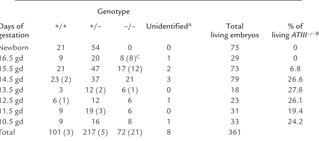
Table 1
Genotypes of embryos derived through timed matings of ATIII+/–heterozygotes
Genotype
Days of +/+ +/– –/– UnidentifiedA Total % of gestation living embryos living ATIII–/–B
Newborn 21 54 0 0 75 0
16.5 gd 9 20 8 (8)C 1 29 0
15.5 gd 21 47 17 (12) 2 73 6.8
14.5 gd 23 (2) 37 21 3 79 26.6
13.5 gd 3 12 (2) 6 (1) 0 18 27.8
12.5 gd 6 (1) 12 6 1 23 26.1
11.5 gd 9 19 (3) 6 0 31 19.4
10.5 gd 9 16 8 1 33 24.2
Total 101 (3) 217 (5) 72 (21) 8 361
[image:4.612.224.539.570.710.2]100% (eight of eight embryos) at 16.5 gd had died and showed extensive subcutaneous hemorrhage (Figure 2). No ATIII–/–mice were observed among the 75 F
2
newborn mice (Table 1). Thirteen dead ATII–/–
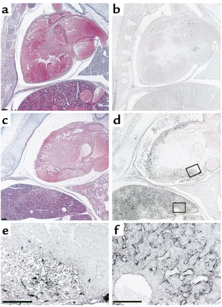
embryos (five embryos at 15.5 gd and eight at 16.5 gd) were examined histologically. In all of them, fibrin(ogen) deposition was detected in the degener-ated myocardium and liver (Figure 3, c–f). In the liver, fibrin(ogen) deposition was observed in the sinusoids, but not in the central veins (Figure 3f). Even in all of five living ATIII–/–embryos at 15.5 gd and one of four
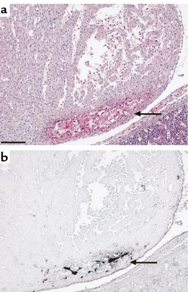
living ATIII–/– embryos examined at 14.5 gd, the
myocardium was partially degenerated where fib-rin(ogen) deposition was detected (Figure 4). In three of five living ATIII–/–embryos at 15.5 gd, the liver was
also partially degenerated where fibrin(ogen) was detected in the sinusoids (data not shown). These observations suggest that the degeneration of the myocardium and liver of dead ATIII–/–embryos is due
to intravascular coagulation rather than the natural progression of the absorption of dead embryos. It seems likely that fibrin deposition among degenerat-ing myocardial fibers is secondary, perhaps from hem-orrhage into the necrotic tissue. In other tissues, including the brain, lung, and kidney, no fibrin(ogen) deposition was observed by immunohistochemical staining, even though there was apparent tissue degen-eration. Extensive hemorrhage was observed subcuta-neously and intracranially (Figure 5a), but no appar-ent fibrin(ogen) deposition was detected by immunohistochemical staining (Figure 5b), suggest-ing that fibrinogen in plasma may have been decreased by consumptive coagulopathy and/or liver dysfunc-tion in the dead ATIII–/–embryos.
Discussion
It has been suggested that the contribution of a given anticoagulant mechanism to the hemostatic balance varies from one vascular bed to another, and thrombosis occurs in an organ-specific fashion
through the critical interplay of genetic and envi-ronmental factors (27). Whereas individuals with heterozygous antithrombin deficiency are suscepti-ble to venous thromboembolic diseases (10–12), a homozygous variant of antithrombin that lacks affinity for heparin causes thrombosis not only in veins but also in arteries (15–17). In this study, we demonstrated that complete antithrombin deficien-cy was lethal to embryos. The reason for embryonic death during mid-late embryogenesis appears to be fibrin deposition in the myocardium and liver, since antithrombin deficiency caused fibrin(ogen) depo-sition around 15.5 gd in the degenerated myocardi-um and liver, but apparently did not in other regions. Although a small amount of fibrin(ogen) deposition undetectable by immunohistochemical staining might be present in several other organs, the myocardium and liver seemed to be more sus-ceptible than other tissues to fibrin(ogen) deposi-tion in antithrombin deficiency.
[image:5.612.64.289.54.204.2]Previous reports have demonstrated that disruption of the tissue-factor pathway inhibitor (TFPI) or pro-tein C (PC) gene also causes fibrin(ogen) deposition
Figure 2
Macroscopic observation of ATIII+/+ (a) and dead ATIII–/– (b)
embryos at 15.5 gd. A dead ATIII–/–embryo at 15.5 gd showed
exten-sive subcutaneous hemorrhage.
Figure 3
Microscopic observation of ATIII+/+(a and b) and dead ATIII–/–(c–f)
embryos at 15.5 gd. (a and c) Hematoxylin-eosin and (band d) immunochemical staining with antifibrin(ogen) Ab. (eand f) Magni-fication of the areas indicated in d. (e) Myocardium. (f) Liver. A dead ATIII–/–embryo at 15.5 gd showed degeneration of the myocardium
[image:5.612.312.533.338.643.2]in the liver (18, 28), although its location was not dis-cussed in detail. In ATIII–/–embryos, fibrin(ogen)
dep-osition was detected in the sinusoids of the liver, but not in the central veins. It has been reported that expression of thrombomodulin and TFPI is not detected in the sinusoids of the liver (29, 30), sug-gesting that anticoagulation in the sinusoid of the liver might be dependent on antithrombin. In con-trast, such disruption of the TFPI or PC gene does not cause severe fibrin(ogen) deposition in the embryon-ic myocardium (18, 28); this is peculiar to ATIII–/–
embryos. Although the mechanism leading to the organ-specific fibrin(ogen) deposition in ATIII–/–
embryos remains to be elucidated, these observations suggest that antithrombin may be an important anti-coagulant protein in the myocardium and sinusoids of the liver during embryogenesis.
Beyond 15.5 gd, dead ATIII–/–embryos had extensive
hemorrhage, with no fibrin(ogen) deposition detectable by immunohistochemical staining, suggesting that fib-rinogen may be decreased by consumptive coagulopathy and/or liver dysfunction. Mice with fibrinogen deficien-cy suffer hemorrhagic symptoms postnatally but are born in normal numbers (31). On the other hand, embryos with TFPI (18) or antithrombin deficiency have a severe hemorrhagic phenotype. Severe hemorrhage of
these anticoagulant-deficient embryos may be attributed to the combined defects of plasma and cellular hemosta-tic components with extensive intravascular coagulation. The plasma levels of antithrombin antigen and activ-ity were significantly reduced in ATIII+/–mice compared
with ATIII+/+mice. However, ATIII+/–mice appear to be
indistinguishable from ATIII+/+mice and did not
expe-rience any apparent thromboembolic events during observation through 4 months of age. Most subjects with heterozygous antithrombin deficiency are asymp-tomatic (32, 33), although antithrombin deficiency is obviously a risk factor for thromboembolic diseases, especially deep venous thrombosis of the lower limb and pulmonary embolism (10–12). The ATIII+/–mouse
will be a useful animal model for studying the mecha-nism of thromboembolic events observed in human heterozygous antithrombin deficiency.
Acknowledgments
[image:6.612.91.280.53.347.2] [image:6.612.318.511.391.666.2]We thank Eriko Yamafuji and Chika Wakamatsu for their excellent technical support. This work is sup-ported by Grants-in-Aid for Scientific Research (10557090, 10670942, 10178102, and 10CE2006) from the Ministry of Education, Science, Sports and Culture, and for Research on Specific Diseases from the Min-istry of Health and Welfare.
Figure 4
Hematoxylin-eosin (a) and immunohistochemical staining with anti-fibrin(ogen) Ab (b) of the myocardium of a living ATIII–/–
embryo at 14.5 gd. Arrows indicate partial degeneration (a) and fibrin(ogen) deposition (b) in the myocardium.
Figure 5
Hematoxylin-eosin (a) and immunohistochemical staining with anti-fibrin(ogen) Ab (b) of the head of a dead ATIII–/–embryo at
1. Lane, D.A., and Caso, R. 1989. Antithrombin: structure, genomic organization, function and inherited deficiency. Baillieres Clin. Haema-tol.2:961–998.
2. O’Reilly, M.S., Pirie-Shepherd, S., Lane, W.S., and Folkman, J. 1999. Antiangiogenic activity of the cleaved conformation of the serpin antithrombin. Science.285:1926–1928.
3. Rosenberg, R.D., and Damus, P.S. 1973. The purification and mecha-nism of action of human antithrombin-heparin cofactor.J. Biol. Chem.
248:6490–6505.
4. Marcum, J.A., Fritze, L., Galli, S.J., Karp, G., and Rosenberg, R.D. 1983. Microvascular heparin-like species with anticoagulant activity. Am. J. Physiol.245:H725–H733.
5. Marcum, J.A., and Rosenberg, R.D. 1984. Anticoagulantly active heparin-like species molecules from vascular tissue. Biochemistry.
23:1730–1737.
6. Rosenberg, R.D., Shworak, N.W., Liu, J., Schwartz, J.J., and Zhang, L. 1997. Heparan sulfate proteoglycans of the cardiovascular system: specific structures emerge but how is synthesis regulated? J. Clin. Invest.99:2062–2070.
7. Kojima, T., Leone, C.W., Marchildon, G.A., Marcum, J.A., and Rosen-berg, R.D. 1992. Isolation and characterization of heparan sulfate pro-teoglycans produced by cloned rat microvascular endothelial cells. J. Biol. Chem.267:4859–4869.
8. Kojima, T., Shworak, N.W., and Rosenberg, R.D. 1992. Molecular cloning and expression of two distinct cDNA-encoding heparan sul-fate proteoglycan core proteins from a rat endothelial cell line. J. Biol. Chem.267:4870–4877.
9. Ishiguro, K., et al. 2000. Syndecan-4 deficiency impairs focal adhesion formation only under restricted conditions. J. Biol. Chem.
275:5249–5252.
10. Egeberg, O. 1965. Inherited antithrombin III deficiency causing thrombophilia. Thromb. Diath. Haemorrh.13:516–530.
11. Demers, C., Ginsberg, J.S., and Hirsh, J. 1992. Thrombosis in antithrombin-III-deficient persons. Report of a large kindred and lit-erature review. Ann. Intern. Med.116:754–761.
12. Lane, D.A., et al. 1997. Antithrombin mutation database: 2nd update. Thromb. Haemost.77:197–211.
13. Lane, D.A., Kunz, G., Olds, R.J., and Thein, S.L. 1996. Molecular genet-ics of antithrombin deficiency. Blood Rev.10:59–74.
14. Bayston, T.A., and Lane, D.A. 1997. Antithrombin: molecular basis of deficiency. Thromb. Haemost.78:339–343.
15. Chowdhury, V., et al. 1994. Homozygous antithrombin deficiency: report of two new cases (99 Leu to Phe) associated with arterial and venous thrombosis. Thromb. Haemost.72:198–202.
16. Okajima, K., et al. 1989. Homozygous variant of antithrombin III that lacks affinity for heparin, AT III Kumamoto. Thromb. Haemost.61:20–24. 17. Boyer, C., Wolf, M., Vedrenne, J., Meyer, D., and Larrieu, M.J. 1986. Homozygous variant of antithrombin III: ATIII Fontainebleau. Thromb. Haemost.56:18–22.
18. Huang, Z.F., Higuchi, D., Lasky, N., and Broze, G.J., Jr. 1997. Tissue
factor pathway inhibitor gene disruption produces intrauterine lethality in mice. Blood.90:944–951.
19. Healy, A.M., Rayburn, H.B., Rosenberg, R.D., and Weiler, H. 1995. Absence of the blood-clotting regulator thrombomodulin causes embryonic lethality in mice before development of a functional car-diovascular system. Proc. Natl. Acad. Sci. USA.92:850–854.
20. Wu, J.K., Scheffield, W.P., and Blajchman, M.A. 1992. Molecular cloning and cell-free expression of mouse antithrombin III. Thromb. Haemost.68:291–296.
21. Tsuzuki, S., et al. 1997. Molecular cloning, genomic organization, pro-moter activity, and tissue-specific expression of the mouse ryudocan gene. J. Biochem.122:17–24.
22. Igakura, T., et al. 1998. A null mutation in basigin, an immunoglob-ulin superfamily member, indicates its important roles in peri-implantation development and spermatogenesis. Dev. Biol.
194:152–165.
23. Terauchi, Y., et al. 1999. Increased insulin sensitivity and hypogly-caemia in mice lacking the p85 alpha subunit of phosphoinositide 3-kinase. Nat. Genet.21:230–235.
24. Liu, M.A. 1995. Overview of DNA vaccines. Ann. NY Acad. Sci.
772:15–20.
25. Chomczynski, P., and Sacchi, N. 1987. Single-step method of RNA iso-lation by acid guanidinium thiocyanate-phenol-chloroform extrac-tion. Anal. Biochem.162:156–159.
26. Yamamoto, K., and Loskutoff, D.J. 1996. Fibrin deposition in tissues from endotoxin-treated mice correlates with decreases in the expres-sion of urokinase-type but not tissue-type plasminogen activator. J. Clin. Invest.97:2440–2451.
27. Rosenberg, R.D., and Aird, W.C. 1999. Vascular-bed-specific hemo-stasis and hypercoagulable states. N. Engl. J. Med.340:1555–1564. 28. Jalbert, L.R., et al. 1998. Inactivation of the gene for anticoagulant
protein C causes lethal perinatal consumptive coagulopathy in mice. J. Clin. Invest.102:1481–1488.
29. Maruyama, I., Bell, C.E., and Majerus, P.W. 1985. Thrombomodulin is found on endothelium of arteries, veins, capillaries, and lymphat-ics, and on syncytiotrophoblast of human placenta. J. Cell Biol.
101:363–371.
30. Yamanobe, F., Mochida, S., Ohno, A., Ishikawa, K., and Fujiwara, K. 1997. Recombinant human tissue factor pathway inhibitor as a pos-sible anticoagulant targeting hepatic sinusoidal walls. Thromb. Res.
85:493–501.
31. Suh, T.T, et al. 1995. Resolution of spontaneous bleeding events but failure of pregnancy in fibrinogen-deficient mice. Genes Dev.
9:2020–2033.
32. McColl, M., et al. 1996. Low thrombosis rate seen in blood donors and their relatives with inherited deficiencies of antithrombin and pro-tein C: correlation with type of defect, family history, and absence of the factor V Leiden mutation. Blood Coagul. Fibrinolysis.7:689–694. 33. Tait, R.C., et al. 1994. Prevalence of antithrombin deficiency in the