INTRODUCTION
Under adverse growth conditions, C. elegans can execute an alternative developmental pathway to give rise to a diapause-like stage referred to as dauer. This specialised developmental stage is associated with profound morphological, metabolic and behavioural changes that allow C. elegans to survive unfavourable growth conditions, as well as promoting its dispersal to more favourable environments (Riddle and Albert, 1997). The integration of signals from the surroundings sensed during the first larval stage dictates whether the larva will progress through reproductive development, or whether this alternative developmental pathway will be executed. High population density initiates the dauer developmental program through signalling by a pheromone, while high temperatures and reduced nutrient resources strongly potentiate this decision (Golden and Riddle, 1982; Golden and Riddle, 1984). However, dauer larvae can recover from this stage when growing conditions improve, thus allowing the animal to develop into a fertile adult without apparent morphological or reproductive consequence.
The genetic and molecular basis of dauer formation has been well characterised and involves three highly conserved signalling pathways. These parallel pathways (TGFβ, insulin-like and cGMP-like) affect signalling within the nervous system of C. elegansto regulate dauer formation, further highlighting the importance of neuronal inputs in the execution of this developmental program (Ren et al., 1996; Bargmann and Horvitz, 1991; Birnby et al., 2000; Patterson and Padgett, 2000; Schackwitz et al., 1996; Wolkow et al., 2000). Previous studies in C. elegans have demonstrated that different amphid neurons are important for several aspects of dauer development, including dauer recovery, and ablation of these neurons often fully phenocopies the abnormal dauer formation phenotypes typical of dauer formation abnormal (Daf) mutants
(Bargmann and Horvitz, 1991; Schackwitz et al., 1996). Recovery from the dauer stage must be equally tightly controlled so that the post-dauer larva can resume its reproductive developmental program and produce progeny in a suitable environment. There is, to date, little information on how C. elegansmaintains or recovers from this stage (Tissenbaum et al., 2000). Recovery during persistent unfavourable conditions would be deleterious and would drastically reduce the fitness of the animal. Therefore, the C. elegansdauer larva must constantly monitor its environment for resource availability, pheromone level (crowding) and probably other external signals, and must integrate all of this sensory information to elicit the appropriate developmental response: to maintain or recover from dauer.
The Notch signalling pathway is well conserved in higher metazoans from C. elegans to humans, and it was shown to be required in these diverse organisms for the specification of various cell fates among a population of equipotent cells (Bray, 2006). Recently, the Notch signalling pathway has been shown to play a novel role in mature adult brain and in non-developmental decisions in C. elegans, Drosophilaand mouse (Chao et al., 2005; Costa et al., 2003; Feng et al., 2001; Ge et al., 2004; Presente et al., 2004; Yu et al., 2001). This novel function of the Notch signalling pathway is consistent with the described expression of some of the components of the Notch signalling pathway in differentiated neuronal cells in adult brain (Lee et al., 1996; Siman and Salidas, 2004). As these neurons are postmitotic, the requirement of the Notch signalling pathway is unlikely to be in the specification or differentiation of neuronal precursor cells.
We noticed that the DSL (Delta/Serrate/LAG-2) Notch ligand LAG-2 is expressed in neurons specifically at the onset of, and during, the dauer stage. As these cells are postmitotic and differentiated, the expression of the Notch ligand in head neurons reflects a possible novel role for this pathway, potentially in neuronal signalling or function. The findings we show here suggest that this initial expression of lag-2activates canonical glp-1Notch signalling in neurons, the function of which is crucial for the maintenance of
Notch signalling is required for both dauer maintenance and
recovery in
C. elegans
Jimmy Ouellet, Shaolin Li and Richard Roy*
The Notch signalling pathway is conserved among higher metazoans and is used repeatedly throughout development to specify distinct cell fates among populations of equipotent cells. Mounting evidence suggests that Notch signalling may also be crucial in neuronal function in postmitotic, differentiated neurons. Here, we demonstrate a novel role for the canonical Notch signalling pathway in postmitotic neurons during a specialised ‘diapause-like’ post-embryonic developmental stage in C. eleganscalled dauer. Our data suggest that cell signalling downstream of the developmental decision to enter dauer leads to the activation of Notch-responding genes in postmitotic neurons. Consistent with this, we demonstrate that glp-1, one of the two C. elegans Notch receptors, and its ligand lag-2are expressed in neurons during the dauer stage, and both genes are required to maintain this stage in a daf-7/TGFβdauer constitutive background. Our genetic data also suggest that a second Notch receptor, lin-12, functions upstream of, or in parallel with, insulin-like signalling components in response to replete growth conditions to promote dauer recovery. Based on our findings, cues associated with the onset of dauer ultimately trigger a glp-1-dependent Notch signalling cascade in neurons to maintain this developmental state. Then, as growth conditions improve, activation of the LIN-12 Notch receptor cooperates with the insulin-like signalling pathway to signal recovery from the dauer stage.
KEY WORDS: C. elegans, Notch, Dauer, Neurons, Insulin-like signalling Development 135, 2583-2592 (2008) doi:10.1242/dev.012435
Department of Biology, McGill University, Montréal, Québec H3A 1B1, Canada.
*Author for correspondence (e-mail: [email protected])
Accepted 9 June 2008
D
E
V
E
LO
P
M
E
N
this stage in daf-7mutant dauer larvae. Moreover, a second Notch receptor, lin-12, is activated later in a lag-2-independent manner, and works upstream of, or in parallel with, the insulin-like signalling pathway to appropriately signal recovery from dauer and the resumption of reproductive development.
MATERIALS AND METHODS Strains and genetics
Strains were cultured as previously described (Brenner, 1974). The
following alleles were used: ins-18(tm339)I, rrf-3(pk1426)II,
rab-7(ok511)II/mIn1, unc-130(oy10, ev505)II, daf-7(e1372, m70, m62)III, daf-2(e1370)III, glp-1(e2141, q224, q231)III, lin-12d(n302, n950)III, unc-32(e189)lin-12(n676n927, n676n930)III, lag-1(om13)IV, lag-2(q420)V and
qIs56 [lag-2::GFP, unc-119(+)]V (Blelloch et al., 1999); deg-1(tu38)X andkyIs51 [odr-2 2b::GFP + pJM23 (lin-15+)] (Chou et al., 2001); and
adEx1269[lin-15(+) odr-1::GFP] (Yu et al., 1997).
lag-2promoter variant plasmids
A 3-kb sequence upstream of the translational start of lag-2was cloned into
pGEM-T (Promega) to generate pMR100. Different fragments were transferred from this vector into the pPD95.67 vector using the various enzymes indicated in Fig. 2A. For the plasmids presented in Fig. 2B,
pMR126 contains the HindIII-AccI(blunted) fragment from pMR103
inserted into the HindIII-SmaI site of pPD95.67. pMR133 (nucleotides
–1643 to –1369 of the lag-2promoter) was created by PCR from pMR126
using the primer designed to delete two of the three predicted forkhead-binding sites. pMR134 contains the PCR fragment amplified from pMR126
(nucleotides –1520 to –1369 of the lag-2promoter) using the primers
rr289-192. pMR137 contains the PCR fragment amplified from pMR126 using the primers rr287-288. pMR145 was created by amplifying fragments from pMR126 with the primers rr287-294 and rr192-303. pMR126, pMR133, pMR134, pMR137 and pMR145 were confirmed by sequencing. All primer sequences used to create these constructs and further cloning details are available on request.
ins-18rescuing construct
The genomic region of the ins-18locus, containing ~3.8 kb of the promoter
region, the coding sequence and 570 bp of the 3⬘UTR, was amplified from
wild-type genomic DNA with the primers rr1072-1073 using Phusion DNA polymerase (NEB). The resulting PCR fragment was cloned into pGEMT-t (Promega) vector (pMR1146).
UNC-130::RFP construct
The genomic region containing the unc-130locus (promoter, ORF and the
3⬘UTR) was amplified from N2 genomic DNA using the primers rr723-724
and cloned into pSKII (pMR1107). The RFP-coding region was amplified
from the plasmid dsRED2 using the primers rr615-616, digested with SacI,
blunted and cloned into the NcoI blunted site (T4 DNA polymerase) of
pMR1107 to create an in frame unc-130 C-terminal translational fusion
(pMR1107.2).
glp-1p::GLP-1::YFP reporter
The YFP variant (Nagai et al., 2002) was amplified from the plasmid pBS7
with the primers NheIGFP-5 and GFPSMT-3 and cloned into the HpaI-cut
pGLP-1 S642N vector (Berry et al., 1997). We used the glp-1(gf)allele to
increase expression through the positive-feedback loop that occurs following receptor activation (Greenwald, 1998).
Dye-filling assay
Dye filling was performed as previously described (Burket et al., 2006). As none of the neurons are exposed to the external environment during the dauer stage, the larvae were stained at the L1 stage, allowed to form dauer and then imaged thereafter.
Neuronal-specific expression of Notch receptor constructs
To create the neuronal promoter constructs, we first cloned the glp-1
wild-type genomic sequence into pSKII and inserted a KpnI site at the 5⬘end to
create pMR1137.4. The neuronal promoters (ser-2prom2,lim-6int3,gpa-2,
unc-25,ceh-6,odr-1) were amplified from wild-type genomic sequence and
primers were designed to introduce either an EagI or a NotI site at the 5⬘end,
in addition to an in frame KpnI site at the 3⬘end for cloning into an Eag
I-KpnI digested pMR1137.4 vector (Burglin and Ruvkun, 2001; Hobert et al.,
1999; Jin et al., 1999; Tsalik et al., 2003; Yu et al., 1997; Zwaal et al., 1997). All primer sequences used to create these constructs and further cloning details can be obtained by request.
Microinjection and transformation
All constructs were injected at concentrations ranging from 5-40 ng/μl, with
either the dominant co-injection marker pRF4 rol-6(D)or the plasmid
pMR352 (Li et al., 2003), which expresses pharyngeal GFP under the
control of the myo-2promoter, at 30-50 ng/μl in the corresponding genetic
background.
lag-2::GFPexpression inunc-130 mutants during reproductive development
L1 larvae of the daf-2(e1370); qIs56and unc-130(oy10); daf-2(e1370);
qIs56 were incubated at 20°C until they reached the L3 stage, after which the animals were examined for GFP expression in the IL2 neurons. The total number represents the average of three independent trials and the bars represent the standard deviation.
Dauer assays
For the dauer recovery assay presented in Table 1, embryos were collected from alkaline/hypochlorite-treated gravid adults and hatched at 15°C overnight. The resulting synchronised L1 larvae were distributed onto seeded plates and placed at 25°C for 48 hours. For each genotype, dauers were transferred on to pre-equilibrated plates and placed at 25°C immediately thereafter. L4 larvae and adults were subsequently scored after 24 hours at 25°C. Each experiment was repeated at least three times.
To test the role of Notch in dauer maintenance in a non-Daf-c background, L1 larvae were incubated at 15°C on plates containing dauer pheromone until larvae formed dauers. The plates were then transferred to the restrictive temperature (25°C) for 24 hours to inactivate the query Notch gene product. Subsequently, 50 dauer larvae were transferred to plates containing a reduced concentration of pheromone, which, at this threshold, permits 70% of the wild-type dauer larvae to recover, while 30% of the dauers maintain this developmental state. The number of L4 larvae and young adults on the plates was scored 24 hours after transfer.
To assess the role of lin-12in wild-type dauer recovery, dauers were
induced by crowding/starvation and 50 animals were transferred onto plates with a fresh bacterial lawn. For the 4-hour time point, we monitored pharyngeal pumping which is an early marker for commitment to recovery. For later time points, morphological changes typical of recovering dauer larvae were scored. In both cases, the experiments were repeated five times. For the suppression of the dauer maintenance defect by the
neuronal-specific expression of glp-1, we subjected transformed gravid adults to
alkaline/hypochlorite and allowed them to hatch at 15°C. The L1 larvae were then incubated at 25°C on seeded plates for two days to allow dauer formation. Then, because the transgenes were maintained as extrachromosomal arrays, we transferred 25 transformed and 25 non-transformed dauer progeny onto the same plate. We counted how many dauer larvae recovered 24 hours later for each genotype and the degree of suppression is represented as follows:
1–(recovered transformed dauer/recovered non-transformed dauer)⫻100.
Each experiment was performed with at least two independent lines and was repeated three times.
Laser ablation
L1 larvae were incubated at 25°C for 24 hours and laser microsurgery was performed on early L2d animals. After the surgery, the ablated animals were incubated for an additional 24 hours at 25°C to allow dauer formation and the ability to maintain dauer was assayed as described above.
Microscopy and image processing
Images were captured using either a Leica DM microscope or a Zeiss LSM Meta confocal microscope, and were processed and assembled in Photoshop
CS (Adobe).
D
E
V
E
LO
P
M
E
N
RESULTS
Although Notch signalling is used in numerous contexts throughout development in C. elegans, its role in postmitotic neurons has not been extensively characterised (Chao et al., 2005). We observed that six head neurons express the gene encoding the Notch DSL ligand
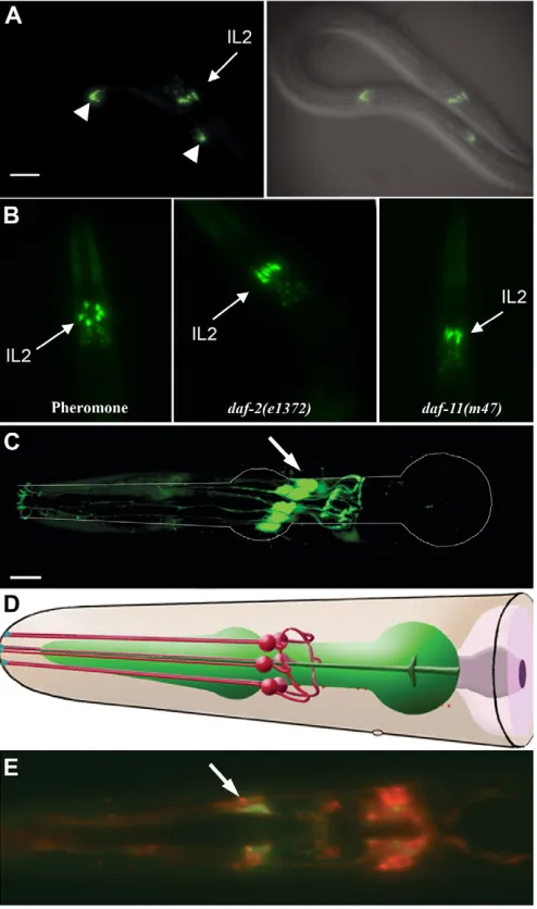
lag-2specifically at the onset of and throughout the dauer stage by using a lag-2::GFPtransgene (Fig. 1A, data not shown). The same level of lag-2::GFPexpression was observed in dauer larvae induced by dauer pheromone, starvation, or by Daf-c mutations in the three known parallel pathways, suggesting that lag-2 expression is activated downstream of the three major signalling pathways involved in dauer formation (Fig. 1B). Moreover, we did not detect any change in the expression level of the lag-2::GFP transgene between newly induced (4 hours post-induction), and older (96 hours post-induction) dauers, suggesting that the expression of the Notch ligand is sustained throughout the dauer stage (data not shown).
On the basis of their position and the morphology of their projections, we identified the lag-2::GFP-expressing cells as being one of the three pairs of Inter Labial (IL) neurons (Fig. 1C,D). To distinguish whether this expression was specific to the IL1 or the IL2 neurons, which are morphologically quite similar, we performed DiI staining with calcium acetate, which stains the amphid and the IL2 neurons, but not the IL1 neurons (Burket et al., 2006). The lag-2::GFP-expressing cells and the DiI-stained neurons overlapped, leading us to conclude that lag-2 is expressed in IL2 neurons during the dauer stage (Fig. 1E). Consistent with lag-2::GFP being expressed in IL2 neurons, we found that its expression was unaffected in dauer larvae that harbour a deg-1(u38)mutation, which causes the degeneration of the IL1 neurons early in post-embryonic development without affecting the IL2 neurons (data not shown) (Chalfie and Wolinsky, 1990). Therefore, based on these results, we conclude that lag-2is expressed in the IL2 neurons at the onset of and throughout the dauer stage, whether they are formed through the normal pheromone-sensing pathway due to crowding, or by Daf-c mutations.
lag-2expression depends on forkhead-binding sites
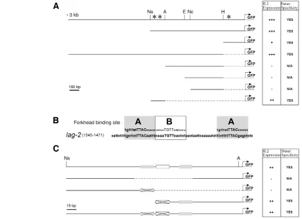
The expression of lag-2::GFPin IL2 neurons at the onset and during dauer prompted us to identify the upstream components required for the dauer-specific neuronal expression of lag-2. Deletion analysis of the lag-2promoter during dauer indicated that a small fragment located 1.3 kb upstream of the lag-2 translational start site was sufficient for dauer-specific expression in IL2 neurons (Fig. 2A). This region contains three highly conserved forkhead transcription factor-binding sites; two of which are nearly identical and match the FoxC consensus (Saleem et al., 2004). We refer to these different classes of binding sites as A and B (Fig. 2B). To determine the importance of these sites for the expression of lag-2, we systematically removed each forkhead-binding site and found that they are required for dauer/IL2-specific GFP expression (Fig. 2C). Detailed analysis of this region indicated that a single A-type forkhead-binding site is sufficient for IL2-specific expression during dauer and this site is present twice in this small interval (Fig. 2C). Consistent with the potential role of this binding site in regulating the appropriate lag-2expression in these neurons, we identified a similar ‘A’ site within the proximal region of the promoter, which was also sufficient to confer dauer/IL2-specific expression of lag-2 (Fig. 2A). Therefore, we propose that, in response to environmental signals, a forkhead transcription factor must act through these sites to trigger lag-2 expression in the IL2 neurons at the onset of dauer.
UNC-130 is required to repress lag-2expression during reproductive development
[image:3.612.52.299.129.547.2]The genome of C. elegans is predicted to encode 15 forkhead transcription factors, six of which have been genetically characterised (Hope et al., 2003; Mango et al., 1994; Miller et al., Fig. 1. The DSL ligand lag-2is expressed in the three pairs of IL2
neurons during the dauer stage.(A) A daf-7(e1372)dauer larva expressing the lag-2::GFPtransgene and the corresponding DIC image overlaid with the GFP expression to show the position of the cells expressing the transgene. White arrowheads indicate the described expression of lag-2::GFP in the distal tip cells (DTC). Scale bar: 25 μm. (B) The head region of dauers expressing lag-2::GFP (qIs56) induced by either pheromone, or in various Daf-c mutants as indicated in the panels. (C) 3D reconstruction of a confocal stack of images of the IL2 neurons in dauer. White lines outline the pharynx of the dauer animal. Scale bars: 10 μm. (D) Diagram of the IL2 neurons indicating their position and their characteristic morphology (adapted with permission from wormatlas.org). (E) Merge of dauer expressing lag-2::GFP(green) in the IL2 neurons that were stained with the lipophilic dye DiI (red). As lag-2::GFPis expressed in the entire cell, whereas DiI only stains the membrane, colocalisation (yellow) is only observed at the membrane, giving a halo-like appearance. In all images, arrowheads indicate the IL2
neurons.
D
E
V
E
LO
P
M
E
N
1993; Nash et al., 2000; Ogg et al., 1997) and only one of them, the C. elegansFoxO homologue DAF-16, has been previously shown to play a crucial role in dauer formation. However, we confirmed that this transcription factor was not responsible for the dauer-specific regulation of lag-2 in the IL2 neurons. First, the identified forkhead-binding sites in the lag-2 promoter do not match the predicted consensus DAF-16-binding site (Furuyama et al., 2000), and, more importantly, the dauer-dependent/IL2-specific lag-2::GFP expression was unaffected in dauers that completely lack DAF-16 (daf-16(mgDf50)null mutant) (data not shown).
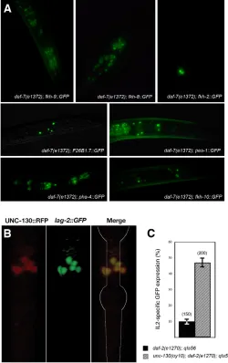
As the forkhead transcription factor required for lag-2 IL2-specific expression during the dauer stage is likely to be expressed in the same neurons as lag-2, we examined the expression pattern of all the C. elegansforkhead genes during the dauer stage by using a series of GFP-reporter strains (Hope et al., 2003; Mango et al., 1994) (Fig. 3A). Although many forkhead reporters are expressed in neurons throughout the head region during the dauer stage, only the unc-130::GFP strain showed strong expression in the presumptive IL2 neurons. We generated an UNC-130::RFP translational fusion
reporter construct and showed that its expression overlapped with that of the lag-2::GFPin the IL2 neurons during dauer (Fig. 3B). This suggests that UNC-130 could regulate the expression of lag-2 in the IL2 neurons specifically during the dauer stage. However, we did not detect any change in lag-2::GFP expression in unc-130 mutants during the dauer stage (data not shown). Therefore, the UNC-130 forkhead transcription factor is not absolutely required for dauer-specific expression of lag-2, and/or it may function redundantly with another factor to initiate the expression of the Notch ligand.
[image:4.612.53.488.61.377.2]As unc-130::RFPis expressed in the same neurons as lag-2, UNC-130 could be required to repress lag-2expression during reproductive development, which would be akin to its described repressor role in sensory neurons (Sarafi-Reinach and Sengupta, 2000). We therefore determined lag-2::GFPexpression in daf-2(e1370)and unc-130(oy10); daf-2(e1370), maintained at the sub-threshold temperature for dauer formation (20°C). We noticed that, under these sensitised conditions, 48.3±4.5% of unc-130(oy10); daf-2(e1370) larvae misexpressed GFP in IL2 neurons during Fig. 2. A cluster of three forkhead-binding sites is sufficient for dauer-specific lag-2expression in the IL2 neurons.(A) A 3-kb region upstream of the lag-2translational start site was subjected to deletion analysis to determine the minimal fragment necessary for IL2/dauer-specific lag-2expression in daf-7animals. Enzyme sites used for the generation of the different promoter variants are indicated: Ns (NspI), A (AccI), E (EcoRI), Nc (NcoI) and H (HhaI). Solid lines represent fragments of the lag-2promoter that were cloned upstream of the GFP-coding sequence; dashed lines represent deleted sequence. Asterisks represent the location of the predicted forkhead-binding sites in the lag-2promoter. (B) Two potential forkhead-binding sites, named A and B, were identified in the minimal fragment required for IL2 neuron/dauer-specific expression. The consensus binding sites for the FoxC1 transcription factor are indicated in the grey box above the lag-2sequence (A binding sites). Capital letters represent the core binding site and small letters indicate nucleotides required for efficient binding. (C) Smaller deletions of the 270 bp fragment were created to determine which forkhead-binding sites are required for IL2 neuron/dauer-specific expression. The white and grey boxes represent the identified forkhead-binding sites and the crosses indicate regions where the core binding site sequence was deleted. The relative intensity of GFP expression in the IL2 neurons is indicated as follows: +++, strong; ++, moderate; +, faint; –, no expression. For the consensus binding sites: w can be A or T; m can be A or C; and n can be A, T, C or G.
D
E
V
E
LO
P
M
E
N
reproductive development (n=200), compared with only 7.1±2.3% indaf-2(e1370)mutants (n=150; Fig. 3C). Although some Unc mutations have been reported to affect dauer formation (Ailion and Thomas, 2000; Ailion and Thomas, 2003), our observations indicate that there is no effect of unc-130 in enhancing dauer formation (data not shown). Therefore, we suggest that the UNC-130 forkhead transcription factor is required to repress lag-2 expression during reproductive development. Then, upon dauer formation, at least in a daf-2 mutant background, UNC-130-mediated repression of lag-2is released and another transcription factor, which may bind to the same region, is required to activate lag-2expression.
Notch signalling is required to maintain the dauer stage in daf-7/TGFβmutants
The expression of lag-2in the IL2 neurons during dauer suggests that Notch signalling might be involved in some aspect(s) of this developmental stage. Because cell division is arrested during the dauer stage, we wanted to determine whether Notch might play a more physiological role that may affect dauer formation, maintenance and/or recovery. By using temperature-sensitive mutations in various effectors of the Notch signalling pathway (as described in Table 1), we found that none of these mutations affected dauer formation in daf-2 or daf-7 dauer constitutive mutants maintained at the restrictive temperature (data not shown), suggesting that Notch signalling is not required to trigger this developmental switch. However, Notch signalling could alternatively be involved in dauer maintenance or recovery. Because insulin-like signalling is required for dauer recovery (Tissenbaum et al., 2000), we examined the effects of mutations in Notch signalling components on maintenance and recovery in a daf-7 mutant background, wherein the signalling system that responds to recovery cues is competent, allowing us to assess how Notch signals impinge on this network and affect this developmental decision.
[image:5.612.49.299.65.463.2]Under these conditions (see Materials and methods), most (81.3±6.43%, n=416) of the daf-7(e1372); lag-2(q420lf) dauer larvae recovered from this stage prematurely, within 24 hours following dauer formation; approximately 10-fold greater than the baseline recovery observed in daf-7(e1372) animals (Table 1). Moreover, laser ablation of the IL2 neurons (which express the Notch ligand lag-2) prior to dauer entry in a daf-7(e1372)mutant background also leads to dauer maintenance defects that are comparable to those observed in a daf-7; lag-2 double mutant background (Table 2). This premature recovery is not due to an inability of these larvae to sense pheromone, as it can be suppressed by maintaining lag-2(q420)mutant dauers on dauer pheromone, indicating that this premature recovery can occur only if pheromone levels are low (Ogg et al., 1997) (data not shown). Taken together, Fig. 3. The forkhead transcription factor UNC-130 is required for
appropriate repression of lag-2expression during reproductive development.(A) Expression in the head region of the various forkhead transcription factors predicted from the C. elegansgenome database during the dauer stage indaf-7(e1372)mutants (Hope et al., 2004). (B) Confocal images depicting the expression of the UNC-130::RFP translational fusion protein in the daf-2(e1370); qIs56( lag-2::GFP) background, and the merge of the two channels during the dauer stage. (C) The percentage of L3 larvae kept at sub-threshold temperature (20°C) that express lag-2::GFPin the IL2 neurons in the mutant background is indicated.
Table 1. Components of a Notch signalling cascade are required during dauer development
Genotype Dauer recovery*
daf-7(e1372) 5.3±0.27 (539)
daf-7(m70) 22.5±1.52 (200)
daf-7(e1372); lag-2(q420) 81.3±6.43 (416)†
lin-12(n696n927) daf-7(e1372) 0 (150)† lin-12(n696n930) daf-7(e1372) 0 (150)† lin-12d(n302gf) daf-7(e1372) 21.7±1.90 (410)† lin12d(n950gf) daf-7(e1372) 61.5±4.54 (302)† glp-1(e2141) daf-7(e1372) 84.7±3.61 (354)† glp-1(q231) daf-7(e1372) 37.7±2.1 (450)† glp-1(e2141) daf-7(m70) 73.0±6.52 (137)‡
daf-7(e1372); lag-1(om13) 0.42±0.12 (416)† glp-1(e2141) daf-7(e1372); lag-1(om13) 0 (186)¶ daf-7(m70); lag-1(om13) 13.1±2.15 (130)‡
ins-18(tm339); daf-7(e1372) 28.7±1.2 (150)† ins-18(tm339); daf-7(e1372); ins-18::ins-18 1.0±0.3 (100)§ *Results are expressed as the percentage of animals ±s.d. that recover from dauer at 25°C 24 hours after transfer. The total number of dauer larvae scored (n) is indicated in parentheses. Statistical analyses were performed using a Student’s t-test: P<0.005, compared with the indicated genotypes.
†daf-7(e1372). ‡daf-7(m70).
¶glp-1(e2141) daf-7(e1372). §ins-18(tm339); daf-7(e1372).
D
E
V
E
LO
P
M
E
N
[image:5.612.311.564.78.262.2]these findings indicate that the DSL ligand lag-2is expressed in the IL2 neurons at the onset of dauer to appropriately maintain this developmental stage, and that these neurons play an important role in dauer maintenance in daf-7/TGFβDaf-c mutants.
Because of the dauer-specific IL2 expression pattern of lag-2::GFPand the dauer maintenance phenotype associated with lag-2mutations, we next examined the effect of mutations in other components of the Notch signalling pathway on dauer maintenance. We found that two loss-of-function (lf) mutations in the C. elegans Notch receptor gene glp-1cause dauer maintenance defects similar to those observed in lag-2mutants at the restrictive temperature in a daf-7(e1372)background (Table 1). Like in lag-2mutants, the premature recovery typical of glp-1mutant dauer larvae is also suppressed by pheromone (data not shown).
Surprisingly, lin-12 (lf) alleles had opposite effects to glp-1 mutants, where both lin-12(n696n927lf)and lin-12(n696n930lf) alleles completely suppress dauer recovery in a daf-7(e1372)mutant background (Table 1). Furthermore, two different lin-12 gain-of-function (gf) alleles, n302 and n950, caused premature dauer recovery, where the recovery frequency reflected the strength of the individual alleles (Table 1) (Greenwald et al., 1983). Therefore, our data suggest that glp-1 andlin-12signalling play distinct roles during this developmental stage in dauers induced by a daf-7 mutation; GLP-1 enhances dauer maintenance, whereas LIN-12 promotes timely recovery from dauer (see below and Discussion).
The downstream transcription factor LAG-1 is required for dauer recovery
LAG-1 is the C. eleganshomologue of DrosophilaSuppressor of Hairless [Su(H)], and it plays an important role in directing the Notch intracellular domain (NICD) to Notch-responsive target genes (Christensen et al., 1996; Jarriault and Greenwald, 2002). When we scored dauer maintenance in a lag-1(om13lf) background, we found that only 0.6% of the daf-7(e1372); lag-1(om13) dauer larvae recovered prematurely at the restrictive temperature; 10-fold less than the background recovery we observed for daf-7(e1372)mutants alone (Table 1). Furthermore, lag-1(om13)fully suppresses the dauer maintenance defect associated with a glp-1(e2141) mutation, as none of the glp-1(e2141) daf-7(e1372); lag-1(om13)dauer larvae recovered prematurely at the restrictive temperature (Table 1). As lag-1is downstream of both Notch receptors, its compromise causes a consequent decrease in the expression of Notch gene targets involved in recovery, thus rendering the animal incapable of recovering from dauer, consistent with the phenotype observed in lin-12 (lf)mutants.
GLP-1 Notch receptor is expressed in postmitotic neurons
The nervous system is known to play a crucial role during dauer development, so we predicted that the Notch-responding cells involved in dauer maintenance would most likely be neurons
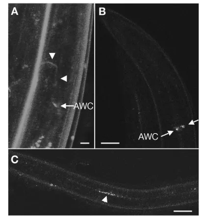
[image:6.612.44.562.71.131.2](Bargmann and Horvitz, 1991). Determination of GLP-1 protein expression during the dauer stage using antibody staining proved to be difficult because of the relatively impermeable specialised dauer cuticle. Therefore, to detect GLP-1 in dauer larvae, we constructed a glp-1p::GLP-1::YFP translational fusion reporter transgene. By using this strategy, we were able to capture GLP-1::YFP expression in head neurons located near the terminal bulb during the dauer stage (Fig. 4A, white arrow). The axons of these neurons project anteriorly toward the nerve ring placing them in close proximity to the IL2 processes (Fig. 4A, white arrowheads). In parallel, we monitored the expression of this transgene in a rab-7(ok511) mutant background that disrupts endosome fusion, a process necessary for Notch turnover (Sakata et al., 2004). In these Table 2. Laser microsurgery demonstrates a role for the IL2 and the AWC neurons in dauer maintenance
Genotype Neurons ablated Dauer recovery*
daf-7(e1372); odr-2 (2b)::GFP‡ IL2 5/7†
Mock 2/17
glp-1(e2141) daf-7(e1372); odr-1::GFP¶ AWC 1/7†
Mock 9/17
*The number of dauer larvae that recover 24 hours after their transfer onto a fresh, pre-equilibrated plate over the total number of transferred dauers.
†Statistical analyses were performed: P<0.005, compared with mock-treated animals. ‡Transgene from Chou et al. (Chou et al., 2001).
¶Transgene from Yu et al. (Yu et al., 2001).
Fig. 4. GLP-1 is expressed in differentiated neurons in the head during dauer.(A) The glp-1p::GLP-1::YFPtransgene is expressed in two head neurons during dauer in a daf-7(e1372)background. The cell bodies (indicated by arrow) are located in close proximity to the terminal bulb. The axon of these neurons (arrowheads) projects toward the anterior into the nerve ring, where they are within close proximity of the IL2 axons. Scale bar: 5 μm. (B,C) The same construct was injected in a rab-7(ok511)II/mIn1; daf-7(e1372)mutant, which disrupts the endocytic-mediated receptor recycling pathway. YFP-containing vesicles accumulate in the same neurons as in A. (C) Confocal
micrograph depicting the accumulation of YFP vesicles along the length of the animal in neurons within the ventral cord. Scale bar: 20 μm. rol-6was used as co-transformation marker, which accounts for the abnormal morphology of the ventral cord neuronal processes. Anterior
is to the left in C.
D
E
V
E
LO
P
M
E
N
[image:6.612.333.537.355.578.2]mutant animals, the GLP-1::YFP receptor would be internalised, but not degraded, and therefore YFP-containing vesicles should accumulate in the cytoplasm in a time-dependent manner, thereby facilitating our identification of these neurons. As predicted, the GLP-1::YFP-expressing head neurons described above accumulate YFP during the dauer stage in two bilateral head neurons (Fig. 4B, white arrows). GLP-1::YFP also accumulated throughout the length of the animal in the ventral cord, in axons that appear to emanate from neurons located in the head region (Fig. 4C, white arrowhead). Therefore, both our genetic data, and our description of this novel GLP-1 expression pattern in neurons, suggest that GLP-1/Notch signalling is required in neurons in order to maintain developmental quiescence during the dauer stage in daf-7/TGFβmutants.
To identify these neurons, we expressed glp-1under the control of well-described neuronal promoters, the expression of which overlaps in various neurons (Burglin and Ruvkun, 2001; Hobert et al., 1999; Jin et al., 1999; Tsalik et al., 2003; Yu et al., 1997; Zwaal et al., 1997), and we determined their capacity to rescue the dauer maintenance defect typical ofglp-1(e2141) daf-7(e1372)(Table 3). We found that the expression of glp-1specifically in gpa-2-and odr-1-expressing neurons efficiently and reproducibly suppressed the dauer maintenance defect associated with glp-1(e2141)(Table 3). Based on the known expression of gpa-2andodr-1, and on the position and morphology of the glp-1::YFPexpressing neurons, we conclude that glp-1is required in the AWC neurons for proper dauer maintenance. These neurons are crucial for dauer recovery, as ablation of the AWC neurons in a glp-1(e2141) daf-7(e1372) mutant background suppressed their premature recovery/dauer maintenance defect (Table 2). Moreover, although these neurons are remodelled during the dauer stage (Albert and Riddle, 1983), no morphological defects were detected in the AWC neurons of glp-1(e2141) daf-7(e1372)when compared with those of daf-7(e1372)mutants (data not shown), suggesting that glp-1is not required for the dauer-specific remodelling of these neurons. However, as we observed glp-1::YFPin the ventral nerve cord, which does not receive processes from the AWC neurons, it is possible that glp-1may be required in additional neurons (Fig. 4C).
Notch regulates dauer decisions in wild-type (non-‘dauer constitutive’) animals
Notch signalling therefore plays two roles during dauer development: one in maintenance that is mediated by glp-1; and a second in mediating recovery when conditions are replete, which is
lin-12-dependent. However, due to the complexity of the crosstalk between the dauer formation pathways, we were only able to verify these roles in a daf-7mutant background. In order to determine whether these Notch receptors were also important for these dauer decisions outside of the daf-7mutant background, we tested their function in dauers that were induced either by high concentrations of dauer pheromone or by crowded/starved conditions.
[image:7.612.338.505.338.635.2]Using pheromone, we induced animals to form dauers and then allowed them to recover by transferring these pheromone-induced or ‘wild-type’ dauers onto plates that contained a lower concentration of dauer pheromone that permitted 70% of the wild-type dauers to recover. Under these conditions, 99.6±0.9% of the glp-1(e2141)recovered from dauer, as compared with 72.7±7.9% for wild type (Fig. 5A; P<0.005 using a Mann-Whitney test, five independent trials, n=50/trial). We did not observe a significant difference in recovery for the weaker glp-1(q231) allele in these wild-type dauers, consistent with its attenuated effect in the daf-7(e1372) background (Table 1). In addition, no significant change in the frequency of premature dauer recovery was detected using either lin-12 (lf)or (gf)mutations in this assay (Fig. 5A). It is likely that under these conditions, even in the presence of reduced pheromone concentrations, compromise of the lin-12 Notch signalling pathway cannot overcome the dauer recovery program
Table 3. glp-1 expression in AWC neurons is sufficient to correct glp-1-dependent dauer maintenance defects Transgene injected* Rescue of dauer maintenance defect ±s.d.†
myo-2p::GFP 6.1±5.4%
gpa-2p::glp-1 70.0±9.6%‡
odr-1p::glp-1 76.5±7.5%‡
ser-2prom2::glp-1 14.3±3.8%
lim-6int3::glp-1 5.0±4.5%
unc-25p::glp-1 15.5±6.2%
ceh-6p::glp-1 6.6±12.6%
Neuronal promoters were used to drive GLP-1 expression in various types of neurons to determine whether neuron-specific GLP-1 expression would suppress the dauer maintenance defects associated with glp-1 lfmutations. p, promoter.
*All constructs were injected with myo-2p::GFPas a co-transformation marker in the parental strain glp-1(e2141) daf-7(e1372).
†See Materials and methods; s.d., standard deviation; n=100.
‡Statistical analyses were performed: P<0.005, compared with control myo-2::GFP
[image:7.612.48.299.588.671.2]-expressing transgenic lines.
Fig. 5. glp-1and lin-12regulate maintenance and recovery in wild-type dauer larvae.(A) A scatter plot representing the effects of Notch mutations on the frequency of dauer recovery in pheromone-induced dauers. Each X represents an independent trial of 50 animals, the mean of which is indicated by the solid line. *P<0.05, using Mann-Whitney test, compared with wild type. (B) Time course analysis of dauer recovery in wild-type dauers induced by starvation/crowding for both wild type (N2) and lin-12(n676n930lf)(see Materials and methods
for details).
D
E
V
E
LO
P
M
E
N
that is presumably triggered by the resumption of insulin-like signalling. Therefore, from these data we can conclude that even in ‘wild-type’ dauer larvae, glp-1functions during the dauer stage to properly maintain this developmental state.
To further investigate the potential role of the LIN-12 Notch receptor in promoting dauer recovery in a non-Daf-c background, we induced dauer by crowding/starvation, and then transferred these ‘wild-type’ dauers onto plates with bacteria and scored the rate of recovery over time. As shown in Fig. 5B, at the early time point (4 hours after transfer) only 42.5±4.8% of the lin-12(n676n930lf) animals showed signs of recovery (four independent trials, n=50 each trial), which was scored by the resumption of pharyngeal pumping (one of the earliest characteristics of recovering dauer larvae), as compared with 96.6±1.6% for wild type. After 24 hours, 88.6±6.1% of the lin-12(n676n930lf)larvae recovered from dauer compared with 100% in wild type. Dauer recovery was not entirely inhibited in these mutants because all of the lin-12(n676n930lf) dauers had recovered by 48 hours after the transfer, indicating that although signalling through the LIN-12 Notch receptor promotes timely recovery, other pathways may be involved in this developmental transition.
The Notch and insulin-like signals function antagonistically during dauer
To test whether GLP-1/Notch was also required for dauer maintenance in dauers induced through reduced insulin-like signalling (Wolkow et al., 2000), we used a daf-2(e1370)mutant that carries a mutation in the insulin-like receptor. We monitored dauer recovery in both daf-2(e1370); lag-2(q420)and daf-2(e1370) lin-12(n302gf)double mutant backgrounds, two Notch mutations that cause premature dauer recovery in daf-7mutants. Unlike our findings with the daf-7/TGFβmutant background, none of the Notch components tested induced premature recovery in a daf-2(e1370) mutant background (n=600 and n=336, respectively). This suggests that the insulin-like pathway is epistatic to both lag-2and lin-12 during dauer development, and that wild-type insulin-like signalling is required for the observed premature dauer recovery associated with the lag-2 (lf)and lin-12 (gf)mutations.
Because the DAF-2 insulin-like receptor is required for dauer recovery, components upstream of daf-2would be likely candidates for a signal involved in blocking premature recovery and thus maintaining dauer. Intriguingly, in C. elegans, there is an unusually large family of insulin-like ligands (Pierce et al., 2001). Two of these putative ligands, ins-1 and ins-18, were shown to act antagonistically to the insulin-like pathway and cause an increase in dauer formation when overexpressed (Pierce et al., 2001). Consistent with ins-18acting as a target of the Notch signalling pathway, we identified five highly conserved, putative lag-1 -binding sites within its promoter and the first intron (data not shown) (Christensen et al., 1996). ins-18may therefore be activated in neurons during the dauer stage by the Notch signalling pathway. Subsequently, it could act as a neuroendocrine signal to prevent dauer recovery by inhibiting the insulin-like receptor DAF-2 throughout the animal. Consistent with this possibility, we found that the double mutant daf-7(e1372); ins-18(tm338)shows dauer maintenance defects quite similar to Notch signalling mutants, albeit at a lower frequency (Table 1), and these defects can be suppressed by expressing a wild-type genomic copy of ins-18 (Table 1). However, we did not observe a difference in ins-18::GFP expression during dauer stage between daf-2 and daf-2; lag-1 mutants (data not shown). Therefore, we cannot conclude whether or not the antagonistic insulin-like ligandins-18is a direct target of
Notch signalling, although our results suggest that expression of ins-18during dauer is required, at least in part, to properly maintain dauer.
DISCUSSION
Studies have shown that various Notch signalling components are expressed in postmitotic neurons in the mature brain and that this expression pattern may correspond to a potentially non-developmental role for Notch in higher neuronal functions (Lee et al., 1996; Siman and Salidas, 2004). In C. elegans, the Notch receptor genelin-12is required in the adult nervous system for specific behaviours (Chao et al., 2005). We have demonstrated that glp-1is required in postmitotic neurons during dauer development in order to signal throughout the entire animal to maintain this developmental state. When conditions improve, lin-12/Notch signalling is required for efficient, timely recovery from this stage. This requirement for the Notch signalling pathway during dauer development is corroborated by microarray analyses indicating that Notch signalling components are significantly upregulated in dauer compared with the corresponding L3 stage (Liu et al., 2004; Wang and Kim, 2003).
Notch functions in two distinct processes during dauer development
[image:8.612.333.535.428.585.2]Our analysis of Notch function in dauer larvae induced either by a Daf-c mutation in the daf-7/TGFβ ligand, or by pheromone/ starvation, indicates that dauer development can be divided into three distinct phases: dauer formation, maintenance and recovery (Fig. 6). Our model predicts that upon dauer formation, UNC-130-dependent lag-2repression is alleviated, and an as yet unknown transcription factor will activate lag-2expression specifically in the
Fig. 6. Proposed model for the requirement of the Notch signalling pathway during dauer development. Based on our observations, glp-1and lin-12play distinct and opposing roles during dauer development. We have shown that lag-2 in the IL2 neurons is downstream of all three known pathways and that this expression is regulated by UNC-130, which is required to repress lag-2 in the IL2 neurons during reproductive growth. LAG-2 expression in IL2 neurons will then activate GLP-1 in the adjacent AWC to block premature recovery and hence promote dauer maintenance. Signals that sense replete growth conditions somehow activate LIN-12, which, in cooperation with the insulin-like receptor DAF-2,promotes recovery from dauer. At present it is not clear whether insulin-like signalling functions in parallel or downstream of the lin-12Notch signalling
pathway. See Discussion for details.
D
E
V
E
LO
P
M
E
N
IL2 neurons. The function(s) of these neurons is not fully understood, but because they access the surrounding environment they are proposed to act in chemosensation (Riddle and Albert, 1997). Then, throughout these adverse conditions, LAG-2 will activate GLP-1/Notch in the AWC neurons in order to maintain this developmental stage. The AWC neurons have been previously implicated in dauer maintenance and recovery (Coburn et al., 1998; Lans et al., 2004; Zwaal et al., 1997), and hyperactivation of gpa-2 in these neurons causes a dauer-constitutive phenotype downstream of the cGMP signalling pathway (Zwaal et al., 1997). The ablation of AWC neurons blocks glp-1-dependent premature dauer recovery, suggesting that a signal emanating from these neurons must be released for the dauer larva to recover, and that this may be regulated by glp-1.
Based on our observations in both starvation/pheromone-induced dauers, and in daf-7 Daf-c dauers, we propose that once environmental conditions improve, a second Notch ligand will activate LIN-12/Notch to promote recovery from this stage. The cells that express both the ligand and the LIN-12/Notch receptor remain unknown, but they may indeed be neurons. This role of LIN-12/Notch in dauer development is consistent with previous findings demonstrating that both sel-12and lin-12(lf)mutations enhanced dauer formation in a daf-7/TGFβ mutant background at sub-threshold temperatures (Levitan and Greenwald, 1998). It is quite plausible that at sub-threshold temperature (20°C), daf-7/TGFβ mutants form transient dauers. However, if components of the lin-12signalling pathway were compromised, these transient dauers would be unable to recover.
Finally, the results we obtained with the lag-1 mutation are consistent with our model, as lag-1(lf)completely suppresses the premature dauer recovery caused by reducedglp-1function in daf-7 mutants. Because the lag-1transcription factor acts downstream of both Notch receptors, we propose that although glp-1downstream targets are downregulated, lin-12targets that are important for dauer recovery are also compromised, thus preventing the dauer larva from recovering prematurely from this stage. Therefore, we have identified two distinct roles of Notch signalling in dauer development: (1) the GLP-1/Notch receptor is required to maintain dauer, probably through blocking dauer recovery in the AWC neurons; and (2) subsequent signalling through LIN-12/Notch promotes recovery.
Like the situation with lag-2, lin-12(gf)mutations do not cause premature dauer recovery when insulin-like signalling is reduced, suggesting that the insulin-like signalling pathway is epistatic to Notch in this process. As the insulin-like signalling pathway is absolutely required for dauer recovery, some of the Notch targets may include these agonistic and antagonistic insulin-like ligands. Indeed, we have shown that mutations in the antagonistic ins-18 insulin-like ligand cause dauer maintenance defects similar to those ofglp-1mutants. A quick survey identified other insulin-like ligands that possess canonical lag-1-binding sites, which could also be regulated by Notch (data not shown) (Christensen et al., 1996; Rebeiz et al., 2002). Except for ins-18, the function of these insulin-like ligands has not been extensively studied, although some are expressed in amphid neurons, which play an important role in dauer development (Bargmann and Horvitz, 1991; Pierce et al., 2001). It will be of considerable interest to understand how these two Notch receptors are differentially, and sequentially, activated during dauer development.
Even if insulin-like ligands are very promising targets, they may not be the only effectors involved in this novel function of the Notch signalling pathway. These additional targets would probably be
different from the Notch-responding genes that become activated during cell fate specification. The identification of these downstream target genes will shed light on the role of Notch signalling in neurons during this specialised larval stage and could potentially provide a basis for the analysis of Notch-mediated processes crucial for neuronal function in higher organisms.
We are grateful to all the Roy lab members for stimulating discussion, and to the anonymous reviewers for many useful suggestions. Some nematode strains used in this work were provided by the CGC, which is funded by the NIH National Cancer for Research Resources (NCRR). J.O. was supported by a studentship from NSERC and this project was funded by NCIC through funds raised from the Terry Fox Run. R.R. is a New Investigator with the Canadian Institutes of Health Research (CIHR).
References
Ailion, M. and Thomas, J. H.(2000). Dauer formation induced by high temperatures in Caenorhabditis elegans. Genetics156, 1047-1067. Ailion, M. and Thomas, J. H.(2003). Isolation and characterization of
high-temperature-induced Dauer formation mutants in Caenorhabditis elegans.
Genetics165, 127-144.
Albert, P. S. and Riddle, D. L.(1983). Developmental alterations in sensory neuroanatomy of the Caenorhabditis elegans dauer larva. J. Comp. Neurol.219, 461-481.
Bargmann, C. I. and Horvitz, H. R.(1991). Control of larval development by chemosensory neurons in Caenorhabditis elegans. Science251, 1243-1246. Berry, L. W., Westlund, B. and Schedl, T.(1997). Germ-line tumor formation caused by activation of glp-1, a Caenorhabditis elegans member of the Notch family of receptors. Development124, 925-936.
Birnby, D. A., Link, E. M., Vowels, J. J., Tian, H., Colacurcio, P. L. and Thomas, J. H.(2000). A transmembrane guanylyl cyclase (DAF-11) and Hsp90 (DAF-21) regulate a common set of chemosensory behaviors in Caenorhabditis elegans.
Genetics155, 85-104.
Blelloch, R., Anna-Arriola, S. S., Gao, D., Li, Y., Hodgkin, J. and Kimble, J. (1999). The gon-1 gene is required for gonadal morphogenesis in Caenorhabditis elegans. Dev. Biol.216, 382-393.
Bray, S. J.(2006). Notch signalling: a simple pathway becomes complex. Nat. Rev. Mol. Cell Biol.7, 678-689.
Brenner, S.(1974). The genetics of Caenorhabditis elegans. Genetics77, 71-94. Burglin, T. R. and Ruvkun, G.(2001). Regulation of ectodermal and excretory
function by the C. elegansPOU homeobox gene ceh-6. Development128, 779-790.
Burket, C. T., Higgins, C. E., Hull, L. C., Berninsone, P. M. and Ryder, E. F. (2006). The C. elegansgene dig-1 encodes a giant member of the
immunoglobulin superfamily that promotes fasciculation of neuronal processes.
Dev. Biol.299, 193-205.
Chalfie, M. and Wolinsky, E.(1990). The identification and suppression of inherited neurodegeneration in Caenorhabditis elegans. Nature345, 410-416. Chao, M. Y., Larkins-Ford, J., Tucey, T. M. and Hart, A. C.(2005). lin-12Notch
functions in the adult nervous system of C. elegans. BMC Neurosci.6, 45. Chou, J. H., Bargmann, C. I. and Sengupta, P.(2001). The Caenorhabditis
elegans odr-2 gene encodes a novel Ly-6-related protein required for olfaction.
Genetics157, 211-224.
Christensen, S., Kodoyianni, V., Bosenberg, M., Friedman, L. and Kimble, J. (1996). lag-1, a gene required for lin-12and glp-1 signaling in Caenorhabditis elegans, is homologous to human CBF1 and Drosophila Su(H). Development
122, 1373-1383.
Coburn, C. M., Mori, I., Ohshima, Y. and Bargmann, C. I.(1998). A cyclic nucleotide-gated channel inhibits sensory axon outgrowth in larval and adult Caenorhabditis elegans: a distinct pathway for maintenance of sensory axon structure. Development125, 249-258.
Costa, R. M., Honjo, T. and Silva, A. J.(2003). Learning and memory deficits in Notch mutant mice. Curr. Biol.13, 1348-1354.
Feng, R., Rampon, C., Tang, Y. P., Shrom, D., Jin, J., Kyin, M., Sopher, B., Miller, M. W., Ware, C. B., Martin, G. M. et al.(2001). Deficient neurogenesis in forebrain-specific presenilin-1 knockout mice is associated with reduced clearance of hippocampal memory traces. Neuron32, 911-926.
Furuyama, T., Nakazawa, T., Nakano, I. and Mori, N.(2000). Identification of the differential distribution patterns of mRNAs and consensus binding sequences for mouse DAF-16 homologues. Biochem. J.349, 629-634.
Ge, X., Hannan, F., Xie, Z., Feng, C., Tully, T., Zhou, H., Xie, Z. and Zhong, Y. (2004). Notch signaling in Drosophila long-term memory formation. Proc. Natl. Acad. Sci. USA101, 10172-10176.
Golden, J. W. and Riddle, D. L.(1982). A pheromone influences larval development in the nematode Caenorhabditis elegans. Science218, 578-580. Golden, J. W. and Riddle, D. L.(1984). A pheromone-induced developmental
switch in Caenorhabditis elegans: temperature-sensitive mutants reveal a wild-type temperature-dependent process. Proc. Natl. Acad. Sci. USA81, 819-823.
D
E
V
E
LO
P
M
E
N
Greenwald, I.(1998). LIN-12/Notch signaling: lessons from worms and flies.
Genes Dev.12, 1751-1762.
Greenwald, I. S., Sternberg, P. W. and Horvitz, H. R.(1983). The lin-12locus specifies cell fates in Caenorhabditis elegans. Cell34, 435-444.
Hobert, O., Tessmar, K. and Ruvkun, G.(1999). The Caenorhabditis elegans lim-6 LIM homeobox gene regulates neurite outgrowth and function of particular GABAergic neurons. Development126, 1547-1562.
Hope, I. A., Mounsey, A., Bauer, P. and Aslam, S.(2003). The forkhead gene family of Caenorhabditis elegans. Gene304, 43-55.
Jarriault, S. and Greenwald, I.(2002). Suppressors of the egg-laying defective phenotype of sel-12 presenilin mutants implicate the CoREST corepressor complex in LIN-12/Notch signaling in C. elegans. Genes Dev.16, 2713-2728. Jin, Y., Jorgensen, E., Hartwieg, E. and Horvitz, H. R.(1999). The
Caenorhabditis elegans gene unc-25 encodes glutamic acid decarboxylase and is required for synaptic transmission but not synaptic development. J. Neurosci.19, 539-548.
Lans, H., Rademakers, S. and Jansen, G.(2004). A network of stimulatory and inhibitory Galpha-subunits regulates olfaction in Caenorhabditis elegans.
Genetics167, 1677-1687.
Lee, M. K., Slunt, H. H., Martin, L. J., Thinakaran, G., Kim, G., Gandy, S. E., Seeger, M., Koo, E., Price, D. L. and Sisodia, S. S.(1996). Expression of presenilin 1 and 2 (PS1 and PS2) in human and murine tissues. J. Neurosci.16, 7513-7525.
Levitan, D. and Greenwald, I.(1998). Effects of SEL-12 presenilin on LIN-12 localization and function in Caenorhabditis elegans. Development125, 3599-3606.
Li, S., Dent, J. A. and Roy, R.(2003). Regulation of intermuscular electrical coupling by the Caenorhabditis elegans innexin inx-6. Mol. Biol. Cell14, 2630-2644. Liu, T., Zimmerman, K. K. and Patterson, G. I.(2004). Regulation of signaling
genes by TGFbeta during entry into dauer diapause in C. elegans. BMC Dev. Biol.
4, 11.
Mango, S. E., Lambie, E. J. and Kimble, J.(1994). The pha-4 gene is required to generate the pharyngeal primordium of Caenorhabditis elegans. Development
120, 3019-3031.
Miller, L. M., Gallegos, M. E., Morisseau, B. A. and Kim, S. K.(1993). lin-31, a Caenorhabditis elegans HNF-3/fork head transcription factor homolog, specifies three alternative cell fates in vulval development. Genes Dev.7, 933-947. Nagai, T., Ibata, K., Park, E. S., Kubota, M., Mikoshiba, K. and Miyawaki, A.
(2002). A variant of yellow fluorescent protein with fast and efficient maturation for cell-biological applications. Nat. Biotechnol.20, 87-90.
Nash, B., Colavita, A., Zheng, H., Roy, P. J. and Culotti, J. G.(2000). The forkhead transcription factor UNC-130 is required for the graded spatial expression of the UNC-129 TGF-beta guidance factor in C. elegans. Genes Dev.
14, 2486-2500.
Ogg, S., Paradis, S., Gottlieb, S., Patterson, G. I., Lee, L., Tissenbaum, H. A. and Ruvkun, G.(1997). The Fork head transcription factor DAF-16 transduces insulin-like metabolic and longevity signals in C. elegans. Nature
389, 994-999.
Patterson, G. I. and Padgett, R. W.(2000). TGF beta-related pathways. Roles in Caenorhabditis elegans development. Trends Genet.16, 27-33.
Pierce, S. B., Costa, M., Wisotzkey, R., Devadhar, S., Homburger, S. A., Buchman, A. R., Ferguson, K. C., Heller, J., Platt, D. M., Pasquinelli, A. A. et al.(2001). Regulation of DAF-2 receptor signaling by human insulin and
ins-1, a member of the unusually large and diverse C. elegansinsulin gene family.
Genes Dev.15, 672-686.
Presente, A., Boyles, R. S., Serway, C. N., de Belle, J. S. and Andres, A. J. (2004). Notch is required for long-term memory in Drosophila. Proc. Natl. Acad. Sci. USA101, 1764-1768.
Rebeiz, M., Reeves, N. L. and Posakony, J. W.(2002). SCORE: a computational approach to the identification of cis-regulatory modules and target genes in whole-genome sequence data. Site clustering over random expectation. Proc. Natl. Acad. Sci. USA99, 9888-9893.
Ren, P., Lim, C. S., Johnsen, R., Albert, P. S., Pilgrim, D. and Riddle, D. L. (1996). Control of C. eleganslarval development by neuronal expression of a TGF-beta homolog. Science274, 1389-1391.
Riddle, D. L. and Albert, P. S. (1997). Genetic and environmental regulation of dauer larva formation. In C. elegansII(ed. D. L. Riddle, T. Blumenthal, B. J. Meyer and J. R. Priess), pp. 739-768. Cold Spring Harbor, NY: Cold Spring Harbor Laboratory Press.
Sakata, T., Sakaguchi, H., Tsuda, L., Higashitani, A., Aigaki, T., Matsuno, K. and Hayashi, S.(2004). Drosophila Nedd4 regulates endocytosis of notch and suppresses its ligand-independent activation. Curr. Biol.14, 2228-2236. Saleem, R. A., Banerjee-Basu, S., Murphy, T. C., Baxevanis, A. and Walter, M.
A.(2004). Essential structural and functional determinants within the forkhead domain of FOXC1. Nucleic Acids Res.32, 4182-4193.
Sarafi-Reinach, T. R. and Sengupta, P.(2000). The forkhead domain gene unc-130 generates chemosensory neuron diversity in C. elegans. Genes Dev.14, 2472-2485.
Schackwitz, W. S., Inoue, T. and Thomas, J. H.(1996). Chemosensory neurons function in parallel to mediate a pheromone response in C. elegans. Neuron17, 719-728.
Siman, R. and Salidas, S.(2004). Gamma-secretase subunit composition and distribution in the presenilin wild-type and mutant mouse brain. Neuroscience
129, 615-628.
Tissenbaum, H. A., Hawdon, J., Perregaux, M., Hotez, P., Guarente, L. and Ruvkun, G.(2000). A common muscarinic pathway for diapause recovery in the distantly related nematode species Caenorhabditis elegans and Ancylostoma caninum. Proc. Natl. Acad. Sci. USA97, 460-465.
Tsalik, E. L., Niacaris, T., Wenick, A. S., Pau, K., Avery, L. and Hobert, O. (2003). LIM homeobox gene-dependent expression of biogenic amine receptors in restricted regions of the C. elegansnervous system. Dev. Biol.263, 81-102. Wang, J. and Kim, S. K.(2003). Global analysis of dauer gene expression in
Caenorhabditis elegans. Development130, 1621-1634.
Wolkow, C. A., Kimura, K. D., Lee, M. S. and Ruvkun, G.(2000). Regulation of
C. eleganslife-span by insulinlike signaling in the nervous system. Science290, 147-150.
Yu, H., Saura, C. A., Choi, S. Y., Sun, L. D., Yang, X., Handler, M., Kawarabayashi, T., Younkin, L., Fedeles, B., Wilson, M. A. et al.(2001). APP processing and synaptic plasticity in presenilin-1 conditional knockout mice.
Neuron31, 713-726.
Yu, S., Avery, L., Baude, E. and Garbers, D. L.(1997). Guanylyl cyclase expression in specific sensory neurons: a new family of chemosensory receptors.
Proc. Natl. Acad. Sci. USA94, 3384-3387.
Zwaal, R. R., Mendel, J. E., Sternberg, P. W. and Plasterk, R. H.(1997). Two neuronal G proteins are involved in chemosensation of the Caenorhabditis elegans Dauer-inducing pheromone. Genetics145, 715-727.