0095-1137/09/$08.00⫹0 doi:10.1128/JCM.00609-09
Copyright © 2009, American Society for Microbiology. All Rights Reserved.
Algorithm Combining Toxin Immunoassay and Stool Culture for
Diagnosis of
Clostridium difficile
Infection
䌤
Bo-Moon Shin,
1Eun Young Kuak,
1Eun Joo Lee,
1and J. Glenn Songer
2*
Department of Laboratory Medicine, Sanggye Paik Hospital, Inje University, Seoul, South Korea,1and Department of
Veterinary Science and Microbiology, The University of Arizona, Tucson, Arizona2
Received 26 March 2009/Returned for modification 14 May 2009/Accepted 13 July 2009
Enzyme immunoassays (EIA) to detect glutamate dehydrogenase or toxins A (TcdA) and B (TcdB), a cytotoxicity assay, and bacteriologic culture have disadvantages when applied individually to diagnosis of
Clostridium difficileinfections. Stool specimens (nⴝ1,596) were subjected to toxin detection via an enzyme-linked fluorescent immunoassay (ELFA; Vidas CDAB assay) and bacteriologic culture for toxigenicC. difficile
in a three-step algorithm with additional toxigenic culture. Isolates (n ⴝ 163) from ELFA-negative stool specimens were examined via ELFA for toxin production. We amplifiedtcdAandtcdBfromC. difficileisolates andtcdBfrom stool specimens that were ELFA positive or equivocal and culture negative, and we compared the results to those obtained with the three-step algorithm. More than 26% of stool specimens (419/1,596) were culture positive, yielding 248 isolates (59.2%) with both toxin genes (tcdA- and tcdB-positive isolates), 88 isolates (21.0%) with eithertcdA ortcdB, and 83 (19.8%) that had no toxin genes (tcdA- andtcdB-negative isolates). Among 49 (culture-negative/ELFA-positive or -equivocal) stool specimens, 53.1% (26/49) represented
tcdB-positive isolates. Therefore, the total number of PCR-positive cases was 362, and 27.1% (98/362) of these were detected through toxigenic culture. The sensitivity, specificity, positive predictive value (PPV), and negative predictive value (NPV) were 63.3%, 96.7%, 90.5%, and 92.4% (ELFA alone); 92.8%, 93.3%, 80.2%, and 97.8% (culture); and 70.7%, 91.4%, 95.5%, and 100% (three-step algorithm ELFA and bacterial culture with toxigenic culture), respectively, with culture and PCR fortcdAandtcdBas the standards. Thus, sensitivity and specificity were highest using culture and ELFA, respectively, but we recommend the three-step algorithm comprising EIA to detect both toxins and toxigenic culture forC. difficileas a practical method for achieving better PPV and NPV.
Clostridium difficile is an important nosocomial pathogen, causing antimicrobial-associated diarrhea and pseudomembra-nous colitis. Toxins A (TcdA) and B (TcdB) mediate the pathogenesis ofC. difficileinfection (CDI), and toxin detection is an important part of diagnosis. A cytotoxicity neutralization assay (CNA) is the reference method for toxin detection, but it is expensive and time-consuming and requires tissue culture facilities (34, 35). Most laboratories now use a commercial enzyme immunoassay (EIA) to detect TcdA and/or TcdB, with the benefits of rapid turnaround time and ease of use (3, 21, 22, 23, 26, 27, 33, 35). The putative⬎90% sensitivity of toxin EIAs is not often realized in practice, but EIA is the only toxin detection method available to many routine medical laborato-ries. The demand for EIA kits detecting both TcdA and TcdB has increased due to increased worldwide prevalence
of TcdA-negative, TcdB-positive (TcdA⫺ TcdB⫹) strains
(1, 12, 24, 29, 32).
A two-step algorithm, based upon EIA-based detection of species-specific antigen glutamate dehydrogenase (GDH-Ag) and toxin detection via CNA, was suggested to have improved sensitivity and specificity in the detection of toxigenicC. diffi-cile(34). However, the GDH-Ag assay detects both nontoxi-genic and toxinontoxi-genic strains, and the aforementioned
shortcom-ings of the CNA assay make it unavailable to many routine laboratories.
Bacteriologic culture can be time-consuming, but it is more straightforward and sensitive than CNA for the detection of toxigenicC. difficile. Furthermore, it provides isolates for char-acterization, yielding information about CDI epidemiology and antimicrobial susceptibility (11, 28, 36). We evaluated the combination of bacteriologic culture and EIA-based detection of TcdA and TcdB as a new strategy for diagnosis of CDI, especially in areas where TcdA⫺TcdB⫹strains are prevalent.
MATERIALS AND METHODS
Specimens.Fecal specimens (n⫽1,596) were collected, between April 2007 and July 2008, from patients admitted to a tertiary teaching hospital in Seoul, South Korea, with clinical signs compatible with CDI.
Assay of stool specimens for TcdA and TcdB.Stool specimens were examined for TcdA and TcdB via an enzyme-linked fluorescent immunoassay (ELFA;
Vidas CDAB assay; bioMe´rieux SA, Marcy-l’Etoile, France). An aliquot (200l)
of well-mixed liquid stool was added to 1 ml diluent and centrifuged for 5 min at
12,000⫻g. Supernatant fluid (300l) was added to the sample well of the
CDAB kit, according to the manufacturer’s instructions. Results were reported as positive, equivocal, or negative, according to the intensity of the fluorescence.
Specimens with a test value ofⱖ0.37 were considered positive, and those with a
test value of⬍0.13 were considered negative. Specimens with a test value
be-tween 0.13 and 0.37 were considered equivocal.
Semiquantitative culture forC. difficile.Fecal specimen (1.0 ml) was mixed with an equal volume of 70% isopropanol and incubated at room temperature
for 30 min. One drop (⬃100l) was then inoculated onto prereduced
Clostrid-ium difficileselective agar (Becton Dickinson, Sparks, MD) and incubated at 37°C under anaerobic conditions (anaerobic pouch; Becton Dickinson, Sparks,
MD) for 48 to 72 h. SuspectC. difficileisolates were identified by colony
mor-phology onC. difficileselective agar, spore staining, and biochemical
character-* Corresponding author. Mailing address: Department of Veteri-nary Science and Microbiology, The University of Arizona, 1117 East Lowell Street, Tucson, AZ 85721. Phone: (520) 621-2962. Fax: (520) 621-6366. E-mail: [email protected].
䌤Published ahead of print on 22 July 2009.
2952
on May 16, 2020 by guest
http://jcm.asm.org/
istics (ANA identification kit; bioMe´rieux, Marcy-l’Etoile, France). Extent of
growth was scored as grade 1 (⬍10 colonies), grade 2 (10 to 50 colonies), grade
3 (⬎50 to 100 colonies), and grade 4 (⬎100 colonies).
Toxigenic culture ofC. difficileisolates for TcdA and TcdB.Isolates ofC. difficilefrom toxin-negative stool specimens were examined for toxin production
via ELFA. Colonies (n⫽2 to 3) from selective medium were suspended in 2.0
ml of sterile 0.45% saline to achieve a concentration approximating McFarland
standard no. 4. An aliquot (200l) was diluted in 1 ml diluent buffer, and 300l
was inoculated into a well. The assay was performed and interpreted as described above.
PCR assay fortcdA andtcdB.Clostridium difficilestrains (n⫽419) were
examined fortcdAandtcdBby a modification of a previously reported PCR
method (15). Template DNA was prepared by suspending 20 colonies in 200l
5% (wt/vol) Chelex-100 (Bio-Rad), boiling for 12 min, and then centrifuging at
12,000⫻gfor 5 min. To detecttcdBin stool specimens (n⫽49), template DNA
was extracted from 200 mg of stool by use of an AccuPrep stool extraction kit
(Bioneer, Daejeon, South Korea). The PCR volume was 100l, containing⬃30
ng of template DNA, 0.15g of each primer, the four deoxynucleoside
triphos-phates (200M each), 10 mM Tris HCl (pH 8.3), 2.5 mM MgCl2, 50 mM KCl,
and 2.5 UTaqpolymerase. Amplification was performed via primers NK9 (5⬘
-CCACCAGCTGCAGCCATA-3⬘) and NK11 (5⬘-TGATGCTAATAATGAATC
TAAAATGGTAAC-3⬘), derived from the repeating sequence of tcdA, and
primers NK104 (5⬘-GTGTAGCAATGAAAGTCCAAGTTTACGC-3⬘) and
NK105 (5⬘-CACTTAGCTCTTTGATTGCTGCACCT-3⬘), derived from the
nonrepeating sequence oftcdB. ATCC strain 43596 was used as thetcdA- and
tcdB-positive control, and ATCC strain 43598 was used as thetcdA-negative,
tcdB-positive variant control. Amplification oftcdAwas performed in a thermal
cycler (Perkin-Elmer, CT) via 40 cycles of 95°C for 15 s, 62°C for 120 s, and 72°C
for 40 s. The thermal profile for the amplification oftcdBwas 40 cycles at 95°C
for 20 s, 62°C for 60 s, and 74°C for 40 s. In both cases, amplification was followed by incubation at 74°C for 5 min to complete extension. Amplified products (10
l) were electrophoresed in a 2% agarose gel, and bands were visualized by UV
transillumination after staining with ethidium bromide. Strains in whichtcdAwas
intact yielded 1,200-bp amplicons, but in those with a varianttcdA(tcdAv), the
products were 700 bp or 500 bp. Strains in whichtcdBwas intact yielded a 204-bp
product.
RESULTS
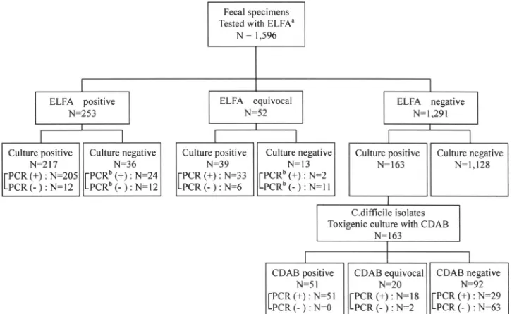
Figure 1 presents the three-step algorithm as a flow chart, including 163 toxigenicC. difficilecultures. Among 1,596 stool specimens, 253 (15.9%), 52 (3.2%), and 1,291 (80.9%) cases were ELFA positive, equivocal, and negative, respectively. The
culture positivity rate was 26.3% (419/1,596). Culture positivity was 85.5% (217/253), 75.0% (39/52), and 12.6% (163/1,291) in ELFA-positive, -equivocal, and -negative groups, respectively. Isolates from Clostridia species other than C. difficile were collected from 18.7% (220/1,177) ofC. difficileculture-negative cases. Toxigenic culture (n⫽163) detected 69 additional cases among ELFA-positive or -equivocal specimens.
Among 419 total isolates, including 163 toxigenic culture-positive strains (i.e., those obtained from ELFA-negative spec-imens), 336 strains (80.2%) were PCR positive. Ninety-eight (60.1%) of the 163 toxigenic culture cases were tcdAand/or
tcdB positive. Direct PCR for tcdB on 49 stool specimens
(ELFA-positive or -equivocal/culture-negative cases) revealed 26tcdB-positive cases (53.1%). Therefore, the total number of PCR-positive cases was 362, and 27.1% (98/362) of these were detected through toxigenic culture.
We placed tcdA-positive and tcdB-positive; tcdA-negative,
tcdB-positive; tcdA-positive, tcdB-negative; and tcdAv, tcdB -positive strains in a hypothetical ELFA--positive group, and
tcdA- andtcdB-negative strains in a hypothetical ELFA-nega-tive group (Table 1). We also placed culture/ELFA conegaELFA-nega-tive cases (n⫽1,128) in a hypothetical ELFA-negative group. We could not absolutely rule out the presence of TcdA or TcdB in culture-negative stool specimens, but it seems justified to conclude that culture is more sensitive than fecal CNA for detection of infection by toxigenic C. difficile (11, 28, 35). Among 419 C. difficileculture-positive cases, tcdA-positive
and tcdB-positive strains were most common (n ⫽ 248
[59.2%]), while those which were tcdA-negative and tcdB -positive (n ⫽ 9 [2.1%]), tcdA-positive and tcdB-negative (n ⫽ 2 [0.5%]),tcdAvandtcdB-positive (n ⫽ 77 [18.4%]), andtcdA- andtcdB-negative (n⫽83 [19.8%]) were detected less frequently. Among 88 (24.3% of 362 hypothetical pos-itive cases)tcdAv/tcdB-positive,tcdA-negative/tcdB-positive, and tcdA-positive/tcdB-negative C. difficile strains, 59 (67.0%) and eight (9.1%) strains were from cases with
pos-FIG. 1. Flow diagram depicting the three-step algorithm, finally confirmed bytcdAandtcdBPCR assays.
on May 16, 2020 by guest
http://jcm.asm.org/
[image:2.585.111.474.69.292.2]itive and equivocal results with ELFA, respectively (Ta-ble 1).
Fifty-two specimens (3.2%) yielding equivocal ELFA results were not included in the calculation of concordance, sensitiv-ity, and specificity. On the basis of these results, the sensitivsensitiv-ity, specificity, positive predictive value, and negative predictive value of the three-step algorithm were 70.7% (256/362), 91.4% (1,128/1,234), 95.5% (256/268), and 100% (1,128/1,128), re-spectively. The corresponding values for the ELFA were 63.3% (229/362), 96.7% (1,193/1,234), 90.5% (229/253), and 92.4% (1,193/1,291), respectively, and for bacteriologic culture were 92.8% (336/362), 93.3% (1,151/1,234), 80.2% (336/419), and 97.8% (1,151/1,177), respectively.
Results of semiquantitative culture were correlated with ELFA positivity (Table 2). The greater the number of colonies on a primary culture plate, the more likely a specimen was to be toxin positive. Odds ratios were 2.109, 4.674, and 5.571 in grades 2, 3, and 4, respectively, compared with grade 1 (P⬍
0.05). The difference among grades was statistically significant (chi-square test for trend,P⬍0.0001), excepting that between grades 3 and 4.
DISCUSSION
A variety of tests is available for detection of C. difficile
toxins, but a lack of guidelines for appropriate use can lead to difficulties in choice of test and to uncertain outcomes. CNA is
generally considered the gold standard for diagnosis of CDI but is not performed by most laboratories; the assay is tedious, requires tissue culture facilities, may lack sensitivity under some conditions, and is difficult to standardize. An assay for GDH-Ag has a high false-positive rate, perhaps due to detec-tion of nontoxigenicC. difficile(23, 27, 33, 34). Bacteriologic culture is strongly recommended by some authors, and its advantages include availability of isolates for determination of toxinogenesis (toxigenic culture), more effective study of epi-demiology, and determination of antimicrobial susceptibility (11, 27, 36). EIA for TcdA and/or TcdB is preferred in most laboratories because a positive test provides indirect evidence that toxigenic C. difficile is in stool specimens; the EIA is readily available and less time-consuming than are CNA and
C. difficileculture (4, 8, 26, 28).
One or two of these methods are usually selected as diag-nostic tools for CDI, depending on the situation in each lab-oratory. Results of a European survey revealed that 93% of laboratories undertook direct detection of toxins in stool
spec-imens and⬃80% used a commercial EIA; 41.6% of the
labo-ratories used both culture and toxin detection (7). A two-step algorithm (GDH-Ag EIA and CNA) was suggested to enhance detection rates (34). It has several advantages, but also limita-tions, in that routine medical laboratories may not be equipped for cell culture and the GDH-Ag test does not detect TcdA or TcdB.
Therefore, we established and evaluated a three-step algo-rithm based upon bacteriologic culture and detection of TcdA and TcdB (in stool specimens and by isolates) by EIA, which makes it more accessible to most laboratories. This allows direct detection of toxin in stool specimens and reliable ruling out of CDI.
In fact, variant strains ofC. difficilehave been described with increasing prevalence worldwide, ranging from 0.2 to 56% in studies from the United States, Europe, and Asia (6, 13, 15, 16, 21, 24, 29, 30), and many recent studies have revealed that TcdA⫺TcdB⫹strains are involved in a wide spectrum of CDI, ranging from colonization to uncomplicated diarrhea to
pseudomembranous colitis (1, 3, 14, 17, 24, 31). TcdA⫹
TcdB⫺ strains have been reported only rarely worldwide.
However, one case was reported (10), and in this work, we encountered two cases (0.5%) involving this strain type. There-fore, a diagnostic method capable of detecting both TcdA and TcdB is likely the best choice, owing to the high prevalence of
TcdA⫺ TcdB⫹ strains in many countries and the possible
[image:3.585.45.283.98.232.2]emergence of TcdB⫺strains.
TABLE 1. Comparison of results ofC. difficileculture, EIA for TcdA and TcdB, and PCR fortcdAandtcdBin the
entire study group (n⫽1,596)
Culture result
PCR
result fora:
ELFA result
(no. of specimens)c
Total (%)
tcdA tcdB Positive Equivocal Negative
⫹ ⫹ ⫹ 146 25 77 248 (15.5)
⫹ ⫹ ⫺ 1 0 1 2 (0.1)
⫹ Variant ⫹ 51 7 19 77 (4.8)
⫹ ⫺ ⫹ 7 1 1 9 (0.6)
⫹ ⫺ ⫺ 12 6 65 83 (5.2)
⫺ ND ⫹b 24 2 0 26 (1.6)
⫺ ND ⫺b 12 11 0 23 (1.4)
⫺ ND ND 0 0 1,128 1,128 (70.8)
Total 253 52 1,291 1,596 (100.0)
a
ND, not done.
b
PCR performed with stool specimens.
c
n⫽1,596.
TABLE 2. Correlation between results of semiquantitativeC. difficileculture and ELFA
Gradea ELFA result
b
(no. of specimens关%兴)
Odds ratio 95% Confidence
interval Pvalue
Positive Equivocal Negative Total
1 18 (28.6) 3 (6.3) 42 (66.7) 63 (100) 1
2 78 (44.1) 26 (14.7) 73 (41.2) 177 (100) 2.109 1.133–3.927 0.0186
3 43 (65.2) 8 (12.1) 15 (22.7) 66 (100) 4.674 2.219–9.846 ⬍0.0001
4 78 (69.0) 2 (1.8) 33 (29.2) 113 (100) 5.571 2.832–10.96 ⬍0.0001
Total 217 (51.8) 39 (9.3) 163 (38.9) 419 (100)
a
Extent of growth of semiquantitative culture forC. difficile. Grade 1,⬍10 colonies; grade 2, 10 to 50 colonies; grade 3,⬎50 to 100 colonies; grade 4,⬎100 colonies.
b
n⫽419.
on May 16, 2020 by guest
http://jcm.asm.org/
[image:3.585.45.542.616.709.2]In our study, 59 (67.0%) and eight (9.1%) strains among 88 cases yieldingtcdAv/tcdB-positive,tcdA-negative/tcdB-positive,
andtcdA-positive/tcdB-negativeC. difficilestrains yielded pos-itive and equivocal ELFA results, respectively. These may have been missed had we used a method detecting TcdA or TcdB alone, although 23.9% (21/88) of these variant strains were not detected with ELFA.
The sensitivities of toxin EIAs are reportedly variable, rang-ing from 55% to⬎90% (2, 22, 26, 35, 36). The limited sensi-tivity of both toxin EIA and CNA might be due to inhibitors in stools, by variable amounts of toxins in stools, and by instability of products and procedures (11, 22, 27, 35, 36). In this work, sensitivity of the dual-toxin EIA was not so high (63.3%). However, the rate of case detection (positive predictive value) via our three-step algorithm (using culture and ELFA) was higher than with another such algorithm, which was based upon detection of GDH-Ag and CNA (95.5% versus 82.1%). There was no significant difference in specificity between the GDH-Ag/CNA algorithm and our ELFA/culture algorithm. Moreover, the processing time with our algorithm (2 to 3 days) was comparable to that of the earlier one.
Culture also allowed identification of additional ELFA-pos-itive cases that would otherwise have been missed, through application of ELFA to toxigenic culture (5, 18, 20). In our study, 98 (60.1%) of 163 toxigenic culture cases were tcdA
and/ortcdBpositive. Among them, 69 strains were ELFA pos-itive (n⫽51) or equivocal (n⫽18), respectively. These may have been missed had we not used toxigenic culture, although 31.6% (31/98) of them were not detected with ELFA.
Interpretation of equivocal ELFA results is not part of the manufacturer’s instructions, in contrast to the toxin A detec-tion kit (CDA2, Vidas; bioMe´rieux, France), but isolates in 86.4% (51/59) of culture-positive/ELFA-equivocal cases (in-cluding ELFA-equivocal cases of toxigenic culture) were PCR positive fortcdAandtcdB. Thus, ifC. difficileculture is posi-tive, ELFA-equivocal results can likely be interpreted as toxin positive. Therefore, if we regarded equivocal ELFA results as positive, the sensitivity of the three-step algorithm would in-crease to 84.8% (307/362).
Positivity rates in the EIA were highly associated with the outcome of semiquantitative culture—the higher the grade, the higher the positivity rates in the ELFA (chi-square test for trend, P⬍ 0.0001). This suggests that detection rates in the
EIA depend on the quantity of TcdA or TcdB produced byC.
difficile, and a false-negative EIA result is associated, at least in part, with lower numbers ofC. difficile isolates (and, thus, its toxins) in stool. The manufacturer suggests that the limit of detection in the ELFA is 3 ng/ml of TcdA and 1 ng/ml of TcdB, and samples in which these limits were not reached would be reported as negative. However, culture positivity in EIA-neg-ative stools, with subsequent demonstration of tcdA and/or
tcdBby a PCR assay, may increase sensitivity. Culture was the most sensitive method for detection of toxigenicC. difficile, but it also missed 7.2% (26/362) of hypothetical positive cases. The prevalence of such cases was very low (3.0%), but it suggested that ELFA-positive or -equivocal/culture-negative cases may be associated with CDI.
In conclusion, we recommended a three-step algorithm com-prising EIA (detecting TcdA and TcdB) and toxigenic bacte-riologic culture. This approach, which is supported by good
positive and negative predictive values, allows direct and rapid assay for toxins and toxigenicC. difficilein stool specimens to confirm or rule out CDI in a reasonable time period. The method is readily accessible for routine microbiology labora-tories, and the availability ofC. difficileisolates will facilitate epidemiologic study and determination of antimicrobial sus-ceptibility.
REFERENCES
1.al-Barrak, A., J. M. Embil, B. Dyck, K. Olekson, D. Nicoll, M. Alfa, and A. Kabani.1999. An outbreak of toxin A negative, toxin B positiveClostridium difficile-associated diarrhea in a Canadian tertiary-care hospital. Can.
Com-mun. Dis. Rep.25:65–69.
2.Aldeen, W. E., M. Bingham, A. Aiderzada, J. Kucera, S. Jense, and K. C. Carroll.2000. Comparison of the Tox A/B test to a cell culture cytotoxicity
assay for the detection ofClostridium difficilein stools. Diagn. Microbiol.
Infect. Dis.36:211–213.
3.Alfa, M. J., A. Kabani, D. Lyerly, S. Moncrief, L. M. Neville, A. al-Barrack, G. K. H. Harding, B. Dyck, K. Olekson, and J. M. Embil.2000.
Character-ization of a toxin A-negative, toxin B-positive strain ofClostridium difficile
responsible for a nosocomial outbreak of Clostridium difficile-associated
diarrhea. J. Clin. Microbiol.38:2706–2714.
4.Anderson, M. A., and J. G. Songer.2008. Evaluation of two enzyme
immu-noassay for detection ofClostridium difficiletoxin A and B in swine. Vet.
Microbiol.128:204–206.
5.Barbut, F., C. Kajzer, N. Planas, and J. C. Petit.1993. Comparison of three enzyme immunoassays, a cytotoxicity assay, and toxigenic culture for
diag-nosis ofClostridium difficile-associated diarrhea. J. Clin. Microbiol.31:963–
967.
6.Barbut, F., V. Lalande, B. Burghoffer, H. V. Thien, E. Grimprel, and J. C. Petit.2002. Prevalence and genetic characterization of toxin A variant strains ofClostridium difficileamong adults and children with diarrhea in France.
J. Clin. Microbiol.40:2079–2083.
7.Barbut, F., M. Delmee, and J. S. Brazier. 2003. A European survey of diagnostic methods and testing protocols for Clostridium difficile. Clin.
Mi-crobiol. Infect.9:989–996.
8.Barbut, F., P. Mastrantonio, M. Delmee, J. S. Brazier, E. Kuijper, I. Poxton, and European Study Group onClostridium Difficile.2007. Prospective study ofClostridium difficileinfection in Europe with phenotypic and genotypic
characterization of the isolates. Clin. Microbiol. Infect.13:1048–1057.
9. Reference deleted.
10.Cohen, S. H., J. Y. Tang, B. Hansen, and J. Silva.1998. Isolation of a toxin
B deficient mutant strain ofClostridium difficilein a case of recurrentC.
difficile-associated diarrhea. Clin. Infect. Dis.26:410–412.
11.Delmee, M., J. Van Broeck, A. Simon, M. Janssens, and V. Avesani.2005.
Laboratory diagnosis ofClostridium difficileassociated diarrhea: a plea for
culture.54:187–191.
12.Drudy, D., N. Harnedy, S. Fanning, R. O’Mahony, and L. Kyne.2007.
Isolation and characterization of toxin A negative, toxin B positive
Clostrid-ium difficilein Dublin, Ireland. Clin. Microbiol. Infect.13:298–304. 13.Geric, B., M. Rupnik, D. Gerding, M. Grabnar, and S. Johnson.2004.
Distribution ofClostridium difficilevariant toxinotypes and strains with
bi-nary toxin genes among clinical isolates in an American hospital. J. Med.
Microbiol.53:887–894.
14.Johnson, S., S. A. Kent, K. J. O’Leary, M. M. Merrigan, S. P. Sambol, L. R. Peterson, and D. N. Gerding.2001. Fatal pseudomembranous colitis
associ-ated with a variantClostridium difficilestrain not detected by toxin A
immu-noassay. Ann. Intern. Med.18:434–438.
15.Kato, H., N. Kato, K. Watanabe, N. Iwai, H. Nakamura, T. Yamamoto, S. Suzuki, S. M. Kim, Y. Chong, and E. B. Wasito.1998. Identification of toxin
A-negative, toxin B-positiveClostridium difficileby PCR. J. Clin. Microbiol.
36:2178–2182.
16.Komatsu, M., H. Kato, M. Aihara, K. Shimakawa, M. Iwasaki, Y. Nagasaka, S. Fukuda, S. Matsuo, Y. Arakawa, M. Watanabe, and Y. Iwatani.2003. High prevalence of antibiotic-associated diarrhea due to toxin A negative, toxin B
positiveClostridium difficilein a hospital in Japan and risk factors for
infec-tion. Eur. J. Clin. Microbiol. Infect. Dis.22:525–529.
17.Kuijper, E. J., J. Weerdt, H. Kato, N. Kato, A. P. Dam, E. R. Vorm, J. Weel, C. Rheenen, and J. Dankert.2001. Nosocomial outbreak ofClostrodium difficileassociated diarrhea due to a clindamycin resistant enterotoxin A
negative strain. Eur. J. Clin. Microbiol. Infect. Dis.20:528–534.
18.Lemee, L., A. Dhalluin, S. Testelin, M. A. Mattrat, K. Maillard, J. F. Leme-land, and J. L. Pons.2004. Multiplex PCR targetingtpi(triose phosphate
isomerase),tcdA(toxin A), andtcdB(toxin B) genes for toxigenic culture of
Clostridium difficile. J. Clin. Microbiol.42:5710–5714. 19. Reference deleted.
20.Lozniewski, A., C. Rabaud, E. Dotto, M. Weber, and F. Mory.2001.
Labo-ratory diagnosis ofClostridium difficile-associated diarrhea and colitis:
use-fulness of premier cytoclone A⫹B enzyme immunoassay for combined
on May 16, 2020 by guest
http://jcm.asm.org/
tection of stool toxins and toxigenicC. difficilestrains. J. Clin. Microbiol.
39:1996–1998.
21.Lyerly, D. M., L. M. Neville, D. T. Evans, J. Fill, and S. Allen. 1998.
Multicenter evaluation of the Clostidium difficileTOX A/B test. J. Clin.
Microbiol.36:184–190.
22.Musher, D. M., A. Manhas, P. Jain, F. Nuila, A. Waqar, N. Logan, B. Marino, and E. A. Graviss.2007. Detection ofClostridium difficiletoxin: comparison of enzyme immunoassay results with results obtained by
cyto-toxicity assay. J. Clin. Microbiol.45:2737–2739.
23.Peterson, L. R., M. M. Olson, C. J. Shanholtzer, and D. N. Gerding.1988. Results of a prospective, 18-month clinical evaluation of culture, cytotoxin
testing, and culturette brand (CDT) latex testing in the diagnosis of
Clos-tridium difficile-associated diarrhea. Diagn. Microbiol. Infect. Dis.10:85–91. 24.Pituch, H., N. van den Braak, W. van Leeuwen, A. van Belkem, G. Marti-rosian, P. Obuch-Woszczatynski, M. Luczak, and F. Meisel-Mikolajczyk.
2001. Clonal dissemination of a toxin A negative/toxin B positiveClostridium
difficilestrain from patients with antibiotic associated diarrhea in Poland.
Clin. Microbiol. Infect.7:442–446.
25. Reference deleted.
26.Post, K. W., B. H. Jost, and J. G. Songer.2002. Evaluation of a test for
Clostridium difficiletoxin A and B for the diagnosis of neonatal swine
enter-itis. J. Vet. Diagn. Investig.14:258–259.
27.Reller, M. E., C. A. Lema, T. M. Perl, M. Cai, T. L. Ross, K. A. Speck, and K. C. Carroll.2007. Yield of stool culture with isolate toxin testing versus a two-step algorithm including stool toxin testing for detection of toxigenic
Clostridium difficile. J. Clin. Microbiol.45:3601–3605.
28.Reyes, R. C., M. A. John, D. L. Ayotte, A. Covacich, S. Milburn, and Z. Hussain. 2007. Performance of TechLab C.DIFF QUIK CHEK and TechLab C.DIFFICILE TOX A/B II for the detection of Clostridium
diffi-cile in stool samples. Diagn. Microbiol. Infect. Dis.59:33–37.
29.Rupnik, M., N. Kato, M. Grabnar, and H. Kato.2003. New types of toxin
A-negative, toxin B-positive strains amongClostridium difficileisolates from
Asia. J. Clin. Microbiol.41:1118–1125.
30.Samra, Z., S. Talmor, and J. Bahar. 2002. High prevalence of toxin A
negative toxin B positiveClostridium difficilein hospitalized patients with
gastrointestinal disease. Diagn. Microbiol. Infect. Dis.43:189–192.
31.Shin, B. M., E. Y. Kuak, S. J. Yoo, W. C. Shin, and H. M. Yoo.2008.
Emerging toxin A⫺B⫹variant strains ofClostridium difficileresponsible for
pseudomembranous colitis at a tertiary care hospital. Diagn. Microbiol.
Infect. Dis.60:333–337.
32.Shin, B. M., E. Y. Kuak, H. M. Yoo, E. C. Kim, K. Lee, K. O. Kang, D. H. Whang, and J. H. Shin.2008. Multicentre study of the prevalence of
toxi-genicClostridium difficilein Korea; results of a retrospective study 2000–
2005. J. Med. Microbiol.57:697–701.
33.Staneck, J. L., L. S. Weckbach, S. D. Allen, J. A. Siders, P. H. Gilligan, G. Coppitt, J. A. Kraft, and D. H. Willis.1996. Multicenter evaluation of four
methods forClostridium difficiledetection: ImmunoCardC. difficile,
cyto-toxin assay, culture, and latex agglutination. J. Clin. Microbiol.34:2718–2721.
34.Ticehurst, J. R., D. Z. Aird, L. M. Dam, A. P. Borek, J. T. Hargrove, and K. C. Carroll.2006. Effective detection of toxigenicClostridium difficileby a two-step algorithm including tests for antigen and cytotoxin. J. Clin.
Micro-biol.44:1145–1149.
35.Turgeon, D. K., T. J. Novicki, J. Quick, L. Carlson, P. Miller, B. Ulness, A. Cent, R. Ashley, A. Larson, M. Coyle, A. P. Limaye, B. T. Cookson, and T. R. Fritsche.2003. Six rapid tests for direct detection ofClostridium difficileand its toxins in fecal samples compared with the fibroblast cytotoxicity assay.
J. Clin. Microbiol.41:667–670.
36.van den Berg, R. J., L. S. B. van Coppernraet, H. J. Gerritsen, H. P. Endtz, E. R. van der Vorm, and E. J. Kuijper.2005. Prospective multicenter eval-uation of a new immunoassay and real-time PCR for rapid diagnosis of
Clostridium difficile-associated diarrhea in hospitalized patients. J. Clin.
Mi-crobiol.43:5338–5340.