Cystic Echinococcosis
Nelson Iván Agudelo Higuita,aEnrico Brunetti,bCindy McCloskeyc
Division of Infectious Diseasesa
and Department of Microbiology,c
Oklahoma University Health Sciences Center, Oklahoma City, Oklahoma, USA; Division of Infectious Disease, San Matteo Hospital Foundation, University of Pavia, WHO Collaborating Centre for Clinical Management of Cystic Echinococcosis, Pavia, Italyb
Echinococcosis is one of the 17 neglected tropical diseases (NTDs) recognized by the World Health Organization. The two major species of medical importance areEchinococcus granulosusandEchinococcus multilocularis.E. granulosusaffects over 1 million people and is responsible for over $3 billion in expenses every year. In this minireview, we discuss aspects of the epidemiology, clinical manifestations, and diagnosis of cystic echinococcosis or cystic hydatid disease caused byE. granulosus.
E
chinococcosis is a zoonotic infection caused by the larval stage of cestode species belonging to the genusEchinococcus. Al-thoughE. granulosuswas initially regarded as the only causative agent of cystic echinococcosis (CE), it was clear that there were different taxa with differences in adult morphology, host specific-ity, and pathogenicity (1). Different strains ofE. granulosuswere identified to precisely portray their specificity for intermediate hosts (e.g., sheep, buffalo, horses, cattle, pigs, camels, and cervids). The lion strain, which was defined based on the definite host, was the exception. Recent advances in phylogenetic systematics have resulted in the recognition of nine species ofEchinococcus:E.granu-losus sensu stricto(G1 to G3),E. equinus(G4),E. ortleppi(G5),E.
canadensis(G6 to G10),E. multilocularis,E. vogeli,E. oligarthrus,
E. felidis, andE. shiquicus(1–3). The taxonomy of cystic
echino-coccosis continues to be under discussion and is far from being completed. For example, the taxonomic status of genotypes G6, G7, G8, and G10 ofE. granulosushas not been solved (4).
Different species ofEchinococcuscause different diseases in hu-mans. Cystic echinococcosis (CE) is caused byE. granulosus sensu
stricto,E. equinus,E. ortleppi, andE. canadensis. Alveolar
echino-coccosis is caused byE. multilocularisand polycystic echinococco-sis byE. vogeliandE. oligarthrus. The most recently described species,E. shiquicus, is found in the Qinghai-Tibet plateau. It in-habits the small intestine of the Tibetan fox (Vulpes ferrilata), and the larval stage is found in the plateau black-lipped pika (Ochotona
curzoniae). To date, no human cases of infection byE. shiquicus
have been described, but its recent isolation from dogs in that region of China emphasizes the need to conduct studies to exam-ine the possibility of human infections (5).
The parasites are maintained in nature by carnivores, which act as definitive hosts (a canine, felid, or hyenid), and by intermediate hosts, which are usually herbivores (e.g., sheep, goats, cattle, cam-els, and cervids) that harbor the larval stage of the parasite (meta-cestode). The adult egg-producing stage of the cestode inhabits the small intestine of the carnivore. The definitive host can be infected with hundreds of worms, with each worm producing thousands of eggs each day. The eggs, which are shed in the stool of the definitive host, are infective upon release. The eggs can remain infective for months or up to a year depending on environmental conditions. The eggs are sensitive to desiccation and heat but can survive freezing temperatures (6).
Once ingested by the intermediate host, the oncosphere hatches from the egg, penetrates the intestinal mucosa, and mi-grates through the bloodstream to internal organs such as the liver. A fluid-filled cyst (metacestode or hydatid cyst) develops in
the affected organ after a period of time that can vary. Protosco-lices bud from the germinal layer (see below) and develop within the cyst and, when ingested by a definitive host, evaginate and attach to the intestinal mucosa, developing into sexually mature adults in a period averaging 4 to 7 weeks. A single cyst can have thousands of protoscolices, and each protoscolex is capable of developing into an adult worm if ingested by the definitive host or, if the cystic fluid is spilled in a cavity such as the peritoneum, into a new cyst (secondary echinococcosis) (6). All mammals in which metacestodes develop act as intermediate hosts, but not all inter-mediate hosts perpetuate the life cycle. For example, humans are considered accidental or aberrant hosts as they are highly unlikely to be involved in disease transmission. Human-to-human trans-mission does not occur (6).
Cysts in each anatomic site, with the exception of bone, are composed of the periparasitic host tissue (pericyst), which en-compasses the endocyst of larval origin. The endocyst has an outer, acellular laminated layer and an inner, or germinal, layer that gives rise to brood capsules and protoscolices. The cyst is filled with clear fluid and, when fertile, with numerous brood capsules and protoscolices. In some stages of cyst development, daughter vesicles of various sizes are present (6,7). In cysts that are degen-erating, one might see abundant free-floating hooklets. These structures represent the “hydatid sand” that sometimes can be seen during imaging procedures when the patient is asked to shift positions (seeFig. 1A).
The minimum time for development of protoscolices is un-known, but, on the basis of animal studies, it is estimated to be 10 months or longer after infection. The growth rate of the echinococcal cyst is poorly understood. In a few studies using ultrasound, such as an observational study carried out in Kenya, 43% of the cysts grew at a rate of 6 to 15 mm per year and 30% grew 1 to 5 mm per year, while approximately 16% of the cysts showed no growth or showed col-lapse. Cysts range in size from those that measure a few centimeters in diameter to much larger cysts containing up to 48 liters of fluid, also
Accepted manuscript posted online16 December 2015
CitationAgudelo Higuita NI, Brunetti E, McCloskey C. 2016. Cystic echinococcosis. J Clin Microbiol 54:518 –523.doi:10.1128/JCM.02420-15.
Editor:C. S. Kraft
Address correspondence to Nelson Iván Agudelo Higuita, [email protected].
Copyright © 2016, American Society for Microbiology. All Rights Reserved.
MINIREVIEW
on May 16, 2020 by guest
http://jcm.asm.org/
called giant cysts (6). It has been suggested that the size of a hydatid cyst can be related to the genotype. For example, a recent study showed that in infected patients, the average diameter ofE. canaden-sisG7 liver cysts was 5.9 cm (range, 3 to 10 cm) compared to 10.7 cm (range, 5 to 21 cm) forE. granulosus(8).
EPIDEMIOLOGY
Cystic echinococcosis has a cosmopolitan distribution and represents a major public health problem in some regions (9). It is considered endemic in areas such as Peru, Chile, Argentina, Uruguay, southern Brazil, the Mediterranean region, central Asia, western China, and East Africa. Cystic echinococcosis is not found in Antarctica and has been eliminated through comprehensive control programs in Ice-land, New ZeaIce-land, Tasmania, Falkland Islands, and Cyprus (10).
The G1 genotype ofEchinococcus granulosus sensu strictois re-sponsible for the vast majority (88%) of human cases worldwide. It has a cosmopolitan distribution and is associated with transmis-sion from sheep as an intermediate host (11).E. canadensisG6 and G7 are responsible for 7.3% and 3.7% of infections worldwide, respectively. There have been no human cases ofE. equinus de-scribed in the literature (11).
The human incidence can exceed 50 per 100,000 person-years in areas of endemicity, and prevalence rates as high as 5% to 10% can be found in parts of Peru, Argentina, east Africa, and China (10). Every year, echinococcosis is responsible for the loss of at least 1 million disability-adjusted life years (DALYs) and of $3 billion dollars in ex-penses, including treatment and livestock losses (9).
Cystic echinococcosis typically occurs in poor pastoral regions in which sheep or other livestock are raised and in which dogs are kept, for herding or property guarding, in close proximity to households. Dogs in such regions are frequently fed offal, and, for religious and other reasons, their populations might not be cur-tailed (12). The prevalence of cystic echinococcosis increases with age, and women are affected more frequently than men; this might be related to domestic activities that bring them in closer contact with dogs through feeding, herding, or milking livestock (12).
CLINICAL MANIFESTATIONS
The incubation period and the clinical presentation of CE are highly variable. The latter is dependent upon several features such as the involved organ, the location of the cyst within the organ, and its relation with surrounding structures, its size, and the in-tegrity of the wall. Other factors, such as the genotype, have been suggested to have a role. For example, cysts belonging to the cervid genotype (G8) have been reported to most frequently localize in the lung, tend to grow slowly, and are less likely to cause compli-cations (13). A recent case series suggested thatE. canadensisG6 might have a higher affinity for the human brain (14).
Cystic echinococcosis is usually asymptomatic unless compli-cations occur. Rupture with resultant infection or anaphylaxis, fistula development with adjacent structures (e.g., in the biliary tract, intestine, and bronchus) or mass effect on neighboring structures are the major mechanisms by which a cyst usually be-comes symptomatic (15). It is not uncommon for cysts to be dis-covered incidentally by imaging studies done for other reasons.
Most patients (40% to 80% of cases) have a single cystic lesion located in a single organ. The liver is affected in 70% of the cases, the right lobe more commonly than the left. The lung is the next most frequently affected organ and is affected in about 20% of the cases (6,15). Cysts can localize in virtually any organ and struc-ture, such as abdominal or pleural cavities, kidney, spleen, bone, brain, eye, ovary, testis, and pancreas (6,15). Rare immune-me-diated reactions such as urticaria, asthma, membranous nephrop-athy, and anaphylaxis have also been well described (15).
A proportion of patients present with cyst-related complications that require medical attention in a timely manner. For cysts located in the liver, a cysto-biliary fistula is the most common complication (13% to 37% of cases) (16). The high complication rate has been attributed to the increased intracystic pressure (30 to 80 cm H2O)
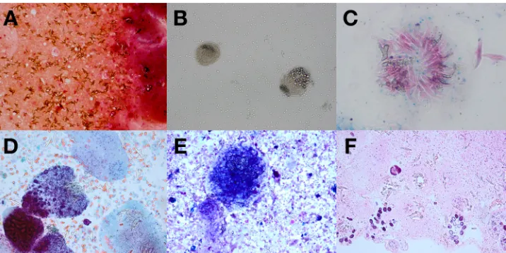
compared to the low intracystic pressure present in nonparasitic he-patic cysts (17). Cysto-biliary fistulas can be classified as frank or occult. A frank cysto-biliary fistula is generally easy to diagnose, both clinically and radiographically. An occult cysto-biliary fistula is diffi-FIG 1(A) Gram stain of cyst fluid (magnification,⫻200). (B) Wet-mount preparation of cyst fluid (magnification,⫻200). (C) Ziehl-Neelsen stain of cyst fluid (oil immersion; magnification,⫻1,000). (D) Papanicolaou stain of cyst fluid (magnification,⫻400; polarized). (E) Diff-Quick stain of cyst fluid (magnification, ⫻400; polarized). (F) Hematoxylin and eosin stain of formalin-fixed paraffin-embedded cell block of cyst fluid (magnification,⫻200). (Images courtesy of Diane Hensel [panels A and B], Cindy McCloskey [panel C], and Rachel Conrad [panels D to F], University of Oklahoma Health Sciences Center, Oklahoma City, OK, USA; reproduced with permission.)
Minireview
on May 16, 2020 by guest
http://jcm.asm.org/
[image:2.585.114.472.66.245.2]cult to detect; if not identified preoperatively, the postoperative course is usually complicated. Several scoring systems have been used to predict its presence preoperatively (16,18).
DIAGNOSIS
The diagnosis of cystic echinococcosis rests mainly on imaging. The World Health Organization Informal Working Group on Echinococcosis (WHO-IWGE) published in 2003 a standardized classification of ultrasound images based on the first classification developed by H. A. Gharbi, W. Hassine, M. W. Brauner, and K. Dupuch in 1981 (19). This classification is intended to be used both in the epidemiological field and in clinical settings to dictate stage-specific management.
The classification allocates cystic echinococcosis cysts into three relevant groups: active (CE1 and CE2), transitional (CE3), and inactive (CE4 and CE5) (seeFig. 2). The cystic lesion (CL) stage consists of unilocular cystic lesions without pathognomonic signs on ultrasound and whose parasitic nature needs to be con-firmed with further investigations. The group consisting of CE3 (transitional) cysts has recently been separated into CE3a (with detached endocyst) and CE3b (predominantly solid, with daugh-ter vesicles) on the basis of their different metabolic profiles and different responses to nonsurgical treatments (20). CE3a cysts are true transitional cysts because they may be active or inactive, while CE3b cysts are active. This classification was developed on the basis of analysis of cystic echinococcosis cysts in the liver, but, in a number of cases, it can also be applied in the management and follow-up of cysts located elsewhere. One of the advantages of the classification is that, at least for hepatic CE, it facilitates a more rational treatment approach based on cyst stage and size (20).
Cysts that are not accessible to ultrasound can be studied using other imaging modalities such as computed tomography (CT) or magnetic resonance imaging (MRI). MRI is better than CT at
detecting the structural, stage-defining features of cysts seen on ultrasound. There is a very good level of agreement between ultra-sound imaging and MRI for liver cystic echinococcosis stages CE1 to CE4. MRI has the shortcoming of being unable to identify cer-tain details of the cyst wall such as calcification. If an ultrasound cannot be performed, a MRI with T2-weighted sequences (in par-ticular, True Fisp and HASTE [half-Fourier acquisition single-shot turbo spin echo] sequences) should be obtained as these se-quence best detect liquid content in the cyst matrix (21).
MRI imaging with MR cholangiopancreatography (MCRP) also has a role in the preoperative evaluation of complications such as cysto-biliary fistulas. It performs as well as endoscopic retrograde cholangiopancreatography (ERCP) in the recognition of biliary ob-struction and is not invasive. Cystobiliary fistulas become apparent only after rupture of the endocyst (mostly stages CE2 and CE3b), and the sensitivity and specificity of MRCP to detect a cysto-biliary fistula in these stages are 75% and 95%, respectively (22).
Clinical laboratory analysis, including chemistry and hematol-ogy testing, is nonspecific in patients with cystic echinococcosis. For those with biliary obstruction, elevated levels of bilirubin, transaminases, and gamma-glutamyl transferase may be ob-served. In the setting of cyst leakage into the biliary tree or cyst rupture, significant elevation in gamma-glutamyl transferase and alkaline phosphatase may also be observed, together with eosino-philia, which is usually absent in intact cysts.
Immunodiagnostics play an ancillary role in diagnosis due to limitations in sensitivity and specificity. However, serology may be useful to support or confirm a diagnosis of cystic echinococco-sis. World Health Organization/World Organisation for Animal Health recommendations include sequential testing based on a screening and confirmatory test model (4). Primary screening methodologies include enzyme-linked immunosorbent assays (ELISA), indirect hemagglutination antibody tests (IHAT), latex FIG 2World Health Organization Informal Working Group on Echinococcosis classification of ultrasound images for cystic echinococcosis cysts (31). CE, cystic echinococcosis; CL, cystic lesion. “A” indicates CE3a, and “B” indicates CE3b (see the text for details). (Reprinted from reference31with permission of the publisher.)
on May 16, 2020 by guest
http://jcm.asm.org/
[image:3.585.66.522.95.304.2]agglutination (LAT), immunofluorescence antibody tests (IFAT), and immunoelectrophoresis (IEP), with ELISA being the most common. These methods have various levels of sensitivity and somewhat poor specificity, especially in patients with other hel-minthic infections. For many assays, the antigen material used for testing is a crude or purified preparation from animal liver hydatid cysts and is likely a significant source of variability in test perfor-mance (6,23).
Many factors, including technical issues such as the quality of the antigen preparation mentioned above, as well as host factors, such as the immune status of the subject, can influence assay sen-sitivity. The sensitivity of the test is also dependent on the integrity of the cyst wall and the development stage of the cyst. Early cysts, such as CE1 cysts, usually have their antigens sequestered from the host’s immune system and might present with a negative serologic test result, and patients with inactive CE4 and CE5 cysts are also often seronegative. Cystic lesions located in sanctuary sites, such as the eye or brain, are also usually missed by serologic evaluation. It is therefore important to remember that a negative serologic test result does not exclude the presence of cystic echinococcosis. Se-rology is positive in around 80% to 94% of hepatic echinococcosis cases and 65% of pulmonary cases (23).
Significant cross-reactivity is seen with other parasitic con-ditions, including alveolar echinococcosis, cysticercosis, fasci-oliasis, and filariasis, as well as with other nonparasitic condi-tions, including malignancy. Patients with reactive primary screening serology should have a confirmatory test. Recom-mended methods for secondary testing include immunoblot assays to test for reactivity withE. granulosusantigen subunits, identification of specific IgG subclasses (i.e., IgG1 and/or IgG4), and arc 5 precipitation testing. Although specificity is much im-proved with these assays compared to primary test methods, cross-reactivity with alveolar echinococcosis and cysticercosis is not completely eliminated (6). The use of recombinant antigens for serodiagnosis has also been studied and was shown to improve sensitivity over the use of native antigens or their purified sub-units, but further research is needed to improve the clinical use-fulness of serodiagnosis (24).
New testing modalities that could be used in small laboratories to facilitate epidemiologic studies in countries where the disease is endemic are being developed. A loop-mediated isothermal ampli-fication (LAMP) assay is such an example. The assay is able to accurately detect 5 different species of Echinococcus (E.
granulo-sus sensu stricto,E. equinus,E. ortleppi,E. canadensis, andE. felidis)
(25).
In patients requiring diagnostic aspiration (rarely indicated), the cyst fluid can be examined microscopically to confirm the detection ofEchinococcusspp. by observing protoscolices and/or free hooklets. A wet unstained mount procedure is simple to per-form and is often adequate for diagnosis. Several staining meth-ods, including those employing Ziehl-Neelsen stain, Wheatley trichrome or Ryan trichrome blue stain, Baxby stain, and modi-fied Baxby stain, can improve the visualization of hooklets (whether isolated or associated with protoscolices) (26). Cyst ma-terial can also be identified on histologic tissue sections (seeFig. 1BtoF). The acellular laminated layer and thin cellular, germinal layer with brood capsules and protoscolices can often be visual-ized in mature echinococcal cysts.
TREATMENT AND PREVENTION
The management of cystic echinococcosis is complex and is be-yond the scope of this review. The WHO-IWGE published an “Expert Consensus for the Diagnosis and Treatment of Cystic and Alveolar Echinococcosis in Humans” in 2010 (20). Given the lack of trials comparing the different treatment options, the recom-mendations are based on the opinions of experts in the field. Even among those respected authorities, there is great variation in the management of the disease worldwide (27).
Surgery has been considered the gold standard, but alternatives exist for selected patients and are now considered the first man-agement options. In general, there are four different manman-agement modalities: percutaneous therapy, surgery, chemotherapy, and observation without intervention (watch and wait). The expertise of the personnel and the availability of resources, the stage, size, and location of the cyst, and the presence of symptoms/complica-tions are the main elements taken into consideration in choosing among these options. There is no test of cure, and long-term fol-low-up with imaging is required to evaluate the efficacy of treat-ment, as serology results may remain positive for years even after successful treatment. When possible, patients should be evaluated in a reference center.
Although benzimidazoles have been the cornerstone of medi-cal therapy since the late 1980s, many issues (e.g., duration of therapy) remain unresolved (28). Many other compounds have been tested experimentally without success, and, despite the effect of the synergy between benzimidazoles and other agents such as metformin seenin vitro, there is still a great need to invest in the development of new chemotherapeutic agents (28,29).
The prevention of the disease depends on the interruption of the life cycle ofE. granulosus. For example, regular screening and treatment of infected dogs have been successful in eradicating the disease in areas of endemicity such as New Zealand (10). Restrict-ing the feedRestrict-ing of home-slaughtered livestock to dogs and vacci-nating the intermediate host (e.g., sheep) are other available con-trol measures.
CONCLUSION
Cystic echinococcosis is a complex disease. It continues to be a major public health problem in many countries despite being, in principle, preventable, treatable, and eradicable. There are many unanswered questions and unresolved problems. For example, the evidence available to guide treatment is poor and well-designed trials that could guide therapy are overdue. In the interim, adop-tion of the stage-specific approach advocated by the WHO-IWGE, at least for liver CE, would greatly reduce mismanagement (27). There is also a clear need for research into development of diag-nostics and prevention/control programs that takes into account the social, political, and economical situation of the affected com-munities (30).
FUNDING INFORMATION
This work was partly funded by the European Union through grant no. 602051 (project title: HERACLES—Human cystic Echinococcosis ReseArch in CentraL and Eastern Societies), FP7-HEALTH-2013-INNOVATION-1 (to E. Brunetti).
REFERENCES
1.Nakao M, Lavikainen A, Yanagida T, Ito A.2013. Phylogenetic system-atics of the genus Echinococcus (Cestoda: Taeniidae). Int J Parasitol43:
1017–1029.http://dx.doi.org/10.1016/j.ijpara.2013.06.002.
Minireview
on May 16, 2020 by guest
http://jcm.asm.org/
2.Xiao N, Qiu J, Nakao M, Li T, Yang W, Chen X, Schantz PM, Craig PS, Ito A.2005. Echinococcus shiquicus n. sp., a taeniid cestode from Tibetan fox and plateau pika in China. Int J Parasitol35:693–701.http://dx.doi.org /10.1016/j.ijpara.2005.01.003.
3.Hüttner M, Nakao M, Wassermann T, Siefert L, Boomker JD, Dinkel A, Sako Y, Mackenstedt U, Romig T, Ito A.2008. Genetic characterization and phylogenetic position of Echinococcus felidis (Cestoda: Taeniidae) from the African lion. Int J Parasitol38:861– 868.http://dx.doi.org/10 .1016/j.ijpara.2007.10.013.
4.Lymbery AJ, Jenkins EJ, Schurer JM, Thompson RC.2015. Echinococ-cus canadensis, E borealis, and E intermedius. What’s in a name? Trends Parasitol31:23–29.http://dx.doi.org/10.1016/j.pt.2014.11.003. 5.Boufana B, Qiu J, Chen X, Budke CM, Campos-Ponce M, Craig PS.
2013. First report of Echinococcus shiquicus in dogs from eastern Qing-hai-Tibet plateau region, China. Acta Trop127:21–24.http://dx.doi.org /10.1016/j.actatropica.2013.02.019.
6.Eckert J, Gemmell MA, Meslin F-X, Pawłowski ZS (ed).2001. WHO/ OIE manual on echinococcosis in humans and animals: a public health problem of global concern. World Organisation for Animal Health (Office International des Epizooties), Paris, France, and World Health Organiza-tion, Geneva, Switzerland.
7.Díaz A, Casaravilla C, Allen JE, Sim RB, Ferreira AM.2011. Under-standing the laminated layer of larval Echinococcus II: immunology. Trends Parasitol27:264 –273.http://dx.doi.org/10.1016/j.pt.2011.01.008. 8.Schneider R, Gollackner B, Schindl M, Tucek G, Auer H.2010. Echi-nococcus canadensis G7 (pig strain): an underestimated cause of cystic echinococcosis in Austria. Am J Trop Med Hyg82:871– 874.http://dx.doi .org/10.4269/ajtmh.2010.09-0639.
9.Budke CM, Deplazes P, Torgerson PR. 2006. Global socioeconomic impact of cystic echinococcosis. Emerg Infect Dis12:296 –303.http://dx .doi.org/10.3201/eid1202.050499.
10. Craig PS, McManus DP, Lightowlers MW, Chabalgoity JA, Garcia HH, Gavidia CM, Gilman RH, Gonzalez AE, Lorca M, Naquira C, Nieto A, Schantz PM.2007. Prevention and control of cystic echino-coccosis. Lancet Infect Dis 7:385–394. http://dx.doi.org/10.1016 /S1473-3099(07)70134-2.
11. Alvarez Rojas CA, Romig T, Lightowlers MW. 2014. Echinococcus granulosus sensu lato genotypes infecting humans–review of current knowledge. Int J Parasitol 44:9 –18. http://dx.doi.org/10.1016/j.ijpara .2013.08.008.
12. Craig PS, Li T, Qiu J, Zhen R, Wang Q, Giraudoux P, Ito A, Heath D, Warnock B, Schantz P, Yang W. 2008. Echinococcosis and Tibetan communities. Emerg Infect Dis14:1674 –1675.http://dx.doi.org/10.3201 /eid1410.071636.
13. Moro P, Schantz PM.2006. Cystic echinococcosis in the Americas. Para-sitol Int55(Suppl):S181–S186.http://dx.doi.org/10.1016/j.parint.2005.11 .048.
14. Sadjjadi SM, Mikaeili F, Karamian M, Maraghi S, Sadjjadi FS, Shariat-Torbaghan S, Kia EB.2013. Evidence that the Echinococcus granulosus G6 genotype has an affinity for the brain in humans. Int J Parasitol43:875– 877.http://dx.doi.org/10.1016/j.ijpara.2013.06.008.
15. Ammann RW, Eckert J.1996. Cestodes echinococcus. Gastroenterol Clin North Am25:655– 689.http://dx.doi.org/10.1016/S0889-8553(05)70268-5. 16. Demircan O, Baymus M, Seydaoglu G, Akinoglu A, Sakman G.2006.
Occult cystobiliary communication presenting as postoperative biliary leakage after hydatid liver surgery: are there significant preoperative clin-ical predictors? Can J Surg49:177–184.
17. Yalin R, Aktan AO, Yegen C, Dosluoglu HH. 1992. Significance of intracystic pressure in abdominal hydatid disease. Br J Surg79:1182–1183.
http://dx.doi.org/10.1002/bjs.1800791127.
18. Saylam B, Coskun F, Demiriz B, Vural V, Comcali B, Tez M.2013. A new and simple score for predicting cystobiliary fistula in patients with hepatic hydatid cysts. Surgery153:699 –704.http://dx.doi.org/10.1016/j .surg.2012.11.017.
19. Anonymous.2003. International classification of ultrasound images in cystic echinococcosis for application in clinical and field epidemiological settings. Acta Trop85:253–261.http://dx.doi.org/10.1016/S0001-706X(02)00223-1. 20. Brunetti E, Kern P, Vuitton DA.2010. Expert consensus for the diagnosis
and treatment of cystic and alveolar echinococcosis in humans. Acta Trop
114:1–16.http://dx.doi.org/10.1016/j.actatropica.2009.11.001.
21. Stojkovic M, Rosenberger K, Kauczor HU, Junghanss T, Hosch W.
2012. Diagnosing and staging of cystic echinococcosis: how do CT and MRI perform in comparison to ultrasound? PLoS Negl Trop Dis6:e1880.
http://dx.doi.org/10.1371/journal.pntd.0001880.
22. Hosch W, Stojkovic M, Janisch T, Heye T, Werner J, Friess H, Kauff-mann GW, Junghanss T.2008. MR imaging for diagnosing cysto-biliary fistulas in cystic echinococcosis. Eur J Radiol66:262–267.http://dx.doi .org/10.1016/j.ejrad.2007.08.002.
23. Biava MF, Dao A, Fortier B.2001. Laboratory diagnosis of cystic hydatic disease. World J Surg25:10 –14.http://dx.doi.org/10.1007/s002680020002. 24. Hernández-González A, Santivanez S, Garcia HH, Rodriguez S, Munoz
S, Ramos G, Orduna A, Siles-Lucas M.2012. Improved serodiagnosis of cystic echinococcosis using the new recombinant 2B2t antigen. PLoS Negl Trop Dis6:e1714.http://dx.doi.org/10.1371/journal.pntd.0001714. 25. Wassermann M, Mackenstedt U, Romig T. 2014. A loop-mediated
isothermal amplification (LAMP) method for the identification of species within the Echinococcus granulosus complex. Vet Parasitol200:97–103.
http://dx.doi.org/10.1016/j.vetpar.2013.12.012.
26. Clavel A, Varea M, Doiz O, Lopez L, Quilez J, Castillo FJ, Rubio C, Gomez-Lus R.1999. Visualization of hydatid elements: comparison of several techniques. J Clin Microbiol37:1561–1563.
27. Nabarro LE, Amin Z, Chiodini PL.2015. Current management of cystic echinococcosis: a survey of specialist practice. Clin Infect Dis60:721–728.
http://dx.doi.org/10.1093/cid/ciu931.
28. Vuitton DA.2009. Benzimidazoles for the treatment of cystic and alveolar echinococcosis: what is the consensus? Expert Rev Anti Infect Ther7:145– 149.http://dx.doi.org/10.1586/14787210.7.2.145.
29. Loos JA, Cumino AC.2015. In vitro anti-echinococcal and metabolic effects of metformin involve activation of AMP-activated protein kinase in larval stages of Echinococcus granulosus. PLoS One10:e0126009.http: //dx.doi.org/10.1371/journal.pone.0126009.
30.Brunetti E, Garcia HH, Junghanss T. 2011. Cystic echinococcosis: chronic, complex, and still neglected. PLoS Negl Trop Dis5:e1146.http: //dx.doi.org/10.1371/journal.pntd.0001146.
31. World Health Organization.2001. PAIR: puncture, aspiration, injection, re-aspiration—an option for the treatment of cystic echinococcosis. World Health Organization, Geneva, Switzerland. http://apps.who.int /iris/bitstream/10665/67207/1/WHO_CDS_CSR_APH_2001.6.pdf.
on May 16, 2020 by guest
http://jcm.asm.org/
Nelson Iván Agudelo Higuitais an Assistant Professor of Medicine at the University of Okla-homa Health Sciences Center. He also serves as the Associate Program Director for the Internal Medicine Residency Program. He earned his doctoral degree from the Autonomous Univer-sity of Honduras. He then completed a resi-dency in Internal Medicine and a fellowship in Infectious Diseases, both at the University of Oklahoma Health Sciences Center. He has spe-cial interest in tropical diseases and other geo-graphic and travel-related infections.
Minireview