0095-1137/10/$12.00
doi:10.1128/JCM.02224-09
Copyright © 2010, American Society for Microbiology. All Rights Reserved.
Identification, Molecular Characterization, and Occurrence of Two
Bovine Hemoplasma Species in Swiss Cattle and Development
of Real-Time TaqMan Quantitative PCR Assays for
Diagnosis of Bovine Hemoplasma Infections
䌤
Marina L. Meli,
1* Barbara Willi,
1Ute M. Dreher,
1Valentino Cattori,
1Gabriela Knubben-Schweizer,
2Karl Nuss,
2Ueli Braun,
2Hans Lutz,
1and Regina Hofmann-Lehmann
1Clinical Laboratory, Vetsuisse Faculty, University of Zurich, CH-8057 Zurich, Switzerland,
1and Department of
Farm Animals, Vetsuisse Faculty, University of Zurich, CH-8057 Zurich, Switzerland
2Received 13 November 2009/Returned for modification 20 February 2010/Accepted 23 July 2010
Concomitantly with an outbreak of fatal anaplasmosis in a cattle herd in Switzerland in 2002, we detected
two bovine hemoplasma species in diseased animals:
Mycoplasma wenyonii
(formerly
Eperythrozoon wenyonii
)
and a second, novel bovine hemoplasma species later designated “
Candidatus
Mycoplasma haemobos”
(syn-onym, “
Candidatus
Mycoplasma haemobovis”). The second species was characterized by a shorter 16S rRNA
gene. The aims of the present study were to provide a detailed molecular characterization of this species, to
develop specific quantitative real-time PCR assays for the two bovine hemoplasma species, and to apply these
assays in order to evaluate the prevalence and clinical significance of the hemoplasmas. Sequencing of the
near-complete 16S rRNA gene of the second hemoplasma revealed that it was 94% identical to that of
Mycoplasma haemofelis
, an anemia-inducing feline hemoplasma species, but less than 85% identical to that of
the bovine hemoplasma
M. wenyonii
. Using the newly developed assays, a total of 159 animals from the
anaplasmosis outbreak were reexamined. In addition, we tested 57 clinically ill and 61 healthy Swiss cattle, as
well as 47 calves. Both hemoplasmas were highly prevalent in adult cattle but occurred rarely in calves.
Animals from the herd with the fatal anemia outbreak were more frequently infected with
M. wenyonii
and
exhibited higher
M. wenyonii
blood loads than animals with unrelated diseases and healthy animals.
Coinfec-tions may increase the pathogenicity and clinical significance of bovine hemoplasmosis.
In connection with an outbreak of anaplasmosis in a cattle
herd in eastern Switzerland in 2002, more than 300 animals
were culled. Most of these cattle exhibited pronounced
ane-mia. The anemia was statistically associated with the detection
of
Anaplasma marginale,
Babesia
spp.,
Theileria
spp., and
My-coplasma wenyonii
in the blood of diseased animals (5).
M. wenyonii, first described in 1934, was formerly known as
Eperythrozoon wenyonii
(1, 13). The species was recently
re-classified within the group of hemotropic mycoplasmal species
based on the 16S rRNA gene sequence (11–13).
M. wenyonii
is
a cell wall-free bacterium that parasitizes bovine red blood
cells (11). In our study of the above-mentioned outbreak, we
reported two distinct hemotropic mycoplasma species:
M.
we-nyonii
and a second, unknown, agent (5). The 16S rRNA gene
of the second agent was shorter than that in
M. wenyonii
and
was 95% identical to the 16S rRNA gene found in
Mycoplasma
haemofelis, the causative agent of feline infectious anemia (24,
25). A similar bovine hemoplasma species has since been
de-tected in China and Japan using molecular assays, and the
name “Candidatus
Mycoplasma haemobos” has been proposed
(20). Other bovine hemoplasmas that were found to be distinct
from
M. wenyonii
using other methods are described elsewhere
(3, 30–32). Characterization included morphological and
im-munogenic differences, as well as different localization of the
agents within the host (28, 29, 31, 32, 38).
The clinical relevance of
M. wenyonii
is controversial (16,
18); in the United States, infection with
M. wenyonii
is
consid-ered to be of low pathogenicity. However, a study with
sple-nectomized calves showed that a preexisting
M. wenyonii
in-fection followed by an
A. marginale
superinfection led to severe
anemia, with packed-cell volumes (PCV) dropping from 30%
to 9.5% when
M. wenyonii
was found in the blood and about 2
weeks before
A. marginale
appeared (13). The clinical
rele-vance of “Candidatus
Mycoplasma haemobos” remains unclear
(14, 20).
The aims of the present study were to characterize the two
bovine hemotropic mycoplasma species identified in 2002
using molecular techniques, to develop specific real-time
TaqMan PCR assays for the detection and quantification of
these species, and to determine the occurrence of the two
bovine hemoplasmas in sick and healthy cattle in order to
evaluate their clinical importance.
MATERIALS AND METHODS
Samples. We studied a total of 216 EDTA-anticoagulated blood samples collected from diseased animals. In total, 159 samples came from cattle origi-nating from a large dairy herd that had experienced an anaplasmosis disease outbreak in Switzerland in August 2002 (5). The remaining 57 samples originated from cows that were brought to the clinic for farm animals at the University of Zurich between January and May 2004 for the treatment of diseases other than anaplasmosis. In addition, whole blood samples were available from 61 healthy
* Corresponding author. Mailing address: Clinical Laboratory,
Vet-suisse Faculty, University of Zurich, Winterthurerstrasse 260, CH-8057
Zurich, Switzerland. Phone: 41 (44) 635 83 85. Fax: 41 (44) 635 89 06.
E-mail: [email protected].
䌤
Published ahead of print on 4 August 2010.
3563
on May 16, 2020 by guest
http://jcm.asm.org/
animals originating from different Swiss farms. Furthermore, we also tested EDTA-anticoagulated blood samples from 12 healthy calves from a herd owned by the University of Zurich as well as 35 calves from 20 different herds in northeastern Switzerland.
PCR analysis and sequencing.DNA was purified from 200l of whole blood with a MagNA Pure LC DNA isolation kit I (Roche Diagnostics, Rotkreuz, Switzerland).
The near-complete sequence of the 16S rRNA gene of the new hemoplasma isolate was determined from three cows. Two of the cows were involved in the outbreak; one was infected only with the new hemoplasma isolate (isolate CH311), while the second was coinfected with the new isolate and withA. marginale(isolate CH307). The third cow originated from the group of animals that were transported to the University of Zurich clinic (isolate CH88). Briefly, the species-specific primers MHBforw (5⬘-GAA TTA ATG CTG ATG GTA TGC CTA A-3⬘, 25 bp) and MHBrev (5⬘-CCA ATC AGA ATG TTC ACT CTA GAT GC-3⬘, 26 bp) were used to amplify 2.5l of template DNA in a 25-l reaction mixture containing 5⫻Phusion HF buffer (Finnzymes, Espoo, Finland), 500 nM each primer, 200 nM each deoxynucleoside triphosphate (dNTP) (Sig-ma-Aldrich, Buchs, Switzerland), and 1 U Phusion DNA polymerase (Finnzymes). The thermal program comprised 98°C for 3 min; 35 cycles of 98°C for 10 s, 52°C for 30 s, and 72°C for 1 min; and finally 72°C for 10 min. PCR products of 1,393 bp were gel purified and cloned into the vector pCR4-TOPO (Invitrogen, Basel, Switzerland). Sequencing was performed with vector-specific M13 forward and reverse primers and two internal primers, Mwen_short.forw (5⬘-CCA TGT GAA CGA AGA AGG TCT TT-3⬘, 23 bp) and Mwen_short.rev (5⬘-AGT TTG CTG TCA CTT ATT CAT GAG GTA-3⬘, 27 bp), using the BigDye Terminator cycle sequencing ready reaction kit v1.1 (Applied Biosys-tems, Rotkreuz, Switzerland). Sequences were then analyzed on an ABI PRISM 310 Genetic Analyzer (Applied Biosystems). Three clones from each animal were sequenced.
For phylogenetic and molecular evolutionary analysis, the sequences were aligned with known hemoplasma sequences from GenBank using ClustalW (26) and manually adjusted when necessary. Only nucleotides that were available for all included sequences were used in the phylogenetic analysis. A bootstrap phylogenetic tree was used to demonstrate the relationship of the new bovine species to other hemoplasma species. The tree was created by the neighbor-joining method (17) using a distance matrix corrected for nucleotide substitu-tions based on the Kimura two-parameter model. The data set was resampled 1,000 times to generate bootstrap values. Phylogenetic and molecular evolution-ary analyses were conducted using MEGA version 4 (21).
Development of real-time PCR assays specific for the two bovine hemotropic mycoplasma species.To develop specific real-time PCR assays, two sets of primers and probes were designed. The first set, specific forM. wenyonii, was designed based on alignments ofM. wenyonii16S rRNA gene sequences from five cattle (5) (accession numbers GQ259756 to GQ259760) and on a published sequence (AY946266): MwenyoniiF (5⬘-CCA CGT GAA CGA TGA AGG TCT T-3⬘, 22 bp), MwenyoniiR (5⬘-GGC ACA TAG TTA GCT GTC ACT TAT TCA A-3⬘, 28 bp), and Mweny_P (5⬘-FAM-AGT ACC ATC AAG GCG CGC TCA TTT CCT AG-TAMRA-3⬘, 29 bp). Sequence comparisons revealed that the assay may potentially amplifyMycoplasma ovis(AF338268) and also a swine hemotropic mycoplasma (DQ346727) but not the feline and canine hemoplasma species or “CandidatusMycoplasma haemobos.” The second set of primers was designed based on alignments of 16S rRNA gene sequences from the bovine mycoplasma isolates obtained from three animals (5) (GQ259761 to GQ259763), which were compared to the 16S rRNA sequence ofM. haemofelis(AY069948 and U88563) andM. haemocanis(AF407208 and AF197337). The assay was designed to amplify “CandidatusMycoplasma haemobos,” but not the feline and canine sequences or the commonM. wenyonii, using the following primers and probe: Mwen_short.forw (5⬘-CCA TGT GAA CGA TGA AGG TCT TT-3⬘, 23 bp), Mwen_short.rev (5⬘-AGT TTG CTG TCA CTT ATT CAT GAG GTA-3⬘, 27 bp), and Mwen_short.p (5⬘-VIC-CTA TCA GTT RTT ATC CCT CAT AA-MGB-3⬘, 23 bp). For both systems, the 25-l PCR mixture comprised 12.5l of 2⫻quantitative PCR (qPCR) Mastermix (Eurogentec), 900 nM each primer, 250 nM probe, and 5l of template DNA. Assays were performed using the ABI PRISM 7700 sequence detection system (Applied Biosystems).
DNA standards for absolute quantitation.The sequenced pCR4-TOPO plas-mids (see above) containing the almost-complete 16S rRNA genes of “ Candi-datusMycoplasma haemobos” (isolates CH311 and CH307) andM. wenyonii (isolate CH82), including the target sequences encompassed by the real-time PCR assays, were linearized by restriction digestion using NotI and gel purified (QIAquick gel extraction kit; Qiagen). The plasmid DNA copy numbers were then calculated by spectrophotometry (NanoDrop ND-1000; Witec Ag, Littau, Switzerland) and agarose gel electrophoresis (Gene Tools; Syngene, Cambridge,
United Kingdom). Ten-fold serial dilutions (108to 100copies/reaction) of the DNA standards were prepared and analyzed as previously described (22).
Analytic specificities, sensitivities, and amplification efficiencies of the real-time PCR assays.The specificities of both assays were evaluated by testing template DNA from the hemotropic mycoplasma species “Candidatus Myco-plasma haemominutum” andM. haemofelis(feline),M. haemocanisand “ Can-didatusMycoplasma heamatoparvum” (canine), and “CandidatusMycoplasma haemolamae” (camelidae), as well as the nonhemotropic mycoplasma speciesM. arginini,M. agalactiae,M. penetrans, andM. pneumoniae. The sensitivities of the assays were assessed by analyzing 10-fold dilutions of standard DNA templates produced using the cloned 16S rRNA sequences. The sensitivity of the assay was assessed in an endpoint dilution experiment using standard DNA template as previously described (22). The amplification efficiency was determined by assess-ing the slope (s) of the curve (threshold cycle versus dilution) obtained by PCR amplification of serial 10-fold dilutions of the standard DNA and a positive DNA sample in triplicates. Amplification efficiency was calculated as 101/⫺s⫺
1 (7).
Statistical analysis.Data were compiled and analyzed with Excel 2007 (Mi-crosoft), Analyze-it clinical laboratory version 2.20 (Analyze-it Software, Ltd., Leeds, United Kingdom), and GraphPad Prism version 3.00 (GraphPad Soft-ware, Inc., San Diego, CA). The frequency of infection between groups was assessed using a two-tailed chi-square test. Blood loads (copy numbers) were tested for statistically relevant differences among cattle groups using a Kruskal-Wallis one-way analysis of variance (ANOVA) by ranks and a Dunn posttest for multiple comparisons.
Nucleotide sequence accession numbers.The nucleotide sequences have been submitted to GenBank under accession numbers EF616467, EF616468, and GQ259756 to GQ259763.
RESULTS
Molecular characterization of the second bovine
hemo-plasma species.
The second, unknown bovine hemoplasma
[image:2.585.352.494.69.240.2]isolate identified during the outbreak of fatal anemia (5) was
characterized by a shorter 16S rRNA gene fragment (Fig. 1).
Sequencing of the amplified PCR products (193 and 172 bp)
revealed that that the longer amplicon (193 bp; accession
num-bers GQ259756 to GQ259760) was
⬎
99% identical to
pub-lished
M. wenyonii
sequences (AF016546). Interestingly, the
shorter amplicon (172 bp; accession numbers GQ259761 to
GQ259763) was 95% identical to
M. haemofelis
(U88563 and
FIG. 1. Bovine hemoplasma 16S rRNA gene products identified
using conventional PCR. Agarose gel electrophoresis (3%) of PCR
products that were amplified with primers specific for both feline
and bovine hemoplasma 16S rRNAs (MychaeF and MychaeR [25])
is shown. Abbreviations: M, molecular size marker (100-bp ladder);
C, “
Candidatus
Mycoplasma haemominutum”; H,
M. haemofelis
; K1
to K5, five different samples from cows from the herd with fatal
anemia (5).
on May 16, 2020 by guest
http://jcm.asm.org/
U95297). The sequences of nearly the entire 16S rRNA genes
of three isolates (CH88, CH307, and CH311) were determined.
The sequence of the CH88 isolate was 100% identical to that of
isolate CH311 (EF616468). Isolates CH307 (EF616467) and
CH311 were 98% identical to each other. Comparisons of the
gene sequences obtained with those in the GenBank database
showed the highest identity (Table 1) to “Candidatus
Myco-plasma haemobos” from China and Japan (EF460765 and
EU367965) and to three bovine mycoplasma sequences from
German cattle (FN392887 to FN392889). Identities to other
hemotropic mycoplasma species are summarized in Table 1. A
bootstrap phylogenetic tree confirmed the close evolutionary
relationship between the Swiss isolates, “Candidatus
Myco-plasma haemobos” from China and Japan, and the bovine
German isolates, which formed a separate cluster (Fig. 2).
To-gether with
M. haemocanis
and
M. haemofelis, they branched
away from the group of
M. wenyonii.
Real-time PCR assays.
We developed two real-time PCR
[image:3.585.42.283.97.396.2]assays for the specific detection of
M. wenyonii
and
“Candida-tus
Mycoplasma haemobos.” We tested the specificities of the
assays using isolates from different mycoplasma species, as
noted in Materials and Methods. Neither of the assays
ampli-fied any of the other tested mycoplasmas. The assay for
M.
wenyonii
did not amplify “Candidatus
Mycoplasma haemobos”
and vice versa (data not shown). Amplification of 10-fold serial
dilutions of both standards (“Candidatus
Mycoplasma
haemo-bos” and
M. wenyonii) showed a linear range over 8 orders of
magnitude. The highest dilution still yielding a positive signal
contained an average of one copy per reaction; in an endpoint
TABLE 1. Sequence identity between “
Candidatus
Mycoplasma
haemobos” Swiss isolates and other hemotropic
mycoplasma species
Organism (accession no.)
% Sequence identity with “CandidatusMycoplasma
haemobos” isolate (accession no.):
CH307 (EF616467)
CH311 (EF616468)
“
Candidatus
Mycoplasma haemobos,”
China (EF460765)
98
99
“
Candidatus
Mycoplasma haemobos,”
Japan (EU367965)
98
99
Mycoplasma
sp. strain BovHM-2, Germany
(FN392887–FN392889)
98
99
M. haemocanis
(AY529641)
94
94
M. haemofelis
(AY150984)
94
94
M. coccoides
(AY171918)
90
90
“
Candidatus
Mycoplasma turicensis”
(AY831867)
88
88
M. haemomuris
(U82963)
87
87
“
Candidatus
Mycoplasma haemominutum”
(AY150980)
85
82
“
Candidatus
Mycoplasma haemolamae”
(AF306346)
85
85
“
Candidatus
Mycoplasma kahanei”
(AF338269)
85
85
“
Candidatus
Mycoplasma hematoparvum”
(AY532390)
84
84
M. wenyonii
(AY946266)
84
84
M. suis
(AY492086)
83
84
“
Candidatus
Mycoplasma
haemodidelphidis” (AF178676)
83
84
M. ovis
(AF338268)
83
84
FIG. 2. Phylogenetic analysis of near-complete 16S rRNA gene sequences from “
Candidatus
Mycoplasma haemobos” and related
hemoplas-mas. A bootstrap phylogenetic tree demonstrating the relationship of the Swiss “
Candidatus
Mycoplasma haemobos” isolates (CH307 and CH311)
to other hemoplasma species using the neighbor-joining method is shown. The numbers at the nodes were generated from 1,000 bootstrap
resamplings. The bar represents the mean number of differences per 100 sites. GenBank accession numbers are shown in parentheses.
on May 16, 2020 by guest
http://jcm.asm.org/
[image:3.585.83.477.397.681.2]dilution experiment, 9 out of 13 replicates (69%) for
“Candi-datus
Mycoplasma haemobos” and 7 out of 12 replicates (58%)
for
M. wenyonii
of this dilution were positive. The sensitivity of
both assays was thus approximately one 16S rRNA gene copy
per reaction, corresponding to 200 16S rRNA genome
equiv-alents per ml blood. The amplification efficiency of the
real-time PCR assay using the standard dilutions was 96.6% (slope
of the curve,
⫺
3.41) for “Candidatus
Mycoplasma haemobos”
and 92.9% (slope of the curve,
⫺
3.50) for
M. wenyonii.
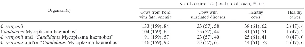
Occurrence of
M. wenyonii
and “
Candidatus
Mycoplasma
haemobos” in healthy and sick animals.
To assess the
poten-tial clinical importance of bovine hemotropic mycoplasmas, we
tested different populations of cattle for
M. wenyonii
and
“Can-didatus
Mycoplasma haemobos”. Both bovine hemoplasmas
were frequently detected in diseased and healthy adult cattle,
but just a few calves (3 out of 47) were found to be infected
(Table 2). In the cattle herd with fatal anemia, the frequencies
of
M. wenyonii
and “Candidatus
Mycoplasma haemobos”
in-fection were significantly higher than those in cattle with
un-related diseases (P
[
2]
⬍
0.0001 and
P
[
2]
⫽
0.0044,
respec-tively) and or in healthy cows (P
[
2]
⫽
0.0007 and
P
[
2]
⫽
0.0467, respectively) (Table 2). The frequency of infection was
significantly higher in the herd with fatal anemia when
coin-fection with both
M. wenyonii
and “Candidatus
Mycoplasma
haemobos” was taken into account. This was also true when
the frequency of either infection was compared to that in cattle
with unrelated diseases (P
[
2]
⫽
0.0285 and
P
[
2]
⬍
0.0001,
respectively) and healthy cows (P
[
2]
⫽
0.0307 and
P
[
2]
⫽
0.0001, respectively) (Table 2). We identified no significant
difference between cattle with unrelated diseases and healthy
animals when we assessed the frequencies of both hemoplasma
species considered either individually or together (P
[
2]
⬎
0.05 in both cases). In the diseased cattle presented at our
clinic, we failed to identify an association between the
occur-rence of the hemoplasmas and low PCV (data not shown).
Bacterial loads of
M. wenyonii
and “
Candidatus
Mycoplasma
haemobos” in healthy and sick animals.
We quantified the
mycoplasmal blood load in all samples that tested positive
using real-time PCR, and we compared the loads for the
fol-lowing three cattle populations: herd with fatal anemia, cattle
with unrelated diseases, and healthy cows. When comparing
singly infected and coinfected animals from all groups, we
observed no significant difference in the bacterial load for
either
M. wenyonii
or “Candidatus
Mycoplasma haemobos”
(data not shown). To assess the contributions of the single
groups, we separately analyzed singly infected and coinfected
animals. In the case of
M. wenyonii, the bacterial blood load
was significantly higher in the cattle herd with fatal anemia
than in cattle with unrelated diseases and healthy animals,
when considering both singly and coinfected animals
(Kruskal-Wallis
P
⬍
0.001 and Dunn’s posttest
P
⬍
0.05 for single
infection; Kruskal Wallis
P
⬍
0.001 and Dunn’s test
P
⬍
0.01
for coinfection) (Fig. 3). No significant differences were found
in the bacterial loads of “Candidatus
Mycoplasma haemobos”
among the three groups (Fig. 3).
DISCUSSION
In this study we characterized a second hemoplasma species
found in Swiss cattle, which was characterized by a shorter 16S
rRNA gene than in
M. wenyonii
(5). Sequence analysis of the
almost-complete 16S rRNA gene showed that it shared more
than 98% identity with “Candidatus
Mycoplasma haemobos”
(20) (synonym, “Candidatus
Mycoplasma haemobovis” [6, 33,
36]). It further showed more than 94% identity with
mycoplas-mas of feline and canine origin (M. haemofelis
and
M.
haemo-canis) and less than 80% identity with
M. wenyonii.
Further-more, we developed two species-specific real-time qPCR
assays to differentiate the two mycoplasmas. A similar
real-time PCR assay based on melting curve analysis has been
published by a Japanese group (14) since the submission of this
paper.
In cats and dogs, different hemotropic mycoplasmas are
characterized by different pathogenic potentials (2, 4, 10, 19,
34, 35), and we hypothesized that the same may also hold true
for the two bovine hemoplasmas. Furthermore, bovine
hemo-plasmas were significantly more common in animals with
se-vere anemia than in cattle with less pronounced anemia (5).
Therefore, we analyzed three cattle populations, namely, the
herd that was affected by the fatal anemia outbreak (5),
ani-mals with unrelated diseases, and healthy aniani-mals.
M. wenyonii
infection was found at a high frequency in the Swiss cattle
population. This is in accordance with PCR-based observations
from the United States and from Great Britain (11–13). In
addition, “Candidatus
Mycoplasma haemobos” was also
fre-quently present in Swiss cattle. “Candidatus
Mycoplasma
hae-mobos” was reported in clinically ill cattle from Japan (20);
however, no healthy animals were included in that study.
Whereas the prevalence of
M. wenyonii
in Japanese cattle (14)
was found to be comparable to the prevalence in Swiss cattle,
both the prevalence of “Candidatus
Mycoplasma haemobos”
and the frequency of coinfected animals were found to be
much higher in our study. This might reflect geographical
dif-ferences and/or different sensitivities of the assays.
[image:4.585.46.543.82.160.2]Additionally,
M. wenyonii
was found more often and the
bacterial load was significantly higher in animals from the herd
with fatal anemia than in any other animals tested. This may be
due to the epidemiological and clinical contexts of the herd.
TABLE 2. Frequency distribution of the two bovine hemoplasma species in the examined cattle populations, assessed using real-time PCR
Organism(s)
No. of occurrences (total no. of cows), %, in:
Cows from herd with fatal anemia
Cows with unrelated diseases
Healthy cows
Healthy calves
M. wenyonii
133 (159), 84
33 (57), 58
38 (61), 62
2 (47), 4
“
Candidatus
Mycoplasma haemobos”
104 (159), 65
25 (57), 44
31 (61), 51
1 (47), 2
M. wenyonii
and “
Candidatus
Mycoplasma haemobos”
91 (159), 57
23 (57), 40
25 (61), 41
0 (47), 0
M. wenyonii
and/or “
Candidatus
Mycoplasma haemobos”
146 (159), 92
35 (57), 61
44 (61), 72
3 (47), 6
on May 16, 2020 by guest
http://jcm.asm.org/
These cattle were coinfected with a large number of pathogens
and infested with potential arthropod vectors. We identified
several factors that may have facilitated the rapid spread of the
infectious agents from one animal to another in this herd: the
animals had experienced an infestation of ticks and lice, and
were roaming free within a limited area, and had access to
automatically rotating brushes (5). Little is known about the
means of transmission of
M. wenyonii
and other mycoplasma
species, but there is some evidence that flies, lice, and fleas may
serve as vectors for mechanical transmission (5, 15), while
blood-sucking arthropods such as ticks may serve as biological
vectors (8, 23, 37). The fact that just a very small portion of the
analyzed calves was found to be infected might substantiate
this hypothesis, as they are normally less exposed to vectors in
the first months of life. Alternatively this could reflect an
im-munological phenomenon, e.g., protection due to maternally
derived antibodies.
The exact role of each pathogen in the herd with fatal
ane-mia is currently unclear; however, in animals infected with
different pathogens, the immune system may be overwhelmed
or even impaired (e.g., due to
Theileria
or
Anaplasma
infec-tions [9, 27]). This may have favored other infecinfec-tions and/or
could have increased the pathogenic potential of hemoplasmas
or other infectious agents.
In cattle with unrelated diseases that were seen at our clinic,
we were unable to identify a correlation between the
occur-rence of the hemoplasmas and a decrease in PCV. However, in
these cattle, the stage of infection was unknown. For other
hemoplasma species, it has been hypothesized that during
chronic carrier stages, clinical signs may be absent, while acute
infection may lead to anemia in infected animals (10). Thus,
the cattle that were referred to our clinic for unrelated reasons
might have been in the chronic phase, while the cattle in the
herd with the acute fatal anemia outbreak may have been in
the acute stage of infection.
In conclusion, we identified and molecularly characterized
“Candidatus
Mycoplasma haemobos” in the Swiss cattle
pop-ulation. Using real-time qPCR, we were able to detect both
bovine hemoplasmal species in all tested groups of cattle,
in-dependent of their clinical status. The pathogenic potential of
bovine hemoplasmas is still difficult to interpret; the presence
of coinfections may contribute to their pathogenicity in a
syn-ergistic manner, in particular when leading to
immunosuppres-sion.
ACKNOWLEDGMENTS
We thank Y. Bosshart, C. Bro
¨nnimann, E.-E. Go
¨nczi, U. Egger, U.
Frick, B. Glaus, E. Gra
¨ssli, M. Huder, S. Keo, H. A. Knorr, B. Lange,
B. Pineroli, T. Meili Prodan, M. Nussbaumer, E. Schuler, E. Rogg, J.
Wa
¨lchli, B. Weibel, and B. Wenger for their excellent technical
assis-tance. We are grateful to W. Regli, DIAVET Labor AG, Ba
¨ch,
Swit-zerland, for providing blood samples from healthy animals. Laboratory
work was performed using the logistics of the Center for Clinical
Studies at the Vetsuisse Faculty of the University of Zurich.
[image:5.585.136.449.71.313.2]This work was financially supported by the Swiss Organization for
Ruminant Medicine (SVW), the Swiss Federal Veterinary Office
(BVET) (Bern, Switzerland), and UBS AG. R.H.L. is the recipient of
a professorship from the Swiss National Science Foundation (grants
PP00B-102866 and PP00P3-119136).
FIG. 3. Box plot of the distribution of bacteremia as assessed using real-time qPCR in the following three examined cattle populations: animals
from a herd with a fatal anemia outbreak, cows with unrelated diseases, and healthy animals. (A) Singly infected animals; (B) coinfected animals.
MW,
M. wenyonii
;
C
Mhb, “
Candidatus
Mycoplasma haemobos.” Boxes extend from the 25th to the 75th percentile, a horizontal line represents
the median, and the error bars extend down to the smallest and up to the largest value. Only hemoplasma-positive cattle were included in our
analysis. Groups were compared using Kruskal-Wallis statistics and Dunn’s posttests. A significant difference in the bacterial loads (copy number)
was apparent among the three groups for
M. wenyonii
in both singly infected and coinfected animals. Significant differences in bacterial loads
between groups are indicated (
*
,
P
⬍
0.05;
**
,
P
⬍
0.01;
***
,
P
⬍
0.001).
on May 16, 2020 by guest
http://jcm.asm.org/
REFERENCES
1.Adler, S., and V. Ellenbogen.1934. A note on two new blood parasites of cattle,EperythrozoonandBartonella. J. Comp. Pathol. Ther.47:219–221. 2.Berent, L. M., J. B. Messick, and S. K. Cooper.1998. Detection of
Haemo-bartonella felisin cats with experimentally induced acute and chronic infec-tions, using a polymerase chain reaction assay. Am. J. Vet. Res.59:1215– 1220.
3.Donatien, A., and F. Lestoquard.1934. Sur uneBartonella nouvelle du boeuf, Bartonella bovisn. sp. Bull. Soc. Pathol. Exot.27:652–654.
4.Foley, J. E., S. Harrus, A. Poland, B. Chomel, and N. C. Pedersen.1998. Molecular, clinical, and pathologic comparison of two distinct strains of Haemobartonella felisin domestic cats. Am. J. Vet. Res.59:1581–1588. 5.Hofmann-Lehmann, R., M. L. Meli, U. M. Dreher, E. Gonczi, P. Deplazes,
U. Braun, M. Engels, J. Schupbach, K. Jorger, R. Thoma, C. Griot, K. D. Stark, B. Willi, J. Schmidt, K. M. Kocan, and H. Lutz.2004. Concurrent infections with vector-borne pathogens associated with fatal hemolytic ane-mia in a cattle herd in Switzerland. J. Clin. Microbiol.42:3775–3780. 6.Hornok, S., M. L. Meli, A. Erdos, I. Hajtos, H. Lutz, and R.
Hofmann-Lehmann.2009. Molecular characterization of two different strains of hae-motropic mycoplasmas from a sheep flock with fatal haemolytic anaemia and concomitantAnaplasma ovisinfection. Vet. Microbiol.136:372–377. 7.Klein, D., P. Janda, R. Steinborn, M. Muller, B. Salmons, and W. H.
Gunzburg.1999. Proviral load determination of different feline immunode-ficiency virus isolates using real-time polymerase chain reaction: influence of mismatches on quantification. Electrophoresis20:291–299.
8.Lappin, M. R., B. Griffin, J. Brunt, A. Riley, D. Burney, J. Hawley, M. M. Brewer, and W. A. Jensen.2006. Prevalence ofBartonella species, haemo-plasma species,Ehrlichiaspecies,Anaplasma phagocytophilum, and Neorick-ettsia risticiiDNA in the blood of cats and their fleas in the United States. J. Feline Med. Surg.8:85–90.
9.Larsen, H. J., G. Overnes, H. Waldeland, and G. M. Johansen.1994. Im-munosuppression in sheep experimentally infected withEhrlichia phagocy-tophila. Res. Vet. Sci.56:216–224.
10.Messick, J. B.2003. New perspectives about Hemotrophic mycoplasma (for-merly, Haemobartonella andEperythrozoonspecies) infections in dogs and cats. Vet. Clin. North Am. Small Anim. Pract.33:1453–1465.
11.Neimark, H., K. E. Johansson, Y. Rikihisa, and J. G. Tully.2001. Proposal to transfer some members of the generaHaemobartonellaandEperythrozoon to the genusMycoplasmawith descriptions of ‘CandidatusMycoplasma hae-mofelis’, ‘CandidatusMycoplasma haemomuris’, ‘CandidatusMycoplasma haemosuis’ and⬘CandidatusMycoplasma wenyonii’. Int. J. Syst. Evol. Mi-crobiol.51:891–899.
12.Neimark, H., K. E. Johansson, Y. Rikihisa, and J. G. Tully.2002. Revision of haemotrophicMycoplasmaspecies names. Int. J. Syst. Evol. Microbiol.
52:683.
13.Neimark, H., and K. M. Kocan.1997. The cell wall-less rickettsia Eperyth-rozoon wenyoniiis aMycoplasma. FEMS Microbiol. Lett.156:287–291. 14.Nishizawa, I., M. Sato, M. Fujihara, S. Sato, and R. Harasawa.2010.
Dif-ferential detection of hemotropicMycoplasmaspecies in cattle by melting curve analysis of PCR products. J. Vet. Med. Sci.72:77–79.
15.Prullage, J. B., R. E. Williams, and S. M. Gaafar.1993. On the transmissi-bility ofEperythrozoon suisbyStomoxys calcitransandAedes aegypti. Vet. Parasitol.50:125–135.
16.Purnell, R. E., D. W. Brocklesby, and E. R. Young.1976.Eperythrozoon wenyoni, a possible cause of anaemia in British cattle. Vet. Rec.98:411. (Letter.)
17.Saitou, N., and M. Nei.1987. The neighbor-joining method: a new method for reconstructing phylogenetic trees. Mol. Biol. Evol.4:406–425. 18.Smith, J. A., M. A. Thrall, J. L. Smith, M. D. Salman, S. V. Ching, and J. K.
Collins.1990.Eperythrozoon wenyoniiinfection in dairy cattle. J. Am. Vet. Med. Assoc.196:1244–1250.
19.Sykes, J. E., L. M. Ball, N. L. Bailiff, and M. M. Fry.2005. ‘Candidatus Mycoplasma haematoparvum’, a novel small haemotropic mycoplasma from a dog. Int. J. Syst. Evol. Microbiol.55:27–30.
20.Tagawa, M., K. Matsumoto, and H. Inokuma.2008. Molecular detection of
Mycoplasma wenyoniiand ‘CandidatusMycoplasma haemobos’ in cattle in Hokkaido, Japan. Vet. Microbiol.132:177–180.
21.Tamura, K., J. Dudley, M. Nei, and S. Kumar.2007. MEGA4: Molecular Evolutionary Genetics Analysis (MEGA) software version 4.0. Mol. Biol. Evol.24:1596–1599.
22.Tandon, R., V. Cattori, M. A. Gomes-Keller, M. L. Meli, M. C. Golder, H. Lutz, and R. Hofmann-Lehmann.2005. Quantitation of feline leukaemia virus viral and proviral loads by TaqMan real-time polymerase chain reac-tion. J. Virol. Methods130:124–132.
23.Taroura, S., Y. Shimada, Y. Sakata, T. Miyama, H. Hiraoka, M. Watanabe, K. Itamoto, M. Okuda, and H. Inokuma.2005. Detection of DNA of ‘ Can-didatusMycoplasma haemominutum’ andSpiroplasmasp. in unfed ticks collected from vegetation in Japan. J. Vet. Med. Sci.67:1277–1279. 24.Tasker, S., S. H. Binns, M. J. Day, T. J. Gruffydd-Jones, D. A. Harbour, C. R.
Helps, W. A. Jensen, C. S. Olver, and M. R. Lappin.2003. Use of a PCR assay to assess the prevalence and risk factors forMycoplasma haemofelisand ‘CandidatusMycoplasma haemominutum’ in cats in the United Kingdom. Vet. Rec.152:193–198.
25.Tasker, S., C. R. Helps, M. J. Day, T. J. Gruffydd-Jones, and D. A. Harbour.
2003. Use of real-time PCR to detect and quantifyMycoplasma haemofelis and ‘Candidatus Mycoplasma haemominutum’ DNA. J. Clin. Microbiol.
41:439–441.
26.Thompson, J. D., D. G. Higgins, and T. J. Gibson.1994. CLUSTAL W: improving the sensitivity of progressive multiple sequence alignment through sequence weighting, position-specific gap penalties and weight matrix choice. Nucleic Acids Res.22:4673–4680.
27.Tuomi, J.1967. Experimental studies on bovine tick-borne fever. 1. Clinical and haematological data, some properties of the causative agent, and ho-mologous immunity. Acta Pathol. Microbiol. Scand.70:429–445. 28.Tuomi, J., and R. Tanskanen.1980. Antigenic non-relationship of two
bo-vineEperithrozoademonstrated by the immunofluorescent method. Acta Vet. Scand.21:699–701.
29.Tuomi, J., and C.-H. von Bonsdorff.1967. Ultrastructure of a microorganism associated with bovine platelets. Experientia23:111–114.
30.Uilenberg, G.2009. ‘CandidatusMycoplasma haemobos’. Vet. Microbiol.
138:200–201.
31.Uilenberg, G.1967.Eperythrozoon tuomii, n.sp. (Rickettsiales), the 3rd species ofEperythrozoonof cattle in Madagascar. Rev. Elev. Med. Vet. Pays Trop.
20:563–569.
32.Uilenberg, G.1965. Notes on theEperythrozoonof cattle in Madagascar. Rev. Elev Med. Vet. Pays Trop.18:73–81.
33.Wengi, N., B. Willi, F. S. Boretti, V. Cattori, B. Riond, M. L. Meli, C. E. Reusch, H. Lutz, and R. Hofmann-Lehmann.2008. Real-time PCR-based prevalence study, infection follow-up and molecular characterization of ca-nine hemotropic mycoplasmas. Vet. Microbiol.126:132–141.
34.Westfall, D. S., W. A. Jensen, W. J. Reagan, S. V. Radecki, and M. R. Lappin.
2001. Inoculation of two genotypes ofHemobartonella felis(California and Ohio variants) to induce infection in cats and the response to treatment with azithromycin. Am. J. Vet. Res.62:687–691.
35.Willi, B., F. S. Boretti, C. Baumgartner, S. Tasker, B. Wenger, V. Cattori, M. L. Meli, C. E. Reusch, H. Lutz, and R. Hofmann-Lehmann.2006. Prev-alence, risk factor analysis, and follow-up of infections caused by three feline hemoplasma species in cats in Switzerland. J. Clin. Microbiol.44:961–969. 36.Willi, B., F. S. Boretti, S. Tasker, M. L. Meli, N. Wengi, C. E. Reusch, H.
Lutz, and R. Hofmann-Lehmann.2007. FromHaemobartonellato hemo-plasma: molecular methods provide new insights. Vet. Microbiol.125:197– 209.
37.Woods, J. E., M. M. Brewer, J. R. Hawley, N. Wisnewski, and M. R. Lappin.
2005. Evaluation of experimental transmission of ‘CandidatusMycoplasma haemominutum’ andMycoplasma haemofelisbyCtenocephalides felisto cats. Am. J. Vet. Res.66:1008–1012.
38.Zwart, D., P. Leeflang, and C. J. A. H. V. van Vorstenbaosch.1969. Studies on theEperithrozoonassociated with bovine thrombocytes. Zentralbl. Bak-teriol. Orig.210:82–105.