• No results found

Rapid Identification of Mycobacteria to the Species Level Using INNO LiPA Mycobacteria, a Reverse Hybridization Assay

N/A
N/A
Protected

Academic year: 2020

Share "Rapid Identification of Mycobacteria to the Species Level Using INNO LiPA Mycobacteria, a Reverse Hybridization Assay"

Copied!
6
0
0

Loading.... (view fulltext now)

Full text

(1)

Copyright © 2001, American Society for Microbiology. All Rights Reserved.

Rapid Identification of Mycobacteria to the Species Level Using

INNO-LiPA Mycobacteria, a Reverse Hybridization Assay

P. N. SUFFYS,1* A.DASILVA ROCHA,1M.DEOLIVEIRA,1C. E. DIAS CAMPOS,2, A. M. WERNECK BARRETO,2 F. PORTAELS,3L. RIGOUTS,3G. WOUTERS,4G. JANNES,4G. VAN REYBROECK,4

W. MIJS,4ANDB. VANDERBORGHT4,5

Biochemistry and Molecular Biology Department, Oswaldo Cruz Institute, Oswaldo Cruz Foundation,1Reference Center Professor He´lio Fraga,2and Laboratory of Molecular Biology, HUCFF, Federal University of Rio de Janeiro,5Rio de Janeiro,

Brazil, and Institute of Tropical Medicine, Antwerp,3and Innogenetics, Zwijnaarde,4Belgium Received 25 May 2001/Returned for modification 16 July 2001/Accepted 13 September 2001

INNO-LiPA Mycobacteria (LiPA; Innogenetics, Zwijnaarde, Belgium) is a kit for the simultaneous detection and identification ofMycobacteriumspecies in culture and identifies theMycobacterium tuberculosiscomplex, theM. aviumcomplex (MAC), and the followingMycobacteriumspecies:M. kansasii,M. avium,M. intracellulare,

M. scrofulaceum,M. gordonae,M. xenopi, and theM. chelonae-M. abscessuscomplex. The assay, which targets the 16S-23S rRNA spacer region, was evaluated on 157 mycobacterial strains that had been identified by conven-tional techniques and PCR-restriction enzyme analysis of thehsp65gene (PRA). Forty-seven reference strains consisting of 37 different species and 110 human clinical isolates were submitted to the test, and all were hybridized with the Mycobacterium genus probe (MYC) on the LiPA strip (100% sensitivity). Ninety-four isolates hybridized to their corresponding species- or complex-specific probes; only one isolate phenotypically identified asM. gordonaedid not react with its specific probe (99.4% accuracy). Thirty-seven MAC strains were phenotypically identified to the complex level and to the species level by LiPA asM. avium(n18) or M. intracellulare(n7) or as belonging to theM. avium-M. intracellulare-M. scrofulaceumcomplex (n12). Of the last 12 strains, 10 hadM. aviumPRA patterns and 2 hadM. intracellularePRA patterns. Three isolates that had been identified as a single species by conventional identification were proven to be mixed cultures by the LiPA assay. The whole procedure can be performed in 1 working day, starting with the supernatant of a small amount of bacterial mass that had been treated by freezing and then boiling.

To date, more than 70 mycobacterial species have been identified, and occasionally, isolates with unknown character-istics, mostly from immunodeficient patients, are being de-scribed. Differentiation of pathogenic and nonpathogenic my-cobacteria other than Mycobacterium tuberculosis (MOTT) from members of theM. tuberculosiscomplex (MTC) is needed for patient management, considering that many MOTT are resistant to the antibiotics used for treatment of tuberculosis (27).

Identification of mycobacterial isolates to the species level is performed by analysis of phenotypic and biochemical charac-teristics of the organisms after culture in solid media, which is a time-consuming process, or by high-pressure liquid chroma-tography analysis, which requires expensive equipment. Devel-opment of molecular tests has speeded up diagnosis, but most methods suffer from specific drawbacks. The AccuProbe sys-tem (Gen-Probe) differentiates only the MTC,M. avium, M. intracellulare, M. gordonae, and M. kansasii. In-house PCR-based identification systems have been developed but either identify a limited number of mycobacterial species or are dif-ficult to use on a routine basis (4, 11). Restriction enzyme analysis of PCR products of specific genes is still widely used for identification of mycobacteria to the species level (7, 19, 23) and on clinical isolates (22, 26). Sequencing of conserved genes

is sensitive and accurate but still expensive and technically demanding (9, 18, 21, 28), although commercially available sequencing systems might simplify the procedure (16). Finally, recently developed high-density DNA probe systems need spe-cialized equipment (8).

Recently, INNO-LiPA Mycobacteria (LiPA), a commer-cially available assay targeting the 16S-23S rRNA spacer re-gion, was developed for the detection ofMycobacteriumspp. and identification of members of the MTC, M. kansasii, M. xenopi,M. gordonae, M. avium, M. intracellulare, M. scrofula-ceum, and M. chelonae. The assay has been evaluated with BACTEC 12B bottles, and LiPA results have been compared with results obtained by using DNA probes, conventional bio-chemical tests, and PCR-restriction fragment length polymor-phism (RFLP) analysis; the assay demonstrated clear-cut re-sults and was rapid and easy to perform (14). All samples tested were, however, local clinical isolates, and the specificity of the test was not evaluated on a large number of different species. Furthermore, genetic variability has been observed in mycobacterial isolates with distinct geographic origins (13, 17). We therefore evaluated the assay with reference strains and clinical isolates from different regions of Brazil belonging to 42 different species.

MATERIALS AND METHODS

Culture of mycobacterial strains and DNA extraction.Forty-seven mycobac-terial reference strains belonging to 37 species were used in this study and are

listed in Table 1. Clinical mycobacterial isolates (n⫽110, see Table 2) were

obtained as Lowenstein-Jensen cultures mainly from the National Reference Center Professor He´lio Fraga (Rio de Janeiro, Brazil) and from Laborato´rio

* Corresponding author. Mailing address: Laboratory of Molecular and Diagnosis of Infectious Diseases, DBBM, IOC, Fiocruz, Av. Brasil 4365, Manguinhos 21045-900, Rio de Janeiro, Brazil. Phone: 55-21-5984289. Fax: 55-21-2709997. E-mail: [email protected].

4477

on May 15, 2020 by guest

http://jcm.asm.org/

(2)

Richet (Rio de Janeiro, Brazil). Conventional identification had been performed according to standard procedures (10), and high-quality DNAs from mycobac-terial reference cultures were prepared as described earlier (20). For clinical isolates, the DNA extraction procedure was simplified by suspending a loop of mycobacterial mass taken from a culture in Lowenstein-Jensen medium in 0.5 ml of 10 mM Tris-HCl–1 mM EDTA–1% Triton X-100 and submitting it to three

cycles of freezing and boiling (5 min,⫺70°C; 10 min, 100°C). Nucleic acids and

processed cultures were stored at⫺20°C until further use.

PCR-RFLP analysis ofhsp65.PCR-restriction enzyme analysis of a 441-bp

amplified fragment of thehsp65gene (PRA) was performed as modified by

Telenti et al. (23). Briefly, 10 ng of purified mycobacterial DNA or that amount

present in 2␮l of the supernatant of a frozen and boiled bacterial mass was

amplified using 1.25 U ofTaqpolymerase by submitting the sample to 45 cycles

of 1 min at 94°C, 1 min at 65°C, and 1 min at 72°C, followed by a final extension

step at 72°C for 7 min. When an amplicon was present, 20␮l was digested with

10 U of eitherBstEII orHaeIII and the restriction pattern was analyzed by

electrophoresis in a mixture of 4% agarose (Sigma) and 1% NuSieve agarose (FMC BioProducts, Rockland, Maine) and by ethidium bromide staining. Each gel had a 50- or 100-bp-ladder molecular weight marker (Gibco BRL) in at least two lanes. Patterns were compared with those published in the literature (3, 6, 22, 23) or analyzed against a pattern database constructed with GelCompar (3).

LiPA.The same amount of the DNA sample that was submitted to PCR-RFLP

was added to a 40-␮l PCR mixture containing deoxynucleoside triphosphates

(supplied in the kit as amplification buffer), biotinylated primers and MgCl2

(supplied as primer solution), and 1.25 U ofTaqpolymerase (Amersham

Phar-macia, Little Chalfont, Buckinghamshire, United Kingdom). After 1 min at 95°C, amplification was performed by submitting mixtures to 30 s at 95°C, 30 s at 62°C, and 30 s at 72°C for 40 cycles. Amplification was verified in 2% agarose and by staining with ethidium bromide, and generally, a band with a size ranging from

250 to 500 bp was obtained. A 10-␮l aliquot of the amplified product was mixed

with 10␮l of denaturing solution and incubated for 5 min at room temperature

[image:2.587.46.543.84.466.2]

in a hybridization trough in a 12-trough tray (both supplied with the kit). Two milliliters of hybridization solution (at 62°C) and a membrane strip were added to each sample, and the tray was then placed in a shaking water bath (Gemini twin; Robbins Scientific, Sunnyvale, Calif.) at 62°C and incubated for 30 min. This hybridization step was followed by two 1-min incubations with 2 ml of wash solution at room temperature and one 10-min incubation at 62°C. All further incubations were performed at room temperature in an orbital shaker. The strips were rinsed with 2 ml of rinse solution, followed by incubation with alkaline phosphatase-streptavidin conjugate solution for 30 min and washing with 2 ml of rinse solution and 2 ml of substrate buffer. Finally, 2 ml of substrate solution was added and the strips were incubated for 30 min; the color reaction was stopped by washing the strips twice with 2 ml of deionized water, and identification of the sample was performed by comparing the strip against an interpretation chart supplied with the kit. In each test run, one LiPA control sample was hybridized against a strip as a control for the hybridization conditions. The LiPA test was repeated when hybridization signals suggested the presence of two mycobacterial species in a sample or when discrepant results were obtained by the different

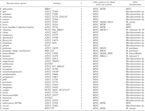
TABLE 1. LiPA patterns and LiPA interpretations obtained with 47 reference isolates of 37 different mycobacterial species

Mycobacteriumspecies Strain(s) n LiPA probe(s) for whichstrain was positive interpretationLiPA

M. africanum MB3a 1 MYC, MTB MTC

M. agri ATCC 27406 1 MYC Mycobacteriumsp.

M. aichiense ATCC 27280 1 MYC Mycobacteriumsp.

M. asiaticum ATCC 25276, Z26114b 2 MYC Mycobacteriumsp.

M. aurum ATCC 23366 2 MYC Mycobacteriumsp.

M. avium ATCC 25291 1 MYC, MAIS, MAV M. avium

M. bovis ATCC 19210 1 MYC, MTB MTC

M. bovisbacillus Calmette-Gue´rin ATCC 35736 1 MYC, MTB MTC

M. chelonae NCTC 946, MB41a 2 MYC, MCH-1 M. chelonaeII, IV

M. chitae ATCC 19627 2 MYC Mycobacteriumsp.

M. chubuense ATCC 27278 1 MYC Mycobacteriumsp.

M. diernhoferi ATCC 19340 2 MYC Mycobacteriumsp.

M. flavescens ATCC 14474 1 MYC Mycobacteriumsp.

M. fortuitum ATCC 6841 1 MYC Mycobacteriumsp.

M. gilvum S132b 1 MYC Mycobacteriumsp.

M. gordonae ATCC 14470 1 MYC, MGO M. gordonae

M. gordonaesubsp.ureolyticum MIS 222b 1 MYC, MGO M. gordonae

M. intracellulare ATCC 13950 1 MYC, MAIS, MIN M. intracellulare

M. kansasii ATCC 12478 1 MYC, MKA-1 M. kansasiiI

M. komossense ATCC 33013 1 MYC Mycobacteriumsp.

M. mageritense ATCC 700351c 1 MYC Mycobacteriumsp.

M. malmoense Z10792b 1 MYC Mycobacteriumsp.

M. marinum ATCC 927, MIS14b 2 MYC Mycobacteriumsp.

M. neoaurum ATCC 25790 1 MYC Mycobacteriumsp.

M. nonchromogenicum ATCC 19530a 2 MYC Mycobacteriumsp.

M. parafortuitum ATCC 19686 1 MYC Mycobacteriumsp.

M. peregrinum ATCC 14467 1 MYC Mycobacteriumsp.

M. phlei ATCC 11758 1 MYC Mycobacteriumsp.

M. porcinum ATCC 33776 1 MYC Mycobacteriumsp.

M. rhodesiae ATCC 27024 1 MYC Mycobacteriumsp.

M. smegmatis ATCC 19420 1 MYC Mycobacteriumsp.

M. szulgai NCTC 10831, SCS74/13b 2 MYC Mycobacteriumsp.

M. terrae ATCC 15755 1 MYC Mycobacteriumsp.

M. thermoresistibile ATCC 19527 1 MYC Mycobacteriumsp.

M. tokaiense T47502b 1 MYC Mycobacteriumsp.

M. triviale ATCC 23292 1 MYC Mycobacteriumsp.

M. tuberculosisH37Rv ATCC 27294 1 MYC, MTB MTC

M. vaccae ATCC 15483 1 MYC Mycobacteriumsp.

M. xenopi MYC527b 1 MYC, MXE M. xenopi

aUniversity of Ghent, Ghent, Belgium.

bNational Institute of Public Health and the Environment, Bilthoven, The Netherlands.

cUniversity of Madrid, Madrid, Spain.

on May 15, 2020 by guest

http://jcm.asm.org/

(3)

identification procedures. Also, 3 of the 12 samples that hybridized with the

genus andM. avium-M. intracellulare-M. scrofulaceumprobes only were

resub-mitted to the test and had their hybridization patterns confirmed.

Sequencing and analysis.For some isolates, the amplified 16S-23S spacer region generated as described above was purified using a QIAquick PCR puri-fication kit (Qiagen, Chatsworth, Calif.) and sequenced using an ABI PRISM BigDye Terminator Cycle Sequencing Ready Reaction Kit on an ABI PRISM 377 sequencer (Applied Biosystems, Foster City, Calif.). Analysis of sequences was performed by comparison against sequences in GenBank using Seqed and Fasta of the Wisconsin Package (version 9.1; Genetics Computer Group, Mad-ison, Wis).

RESULTS

No difference in signal intensity or background was observed when the LiPA assay was performed on purified DNA or on the supernatant of a heat shock-treated bacterial mass (data not shown). For all isolates analyzed by hybridization, a clear amplicon of the expected size was visible on agarose gel.

Initially, we tested 49 reference strains, including the type strains ofM. ulcerans (ATCC 19423) and M. simiae(ATCC 25275), and all hybridized to theMycobacteriumgenus probe (MYC). TheM. ulceransandM. simiaestrains, however, gave anM. intracellularepattern on LiPA assay, and PRA analysis

demonstrated that they had been contaminated withM. intra-cellulare, so they were not included in the study. Twelve strains also hybridized to their corresponding complex- or species-specific probes (Table 1): reference strains ofM. tuberculosis, M. bovis, andM. africanumstrains hybridized correctly with the probe for the MTC, whileM. gordonae,M. avium,M. intracel-lulare, andM. xenopireacted with their specific probes. ForM. kansasii and M. chelonae, species for which more than one probe was present on the strip, hybridization occurred with MKA-1 and MCH-1, respectively. All other reference strains, includingM. fortuitum, reacted with the MYC probe only.

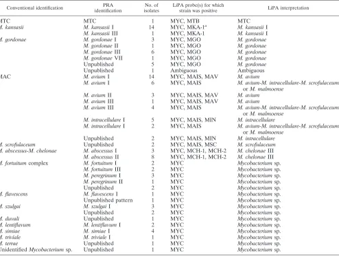
All human clinical isolates reacted with the genus-specific Mycobacteriumprobe (MYC) (Table 2). For species or species groups that have specific probes in the LiPA assay, concordant results were found: isolates of MTC (n⫽1),M. kansasii(n⫽

[image:3.587.47.541.93.466.2]

15),M. gordonae(n ⫽ 16),M. scrofulaceum(n ⫽2), M. ab-scessus(n⫽11), and theM. avium complex(MAC) (n⫽37) all hybridized with the appropriate probes on LiPA assay. TheM. kansasiiisolates were positive with the MKA-1 probe, while theM. abscessusisolates reacted with the MCH-1 and MCH-2 probes. For oneM. kansasiiisolate, a weak cross-reaction was TABLE 2. Conventional identification, PRA and LiPA patterns, and interpretations obtained with

110 clinical human isolates of mycobacteria

Conventional identification identificationPRA isolatesNo. of LiPA probe(s) for whichstrain was positive LiPA interpretation

MTC MTC 1 MYC, MTB MTC

M. kansasii M. kansasiiI 14 MYC, MKA-1a M. kansasiiI

M. kansasiiIII 1 MYC, MKA-1 M. kansasiiI

M. gordonae M. gordonaeI 3 MYC, MGO M. gordonae

M. gordonaeII 1 MYC, MGO M. gordonae

M. gordonaeIII 6 MYC, MGO M. gordonae

M. gordonaeVII 1 MYC, MGO M. gordonae

Unpublished 5 MYC, MGO M. gordonae

Unpublished 1 Ambiguous Ambiguous

MAC M. aviumI 14 MYC, MAIS, MAV M. avium

M. aviumI 6 MYC, MAIS M. avium-M. intracellulare-M. scrofulaceum

orM. malmoense

M. aviumII 3 MYC, MAIS, MAV M. avium

M. aviumIII 1 MYC, MAIS, MAV M. avium

M. aviumIII 4 MYC, MAIS M. avium-M. intracellulare-M. scrofulaceum

orM. malmoense M. intracellulareI 5 MYC, MAIS, MIN M. intracellulare

M. intracellulareI 2 MYC, MAIS M. avium-M. intracellulare-M. scrofulaceum

orM. malmoense

Unpublished 2 MYC, MAIS, MIN M. intracellulare

M. scrofulaceum Unpublished 2 MYC, MAIS, MSC M. scrofulaceum

M. abscessus-M. chelonae M. abscessusI 3 MYC, MCH-1, MCH-2 M. chelonaeIII

M. abscessusII 8 MYC, MCH-1, MCH-2 M. chelonaeIII

M. fortuitumcomplex M. fortuitumI 2 MYC Mycobacteriumsp.

M. fortuitumIII 2 MYC Mycobacteriumsp.

M. peregrinumI 3 MYC Mycobacteriumsp.

M. peregrinumII 1 MYC Mycobacteriumsp.

Unpublished 2 MYC Mycobacteriumsp.

M. flavescens M. flavescensI 1 MYC Mycobacteriumsp.

Unpublished pattern 1 MYC Mycobacteriumsp.

M. szulgai M. szulgaiI 3 MYC Mycobacteriumsp.

Unpublished 2 MYC Mycobacteriumsp.

M. duvali Unpublished 1 MYC Mycobacteriumsp.

M. lentiflavum M. lentiflavumI 2 MYC Mycobacteriumsp.

M. simiae M. simiaeI 4 MYC Mycobacteriumsp.

M. trivı´ale M. trivialeI 1 MYC Mycobacteriumsp.

M. terrae Unpublished 1 MYC Mycobacteriumsp.

UnidentifiedMycobacteriumsp. Unpublished 1 MYC Mycobacteriumsp.

aThe MAV probe was weakly visible for one isolate.

on May 15, 2020 by guest

http://jcm.asm.org/

(4)

visible with the MAV probe in the absence of theM. avium-M. intracellulare-M. scrofulaceumsignal. The LiPA test could not be repeated on this isolate, and we consider this result a hy-bridization artifact that was probably due to a small variation in hybridization temperature, which is critical during the assay, or due to the fact that the strip was not completely submerged into the assay buffers. Clinical isolates ofM. fortuitum(n⫽10), M. flavescens(n⫽2),M. terrae(n⫽1),M. lentiflavum(n⫽2), M. duvali(n⫽1), M. szulgai(n⫽5),M. triviale(n⫽1), M. simiae(n ⫽ 4), and one unidentifiedMycobacterium species were identified to the genus level only. One isolate identified as M. gordonae by conventional methods gave different results during different test runs: in two tests, only the MYC probe was positive, and in one other run, anM. avium isolate was found on LiPA assay.

All isolates were also characterized by PRA, as summarized in Table 2. All clinicalM. kansasiiisolates had theM. kansasii I-type PRA pattern, except for one isolate that had the M. kansasiiIII pattern. Of the 16M. gordonaeisolates, 3 had the M. gordonaeI PRA pattern, 1 had theM. gordonaeII pattern, 6 had theM. gordonaeIII pattern, 1 had theM. gordonaeVII pattern, and 5 were characterized by three new PRA patterns for this species (data not shown). At the time of the study, no conventional identification procedures were performed to dif-ferentiate between isolates ofM. chelonae and M. abscessus, but among the strains identified as theM. chelonae-M. absces-suscomplex, three had the PRA pattern ofM. abscessusI and eight had the pattern ofM. abscessusII. The two strains that reacted with theM. scrofulaceumprobe on LiPA assay had a so-far-undescribed PRA pattern (data not shown), and their identities were confirmed twice by conventional testing. Among the isolates of the M. fortuitum-M. peregrinum complex, two had theM. fortuitumI PRA pattern, two had theM. fortuitum III pattern, three had theM. peregrinumI pattern, one had the M. peregrinumII pattern, and two had unpublished PRA pat-terns (data not shown). One strain had theM. duvalipattern, one had theM. terraepattern, one had theM. flavescens pat-tern, and twoM. szulgaistrains had a so-far-undescribed PRA pattern (data not shown).

Thirty-seven clinical isolates that had been identified phe-notypically as MAC were identified by the LiPA assay asM. avium (n ⫽ 18), M. intracellulare (n ⫽ 7), or M. avium-M. intracellulare-M. serofulaceum(probes MYC and MAIS posi-tive;n⫽12). From the 18 isolates hybridizing with the specific M. aviumprobe on LiPA assay, 14 wereM. aviumI, 3 wereM. aviumII, and 1 wasM. aviumIII on PRA. Among the seven isolates reacting with the specific M. intracellulare probe on LiPA assay, five were identified asM. intracellulareI on PRA and two had this pattern but without theHaeIII 60-bp band (data not shown). From the 12 strains that reacted with the MAIS probe only, 6 wereM. aviumI, 4 wereM. aviumIII, and 2 wereM. intracellulareI.

Initially, for seven isolates from patients, discrepant results obtained by different identification procedures were caused by mixed cultures: in two cultures, the presence of at least two different species was detected by genetic analysis, while in five others, the presence of different species in a single culture was suggested upon repetition of conventional identification. Among the cultures that had been characterized as a single species by conventional procedures but had mixed signals by

the LiPA assay, one isolate was an M. gordonae strain that hybridized two times with the MGO, MAIS, and MAV probes on LiPA assay and had a combination ofM. gordonaeIII and M. aviumIII patterns on PRA. Another isolate, phenotypically identified asM. fortuitum, was initially positive on two LiPA assays for the MYC, MAIS, MAV, and MIN probes but had a PRA pattern suggestive of M. fortuitum I with background; upon subcloning, the isolate was reidentified asM. fortuitum, had a clearM. fortuitumI PRA pattern, and was MYC probe positive only on LiPA assay. For five clinical isolates, discrep-ant results were obtained when conventional identification re-sults were compared to the rere-sults obtained by PRA and the LiPA assay. One isolate that had been identified asM. scrofu-laceumby a conventional method was identified asM. gordonae III and was positive for the MGO probe by the LiPA assay, an isolate identified asM. gordonaewas identified asM. kansasiiI and was positive for the MKA-1 probe by the LiPA assay, and three isolates conventionally identified as MAC gave genetic patterns for another species. One of the isolates of the last group had patterns for MTC by both genetic assays, one wasM. peregrinumI andMycobacteriumspecies by PRA and the LiPA assay, respectively, and the other wasM. szulgaiand Mycobac-teriumspecies by PRA and the LiPA assay, respectively. For these isolates, the biochemical identification was repeated on subculture, confirming the results obtained by PRA and the LiPA assay.

DISCUSSION

In this study, we evaluated the LiPA line probe assay on both clinical isolates and reference strains. Recently, Miller et al. (14) examined the performance of the LiPA test, demonstrat-ing correct identifications for 59 of 60Mycobacteriumisolates, but that study concentrated on cultures received in a single lab in the United States, half of which wereM. tuberculosis. Ge-netic variability of mycobacteria has been associated with dif-ferences in geographic origin (5, 13), and although MOTT in Brazil are mostly MAC,M. kansasii, andM. fortuitum(1), all assays required for conventional identification to the species level are not routinely performed, so the number of infective or colonizingMycobacteriumspecies in the country is underesti-mated (2, 3). Also, an earlier study using PRA on a relatively small number of clinical isolates suggests the existence of par-ticular Brazilian allelic types of mycobacteria (2). The LiPA assay in this study was performed on clinical isolates obtained from a laboratory that receives clinical specimens from differ-ent regions of the country.

All mycobacterial samples reacted positively with the Myco-bacteriumgenus probe on LiPA assay (100% sensitivity), so, in contrast to what occurs in acid-fast staining, a single technol-ogy determines both the genus and clinically important species. All isolates for which species- or complex-specific probes are present on the strip were correctly identified by a positive reaction of the specific probes, except for one M. gordonae isolate. The LiPA assay for this isolate was performed three times, once on a DNA sample that yielded an unpublished PRA pattern that was observed in two other isolates that were positive for the MGO probe on LiPA assay. We have no simple explanation for this result, but PCR contamination or DNA degradation could be involved. The total accuracy for

on May 15, 2020 by guest

http://jcm.asm.org/

(5)

fication is therefore 99.4% (156 of 157 samples correctly iden-tified).

The LiPA assay was able to further identify to the species level 25 out of 37 strains that had been conventionally charac-terized as MAC; these isolates were identified by the LiPA assay asM. aviumandM. intracellulareand had PRA patterns corresponding to these species (Table 2). Twelve strains, be-sides being positive for the MYC probe, were positive for the MAIS probe only, and although four of these had theM. avium III PRA pattern that has been detected so far only in strains from Brazil (12), six hadM. aviumI and two hadM. intracel-lulareI PRA patterns. Three out of the 53 isolates that could not be correctly identified by Miller et al. (14) wereM. intra-cellularestrains (from a total of 15 MAC strains) that were identified by the LiPA assay asM. avium-M. intracellulare-M. scrofulaceum. The observation that one out of three MAC strains in our study could be identified to the species level by PRA but to the M. avium-M. intracellulare-M. scrofulaceum complex level only by the LiPA assay could be explained by the different genetic targets of both assays. Some strains may be particular to Brazil since certain PRA genotypes of MAC iso-lates have so far been reported only in this country (12, 15). We suspect these strains to beM. avium-M. intracellulare interme-diates, based on the large number of MAC strains that have been sequenced for development of the species-specific probes (data not shown). Nonetheless, the fact that these variants have been positive for the MAIS probe demonstrates that the assay is sufficiently sensitive to pick up genetic variants within MAC.

Essentially as reported by Miller et al. (14) but in contrast to a recent study performed in Italy (24), all isolates that hybrid-ized with the MCH-1 and MCH-2 probes wereM. abscessus and allM. kansasiistrains (except one) that were positive for the MKA-1 probe on LiPA assay were determined to be M. kansasiiI on PRA. We tested only a single clinical isolate ofM. tuberculosis, but all 26 strains tested by Miller et al. (14) hy-bridized with the correct probes.

The fact that M. fortuitum cannot be differentiated from other species for which no specific probes are present on the strip may be a limitation of the assay because specific thera-peutic schemes for this organism are available (27). In Brazil, 11% of MOTT of pulmonary origin isolated areM. fortuitum strains (1), and about 50% of the clinical isolates that reacted only with the species-specific probe in this study wereM. for-tuitumstrains. This species, however, is genetically heteroge-neous (W. Mijs, L. Rigouts, F. Portaels, and R. Rossau, Abstr. 21st Annu. Congr. Eur. Soc. Mycobacteriol., 2000), so inclu-sion of species-specific probes in the LiPA assay may be tech-nically complicated.

Although 18 out of the 110 clinical isolates had so-far-un-published PRA patterns, we feel confident about the identities of 109 samples because conventional identification procedures were performed in a reference center whose staff has consid-erable experience in the matter. Our results also demonstrate that discordance between the results of conventional and ge-netic identification procedures was mostly due to the presence of two different mycobacterial species in a culture. In the present study, no single colonies were picked for resubmitting to the different identification procedures but colonies with different morphologies were sometimes observed during

con-ventional identification (data not shown). The LiPA assay was able to detect mixtures when mycobacterial species for which specific probes are present on the strip were involved, as dem-onstrated by the mixed signals described in Results; the assay did not, however, allow simultaneous identification ofM. for-tuitumand another species because no species-specific probe was present for the former species. We imagine that the result obtained by different identification procedures on mixed cul-tures will be influenced by the relative quantity of each species present in the clinical sample, by the growth rate of the organ-isms, and by the fraction of the culture that is being submitted to subculturing or DNA extraction. At least for species that have a specific probe, the LiPA assay seems to be more sen-sitive than PRA for detecting mixed cultures. This conclusion is in agreement with the reported higher sensitivity of the line probe assay than those of sequencing and phenotypic assays for detecting mixed populations of human immunodeficiency virus (25). In any case, we suggest that preferentially clonal organ-isms should be used for comparison of identification proce-dures.

When this paper was in preparation, Tortoli et al. (24) re-ported an assessment of the performance of the LiPA line probe assay on 238 strains isolated in seven Italian laborato-ries. The kit correctly identified 99.6% of the strains tested, and the discrepant result was for an unresolved MAC strain identified by the LiPA assay as a MAC intermediate.

Conclusion.Evaluation of the LiPA assay was performed on a large number of clinical isolates and on reference isolates from differentMycobacteriumspecies, which were character-ized by phenotypic methods and PRA. Discordance between the results of different identification procedures was due to the presence of two different organisms in a culture. The LiPA assay, which targets the 16S-23S rRNA spacer region, was able to correctly identify 156 out of 157 isolates or strains, resulting in an accuracy of 99.4%. One M. gordonae isolate showed ambiguous results. A correlation was found between identifi-cations obtained by the LiPA assay, which targets the 16S-23S rRNA spacer region, and PRA, which targets thehsp65gene, although allelic differences were observed. In general, the LiPA assay is a reliable genetic test for the identification and differentiation ofMycobacteriumspecies.

ACKNOWLEDGMENTS

We thank Mario Vaneechoutte, Maria Jesus Garcia, Dick van Soolingen, and Richet Laboratory for donation of some of the isolates.

This study was supported by the CNPq, Faperj, and PRONEX.

REFERENCES

1.Barreto, A. M. W., and C. E. D. Campos.2000. Micobacte´rias na˜o

tubercu-losas no Brasil. Bol. Pneumol. Sanit.8:23–32.

2.Barreto, A. M. W., and F. M. Martins.1999. Laborato´rio de tuberculose do

CRPHF: quinze anos de atividades. Bol. Pneumol. Sanit.7:41–51.

3.da Silva Rocha, A., C. da Costa Leite, H. M. Torres, A. B. de Miranda, M. Q. Pires Lopes, W. M. Degrave, and P. N. Suffys,1999. Use of PCR-restriction

fragment polymorphism analysis of thehsp65gene for rapid identification of

mycobacteria in Brazil. J. Microbiol. Methods37:223–229.

4.De Beenhouwer, H., Z. Liang, P. de Rijk, C. van Eekeren, and F. Portaels.

1995. Detection and identification of mycobacteria by DNA amplification and oligonucleotide-specific capture plate hybridization. J. Clin. Microbiol.

33:2994–2998.

5.Devallois, A., M. Picardeau, C. N. Paramasivan, V. Vincent, and N. Rastogi.

1997. Molecular characterization ofMycobacterium aviumcomplex isolates

giving discordant results in AccuProbe tests by PCR-restriction enzyme anal-ysis, 16S rRNA gene sequencing, and DT1-DT6 PCR. J. Clin. Microbiol.

35:2767–2772.

on May 15, 2020 by guest

http://jcm.asm.org/

(6)

6.Devallois, A., K. S. Goh, and N. Rastogi. 1997. Rapid identification of mycobacteria to species level by PCR-restriction fragment length

polymor-phism analysis of thehsp65gene and proposition of an algorithm to

differ-entiate 34 mycobacterial species. J. Clin. Microbiol.35:2969–2973.

7.Dobner, P., K. Feldmann, M. Rifai, T. Lo¨scher, and H. Rinder.1996. Rapid identification of mycobacterial species by PCR amplification of

hypervari-able 16S rRNA gene promoter region. J. Clin. Microbiol.34:866–869.

8.Gingeras, T. R., G. Ghandour, E. Wang, A. Berno, P. M. Small, F. Drob-niewski, D. Alland, E. Desmond, M. Holodniy, and J. Drenkow.1998. Si-multaneous genotyping and species identification using hybridization pattern recognition analysis of generic Mycobacterium DNA arrays. Genome Res.

8:435–448.

9.Kapur, V., L.-L. Li, M. R. Hamrick, B. B. Plikaytis, T. M. Shinnick, A. Telenti, W. R. Jacobs, Jr., A. Banerjee, S. Cole, K. Y. Yuen, J. E. Clarridge, B. N. Kreiswirth, and J. M. Musser.1995. RapidMycobacteriumspecies assignment and unambiguous identification of mutations associated with

antimicrobial resistance inMycobacterium tuberculosisby automated DNA

sequencing. Arch. Pathol. Lab. Med.119:131–138.

10.Kent, P. T., and G. P. Kubica.1985. Public health mycobacteriology: a guide for the level II laboratory. Centers for Disease Control, Atlanta, Ga. 11.Kox, L. F. F., H. M. Jansen, S. Kuijper, and A. H. J. Kolk.1997. Multiplex

PCR assay for immediate identification of the infecting species in patients

with mycobacterial disease. J. Clin. Microbiol.35:1492–1498.

12.Lea˜o, S. C., M. R. S. Briones, M. P. Sircili, S. C. Balian, N. Mores, and J. S. Ferreira-Neto.1999. Identification of two novelMycobacterium aviumallelic variants in pig and human isolates from Brazil by PCR-restriction enzyme

analysis. J. Clin. Microbiol.37:2592–2597.

13.Legrand, E., K. S. Goh, C. Sola, and N. Rastogi.2000. Description of a novel

Mycobacterium simiaeallelic variant isolated from Caribbean AIDS patients by PCR-restriction enzyme analysis and sequencing of hsp65 gene. Mol. Cell.

Probes14:355–363.

14.Miller, N., S. Infante, and T. Cleary.2000. Evaluation of the LiPA MYCO-BACTERIA assay for identification of mycobacterial species from BACTEC

12B bottles. J. Clin. Microbiol.38:1915–1919.

15.Oliveira, R. S., M. P. Sircili, S. Y. M. Ueki, M. A. S. Telles, B. Schnabel, M. R. S. Briones, and S. C. Lea˜o.2000. PCR-restriction enzyme analysis of a bone marrow isolate from a human immunodeficiency virus-positive

pa-tient discloses polyclonal infection with twoMycobacterium aviumstrains.

J. Clin. Microbiol.38:4643–4645.

16.Patel, J. B., D. G. B. Leonard, X. Pan, J. M. Musser, R. E. Berman, and I. Nachamkin.2000. Sequence-based identification ofMycobacteriumspecies using the MicroSeq 500 16S rDNA bacterial identification system. J. Clin.

Microbiol.38:246–251.

17.Picardeau, M., G. Prod’hom, L. Raskine, M. P. LePennec, and V. Vincent.

1997. Genotypic characterization of five subspecies ofMycobacterium

kan-sasii. J. Clin. Microbiol.35:25–32.

18.Rogall, T., T. Flohr, and E. C. Bo¨ttger.1990. Differentiation of

Mycobacte-riumspecies by direct sequencing of amplified DNA. J. Gen. Microbiol.

136:1915–1920.

19.Roth, A., U. Reischl, A. Streubel, L. Naumann, R. M. Kroppenstedt, M. Habicht, M. Fischer, and H. Mauch.2000. Novel diagnostic algorithm for identification of Mycobacteria using genus-specific amplification of the 16S– 23S rRNA gene spacer and restriction endonucleases. J. Clin. Microbiol.

38:1094–1104.

20.Santos, A. R., A. B. de Miranda, L. M. Lima, P. N. Suffys, and W. M. Degrave,1992. Method for high yield preparation in large and small scale of

nucleic acids from mycobacteria. J. Microbiol. Methods15:83–94.

21.Soini, H., E. C. Bo¨ttger, and M. K. Viljanen.1994. Identification of myco-bacteria by PCR-based sequence determination of the 32-kilodalton protein

gene. J. Clin. Microbiol.32:2944–2947.

22.Taylor, T. B., C. Patterson, Y. Hale, and W. W. Safranek.1997. Routine use of PCR-restriction fragment length polymorphism analysis for identification

of mycobacteria growing in liquid media. J. Clin. Microbiol.35:79–85.

23.Telenti, A., F. Marchesi, M. Balz, F. Bally, E. C. Bo¨ttger, and T. Bodmer.

1993. Rapid identification of mycobacteria to the species level by polymerase

chain reaction and restriction enzyme analysis. J. Clin. Microbiol.31:175–

178.

24.Tortoli, E., A. Nanetti, C. Piersimoni, P. Cichero, C. Farina, G. Mucignat, C. Scarparo, L. Bartolini, R. Valentini, D. Nista, G. Gesu, C. P. Tosi, M. Crovatto, and G. Brusarosco.2001. Performance assessment of new

multi-plex probe assay for identification of mycobacteria. J. Clin. Microbiol.39:

1079–1084.

25.Van Laethem, K., K. Van Vaerenbergh. J.-C. Schmit, S. Sprecher, P. Her-mans, V. De Vroey, R. Schuurman, T. Harrer, M. Witvrouw, E. Van Wijn-gaerden, L. Stuyver, M. Van Ranst, J. Desmyter, E. De Clercq, and A.-M. Vandamme.1999. Phenotypic assays and sequencing are less sensitive than point mutation assays for detection of resistance in mixed HIV-1 genotypic

populations. J. Acquir. Immune Defic. Syndr.22:107–118.

26.Wilson, R. W., V. A. Steingrube, B. A. Brown, and R. J. Wallace, Jr.1998. Clinical application of PCR-restriction enzyme pattern analysis for rapid

identification of aerobic actinomycete isolates. J. Clin. Microbiol.36:148–

152.

27.Wright, P. W., and R. J. Wallace, Jr.1994. Nontuberculous mycobacteria

with and without HIV infection, p. 183–206.InL. N. Friedman (ed.),

Tu-berculosis: current concepts and treatment. CRC Press, Inc., Boca Raton, Fla.

28.Zolg, J. W., and S. Philippi-Schulz.1994. The superoxide dismutase gene, a target for detection and identification of mycobacteria by PCR. J. Clin.

Microbiol.32:2801–2812.

on May 15, 2020 by guest

http://jcm.asm.org/

Figure

TABLE 1. LiPA patterns and LiPA interpretations obtained with 47 reference isolates of 37 different mycobacterial species
TABLE 2. Conventional identification, PRA and LiPA patterns, and interpretations obtained with110 clinical human isolates of mycobacteria

References

Related documents

The data cover the daily trading stock indices from May 12, 1998 to July 31, 2015 with a total of 4494 observations, including the Shanghai stock index (China A-shares), the Hang

In conclusion, although the diagnosis is mainly clinical, molecular genetic tests can help make an early diag- nosis, before the bone deformities have been submitted and the

Labour mediation is a service provided by the National Agency for Employment, while vocational training, training for detainees and income top-up for unemployed

The paper analyses the cointegration relationships and the causal links between the exchange rate of the US Dollar, on the one side, and different price indices of US products on

Cashew seed fume is unhealthy and its inhalation especially for a long period of time could be very toxic as re- vealed by the significant increased concentration of

the number of comparisons for each pixel is two, while that is one in the proposed algorithm. In the proposed algorithm, there is computational overlapping for calculating

Evaluation results of solving the motif finding problem using our proposed approach on a set of heterogeneous architectures versus the implementation on an