Genetic Multilocus Studies of Different Strains of
Cryptococcus
neoformans
: Taxonomy and Genetic Structure
S. BERTOUT,1F. RENAUD,2D. SWINNE,3M. MALLIE´,1ANDJ.-M. BASTIDE1*
Laboratoire d’Immunologie Parasitologie MENRT UPRES EA 2413, Universite´ Montpellier I, Faculte´ de Pharmacie, 34060 Montpellier Ce´dex 2,1and Centre d’E´tude sur le Polymorphisme des Micro-Organismes,
CEPM/UMR CNRS-ORSTOM 9926, ORSTOM, 34032, Montpellier Ce´dex 1,2France, and Institut de Me´decine Tropicale Prince Le´opold, 2000 Antwerp, Belgium3
Received 18 August 1998/Returned for modification 15 October 1998/Accepted 30 November 1998
The genotypes of 107 strains ofCryptococcusisolated from the environment or from patients from various
geographical areas were determined by multilocus enzyme electrophoresis (MLEE). We analyzed the relation-ships between genotype structure and serotype and between genotype structure and strain origin. Twelve of the 14 enzyme-encoding loci studied were polymorphic, giving rise to 48 electrophoretic types. The genotypes of
C. neoformansandC. laurentiiwere very similar. MLEE could not distinguish between these two pathogenic
species. A correlation between the genetic multilocus structure and the origin of the sample (from the environment or patients) existed. A second analysis detected a correlation between genotype distribution and serotype. The second analysis considered three serotype groups (B, C, and A plus D plus A/D), proving that serotypes A, D, and A/D are closely related. MLEE is a useful epidemiological tool for improving our understanding of the biology of this fungus.
Cryptococcus neoformansis a yeast-like fungus. This species has two varieties (14):C. neoformansvar.neoformans, which is ubiquitous and which groups together serotypes A and D and the very controversial serotype A/D (2, 4, 28), andC. neofor-mansvar.gattii(serotypes B and C), which is found mainly in tropical and subtropical regions.
This opportunistic pathogen causes cryptococcosis, a deep mycosis with a specific tropism for the lungs and brain. It affects immunocompromised individuals and patients suffering from severe hematological malignancies (10). Its incidence has increased over the last 10 years because of the increase in the number of patients with AIDS and those undergoing trans-plantation (10).
Epidemiological studies of cryptococcosis are essential if we are to understand the biology of this fungus so as to obtain improved treatments for this infection (9, 22). Serotyping was used as an epidemiological tool for many years (1, 9). The development of DNA typing methods increases the power of discrimination of strains. Random amplified polymorphic DNA analysis (5, 12, 22), karyotyping (11, 17), and fingerprint-ing (12, 22) are routinely used to demonstrate intraspecific diversity in the genus Cryptococcus. Multilocus enzyme elec-trophoresis (MLEE), first used for the discrimination of Can-didastrains (18, 19), has provided good results forAspergillus
(20, 21) andCryptococcus (5, 6). It is a very effective typing method (15, 21) and provides information about both the phe-notype and the gephe-notype (5, 6, 18, 19, 20).
We typed 107 strains ofCryptococcusof very different origins (patients and environment) and from different geographical areas by MLEE (5). Cryptococcus laurentiiis slightly patho-genic for immunocompromised individuals, and some strains were included in this study to explore the genetic diversity of the genusCryptococcus.
This study was performed to obtain taxonomic information about the two species C. neoformans and C. laurentii and to identify any relationships between genotype information and the serotype or origin of the strains. This should improve our understanding of the biology of this fungus.
MATERIALS AND METHODS
The 107 strains tested in the present study were kindly donated by the Institute of Tropical Medicine, Antwerp, Belgium. We used 48 strains that originated from the environment (e.g., soil, fruits, and eucalyptus), 57 strains that originated from patients, and 2 reference strains (Table 1). The isolates were grown on Sabouraud agar (Biome´rieux, Marcy l’Etoile, France) at 33°C.
Preparation of culture lysates.The isolates were grown in 500-ml flasks con-taining 180 ml of Sabouraud agar medium (pH 8) at 33°C for 72 h. Yeasts were collected from the surface with glass beads (diameter, 8 mm) in 12 ml of sterile deionized water.
The resulting suspension was centrifuged at 4,0003gfor 30 min, and the yeasts were suspended in 8 ml of distilled water. The cells were mechanically disrupted in the cold by the glass bead method (bead diameter, 0.25 mm) for 2 min in a Science tech MSK homogenizer (B. Braun, San Francisco, Calif.). The cellular debris was removed by centrifugation at 15,0003gfor 15 min at 0°C. The supernatant of each lysate was aliquoted and was stored at220°C before use in MLEE.
Enzyme electrophoresis.Starch gel electrophoresis and specific enzyme stain-ing were performed as described previously (16, 18, 19, 20). In this study, the following 14 enzyme systems were tested: peptidase A (PEP A; EC 3.4.11; substrate, Val-Leu), peptidase B (PEP B; EC 3.4.11; substrate, Leu-Gly-Gly),
* Corresponding author. Present address: Laboratoire d’Immunologie et Parasitologie, Faculte´ de Pharmacie, 15, Av. Charles Flahault, 34060 Montpellier Ce´dex 2, France. Phone: 33.4.67.63.52.02. Fax: 33.4.67.41.16.17. E-mail: [email protected]
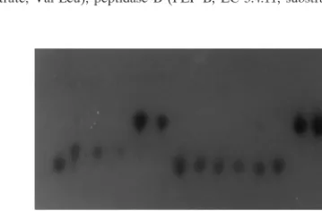
.fr. system.FIG. 1. Polymorphism observed inC. neoformanswith GPI as the enzymatic
715
on May 15, 2020 by guest
http://jcm.asm.org/
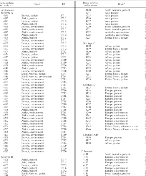
[image:1.612.339.519.592.710.2]TABLE 1. Origins and ETs ofCryptococcusstrains
Strain, serotype,
and strain no. Origina ET Strain, serotype,and strain no. Origina ET
C. neoformans Serotype A
4091 Europe, patient ET 1
4092 Africa, patient ET 2
4093 Europe, patient ET 2
4094 Africa, patient ET 2
4095 Europe, environment ET 2
4096 Africa, environment ET 2
4097 Africa, environment ET 2
4098 Africa, patient ET 2
4099 Africa, patient ET 3
4100 Europe, environment ET 2
4101 Europe, environment ET 2
4102 Europe, environment ET 3
4103 Europe, environment ET 2
4104 Africa, patient ET32
4124 Europe, patient ET 2
4125 Europe, patient ET35
4127 Europe, environment ET10
4128 Africa, environment ET10
4129 Africa, environment ET36
4141 Africa, patient ET13
4142 Europe, environment ET40
4143 South America, patient ET41
4186 South America, environment ET14
4187 Europe, environment ET15
4188 Europe, environment ET13
4189 Europe, environment ET14
4190 Europe, environment ET13
4191 Europe, environment ET16
4192 Europe, environment ET15
4193 Europe, environment ET16
4194 Europe, environment ET17
4195 Europe, environment ET18
4196 Europe, environment ET18
4197 Europe, environment ET14
4198 Africa, patient ET18
4199 Africa, environment ET18
4200 Africa, environment ET14
4201 Africa, environment ET18
4202 Africa, environment ET18
4203 Africa, environment ET14
4204 Africa, environment ET18
4205 Africa, environment ET18
4206 Africa, environment ET14
4207 Europe, environment ET19
4208 Europe, environment ET14
4209 Europe, environment ET19
4210 Europe, environment ET14
4211 Europe, environment ET20
4212 Europe, environment ET20
4213 Europe, environment ET20
Serotype B
4105 Africa, patient ET 4
4106 Asia, patient ET 5
4107 Europe, patient ET33
4108 Africa, patient ET 6
4109 Africa, patient ET42
4112 South America, patient ET 8
4137 Africa, patient ET12
4138 United States, patient ET38
4139 Asia, patient ET39
aOrigin indicates the geographical origin of the strain and the origin of the sample.
4140 South America, patient ET 48
4214 Asia, patient ET 21
4215 Asia, patient ET 20
4216 Asia, patient ET 20
4217 Asia, patient ET 21
4218 Asia, patient ET 21
4220 South America, patient ET 22
4221 South America, patient ET 22
4222 Australia, environment ET 12
4223 Australia, environment ET 23
4224 United States, patient ET 24
Serotype C
4110 Africa, patient ET 43
4119 United States, patient ET 34
4120 Africa, patient ET 44
4121 Africa, patient ET 28
4225 Africa, patient ET 23
4226 Africa, patient ET 25
4227 Africa, patient ET 26
4228 Africa, patient ET 25
4229 Africa, patient ET 25
4230 Africa, patient ET 25
4231 United States, patient ET 27
4232 United States, patient ET 27
4233 United States, patient ET 20
Serotype D
4113 United States, patient ET 28
4114 Europe, patient ET 9
4115 Europe, patient ET 31
4234 Europe, patient ET 28
4235 Europe, patient ET 27
4236 Europe, patient ET 28
4237 Europe, patient ET 20
4238 Europe, patient ET 27
4239 Europe, patient ET 20
4240 Europe, environment ET 29
4241 Europe, environment ET 20
4242 Europe, environment ET 27
4243 Europe, environment ET 23
4244 Europe, environment ET 30
4245 United States, reference strain ET 31 4246 United States, reference strain ET 28
Serotype A/D
4116 Europe, patient ET 34
4117 Africa, patient ET 2
4118 Africa, patient ET 32
C. laurentii
4111 South America, patient ET 7
4130 Europe, environment ET 45
4131 Europe, environment ET 10
4132 Europe, environment ET 37
4133 Africa, patient ET 10
4134 Europe, environment ET 46
4135 Europe, environment ET 11
4136 South America, patient ET 47
4137 South America, patient ET 12
4138 South America, environment ET 38
on May 15, 2020 by guest
http://jcm.asm.org/
peptidase C (PEP C; EC 3.4.11; substrate, Leu-Ala), glucose phosphate isomer-ase (GPI; EC 5.3.1.9), malate dehydrogenisomer-ase (MDH; EC 1.1.1.37), glucose-6-phosphate dehydrogenase (G6PD; EC 1.1.1.27), phosphoglucomutase (PGM; EC 2.7.5.1), alcohol dehydrogenase (ADH; EC 1.1.1.1), fructokinase (FK; EC 2.7.1.4), fumarase (FUM, EC 4.2.1.2), isocitrate dehydrogenase (IDH; EC 1.1.1.42), 6-phosphogluconate dehydrogenase (PGD; EC 1.1.1.43), pyruvate ki-nase (PK; EC 2.7.1.40), and sorbitol dehydrogeki-nase (SDH; EC 1.1.1.14).
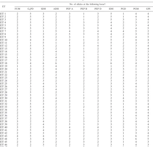
Twelve loci were polymorphic in the 107 strains examined, and each polymor-phic locus had three to six alleles (see Fig. 1). We identified 58 alleles that gave 48 different electrophoretic types (ETs) (see Table 2). Alleles were numbered in decreasing order of mobility. The various polymorphic enzyme-encoding loci were characterized for each isolate. Distinct multilocus variants were designated ETs.
Statistical analysis. (i) Factorial correspondence analysis.We analyzed the results by factorial correspondence analysis (FCA) using the PRAXIS-p.c., ver-sion 2.0, software package (Praxe`me, R&D, Biome´trie, Centre National de la Recherche Scientifique, Montpellier, France).
FCA involves the construction of a contingency table (samples versus alleles), in which each isolate is represented in terms of its allelic composition. This method of analysis simultaneously characterizes the ET according to all the genetic variables (alleles) and determines the contribution of each allele to the overall variability of the sample. The calculation of variance is based on thex2 test, which measures the extent to which a sample distribution deviates from a theoretical distribution and is an integral part of the correspondence analysis (3, 7). FCA was presented as a plane projection of the two most informative axes accounting for the genetic structure of the ETs.
(ii) Canonical correspondence analysis.We used canonical correlation anal-ysis (CCA) to compare and correlate genetic information and the origin or serotype of strains of various ETs using the PRAXIS-p.c., version 2.0, software package. This CCA was performed with two contingency tables: one (X) in which each ET was represented in terms of its allelic composition and a second one (Y) in which the strains were organized into various groups (sample origin and serotype) (23–25). The independence ofXandYwas testing by the approxima-tion-to-permutation test.
ET No. of alleles at the following locus
a:
FUM G6PD SDH ADH PEP A PEP B PEP D IDH PGD PGM GPI FK
ET 1 2 5 3 2 3 1 3 5 1 4 4 2
ET 2 2 5 3 2 2 1 2 5 1 4 4 2
ET 3 2 5 3 2 2 1 2 5 3 3 4 2
ET 4 2 5 3 2 5 3 5 4 2 3 5 2
ET 5 2 5 3 2 5 3 5 4 2 3 4 2
ET 6 2 5 3 2 5 3 5 4 2 4 5 2
ET 7 3 2 1 5 4 2 4 4 4 5 2 2
ET 8 2 3 3 2 5 3 5 4 2 3 5 2
ET 9 2 5 3 2 3 1 3 5 2 4 5 2
ET 10 2 4 3 2 2 1 2 5 1 4 4 2
ET 11 2 1 5 1 1 2 1 5 2 3 3 2
ET 12 2 5 3 2 4 3 4 5 2 3 4 2
ET 13 2 5 3 2 2 1 2 5 2 3 4 2
ET 14 1 3 2 4 2 3 2 1 3 2 3 1
ET 15 2 5 3 2 1 1 1 5 2 4 3 2
ET 16 1 3 2 4 3 3 2 1 3 2 3 1
ET 17 2 5 3 2 3 1 3 5 3 3 4 2
ET 18 3 4 6 6 1 1 1 6 2 4 1 1
ET 19 1 3 2 4 2 3 2 1 3 2 3 2
ET 20 1 2 2 4 2 2 2 2 3 1 2 1
ET 21 1 2 3 4 2 2 2 2 3 1 2 2
ET 22 2 3 3 2 4 2 4 3 2 3 4 2
ET 23 1 2 2 4 2 2 2 2 4 2 2 1
ET 24 2 2 3 2 2 2 2 3 3 3 2 2
ET 25 3 3 4 3 3 2 3 3 3 4 5 3
ET 26 3 3 4 3 2 2 2 3 3 4 5 3
ET 27 1 2 2 4 2 2 2 2 3 1 2 2
ET 28 2 5 3 2 2 1 2 5 3 3 5 2
ET 29 2 5 3 2 2 1 2 5 1 3 5 2
ET 30 1 2 2 4 2 2 2 2 4 2 2 2
ET 31 2 5 3 2 3 1 3 5 3 3 5 2
ET 32 2 5 3 2 2 1 2 5 1 3 4 2
ET 33 3 5 4 2 2 2 2 5 1 4 4 3
ET 34 2 5 3 2 2 1 2 5 3 3 4 2
ET 35 2 3 3 2 2 1 2 5 1 4 4 2
ET 36 2 3 3 2 2 1 2 3 1 4 4 2
ET 37 3 2 3 2 2 2 2 3 3 3 3 2
ET 38 3 2 4 2 3 3 3 5 3 4 4 3
ET 39 2 2 4 2 5 3 5 3 2 3 4 2
ET 40 3 2 3 2 4 2 4 3 3 3 3 2
ET 41 3 5 4 2 2 2 2 3 3 3 3 2
ET 42 2 5 3 2 2 2 2 5 1 3 4 2
ET 43 4 5 4 3 2 2 2 5 1 6 5 3
ET 44 3 3 4 3 3 2 3 3 1 6 5 3
ET 45 2 3 3 2 2 3 2 3 1 4 3 2
ET 46 2 2 3 2 2 3 2 3 1 4 3 2
ET 47 3 2 4 2 3 2 3 3 1 3 3 2
ET 48 2 2 4 2 2 3 —b 3 — 3 4 2
aFor definitions of the abbreviations, see the text. Alleles are encoded in decreasing order of their anodal mobility. b—, no enzyme activity was detected for this locus.
on May 15, 2020 by guest
http://jcm.asm.org/
[image:3.612.55.550.81.556.2]RESULTS
MLEE gave clearly reproducible results. Identical results were obtained when isolates were subcultured many times on Sabouraud’s medium and when electrophoresis was performed with samples from every fourth isolate passage.
Genetic diversity and taxonomy.Twelve loci were
polymor-phic in the 107 strains examined, and each polymorpolymor-phic locus had three to six alleles (Fig. 1). We identified 58 alleles giving 48 different ETs (Table 2). In the first FCA, strains formed three clusters (Fig. 2) in the most informative plane (account-ing for 26.78% of the total genetic variability). Cluster I con-sisted of one strain ofC. laurentii, and the GPI 1 allele was responsible for this structure (Table 2). Cluster II consisted of
C. neoformansstrains only, and cluster III contained bothC. neoformansandC. laurentiistrains. This projection shows the extensive diversity of theC. neoformansspecies. TheC. neo-formansstrains of cluster II are separated from those of cluster III by axis 1, which is the most informative (15.78% of the total variability). This shows that the strains from the various clus-ters have very different genotypes. The inclusion ofC. laurentii
strains in aC. neoformans cluster suggests that these species are genetically related and that there is greater genotype di-versity in theC. neoformans sample than in the C. laurentii
sample. The alleles responsible for this structure are indicated
in Table 2 and are confirmed in Table 3 because the least frequent alleles are the most discriminatory.
We investigated genotype differences between the ETs of cluster III using a second FCA. We obtained three clusters from the original group III (Fig. 3). Clusters III-1 and III-2 are separated by axis 1 (accounting for 17.31% of the total vari-ability). Cluster III-1 contains exclusively human serotype B strains, and cluster III-2 contains only human serotype C strains. Cluster III-3 is separated from the other two clusters by axis 2 (accounting for 15.36% of the total variability) and consists of bothC. neoformans(serotypes A, B, C, D, and A/D) andC. laurentiistrains. PEP A5 (the numbers after the locus designation indicate allele numbers) was present only in iso-lates from cluster III-1, and ADH 3 was present only in those from cluster III-2 (Table 2).
Genetic structure and serotype.A CCA was carried out to
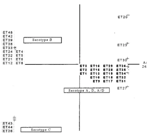
identify a relationship between genetic structure and serotype. No link between serotype and genotype was found, and the permutation test revealed no significant relationship between genetic structure and serotype (P.0.1). Serotypes A, D, and A/D are genetically similar, so we put them in the same cluster, in agreement with previous studies (22). We considered three serotyping groups (B, C, and A plus D plus A/D) (Fig. 4), and a correlation between the genotype structures of the strains and their serotypes was identified on the basis of a permutation test (P,0.02).
Genetic structure and origin.A second CCA showed a
clus-tering of the sample after the correlation between allelic com-position and sample origin (patients and the environment) was tested. This was confirmed by a permutation test (P,0.02). Isolates were grouped into three clusters on the projection (Fig. 5). One cluster contained strains isolated from both pa-tients and the environment, the second contained strains from patients only, and the third contained strains from the envi-ronment only.
DISCUSSION
[image:4.612.321.539.71.262.2]We carried out a genetic analysis of 107 strains of Crypto-coccus. The results obtained showed that there is extensive variation in this fungus, as reported in several other studies (1,
FIG. 2. First plane projection of FCA in two informative axes of all ETs observed. Axes 1 and 2 represent 15.78 and 11.00% of the overall variability, respectively. ETs represent the observed ET (Table 2). The ET group at the
[image:4.612.58.291.72.235.2]upper left (cluster III) includesC. laurentiistrains. FIG. 3. First plane projection of FCA in two informative axes of ETs of cluster III (Fig. 2). Axes 1 and 2 represent 17.31 and 15.38% of the overall variability, respectively. ETs represent the observed ETs (Table 2). The ET group at the lower (cluster III-3) includesC. laurentiistrains.
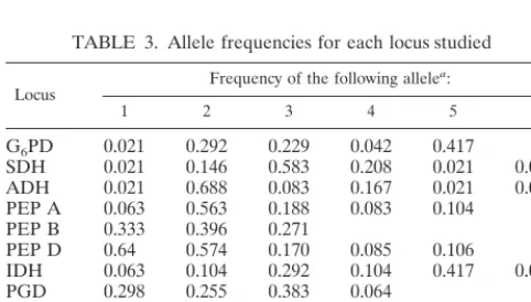
TABLE 3. Allele frequencies for each locus studied
Locus Frequency of the following allele a:
1 2 3 4 5 6
G6PD 0.021 0.292 0.229 0.042 0.417
SDH 0.021 0.146 0.583 0.208 0.021 0.021 ADH 0.021 0.688 0.083 0.167 0.021 0.021 PEP A 0.063 0.563 0.188 0.083 0.104
PEP B 0.333 0.396 0.271
PEP D 0.64 0.574 0.170 0.085 0.106
IDH 0.063 0.104 0.292 0.104 0.417 0.021
PGD 0.298 0.255 0.383 0.064
PGM 0.063 0.125 0.479 0.271 0.021 0.042
GPI 0.021 0.146 0.271 0.313 0.250
FK 0.104 0.771 0.125
aAlleles are encoded in decreasing order of their anodal mobility.
on May 15, 2020 by guest
http://jcm.asm.org/
[image:4.612.53.294.582.719.2]2, 8, 9, 11, 12, 17). We found 48 allele combinations among 107 isolates by studying 12 polymorphic loci. The level of diversity observed was similar to that reported in previous studies (4, 5) and was due to the large number of enzymes tested and the extensive heterogeneity of the strains studied. MLEE is among the numerous typing methods developed for the differentiation ofC. neoformans isolates. It has a relatively high degree of discriminatory power but also allows assessment of the struc-ture of the sample studied (4), including its genetic diversity. It was therefore used as the main technique for the present study.
Genetic diversity and taxonomy.The first FCA (Fig. 2)
pro-duced three clusters. ET 7 consisted of one strain ofC. lau-rentii. Cluster II contained onlyC. neoformansstrains, whereas cluster III contained both C. neoformans and C. laurentii
strains. There was more genetic divergence between some strains ofC. neoformans than betweenC. laurentii and some strains ofC. neoformans. TheC. neoformansstrains from clus-ters II and III differ more from each other than theC. neofor-mansandC. laurentiistrains of cluster III do. Therefore, these fungi from different species are presumably closely related genetically.
A second FCA (Fig. 3) was used to identify differences between the ETs of cluster III. Most human strains of sero-types B and C were well separated on the projection. Serosero-types A, D, and A/D were grouped together, andC. neoformansand
C. laurentiiwere not always separated on this projection. This raises important taxonomic questions about the notion of spe-cies becauseC. neoformansandC. laurentiihave very similar genotypes. The most informative enzyme systems (FUM, SDH, ADH, PGM, and PEP A) should be tested first to type an isolate and to determine to which cluster it belongs, but these enzymes cannot discriminate between some of the C. neoformansandC. laurentiistrains used in this study.
Correlation between genotype and serotype.Several studies
have shown that multilocus genotypes correlate with variety (C. neoformansvar.neoformansandC. neoformansvar.gattii)
and serotype (5, 6). However, the CCA, which tested for a possible link between serotype and genotype, detected no cor-relation when strains of the five serotypes (serotypes A, B, C, D, and A/D) were considered separately. This absence of a correlation was also confirmed by a nonsignificant value ob-tained in the approximation-to-permutation test (P.0.1). If serotypes A, D, and A/D, which are clustered together in the FCA (Fig. 2), were placed in the same group, there was a correlation between the serotype and the genotype for the three resulting serotype groups (Fig. 4). Thus, MLEE cannot differentiate between serotypes A, D, and A/D, indicating that these serotypes are closely related genetically. Some studies (4, 6) have shown that MLEE differentiates serotypes A, D, and A/D and consider them to be genetically different. The results of our work are consistent with the preliminary results of other studies (27) indicating that strains readily switch from serotype A to serotype D via the intermediate serotype A/D.
Correlation between genotype and strain origin.CCA for
the genotypes and origins of the strains provided completely new information. The sample gave three clusters on the pro-jection. There was a strong relationship between the genotypic information and the origin of the strain, in contrast to the results of other studies (13). This was confirmed by the signif-icant value obtained in the permutation test (P, 0.02). The first cluster, which was genetically diverse, exclusively tained strains isolated from patients. The second cluster con-tained strains only from the environment (Fig. 5). The third cluster contained strains isolated either from patients or from the environment. These results are of epidemiological impor-tance, because most of the environmental strains had particu-lar isoenzyme profiles which were different from those of strains from patients. So, the genotypes of the strains were correlated with their origins. Some studies (11, 12) have dem-onstrated karyotype changes in Cryptococcus after multiple infections in the mouse. This suggests that the fungus acquires a characteristic genotype after infection. Our data suggest that environmental strains are only slightly pathogenic, if at all, for humans. The genotypes of the strains responsible for infection and those isolated from the environment were very different.
[image:5.612.55.292.70.284.2]Thus, MLEE is a powerful discriminatory method. Analysis
FIG. 4. First plane projection of the CCA in two informative axes of all observed ETs. Axes 1 and 2 represent 26.14 and 23.64% of the overall variability, respectively. ETs are grouped as a function of their serotypes. The ETs in boldface type are those of serotype A, D, and A/D strains, the ETs at the upper left are those of serotype B strains, and the ETs at the lower left are those of serotype C strains.
FIG. 5. First plane projection of CCA in two informative axes of the ob-served ETs ofC. neoformans. Axes 1 and 2 represent 52.43 and 47.51% of the overall variability, respectively. ETs are grouped as a function of the sample origin. Boxed ETs represent those of human strains, the shaded ETs at the lower right represent those of environmental strains, and the ETs in boldface type represent those of human or environmental strains.
on May 15, 2020 by guest
http://jcm.asm.org/
of the relationship between ETs should improve our under-standing of the genetic polymorphism and the genotypic sim-ilarity of clinical and environmental C. neoformans isolates. This should help us to understand the transmission of this parasite and identify the risk factors for contamination so that more effective prevention measures can be implemented.
REFERENCES
1.Bellay, T., R. Cherniak, E. B. O’Neill, and T. Kosel.1996. Serotyping of
Cryptococcus neoformansby dot enzyme assay. J. Clin. Microbiol.34:466– 470.
2.Bennett, J. E., K. J. Kwon-Chung, and D. H. Howard.1977. Epidemiologic differences among serotypes ofCryptococcus neoformans. Am. J. Epidemiol.
105:582–586.
3.Benzecri, J.-P., and F. Benzecri.1980. Pratique de l’analyse des donne´es. Analyse des correspondances. Expose´ E´le´mentaire, Dunot, Paris. 4.Brandt, M. E., L. C. Hutwagner, L. A. Klug, W. S. Baughman, D. Rimland,
E. A. Graviss, R. J. Hamill, C. Thomas, P. G. Pappas, A. L. Reingold, R. W. Pinner, and The Cryptococcal Disease Active Surveillance Group.1996. Molecular subtype distribution ofCryptococcus neoformansin four areas in the United States. J. Clin. Microbiol.34:912–917.
5.Brandt, M. E., L. C. Hutwagner, R. J. Kuykendall, R. W. Pinner, and The Cryptococcal Disease Active Surveillance Group.1993. Comparison of mul-tilocus enzyme electrophoresis and random amplified polymorphic DNA analysis for molecular subtyping ofCryptococcus neoformans. J. Clin. Micro-biol.33:1890–1895.
6.Brandt, M. E., S. L. Bragg, and R. W. Pinner.1993. Multilocus enzyme typing ofCryptococcus neoformans. J. Clin. Microbiol.31:2819–2923. 7.Coustau, C., F. Renaud, C. Maillard, N. Pasteur, and B. Delay.1991.
Dif-ferential susceptibility to a trematode parasite among genotypes of the Myti-lus edulis/galloprovincialiscomplex. Genet. Res.57:207–212.
8.Dromer, F., E. Guelho, O. Ronin, and B. Dupont.1993. Serotyping of Cryp-tococcus neoformansby using a monoclonal antibody specific for capsular polysaccharide. J. Clin. Microbiol.31:359–363.
9.Dromer, F., S. Mathoulin, B. Dupont, A. Laporte, and The French Crypto-coccosis Study Group.1996. Epidemiology of cryptococcosis in France: a 9-year survey (1985–1993). Clin. Infect. Dis.23:82–90.
10.Drouhet, E.1997. Milestones in the history ofCryptococcusand cryptococ-cosis. J. Mycol. Med.7:10–27.
11.Fries, B. C., F. Chen, B. P. Currie, and A. Casadevall.1996. Karyotype instability inCryptococcus neoformansinfection. J. Clin. Microbiol.34:1531– 1534.
12.Haynes, K. A., D. J. Sullivan, D. C. Coleman, J. C. K. Clarke, R. Emilianus, C. Atkinson, and K. J. Cann.1995. Involvement of multipleCryptococcus neoformansstrains in a single episode of cryptococcosis and reinfection with novel strains in recurrent infection demonstrated by random amplification of polymorphic DNA and DNA fingerprinting. J. Clin. Microbiol.33:99–102. 13.Kohno, S.1996. Epidemiology of cryptococcosis in Japan, Session II, abstr.
3, p. 41–42.InProgram and abstracts of the 3rd International Conference on
Cryptococcusand Cryptococcosis.
14.Kwon-Chung, K. J., I. Polachek, and J. E. Bennett.1982. Improved diag-nostic medium for separation ofCryptococcus neoformansvar.neoformans
(serotypes A and D) andCryptococcus neoformansvar.gattii(serotypes B and C). J. Clin. Microbiol.15:535–537.
15. Lin, D., P. F. Lehmann, B. H. Hamory, A. A. Padhye, E. Durry, R. W. Pinner, and B. A. Lasker.1995. Comparison of three typing methods for clinical and environmental isolates ofAspergillus fumigatus. J. Clin. Microbiol.33:1596– 1601.
16. Pasteur, N., G. Pasteur, F. Bonhomme, J. Catalan, and J. Britton-Davidian.
1987. Manuel technique de ge´ne´tique par e´lectrophore`se des prote´ines. Techniques et Documentation, Lavoisier, Paris.
17. Perfect, J. R., N. Ketabchi, G. M. Cox, C. W. Ingram, and C. L. Beiser.1993. Karyotyping ofCryptococcus neoformansas an epidemiologic tool. J. Clin. Microbiol.31:3305–3309.
18. Pujol, C., J. Reynes, F. Renaud, M. Mallie´, and J.-M. Bastide.1993. Analyse ge´ne´tique de souches deCandida albicans par e´lectrophore`se des isoen-zymes. J. Mycol. Med.3:14–19.
19. Pujol, C., J. Reynes, F. Renaud, M. Raymond, M. Tibayrenc, F. J. Ayala, F. Janbon, M. Mallie´, and J. M. Bastide.1993. The yeastCandida albicanshas a clonal mode of reproduction in the population of infected human immu-nodeficiency virus-positive patients. Proc. Natl. Acad. Sci. USA90:9456– 9459.
20. Rodriguez, E., T. De Meeu¨s, M. Mallie´, F. Renaud, F. Symoens, P. Mondon, M. A. Piens, B. Lebeau, M. A. Viviani, R. Grillot, N. Nolard, F. Chapuis, A.-M. Tortorano, and J.-M. Bastide.1996. Multicentric epidemiological study ofAspergillus fumigatusisolates by multilocus enzyme electrophoresis. J. Clin. Microbiol.34:2559–2568.
21. Rodriguez, E., F. Symoens, P. Mondon, M. Mallie´, M. A. Piens, B. Lebeau, A. M. Tortorano, F. Chaib, A. Carlotti, J. Villard, M. A. Viviani, F. Chapuis, N. Nolard, R. Grillot, and J.-M. Bastide.1999. Combination of three typing methods for the molecular epidemiology ofAspergillus fumigatusinfections. J. Med. Microbiol.48:1–14.
22. Sorrell, T. C., S. C. Chen, P. Ruma, W. Meyer, T. J. Pfeiffer, D. H. Ellis, and A. G. Brownlee.1996. Concordance of clinical and environmental isolates of
Cryptococcus neoformansvar.gattiiby random amplified polymorphic DNA analysis and PCR fingerprinting. J. Clin. Microbiol.34:1253–1260. 23. Ter Braak, C. J. F.1986. Canonical correspondence analysis: a new
eigen-vector technique for multivariate direct gradient analysis. Ecology67:1167– 1179.
24. Ter Braak, C. J. F.1987. CANOCO—a Fortran program for canonical community ordination. Microcomputer Power, Ithaca, N.Y.
25. Ter Braak, C. J. F.1995. Canonical correspondence analysis and related methods in aquatic ecology. Aquat. Sci.55:255–289.
26. Varma, A., D. Swinne, F. Staib, J. F. Bennet, and K. J. Kwon-Chung.1995. Diversity of DNA fingerprints inCryptococcus neoformans. J. Clin. Micro-biol.33:1807–1814.
27. Viviani, M. A., H. Wen, A. Roverselli, R. Caldarelli-Stefano, M. Cogliati, P. Ferrante, and M. A. Tortorano.1997. Identification by polymerase chain reaction fingerprinting ofCryptococcusserotype AD. J. Med. Vet. Mycol.
35:355–360.
28. Wilson, D. E., J. E. Bennett, and J. W. Bailay.1968. Serologic grouping of
Cryptococcus neoformans. Proc. Soc. Exp. Biol. Med.127:820–823.