Copyright © 2002, American Society for Microbiology. All Rights Reserved.
Frequent Transmission of Enterococcal Strains between Mechanically
Ventilated Patients Treated at an Intensive Care Unit
Bodil Lund,
1,2Christina Agvald-Öhman,
1,3Anna Hultberg,
1,2† and Charlotta Edlund
1,2*
Division of Clinical Bacteriology1and Division of Anesthesiology and Intensive Care,3Huddinge University Hospital,Karolinska Institutet, and Södertörns Högskola University College,2Stockholm, Sweden
Received 26 December 2001/Returned for modification 24 January 2002/Accepted 25 March 2002
The objectives of this investigation were to study the respiratory tract colonization and transmission of enterococci between 20 patients treated with mechanical ventilation at an intensive care unit (ICU), to compare genotyping with phenotyping, and to determine the antibiotic susceptibilities of the isolated enterococci. Samples were collected from the oropharynx, stomach, subglottic space, and trachea within 24 h of intubation,
every third day until day 18, and thereafter every fifth day until day 33. Enterococcal isolates (nⴝ170) were
analyzed by pulsed-field gel electrophoresis and with the PhenePlate (PhP) system. The antimicrobial suscep-tibilities to five agents were determined. Seventeen of the 20 subjects were colonized with enterococci in the respiratory tract; 12 were colonized in the lower respiratory tract. Genotype analyses suggested that 13 patients were involved in a transmission event, including all patients intubated more than 12 days. In conclusion, colonization of resistant enterococci in the respiratory tract of intubated patients treated at an ICU was common. Transmission of enterococci between patients occurred frequently. Prolonged intubation period seems to be a risk factor for enterococcal cross-transmission.
Some reports have proposed that the intensive care unit (ICU) is the part of the hospital with the highest frequency of nosocomial infections (11, 23). Pneumonia is the most com-mon type of nosocomial infection occurring at an ICU (19, 24). According to a recent review by Morehead et al. the incidence of ventilator-associated pneumonia was 9 to 24% for patients intubated longer than 48 h (14).
The main organisms causing nosocomial infections at ICUs in Europe areStaphylococcus aureus,Pseudomonas aeruginosa, coagulase-negative staphylococci, yeasts, and enterococci (19) but may vary between different geographic locations. Entero-cocci are an increasing nosocomial problem worldwide, espe-cially in severely ill and immunocompromised patients, mainly due to intrinsic and acquired antimicrobial resistance. Exoge-nous acquisition of virulence determinants might also increase the pathogenicity of a certain strain (5). Their inherent toler-ance toward unfavorable conditions renders them prone to survive for long periods in the hospital setting even on dry surfaces (17, 25) and thus further facilitates transmission be-tween patients. Although transmission of bacteria bebe-tween pa-tients does not necessarily lead to infection, it is nevertheless an indication that infection control measurements can be im-proved. Measuring the rate of nosocomial infections will un-derestimate the true number of bacterial exchanges between patients and hospital settings.
The aims of the present study were to study the colonization of enterococci in the gastric and respiratory tract of mechan-ically ventilated patients at an ICU using molecular and bio-chemical fingerprinting methods and to study if transmission of
enterococcal clones occurs between patients. The antibiotic susceptibility pattern of the isolated enterococcal strains was also investigated.
MATERIALS AND METHODS
Study design and subjects.The study was conducted from April to August in 1998. Twenty-six consecutive patients, in two periods, admitted to the multidis-ciplinary ICU at Huddinge University Hospital, Stockholm, Sweden, entered the study. Inclusion criteria were adult patients requiring mechanical ventilation for a probable period of 3 days or more. Relatives’ informed consent was collected before patients entered the trial. The study was approved by the Local Ethics Committee at Huddinge University Hospital.
Collection of samples.Samples were collected within 24 h after intubation, every third day until day 18, and then every fifth day until day 33. The sampling locations were the oropharynx, the stomach, the subglottic space, and the tra-chea. Oropharyngeal samples were taken from both the tonsils and the pharynx wall using a sterile swab and a charcoal transport medium and transferred to glycerin-containing broth prior to freezing. Gastric secretions were collected by aspiration of approximately 2 ml via the gastric tube directly into a sterile tube. The first gastric sample was collected directly after application of the gastric tube, and subsequent samples were collected 3 h after enteral feeding ceased. Sub-glottic secretions were aspirated via a special suction device in the endotracheal tube and preceded by disinfection of the outer channel surface with 70% iso-propanyl. Tracheal secretions were aspirated with a sterile suction catheter via the tracheal tube (EVAC Hi Lo; Mallinckrodt, Athlone, Ireland) or a tracheos-toma cannula directly into sterile tubes. The samples were transported in trans-port medium to the Department of Clinical Bacteriology, Huddinge University Hospital, within 30 min of collection and stored at⫺70°C until analyzed.
Microbiological analyses.All the samples were cultured quantitatively and qualitatively, and enterococci were identified by colony morphology, Gram stain-ing, and biochemical tests, as previously described (6). When necessary, subglot-tic and tracheal secretions were lysed by addition of Sputolysin containing di-thiothreitol (ICN Biomedicals Inc., Aurora, Ohio). One enterococcal isolate of each type of colony morphology from each site and time point was stored in glycerin-containing broth at⫺70°C until further analyzed.
Genotyping by PFGE. All collected enterococcal isolates (n⫽170) were further analyzed by pulsed-field gel electrophoresis (PFGE). Chromosomal DNA from the enterococcal isolates was prepared as described by De Lencastre et al. (4) with some modifications as previously described (12). The DNA-containing disks were restricted overnight withSmaI (Promega Corporation, Madison, Wis.) at⫹37°C and loaded in a gel run for 20 h at⫹14°C in a contour-clamped homogenous electric field apparatus (GenePathsystem; Bio-* Corresponding author. Mailing address: Division of Clinical
Bac-teriology, F82, Huddinge University Hospital, SE-141 86 Stockholm, Sweden. Phone: 46-8-5858 11 39. Fax: 46-8-711 39 18. E-mail: charlotta [email protected].
† Present address: Center of Oral Biology, Karolinska Institutet, Stockholm, Sweden.
2084
on May 15, 2020 by guest
http://jcm.asm.org/
Rad Laboratories, Hercules, Calif.). From each gel, at least one representative of isolates that were visually judged to have identical banding patterns within a patient was rerun on a new gel, which was then further analyzed regarding genetic relatedness. Calculation of similarity matrices and creation of dendro-grams was done by aid of Molecular Analyst Software program (Bio-Rad Lab-oratories) using the unweighted pair group method using arithmetic averages. The similarity coefficients were calculated according to the method of Dice. This gave rise to dendrograms consisting of 32Enterococcus faecalisand 27 Entero-coccus faeciumisolates, respectively. The average number of bands per lane was 16. Accepting a three-band difference for probably genetically related strains and a four- to six-band difference for possibly related strains has previously been recommended by Tenover et al. (20). Blind duplicate samples were run in the software analysis to check the appropriateness of the dendrogram created.
Phenotyping with PhP system.All 170 enterococcal isolates were also analyzed according to their phenotype with the PhP system using PHP-FS plates (PhPlate Microplate Techniques AB, Stockholm, Sweden) designed for typing enterococci (10). This biochemical typing system is based on the reactions of 23 biochemical tests, plus one negative control, which are read quantitatively with a microtiter plate reader, three times during the incubation period. Thus, the results of the measurements reflect the kinetics of the biochemical reactions. The PhP system consists of biochemical tests, which give diverse results within a species (13). With the PhP system hundreds of isolates can be analyzed simultaneously. Dendrograms were created with the PhP software, PhPWIN (PhPlate Microplate Techniques AB) using the unweighted pair group method using arithmetic av-erages for clustering. By running duplicate identical reference strains in each round of analysis and subsequently clustering these, the recommended identity level of 97.5% was confirmed.
Determination of antibiotic susceptibility.The MICs of ampicillin (Sigma, St. Louis, Mo.), gentamicin (Biochrom, Berlin, Germany), imipenem (Merck, Rah-way, N.J.), moxifloxacin (Bayer, Elberfeld, Germany), and vancomycin (Eli Lilly, Stockholm, Sweden) for 169 enterococcal isolates were determined using the agar dilution method performed as recommended by the National Committee for Clinical Laboratory Standards (NCCLS) (15).E. faecalisATCC 29212 was used as a reference strain. The break points for resistance were set in accordance with NCCLS recommendations (16). Isolates were considered intermediately resistant or resistant (I⫹R) to ampicillin when the MIC wasⱖ16 mg/liter, I⫹R to imipenem and vancomycin when the MIC wasⱖ8 mg/liter, and high-level resistant to gentamicin when the MIC wasⱖ500 mg/liter. Recommended break-points for moxifloxacin are currently lacking. The level of resistance (I⫹R) was set atⱖ2 mg/liter since this breakpoint is recommended for the related com-pound ciprofloxacin according to the NCCLS (16).
RESULTS
Subjects and collection of samples. A total of 26 patients
were initially included in the study. Six patients were excluded due to death (n⫽ 2) or extubation or allocation to another hospital (n⫽4) within 3 days of inclusion. Thus, data from a total of 20 patients (median age, 58.5 years; range, 34 to 82 years; median APACHE-II score, 24 [range, 9 to 40]) were available for analyses. The median length of intubation was 4.5 days; five patients were intubated forⱖ12 days (patient 6, 8, 15, 17, and 21). The total number of samples collected was 243, deriving from a total of 75 sample occasions.
Microbiological analyses. The quantitative and qualitative
results of the total microflora of the respiratory tracts will be published elsewhere. Seventeen of the included 20 patients were on one or more occasions colonized with enterococci. Sixteen subjects had detectable amounts of enterococci at the first sampling occasion. Twelve patients were, on at least one occasion, colonized with enterococci in the lower airway, i.e., subglottis and/or trachea, with nine already colonized at the onset of intubation. A total of 170 isolates of enterococci were collected for further analysis with PFGE. Ninety-seven of these were identified asE. faecalisdistributed in 15 patients. Sixty-one isolates, from nine individuals, were considered to beE.
faecium. Twelve isolates, all from one subject, were classified asEnterococcus durans.
Genotypic analyses with PFGE. (i) Colonizing patterns
within patients. The first site where a specific enterococcal
genotype could be detected was the oropharynx in seven in-stances, the stomach in three inin-stances, the subglottic space in two instances, and the trachea in five instances. The route of colonization could not be verified for 12 strains since the initial detection of these strains occurred at several sites simulta-neously. Ten patients harbored only one enterococcal geno-type. In three of five patients intubated for at least 12 days, more than one enterococcal genotype or enterococcal species could be detected. In these individuals the enterococcal diver-sity increased with time (Table 1). AllE. duransisolates be-longed to the same genotype.
(ii) Transmission of enterococci between patients.The
den-drograms revealed that seven strains were shared between two or more patients. These genotype analyses suggested that 13 of the 20 patients were involved in an enterococcal transmission event. According to theE. faecalisdendrogram, patient 17 had a strain related to one in patient 16 and another strain related to isolates from patients 13, 21, and 22.E. faecalisisolates from patients 8 and 12 also appeared to be possibly genetically related. The intubation period of the patients harboring a commonE. faecalisclone mostly overlapped each other (data not shown). The genotyping of E. faecium isolates revealed that patients 5 and 17 and patients 15 and 17 were colonized pairwise by probably related genotypes (B:1 and B:2), respec-tively. These two genotypes were also considered possibly ge-netically related to each other. Patients 9 and 10 also had a genotype in common. Gel images of genetically related E. faecalisand E. faecium strains isolated from more than one patient and their corresponding dendrogram based on the phe-notype are shown in Fig. 1.
Phenotypic analyses with PhP system.According to the
phe-notyping results, patients 1, 13, 17, 21, and 22 sharedE. faecalis
strains of the same phenotype. Patients 6, 8, and 15 were suggested to have phenotypically identicalE. faecalisstrains in common, as well as patients 16 and 17. Three E. faecium
phenotypes were isolated from more than one patient each. IndistinguishableE. faecium phenotypes were shared by pa-tients 17 and 24, papa-tients 6 and 15, and papa-tients 5, 9, 10, and 17, respectively. The cophenetic correlation of theE. faecalisand
E. faeciumdendrogram was 0.84 and 0.92, respectively.
Comparison of phenotyping with genotyping. Phenotypic
analyses of enterococcal isolates yielded larger clusters,
includ-TABLE 1. Sequence of detection of strains in specimens from three patients harboring multiple enterococcal clones
Day
Detection of enterococcal clone(s) in specimen from patient:
6 15 17
1 I, II 1 a
3 I, II, III 1 b, c
6 I, II, III 2 b, c
9 I, II, III, IV 1, 2, 3 b, c
12 I, II, V 2, 3 b
15 1, 2, 3 b
18 2, 3 b
23 3
on May 15, 2020 by guest
http://jcm.asm.org/
ing isolates from more patients, than were yielded by PFGE. Strains clustered by PFGE were also clustered with the PhP system with few exceptions.E. faecalisisolates from patients 8 and 12 were considered possibly genotypically related and had a phenotypic similarity of 85% according to the original den-drogram, which included all 97E. faecalis isolates. Similarly, probably genotypically related E. faecium isolates from pa-tients 15 and 17 and papa-tients 6 and 24 had a phenotypic sim-ilarity of 95.4 and 91.7%, respectively, according to the original dendrogram including allE. faeciumisolates. The dendrogram in Fig. 1 illustrates the phenotypic relation between the isolates that were genotypically clustered and the corresponding gel image of each isolate. The dendrogram in Fig. 1 has a cophe-netic correlation of 0.94.
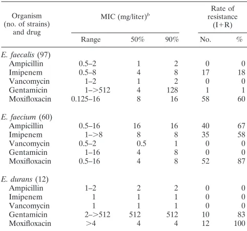
Antibiotic susceptibility of enterococcal isolates.The results
of the MIC determination are shown in Table 2. The resistance to ampicillin and imipenem was highest among theE. faecium
isolates, with rates of 67 and 58%, respectively. The corre-sponding figures for E. faecalis were 0 and 18%. All tested enterococcal isolates were susceptible to vancomycin. OneE. faecalisisolate and 10E. duransisolates were highly resistant to gentamicin (resistance defined as a MIC of ⱖ500 mg/liter). Moxifloxacin resistance was generally high in all three species. Multiple resistances were most common inE. faeciumwhere 50% of all isolates displayed resistance to ampicillin, imipenem and moxifloxacin. The occurrence of multiple resistant strains did not increase with time.
DISCUSSION
Colonization with enterococci in the respiratory tract was common in the studied patient group. The fact that 16 of the 20 patients (80%) were colonized with enterococci already at
time of intubation and that 12 (60%) were colonized by en-terococci in the lower airway on at least one sampling occasion reflects the poor health status of these patients. Treatment in hospital prior to intubation might also influence this
coloniza-FIG. 1. Dendrogram ofE. faecalisandE. faeciumisolates, genetically related according to PFGE analysis, created with PhPWIN software based on analysis of results with the PhP system. The dotted line at 97.5% illustrates the suggested identity level. The corresponding banding pattern after
[image:3.587.108.472.75.290.2]SmaI digestion and subsequent PFGE is shown for each isolate. Strains are regarded as being of the same PFGE type and thereby probably genetically related when band differences between them are three or fewer. Strains within a given PFGE type are denoted with the same capital letter, e.g., A. Strains are regarded as being of related PFGE types, thereby possibly genetically related, when the band difference between them are four or more or six or fewer. Strains with related PFGE types are termed, e.g., B:1 or B:2. Abbreviations: fs,E. faecalis; fm,E. faecium.
TABLE 2. Activities of various drugs against enterococci in this studya
Organism (no. of strains)
and drug
MIC (mg/liter)b resistanceRate of (I⫹R)
Range 50% 90% No. %
E. faecalis(97)
Ampicillin 0.5–2 1 2 0 0
Imipenem 0.5–8 4 8 17 18
Vancomycin 1–2 1 2 0 0
Gentamicin 1–⬎512 4 128 1 1
Moxifloxacin 0.125–16 8 16 58 60
E. faecium(60)
Ampicillin 0.5–16 16 16 40 67
Imipenem 1–⬎8 8 8 35 58
Vancomycin 0.5–2 0.5 1 0 0
Gentamicin 1–16 4 8 0 0
Moxifloxacin 0.5–16 4 8 52 87
E. durans(12)
Ampicillin 1–2 2 2 0 0
Imipenem 1 1 1 0 0
Vancomycin 1 1 1 0 0
Gentamicin 2–⬎512 512 512 10 83
Moxifloxacin ⬎4 4 4 12 100
aActivities of a-lactam (ampicillin), a carbapenem (imipenem), a glycopep-tide (vancomycin), an aminoglycoside (gentamicin), and a new methoxyquin-olone (moxifloxacin) againstE. faecalis, E. faecium, andE. duransgroup strains. b50% and 90%, MICs at which 50 and 90% of isolates are inhibited, respec-tively.
on May 15, 2020 by guest
http://jcm.asm.org/
[image:3.587.300.541.460.686.2]tion pattern, since enterococci are known to be a nosocomial problem. Few data are available for comparison. Bonten et al. (2) demonstrated in a study from 1995 that 16% of patients admitted to an ICU were colonized with E. faecalis in the oropharynx and/or trachea at admission. The higher initial rate ofE. faecaliscolonization in the present study (55%) might be due to the fact that the time point for admission and intubation sometimes do not coincide. However, it is important to be aware that colonization patterns differ between different coun-tries and different wards.
The fact that the oropharynx was the first site of isolation for seven of the genotypes might indicate that the route of colo-nization of enterococci in the respiratory tract starts in the oropharynx. In a study by Sabria-Leal et al. (18) it was sug-gested that gastric colonization ofE. faecaliscould serve as a reservoir of enterococci involved in nosocomial infections. In the present study, enterococci were first isolated also from the other test sites; the preference for oropharynx might, there-fore, be coincidental. In 12 subjects enterococci were first de-tected in more than one site simultaneously, which may be explained by the time lapse between the sampling occasions.
A majority of the patients colonized with enterococci only exhibited one enterococcal genotype each. A possible reason for this may be short intubation periods. Only five of the patients were intubated for a period longer than 12 days. In three of these individuals the number of enterococcal geno-types increased with time. This might suggest an increased risk for divergent colonization with prolonged intubation period. Since only one isolate of each colony morphology type, site, and time point was analyzed, there is a risk for missing a particular isolate at a certain sampling occasion. Noteworthy is that all five patients intubated more than 12 days were involved in an enterococcal transmission event (Fig. 1). This indicates an increased risk for cross-transmission with prolonged intu-bation.
Some previous studies regarding transmission of enterococci between patients in ICUs have focused on nosocomial infec-tions (9, 11) or colonization of vancomycin-resistant entero-cocci (1, 3, 9). This is relevant from a therapeutic point of view but will underestimate the true transmission rate between pa-tients. From a hospital infection control aspect, every trans-mission of bacteria between patients must be regarded as un-desirable. Here we demonstrate frequent transmission of potentially pathogenic bacteria. Enterococci may be consid-ered relevant indicators of bacterial transmission since their tolerance against harsh conditions renders them difficult to control in a healthcare environment. If enterococcal transmis-sion can be reduced, cross-infection of other bacteria is also likely to decline.
Most isolates of the same PFGE type were recovered from patients whose intubation periods at the ICU overlapped each other, which supports the notion that a direct transmission between these patients may have occurred. There were two exceptions to this. According to the genotypic analyses, both patient 6 and patient 24 seemed to have isolates belonging to the same PFGE type (Fig. 1), although patient 6 was dis-charged 3 months prior to the admission of patient 24. The three-band difference between these two isolates might be ex-plained by the long period between their isolation. If these two isolates are genetically related, the strain from patient 6 must
have resided elsewhere in the ward before colonizing patient 24. This is probably also the case for the isolates with identical banding patterns from patients 5 and 17, since the time span between the discharge of patient 5 and the admission of patient 17 was approximately 1 month. This might imply that the extent of cross-transmission of these strains between different patients is greater than the results show.
PFGE is a well-accepted epidemiological tool for determin-ing strain relatedness. However, PFGE is a laborious method requiring special equipment and may be inconvenient for iden-tifying large number of isolates. The PhP system poses an interesting alternative, since it is relatively quick to use com-pared to PFGE. Enterococci are suitable for phenotyping since they are metabolically diverse within their species. Results from the PhP system revealed larger clusters, consisting of strains from more patients than PFGE revealed. All strains suggested to be probably genetically related by PFGE were either phenotypically identical or highly related (⬎90% simi-larity) according to the PhP system. The PhP system may therefore be a suitable method for screening clonal relations between large numbers of isolates. Clones suggested by the PhP system can be further analyzed by PFGE as previously suggested by Kühn et al. (10).
The species with the highest frequency of antibiotic resis-tance wasE. faecium, both with regard to solitude and multiple resistances. The level of resistance is lower in the present study than in a recent study by Hällgren et al. (7). However, in that study antibiotic susceptibilities of enterococci isolated from patients with infections at Swedish ICUs were tested and could thus represent another more-virulent subpopulation of entero-cocci. No vancomycin-resistant enterococci were detected, which is in accordance with previous studies reporting low levels of glycopeptide resistance in Sweden (7, 8, 22). Reports of vancomycin-resistant enterococci outbreaks in Sweden are still sparse (21).
In conclusion, mechanically ventilated patients at a Swedish ICU were commonly colonized with enterococci in the upper and lower respiratory tract already at the time of intubation. Prolonged intubation seemed associated with an increased risk for cross-transmission and acquisition of multiple enterococcal genotypes. Transmission of enterococci between the intubated patients occurred frequently, indicating an increased risk of nosocomial infections and stressing the importance of optimal hygienic precautions in this group of severely ill patients.
ACKNOWLEDGMENT
This work was supported by grants from the Scandinavian Society for Antimicrobial Chemotherapy.
REFERENCES
1.Bakir, M., J. L. Bova, K. A. Newell, J. M. Millis, J. F. Buell, and P. M. Arnow.
2001. Epidemiology and clinical consequences of vancomycin-resistant en-terococci in liver transplant patients. Transplantation72:1032–1037. 2.Bonten, M. J. M., C. A. Gaillard, F. H. van Tiel, S. van der Geest, and E. E.
Stobberingh.1995. Colonization and infection withEnterococcus faecalisin intensive care units: the role of antimicrobial agents. Antimicrob. Agents Chemother.39:2783–2786.
3.Bonten, M. J. M., M. K. Hayden, C. Nathan, J. van Voorhis, M. Matushek, S. Slaughter, T. Rice, and R. A. Weinstein.1996. Epidemiology of colonisa-tion of patients and environment with vancomycin-resistant enterococci. Lancet348:1615–1619.
4.De Lencastre, H., A. E. Brown, M. Chung, D. Armstrong, and A. Tomasz.
1999. Role of transposon Tn5482 in the epidemiology of
on May 15, 2020 by guest
http://jcm.asm.org/
tantEnterococcus faeciumin the pediatric oncology unit of a New York City hospital. Microb. Drug Resist.5:113–129.
5.Eaton, T. J., and M. J. Gasson.2001. Molecular screening ofEnterococcus virulence determinants and potential for genetic exchange between food and medical isolates. Appl. Environ. Microbiol.67:1628–1635.
6.Edlund, C., G. Beyer, M. Hiemer-Bau, S. Ziege, H. Lode, and C. E. Nord.
2000. Comparative effects of moxifloxacin and clarithromycin on the normal intestinal microflora. Scand. J. Infect. Dis.32:81–85.
7.Hällgren, A., H. Abednazari, C. Ekdahl, H. Hanberger, M. Nilsson, A. Samuelsson, E. Svensson, L. E. Nilsson, and the Swedish ICU Study Group.
2001. Antimicrobial susceptibility patterns of enterococci in intensive care units in Sweden evaluated by different MIC breakpoint systems. J. Antimi-crob. Chemother.48:53–62.
8.Hanberger, H., M. Hoffmann, S. Lindgren, and L. E. Nilsson.1997. High incidence of antibiotic resistance among bacteria in 4 intensive care units at a university hospital in Sweden. Scand. J. Infect. Dis.29:607–614. 9.Karanfil, L. V., M. Murphy, A. Josephson, R. Gaynes, L. Mandel, B. C. Hill,
and J. M. Swenson.1992. A cluster of vancomycin-resistantEnterococcus faeciumin an intensive care unit. Infect. Control Hosp. Epidemiol.13:195– 200.
10.Kühn, I., L. G. Burman, S. Hæggman, K. Tullus, and B. E. Murray.1995. Biochemical fingerprinting compared with ribotyping and pulsed-field gel electrophoresis of DNA for epidemiological typing of enterococci. J. Clin. Microbiol.33:2812–2817.
11.Kulski, J. K., R. D. Wilson, R. Bending, and W. Grubb.1998. Antibiotic resistance and genomic analysis of enterococci in an intensive care unit and general wards. Pathology30:68–72.
12.Lund, B., I. Adamsson, and C. Edlund.2002. Gastrointestinal transit survival of anEnterococcus faeciumprobiotic strain administered with or without vancomycin. Int. J. Food Microbiol.77:111–117.
13.Möllby, R., I. Kühn, and M. Katouli.1993. Computerised biochemical fin-gerprinting—a new tool for typing of bacteria. Rev. Med. Microbiol.4:231– 241.
14.Morehead, R. S., and S. J. Pinto.2000. Ventilator-associated pneumonia. Arch. Intern. Med.160:1926–1936.
15.National Committee for Clinical Laboratory Standards.2000. Methods for dilution antimicrobial susceptibility tests for bacteria that grow aerobically;
5th ed. Approved Standards M7-A5, vol. 20, no. 2. National Committee for Clinical Laboratory Standards, Wayne, Pa.
16.National Committee for Clinical Laboratory Standards.2002. Performance standards for antimicrobial susceptibility testing, 12th informational supple-ment. NCCLS document M100-S12, vol. 22, no. 1. National Committee for Clinical Laboratory Standards, Wayne, Pa.
17.Noskin, G. A., V. Stosor, I. Cooper, and L. R. Peterson.1995. Recovery of vancomycin-resistant enterococci on fingertips and environmental surfaces. Infect. Control Hosp. Epidemiol.16:577–581.
18.Sabria-Leal, M., M. A. Pfaller, V. H. Morthland, S. A. Young, R. J. Hollis, L. Werkmeister, R. L. Kleiman-Wexler, and K. S. Ephgrave.1994. Molecular epidemiology of gastric colonization byEnterococcus faecalisin a surgical intensive care unit. Diagn. Microbiol. Infect. Dis.19:197–202.
19.Spencer, R. C.1996. Predominant pathogens found in the European Prev-alence of Infection in Intensive Care Study. Eur. J. Clin. Microbiol. Infect. Dis.15:281–285.
20.Tenover, F. C., R. D. Arbeit, R. V. Goering, P. A. Mickelsen, B. E. Murray, D. H. Persing, and B. Swaminathan.1995. Interpreting chromosomal DNA restriction patterns produced by pulsed-field gel electrophoresis: criteria for bacterial strain typing. J. Clin. Microbiol.33:2233–2239.
21.Torell, E., H. Fredlund, E. Törnquist, E. B. Myhre, L. Sjöberg, and A. Sundsfjord.1997. Intrahospital spread of vancomycin-resistantEnterococcus faeciumin Sweden. Scand. J. Infect. Dis.29:259–263.
22.Torell, E., O. Cars, B. Olsson-Liljequist, B.-M. Hoffman, J. Lindbäck, L. G. Burman, and the Enterococcal Study Group.1999. Near absence of vanco-mycin-resistant enterococci but high carriage rates of quinolone-resistant ampicillin-resistant enterococci among hospitalized patients and nonhospi-talized individuals in Sweden. J. Clin. Microbiol.37:3509–3513.
23.Trilla, A.1994. Epidemiology of nosocomial infections in adult intensive care units. Intensive Care Med. Suppl.20:1–4.
24.Vincent, J. L., D. J. Bihari, P. M. Suter, H. A. Bruining, J. White, M. H. Nicolas-Chanoin, M. Wolff, R. C. Spencer, M. Hemmer, et al.1995. The prevalence of nosocomial infection in intensive care units in Europe. Results of the European Prevalence of Infection in Intensive Care (EPIC) study. JAMA274:639–644.
25.Wendt, C., B. Wiesenthal, E. Dietz, and H. Rüden.1998. Survival of vanco-mycin-resistant and vancomycin-susceptible enterococci on dry surfaces. J. Clin. Microbiol.36:3734–3736.