R E S E A R C H
Open Access
Roflumilast partially reverses smoke-induced
mucociliary dysfunction
Andreas Schmid, Nathalie Baumlin, Pedro Ivonnet, John S. Dennis, Michael Campos, Stefanie Krick
and Matthias Salathe
*Abstract
Background:Phosphodiesterases (PDEs) break down cAMP, thereby regulating intracellular cAMP concentrations
and diffusion. Since PDE4 predominates in airway epithelial cells, PDE4 inhibitors can stimulate Cystic Fibrosis Transmembrane Conductance Regulator (CFTR) by increasing cAMP. Tobacco smoking and COPD are associated with decreased CFTR function and impaired mucociliary clearance (MCC). However, the effects of the PDE4 inhibitor roflumilast on smoke-induced mucociliary dysfunction have not been fully explored.
Methods:Primary normal human bronchial epithelial cells (NHBE) from non-smokers, cultured at the air-liquid interface (ALI) were used for most experiments. Cultures were exposed to cigarette smoke in a Vitrocell VC-10 smoking robot. To evaluate the effect of roflumilast on intracellular cAMP concentrations, fluorescence resonance energy transfer (FRET) between CFP- and YFP-tagged protein kinase A (PKA) subunits was recorded. Airway surface liquid (ASL) was measured using light refraction scanning and ciliary beat frequency (CBF) employing infrared differential interference contrast microscopy. Chloride conductance was measured in Ussing chambers and CFTR expression was quantified with qPCR.
Results:While treatment with 100 nM roflumilast had little effect alone, it increased intracellular cAMP upon stimulation with forskolin and albuterol in cultures exposed to cigarette smoke and in control conditions. cAMP baselines were lower in smoke-exposed cells. Roflumilast prolonged cAMP increases in smoke-exposed and control cultures. Smoke-induced reduction in functional, albuterol-mediated chloride conductance through CFTR was improved by roflumilast. ASL volumes also increased in smoke-exposed cultures in the presence of roflumilast while it did not in its absence. Cigarette smoke exposure decreased CBF, an effect rescued with roflumilast, particularly when used together with the long-acting ß-mimetic formoterol. Roflumilast also enhanced forskolin-induced CBF stimulation in ASL volume supplemented smoked and control cells, confirming the direct stimulatory effect of rising cAMP on ciliary function. In active smokers, CFTR mRNA expression was increased compared to non-smokers and ex-smokers. Roflumilast also increased CFTR mRNA levels in cigarette-smoke exposed cell cultures.
Conclusions:Our results show that roflumilast can rescue smoke-induced mucociliary dysfunction by reversing decreased CFTR activity, augmenting ASL volume, and stimulating CBF, the latter particularly in combination with formoterol. As expected, CFTR mRNA expression was not indicative of apical CFTR function.
Keywords:Roflumilast, FRET, Cilia, Airways, CFTR, Mucociliary clearance
* Correspondence:[email protected]
Division of Pulmonary, Allergy, Critical Care and Sleep Medicine, University of Miami Miller School of Medicine, 1600 NW 10th Ave, RMSB #7058, Miami, FL 33136, USA
Background
The airways are continuously exposed to dust and in-fectious agents. To prevent damage caused by irri-tants, they require effective clearing mechanisms, the physiologically most important one being mucociliary clearance (MCC). Two major determinants of effect-ive MCC are directly regulated by cAMP: Ciliary beat frequency (CBF) [1] and the Cystic Fibrosis Trans-membrane Conductance Regulator (CFTR), a major anion channel regulating airway surface liquid (ASL) volume [2].
Phosphodiesterases (PDEs) belong to a family of en-zymes that catalyze the breakdown of the second mes-sengers cAMP and cGMP to their inactive forms [3]. PDE4 gene products have a higher affinity to cAMP (Km 1–10 μM) than cGMP (Km> 50 μM) and reveal a
broad tissue distribution, including the brain, gastro-intestinal tract, spleen, lung, heart, testis and kidney [4]. The lack of effective drug therapy for chronic obstruct-ive pulmonary disease (COPD) has increased the interest in PDE4 inhibition in recent years [5], as PDEs are not only highly expressed in airway epithelial cells, but also in leukocytes and other inflammatory cells, which are in-volved in the pathogenesis of COPD [3]. Roflumilast, a PDE4 inhibitor with an IC50of 0.6 nM [3], has been
ap-proved in the United States and Europe to reduce the risk of COPD exacerbations in patients with severe COPD associated with chronic bronchitis and a history of exacerbations [6].
CFTR function is decreased in patients with COPD [7, 8] and in smokers [9]. Furthermore, PDE inhibitors have been shown to increase chloride transport through CFTR [10]. PDE4 is one of the major isoforms expressed in the airways [11]. These observations suggest that PDE inhibitors could play a key role in improving MCC by restoring depleted ASL in patients with COPD as well as in smokers.
In this study, we examined the role of roflumilast on parameters of MCC in normal human bronchial epithe-lial (NHBE) cells. We found that roflumilast increased intracellular cAMP concentrations, improved CFTR function and enhanced CBF in cigarette smoke-exposed NHBE cells. For some of these parameters, formoterol had an additive effect. These results indicate that roflu-milast rescues at least part of the dysfunctional MCC after acute smoke exposure.
Methods Chemicals
Roflumilast was provided by Forest Laboratories Inc. (New York, NY, USA). All other chemicals were pur-chased from Sigma (St. Louis, MO, USA), unless other-wise specified.
Cell cultures
Normal human bronchial epithelial (NHBE) cells from non-smokers were isolated from lungs unsuited for transplant and generously donated for research by organ donors. These lungs were recovered with IRB approved consents by the LifeCenter Northwest in Washington State and the Life Alliance Organ Recovery Agency of the University of Miami. After isolation as previously de-scribed [12–15], NHBE cells were dedifferentiated through expansion on plastic and redifferentiated at an air-liquid interface (ALI) on collagen-coated Transwell permeable supports (12 mm, 24 mm and Snapwell, Co-star Corning, Tewksbury, MA, USA). NHBE cells were considered fully differentiated after 3–4 weeks at the ALI as assessed by beating cilia and mucus transport.
Smoke exposure of NHBE ALI cultures
Fully differentiated NHBE cells were exposed to cigarette smoke using a Vitrocell VC-10 smoking robot (Vitrocell Systems GMBH, Waldkirch, Germany). Four cigarettes (University of Kentucky 3R4F) were smoked according to ISO standard 3308: six puffs per cigarette with a 35 mL volume per puff and a waiting time between each puff of 60 s. Roflumilast was added 1 h before and re-added after smoking. CBF, FRET, and CFTR conduct-ance were measured 3 h after smoke exposure. After these measurements, NHBE cells were lysed and lysate stored at−20 °C for quantitative PCR.
Pseudotyped Lentivirus Vectors and Infection of Airway Epithelial Cells
Transwells, virus was added at a ratio of 100 ng per 500,000 cells in bronchial epithelial growth medium (BEGM) containing polybrene (2 μg/ml final concentra-tion). The infection was done overnight, at 37 °C in 5 % CO2. The following day, virus was removed, and BEGM
was changed to ALI medium top and bottom until cells reached confluence, when an air liquid interface was cre-ated. Expression of the fluorescently tagged proteins was monitored using an inverted fluorescence microscope.
Measurement of CBF and FRET in airway epithelial cells
Fully differentiated NHBE cells cultured on 24 mm Transwell supports were placed in a customized, fully enclosed chamber, allowing independent perfusion of the apical and basolateral compartments. The chamber was mounted at room temperature on the stage of an upright Nikon E600fn microscope. Water was added on top of the closed chamber for use of a 63× water immersion objective with a numerical aperture of 1.0. FRET was measured as described previously [13], with images acquired every 10s. CBF was recorded according to published methods [13, 19], using infrared differential interference contrast video microscopy. CBF and FRET were measured in real time and simultaneously in cili-ated cells that expressed both fusion proteins. In addition, CBF was also recorded on an inverted Zeiss Axiovert without apical perfusion before and after apical DPBS supplementation.
Ussing chamber experiments
Snapwell filters containing fully differentiated NHBE cells were rinsed with Krebs-Henseleit solution (KH), and then mounted in Ussing chambers (EasyMount Chambers; Physiologic Instruments, San Diego, CA, USA) containing KH in apical and basolateral chambers. Solutions were maintained at 37 °C by heated water jackets, and were continuously bubbled with a 95 % room air / 5 % CO2mixture to maintain the pH at 7.4.
To monitor short-circuit current (ISC), the transepithelial
membrane potential was clamped at 0 mV with a six-channel voltage clamp (model VCC MC6; Physiologic Instruments) using Ag/AgCl electrodes in agar bridges. Signals were digitized and recorded with DAQplot soft-ware (VVI Softsoft-ware, College Station, PA, USA) via a LabJack A/D converter (LabJack Corp., Lakewood, CO, USA). The input resistance of each filter was measured by the application of 1 mV bipolar pulses of 2-s dur-ation. To eliminate any contribution to the Iscby
epithe-lial sodium channels, 10μM amiloride was added to the apical chamber. Once the Isc stabilized, roflumilast (100
nM) was included in the apical and basolateral perfusate. After a 20 min pre-treatment with roflumilast, 10 μM albuterol or 10 μM forskolin was added to the apical perfusate, and the resulting increase in chloride
conductance was measured as Isc. To assure that
con-ductance changes were related to CFTR, Isc decreases
upon apical addition of 10 μM CFTRinh 172 were
mea-sured as well. To study the combination effect of long acting ß-mimetics and roflumilast, cultures were incu-bated with 100 nM roflumilast, 100 nM formoterol, both or none for 2 h before exposures to smoke or air. In the Ussing chamber, an additional 10 μM of albuterol was added but to assess the overall influence on CFTR con-ductance; then Isc decreases upon addition of the CFTR
inhibitor CFTRinh17 were assessed.
Airway surface liquid (ASL) volume estimates
ASL volumes from NHBE cells were estimated by me-niscus scanning as previously described [20]. Briefly, cul-tures were washed with phosphate buffered saline (PBS) 24 h prior to measurements to clean the filters from mucus accumulation, which can interfere with ASL reading. At the time of measurement, the 12 mm Trans-well supports were placed on a commercially available scanner (Epson perfection V500 PHOTO, Epson Amer-ica Inc., Long Beach, CA). Scanned menisci were used to estimate ASL volume using software generously pro-vided by Dr. Myerburg (University of Pittsburgh). Cali-bration of the 12 mm Transwell supports (Costar Corning, Tewksbury, MA, USA) was done by measuring ASL changes after apical addition of different volumes of PBS.
Bronchoscopic sampling of airway epithelial cells
Bronchial epithelial cells from patients were obtained during bronchoscopy under conscious sedation for other clinical indications as approved by our IRB. After sed-ation, a cytology brush was passed through the broncho-scope to harvest airway epithelial cells. The brushings were done in the left and right main bronchus just below the carina using eight passages with ten strokes each. After each passage, the brush was withdrawn from the bronchoscope and rinsed in 2 ml of PBS. Cells were col-lected from current smokers (ten pack year history of smoking), ex-smokers (quit more than 1 year ago) and non-smokers.
Quantitative PCR
between the targeted gene and GAPDH (ΔCt) was used as the relative level of expression.
Statistics
Results were evaluated by one-way ANOVA. If a signifi-cant difference was found, a parametric (Newman Keuls) or non-parametric analysis (Dunns) using Prism 5 (GraphPad, La Jolla, CA, USA) was used for comparison of groups.p< 0.05 was accepted as significant. Error bars in all figures represent SEM.
Results
Effect of roflumilast on intracellular cAMP
To evaluate the effect of roflumilast on intracellular cAMP concentration ([cAMP]i), we used our previously
described FRET assay of fluorescently tagged PKA sub-units [13] where changes in FRET ratio (ΔFRET ratio) are indicative of changes in [cAMP]i[13]. Apical addition
of 10 μM forskolin as well as 20 μM albuterol increased [cAMP]iin NHBE cells. While roflumilast did not increase
cAMP when added alone, it increased the peak [cAMP]i
response to forskolin and albuterol (Fig. 1a) and prolonged the [cAMP]iincrease after forskolin washout (Fig. 1b).
Roflumilast also increased the [cAMP]i peak response
and duration to 10μM forskolin in cultures exposed to smoke or air (air control) in the Vitrocell VC-10 smok-ing robot. In smoke-exposed cells, the agonist-induced peak ΔFRET ratio response was significantly less com-pared to the air exposed control cells in the presence of roflumilast (Fig. 1d; each data point from three lungs with
n≥21 measurements), while no difference was found for the prolonged cAMP increase between smoke and air con-trol (Fig. 1e). To assess the effect of cigarette smoke on intracellular cAMP concentrations, we compared baseline FRET ratios of smoked and air control cultures (Fig. 1c). We found that cultures exposed to cigarette smoke had a significantly lower baseline FRET ratio compared to air control cultures (0.958 ± 0.023 vs 0.980 ± 0.015; 3 lungs,
n= 30,p< 0.05). While calibrations of these baselines are not possible and these results have to take this caveat into account, the data could indicate that acute cigarette smoke exposure decreased intracellular cAMP concentrations. On the other hand, it did not decrease changes in [cAMP]i
re-sponses to agonist stimulation and brought FRET ratios in smoked cells to the baseline FRET ratios of control cells in the absence of roflumilast. The further augmenting ef-fect of roflumilast on peak forskolin responses, however,
Fig. 1Effect of roflumilast on real-time [cAMP]i,estimated by FRET with and without smoke.aTreatment with 100 nM roflumilast alone (10 min) does not increase [cAMP]i, but roflumilast enhances forskolin- and albuterol-mediated [cAMP]iincreases (10μM forskolin and 20μM albuterol in control cultures without airflow exposure (white bars; forskolin three lungs,n= 21; albuterol one lung,n= 8).bRepresentative tracings of FRET ratios over time as a reflection of changes in [cAMP]iupon stimulation with forskolin in the presence (red) and absence (black) of roflumilast.
cSmoke exposed cells (black bar) show a decreased baseline FRET ratio compared to cultures exposed to airflow (grey bar), possibly indicating a decreased intracellular cAMP level (three different lungs,n= 30). However, FRET ratios under these conditions cannot be calibrated to [cAMP]i.
was reduced by cigarette smoke. On the other hand, roflu-milast still significantly prolonged the increase in [cAMP]i
in smoked cells after forskolin washout.
Effect of roflumilast on apical chloride conductance
The effect of roflumilast on apical chloride conductance was measured by short circuit currents (Isc) in NHBE
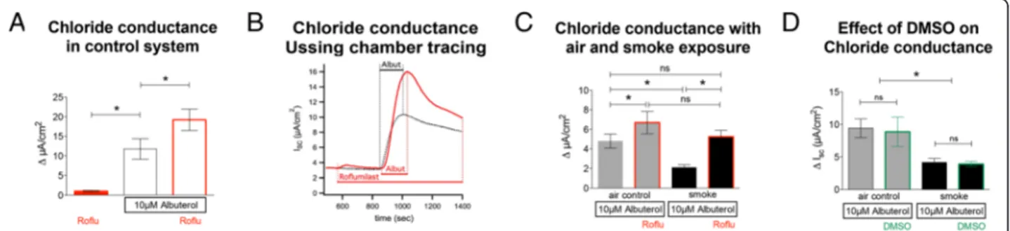
cells. Adding 100 nM roflumilast alone (Fig. 2a) resulted in a minimal current change of 0.99 ± 0.22 μA/cm2 (14 lungs, n= 29), which was significantly less than the changes observed after apical addition of 10μM albute-rol (11.75 ± 2.62 μA/cm2, p< 0.05). Use of a chloride-free apical solution did not enhance the effect of roflu-milast (not shown).
Pretreatment with roflumilast significantly increased the response to albuterol (19.02 ± 2.27μA/cm2;p< 0.05). The dynamics of chloride conductance in cultures treated with roflumilast, albuterol and their combination is demonstrated in Fig. 2b. Effects of 100 nM roflumilast on cultures exposed to control airflow or cigarette smoke in the Vitrocell VC-10 smoking robot are shown in Fig. 2c. Roflumilast increased the short circuit current response to albuterol in air-exposed control cells (6.69 ± 1.14 μA/cm2vs 4.8 ± 0.71 μA/cm2; p< 0.05). Smoke ex-posure significantly decreased chloride conductance compared to controls (2.1 ± 0.26 μA/cm2 vs 4.8 ± 0.71 μA/cm2; p< 0.05) but roflumilast reversed the smoke effect (5.26 ± 0.65 μA/cm2vs 2.1 ± 0.26 μA/cm2; p< 0.05) so that changes in Isc in roflumilast-treated,
smoked-exposed cultures were similar to untreated air-flow controls (5.26 ± 0.65 μA/cm2vs 4.8 ± 0.71 μA/cm2; p> 0.05). As shown in Fig. 2d, DMSO did have no effect on chloride conductance in air control (9.4 ± 1.5μA/cm2
vs 8.9 ± 2.3 μA/cm2) and cigarette smoke exposed cells (4.1 ± 0.6μA/cm2vs 3.9 ± 0.4μA/cm2: 2 different lungs, n= 4).
These data show that roflumilast increased albuterol-stimulated apical chloride efflux and rescued the nega-tive effect of cigarette smoke on this conductance. Addition of roflumilast without additional stimulation with albuterol or forskolin increased Isconly minimally.
Effect of roflumilast on CFTR function in smoke-exposed cells
To assure that the observed effects of roflumilast on Isc
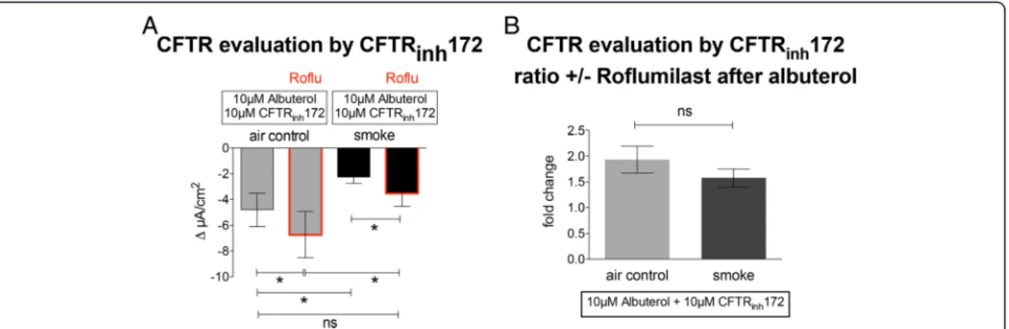
were truly related to Cl−efflux via CFTR, smoke- or air-exposed ALI cultures were pretreated basolaterally with 100 nM roflumilast, mounted in Ussing chambers, stim-ulated with 10μM albuterol and then 10μM CFTR an-tagonist CFTRinh172 was added apically (Fig. 3a). The
observed decrease in Isc is a more specific measure of
CFTR activity. Roflumilast increased CFTR-dependent Cl− secretion in air-exposed cells (−6.74 ± 1.79 μA/cm2 vs −4.81 ± 1.29 μA/cm2; p< 0.05, n= 19). Smoke expos-ure decreased CFTR conductance compared to air con-trol (−2.26 ± 0.51 μA/cm2 vs −4.51 ± 1.29 μA/cm2; p< 0.05) and roflumilast improved CFTR function in smoked cells to a level similar to controls (−3.54 ± 1.00 μA/cm2 vs −4.51 ± 1.29 μA/cm2; p< 0.05; Fig. 3a). The ratio of CFTR conductance in response to albuterol in the absence and presence of roflumilast was not sta-tistically different between smoke- and air-exposed con-trol cells (1.34 ± 0.19 vs 1.9 ± 0,37; p> 0.05; Fig. 3b). These data show that roflumilast enhanced impaired CFTR function in NHBE cell cultures exposed to cigarette smoke.
Fig. 2Effect of roflumilast on apical chloride conductance with and without smoke exposure.aEvaluation of chloride conductance in control cultures without airflow exposure (white bars): Application of roflumilast (100 nM) alone induces a minimal increase in Isc(14 lungs,n= 29). Albuterol (10μM) induces an increase in peak chloride efflux (ΔIsc) that is augmented by pre-treatment with roflumilast (100 nM) (six to ten lungs;
n= 6–10).bRepresentative tracing of an Ussing chamber experiment demonstrates an increase of chloride efflux after administration of 10μM of albuterol (black). Application of roflumilast alone hardly increases the conductance, whereas the addition of albuterol to roflumilast (red) treated cultures significantly increases chloride conductance compared to untreated cultures.cChloride efflux is significantly decreased in cultures exposed to cigarette smoke (black bars) using the Vitrocell VC-10 smoking robot when compared to air control (grey bars). Roflumilast significantly increases apical chloride efflux in air-exposed cells and rescues smoke-associated decreases in chloride conductance (19 lungs;n= 19).dControl experiments, DMSO (vehicle) does not show any changes in the response of chloride conductance to albuterol in air and cigarette
Effect of roflumilast on Airway Surface Liquid (ASL)
To measure airway surface liquid (ASL) volume, a previ-ously described meniscus scanning method [20] was used as outlined in methods. A good correlation of mea-sured volumes with the meniscus scanning method was found when measuring 30 min and 3 h after addition of 5, 10 and 20 μl PBS to control cells (Fig. 4a). ASL mea-surements were taken at different time points after smoke or air exposure (Fig. 4b and c). Baseline measure-ments were taken 1 h after exposure, just prior to roflu-milast addition (100 nM; all n= 6). Measurements of ASL were done after 4, 7, and 24 h of smoke/air expos-ure. While air-exposed cells increased ASL over time after exposure to reach baseline again after 24 h, smoke-exposed cells revealed a blunted response with a de-crease of ASL that trended to fall below baseline after
24 h (however this did not reach statistical significance). Roflumilast enhanced ASL volume increases in both air-and smoke-exposed cells (Fig. 4b air-and c). At 24 h, the roflumilast treated, smoke exposed cells had an ASL vol-ume that was significantly higher than the control cells that were only smoke exposed.
These data show that roflumilast enhances ASL vol-ume in NHBE cell cultures and partially reverses the negative effect of cigarette smoke.
Ciliary Beat Frequency (CBF)
To examine the effect of roflumilast on smoke-mediated changes in CBF, fully differentiated NHBE cells were used after basolateral treatment with 100 nM roflumi-last. DMSO controls were published by us before: DMSO did not show any significant effects on CBF [13]. Fig. 3Effect of roflumilast on CFTR function.aBasolateral pretreatment of cells exposed to either air flow (air control;grey bars) or cigarette smoke (black bars) with 100 nM roflumilast improves CFTR function as measured by the decrease in Iscafter apical application of 10μM CFTRinh172; all cells were stimulated with 10μM albuterol prior to CFTR inhibition. Roflumilast rescues CFTR function in smoke exposed cells to a level not different to the untreated air exposed cells.bAir and cigarette smoke exposed cultures show similar fold increases in CFTR conductance with and without roflumilast (19 lungs;n= 19). *p< 0.05
Baseline measurements were made one hour after roflu-milast addition, before the cells were exposed to cigarette smoke or air in the Vitrocell VC-10 robot (con-trol). Three hours after smoke/air exposure (four hours after treatment with roflumilast), CBF was measured. Baseline CBF of the cultures was 4.5 ± 0.2 Hz before and 4.68 ± 0.2 Hz 1 h after exposure to 100 nM roflumilast (p> 0.05; five lungs, n> 14). Exposure of the cultures to airflow (air control) did not change CBF in untreated cells. However, airflow increased CBF significantly when pre-treated with roflumilast for 4 h (4.19 ± 0.25 Hz vs. 6.63 ± 0.5 Hz; p< 0.05). Three hours after exposure to cigarette smoke, CBF decreased compared to air control (1.28 ± 0.06 Hz vs 4.19 ± 0.24 Hz; n> 14 each; p< 0.05) and non-exposed cultures (4.5 ± 0.2 Hz). Roflumilast in-creased CBF of the smoke-exposed cells significantly at 3 h (3.31 ± 0.43 Hz vs 1.28 ± 0.06 Hz;p< 0.05) and CBF of roflumilast treated, smoke-exposed cultures was not different compared to air-control cultures (3.31 ± 0.43 Hz vs 4.19 ± 0.24 Hz, p> 0.05). After initial CBF measurements, 50 μl PBS was added apically to replen-ish ASL volume. This maneuver equalized CBF in air-and smoke-exposed cells with or without roflumilast (5.78 ± 0.54 Hz vs 6.67 ± 0.22 Hz vs 6.04 ± 0.3 Hz vs 6.82 ± 0.37 Hz;p> 0.05 for all comparisons; Fig. 5a), pos-sibly indicating ASL volume loss as the main reason for the CBF decrease rather than a direct smoke effect on cilia. However, a lower [cAMP] after smoke exposure may contribute here as well.
To evaluate the effect of roflumilast on CBF independ-ent of ASL volume, we measured CBF in submerged
conditions. Treatment with 10 μM forskolin increased CBF in air control cells in the absence and presence of roflumilast (8.75 ± 0.5 Hz vs 10.15 ± 0.55 Hz and 8.73 ± 0.6 Hz vs 10.9 ± 0.62 Hz respectively; n> 10; p< 0.05; Fig. 5b). Similar changes were seen in smoke-exposed cells and smoke-exposed cells treated with roflumilast (4.48 ± 0.31 Hz vs 5.69 ± 0.33 Hz and 5.29 ± 0.25 Hz vs 7.29 ± 0.29 Hz respectively; p< 0.05). CBF of smoke-exposed cells treated with roflumilast and forskolin had a higher CBF than cells treated only with forskolin (7.29 ± 0.29 Hz vs 5.69 ± 0.33 Hz;p< 0.05). This indicates that roflumilast had a stimulatory effect on smoke-exposed cilia directly, independent of ASL volume changes. How-ever, the same phenomenon was not observed in air control cells.
The increased baseline of CBF in these submerged ex-periments compared to the other CBF measurements after apical PBS addition may be related to the fact that the evaluations were done with constant fluid flow in the apical compartment that may stimulate CBF. These data suggest that the CBF changes in Fig. 5b are not re-lated to ASL volume changes, but rather to an increase in cAMP production (see Fig. 1) directly affecting CBF.
In summary, roflumilast has a dual effect on CBF: it directly reverses smoke-induced CBF decreases and in-creases smoke-related reductions in ASL volume, thereby indirectly allowing normal ciliary beating. The smoke-induced lower CBF baselines in Fig. 5a and b may be due to the lower baseline cAMP levels after smoke exposure suggested by the FRET experiments dis-cussed above.
Fig. 5Effect of roflumilast on ciliary beat frequency (CBF).aCBF measurement after roflumilast exposure for 4 h (baseline in white bars on the left of the graph after 1 h; five lungs;n= 14–40). Roflumilast-treated cultures did not show an increase in CBF compared to untreated cultures (baseline). Smoke (4 h after roflumilast) significantly decreases CBF 3 h after exposure, an effect reversed by roflumilast. Exposure to air (air control) increases CBF upon administration of roflumilast (gray bars). Bars on a blue background indicate measurements of CBF after rehydration of the apical surface with 50μl PBS. CBF of cells exposed to smoke vs air equalizes, indicating a role of ASL volume depletion in the CBF changes
Effects of the long acting ß2adrenergic agonist
formoterol with roflumilast
All experiments so far described results using forskolin or the short acting ß2 mimetic albuterol to increase
cAMP levels. In clinical practice, long acting ß2
adrener-gic agonists are used for chronic treatment of COPD, whereas short acting ß2agonists are employed as rescue
inhalers for symptomatic relief [21]. Based on this fact, we examined the effect of formoterol on CFTR and CBF in the absence and presence of roflumilast in control and smoke exposed cultures.
Figure 6a shows specific CFTR conductance evaluated by Isc decreases upon channel inhibition with
CFTRinh172 in the acute presence of 10μM albuterol in
addition to the experimental conditions. Cultures were incubated with 100 mM roflumilast, 100 nM formoterol, both or none for 2 h before adding 10 μM of albuterol. Specific CFTR conductance was significantly decreased in cigarette smoke exposed compared to control cultures (−3 ± 0.79 vs −6.9 ± 1.99μA/cm2). Roflumilast alone did not significantly improve CFTR function either in smoked (−4.58 ± 1.67 μA/cm2) or in control cultures (−9.6 ± 2.8 μA). Formoterol alone, however, improved CFTR function (−6.37 ± 1.2μA/cm2and −12.6 ± 2.3μA/ cm2 respectively; 11 lungs, n= 11). Next, we evaluated the effect of the combination of formoterol and roflumi-last on CBF (Fig. 6b). As expected, CBF was decreased in cigarette smoke exposed versus control cells (2.5 ± 0.7 Hz vs 8.2 ± 0.6 Hz;n= 11;p< 0.05). Neither roflumi-last nor formoterol alone improved CBF. However, the combination of the both significantly increased CBF in
smoked cells to a level similar to control cultures (7.9 ± 0.6 vs 7.9 ± 0.6 Hz; 11 lungs,n= 11).
CFTR expression in cells brushed from patients
We examined the effect of cigarette smoke and roflumilast on CFTR mRNA expression in airway epithelia cells (Fig. 7). First, qPCR was performed on cells from bronchial brushes obtained during bron-choscopies of individuals with different smoking his-tories (Fig. 7a). The groups were patients that never smoked, current smokers (at least 10 pack years), and ex-smokers (quit smoking at least 1 year before collection of the cells). We found that current smokers had a significant increase in CFTR
expres-sion compared to non-smokers and ex-smokers
(44.5 ± 8.1 vs 16.4 ± 2.3 vs 15.3 ± 2.1; expression rela-tive to GAPDH * 1000; each n> 12; p< 0.05). CFTR expression in ex-smokers returned to the level of non-smokers (p> 0.05).
CFTR expression in cultured airway epithelial cells from non-smokers (Fig. 7b) did not increase acutely after exposure to cigarette smoke (four cigarettes) compared to air control (0.89 ± 0.2 vs 0.88 ± 0.1; p> 0.05). When air control cultures were treated with 100 nM roflumi-last, there was also no significant change in CFTR ex-pression (0.88 ± 0.1 vs 1.08 ± 0.1; n= 12; p> 0.05). However, when cells from non-smokers, exposed to smoke were treated with roflumilast, an increase in CFTR expression was observed (0.88 ± 0.18 vs 1.45 ± 0.2; p< 0.05).
Discussion
Our results show that inhibition of PDE4 with roflumi-last improves parameters of mucociliary clearance in NHBE cells. It increases ASL volume by enhancing ap-ical chloride efflux through CFTR. It also stimulates CBF via an indirect effect on ASL volume and a direct effect on cilia. These changes reverse some of the nega-tive effects of cigarette smoke on MCC and provide fur-ther mechanistic evidence that may explain the beneficial effects of this drug on reducing COPD exacerbations.
Following observations that PDE4 inhibition in ro-dents reduced tobacco smoke-mediated neutrophil in-flux in BAL, reduced lung parenchyma infiltration of neutrophils, macrophages and lymphocytes, and inhib-ited endothelial-neutrophil cell interactions [22, 23], ini-tial studies of roflumilast in populations with COPD focused on its anti-inflammatory effects. Roflumilast has been found to reduce sputum neutrophils, eosinophils, and soluble markers of neutrophilic and eosinophilic in-flammation compared with placebo [24]. It also de-creases other inflammatory molecules such as metalloproteinases and TGF-ß [25–27]. Clinically, this translates not only in a decrease in the rate of mild to moderate exacerbations and a prolongation of the time to the next exacerbation, but also into improved spiro-metric values and patient-oriented outcomes such as im-provements in dyspnea scores [28, 29].
MCC is a key mechanism to protect the airways from inhaled particles and infectious agents. Major compo-nents of this apparatus include: 1) effectively beating cilia that move mucus out of the airways and 2) an ad-equate ASL volume allowing cilia to beat efficiently and hydrating mucus optimally. If MCC fails, patients can develop chronic bronchitis, COPD, bronchiectasis and are prone to pulmonary infections [30]. Our studies may provide some understanding on basic non-inflammatory related mechanisms as to why roflumilast positively
influences COPD in patients, namely its effects on ASL volume and ciliary beating.
In our Ussing chamber experiments, 100 nM roflumi-last alone did not increase Isc (Fig. 2a). Since
phospho-diesterase inhibitors do not directly increase [cAMP]i,
but decrease its break down, this finding is not surpris-ing, at least for an acute situation. Chronically, it’s pos-sible that roflumilast can elevate cAMP through intrinsic activities of adenylyl cyclases via some basic stimulation by adenosine for instance. This theory is supported by our findings that roflumilast did not change baseline CBF in control cells, but increased it in cells exposed to control airflow (Fig. 5a). Airflow in-creases ATP release through pannexins [31] and the resulting adenosine increase could stimulate adenylyl cy-clase [32], allowing roflumilast to have a significant effect.
In contradiction to our findings, Lambert et al. pub-lished a study where the sole addition of 1 nM roflu-milast increased Isc close to 10 μA/cm2 [33].
Additionally, roflumilast had an EC50 of 2.9 nM with
a maximal effect of about 40 μA/cm2. In order to achieve these high responses with such doses of roflu-milast, the authors highlighted the use of a low extra-cellular chloride concentration (higher driving force). We repeated these experiments but did not observe the Isc stimulations described by Lambert. In addition
and in contrast to these authors, we saw a significant effect of 10 μM forskolin addition on Isc after
expos-ure to roflumilast. It is unclear why similar experi-ments can lead to such different results. One possible explanation is that we used fully differentiated NHBE cells whereas the cited study used primary bronchial epithelial cells grown in submerged monolayers or Calu3 cells. In addition, others have published in-creases in CBF with sole addition of roflumilast N-oxide [34], an effect only seen upon smoke exposure in our culture system.
Fig. 7Change in CFTR expression in brushed cells obtained during bronchoscopies and NHBE cultures.aExpression of CFTR is increased in brushed cells obtained from airways of smokers compared to non-smokers and ex-smokers. (four to six lungs,n= 12–18).bIn cultured epithelial cells from non-smokers, acute exposure to cigarette smoke does not change CFTR expression compared to control air-exposed cells within 3 h of exposure. Roflumilast (red framed boxes) appears to increase CFTR expression in the smoke-exposed cells after 3 h (four lungs,n= 12) *
Our study showed beneficial effects of roflumilast on NHBE cells exposed to cigarette smoke. Interestingly, smoke did not decrease [cAMP]iresponses as measured
by FRET, even though it might lower baseline [cAMP], consistent with smoke-induced decreases in CFTR func-tion and ASL volume. Baseline FRET measurements, cannot be calibrated, and therefore these results have to be interpreted with caution. On the other hand, roflumi-last rescued decreased apical CFTR conductance and ASL volume by simultaneously increasing [cAMP]i. A
decrease of cAMP in cigarette smoke extract has previ-ously also been described in bronchial fibroblasts [35].
It has already been known that cigarette smoke re-duces CFTR function [7–9] and that this negative effect can be rescued by roflumilast [33]. However, our study goes further by demonstrating that the roflumilast-induced improvement of CFTR function leads to a res-cue of ASL volume and a complex improvement of CBF (direct and indirect effect via ASL volume) as well.
The effect of the [cAMP]ion CBF has been well
docu-mented in mammals [36–38]. Here we show that CBF increases more than 50 % in smoke-exposed cells in the presence of roflumilast and an additional 25 % after addition of forskolin. Roflumilast also increased CBF by about 50 % in air-exposed cultures. This finding is excit-ing as the control cultures exposed to airflow represent the airway epithelium in non-smoking individuals simu-lating air movements during inhalation and exhalation. Furthermore, formoterol, a long acting ß2-adrenergic
agonist and cornerstone of the treatment of symptom-atic COPD [21], enhanced roflumilast’s ability to stimu-late CBF in cigarette smoke exposed cells as shown in Fig. 6b.
While alterations in one parameter of mucociliary clearance not always translate into a complementary change of all parameters, it has been shown that a 16 % change in CBF can be associated with a 56 % improve-ment in mucociliary clearance [39]. Thus, the observed differences with respect to CBF could translate into sig-nificant improvements of mucociliary clearance.
Our data contain some unexpected findings. The ASL volume increased initially upon smoke exposure, likely due to mucus secretion. Those initial changes were not necessarily reflected in CBF, indicating that additional mechanisms must be at work that could include surface viscosity. We also showed that air-way epithelial cell CFTR mRNA expression is in-creased in subjects who were active smokers. However, CFTR function and protein expression have been shown to be depressed not only here but also in patients who actively smoke [40]. Thus, mRNA levels of CFTR are not good predictors of the channel’s apical function. Our results show a
de-creased expression of CFTR in ALI cultures
compared to brushes from airways, a finding that has been previously reported [41].
The pathophysiology of COPD is complex. Tobacco smoke induces many anatomical changes including mucus cell hyperplasia and a lower number of ciliated cells with shortened cilia [42] as well as peri-bronchiolar fibrotic changes. Besides its effects on inflammatory cells, PDE-4 inhibition has been shown to decrease EGF-induced MUC5AC expression in human airway epithelial cells [43], to reduce airway mucous metaplasia via its anti-inflammatory properties [44], and to exhibit antifibrotic effects by targeting fibroblasts [45]. Together, these observations along with our findings of additional positive effects on smoke-induced impairment of MCC, provide a more comprehensive mechanistic understand-ing of the beneficial effects of roflumilast in COPD, in particular for those with chronic bronchitis. While the effect of roflumilast seems clear for improving ciliary function, we do not suggest that this is the only way roflumilast might be beneficial. The effect on ASL vol-ume will affect also mucus hydration and the two effects together will likely have the most significant effect in disease.
Conclusion
Our results show that roflumilast increases ASL volume and CBF in fully differentiated NHBE cells exposed to smoke by increasing [cAMP]iand in part, by enhancing
apical chloride efflux via CFTR.
Abbreviations
ALI:air liquid interface; ASL: airway surface liquid; BEGM: bronchial epithelial growth medium; CBF: ciliary beat frequency; CFP: cyan fluorescent protein; CFTR: cystic fibrosis transmembrane conductance regulator;
FRET: fluorescence resonance energy transfer; KH: Krebs-Henseleit; MCC: mucociliary clearance; NHBE: normal human bronchial epithelium; PBS: phosphate buffered saline; PDE: phosphodiesterase; PKA: phosphokinase A; YFP: yellow fluorescent protein.
Competing interests
The authors declare that they have no competing interests.
Authors’contributions
AS Planning of experiments, collecting and interpreting data, writing manuscript and preparing figures. NB Cell culturing, planning and executing experiments, collecting and interpreting data, reviewing manuscript. PI Planning and executing experiments, collecting and interpreting data, reviewing manuscript. JSD Executing and analyzing experiments and collecting data. MC Initial planning of study, providing samples from bronchial brushes, reviewing manuscript. SK Interpreting data, reviewing manuscript and contributing to resubmission. MS Initial planning and developing of study, overviewing project, planning experiments, interpreting data, and final reviewing of manuscript. All authors read and approved the final manuscript.
Acknowledgments
The study was sponsored by Forest Laboratories, Inc.
Received: 29 January 2015 Accepted: 17 October 2015
References
1. Salathe M. Regulation of mammalian ciliary beating. Annu Rev Physiol. 2007;69:401–22.
2. Matsui H, Grubb BR, Tarran R, Randell SH, Gatzy JT, Davis CW, et al. Evidence for periciliary liquid layer depletion, not abnormal ion composition, in the pathogenesis of cystic fibrosis airways disease. Cell. 1998;95:1005–15. 3. Maurice DH, Ke H, Ahmad F, Wang Y, Chung J, Manganiello VC. Advances in
targeting cyclic nucleotide phosphodiesterases. Nat Rev Drug Discov. 2014;13:290–314.
4. Zhang KY, Ibrahim PN, Gillette S, Bollag G. Phosphodiesterase-4 as a potential drug target. Expert Opin Ther Targets. 2005;9:1283–305. 5. Michalski JM, Golden G, Ikari J, Rennard SI. PDE4: a novel target in the
treatment of chronic obstructive pulmonary disease. Clin Pharmacol Ther. 2012;91:134–42.
6. Giembycz MA, Field SK. Roflumilast: first phosphodiesterase 4 inhibitor approved for treatment of COPD. Drug Des Devel Ther. 2010;4:147–58. 7. Dransfield MT, Wilhelm AM, Flanagan B, Courville C, Tidwell SL, Raju SV,
et al. Acquired cystic fibrosis transmembrane conductance regulator dysfunction in the lower airways in COPD. Chest. 2013;144:498–506. 8. Sloane PA, Shastry S, Wilhelm A, Courville C, Tang LP, Backer K, et al. A
pharmacologic approach to acquired cystic fibrosis transmembrane conductance regulator dysfunction in smoking related lung disease. PLoS One. 2012;7, e39809.
9. Raju SV, Jackson PL, Courville CA, McNicholas CM, Sloane PA, Sabbatini G, et al. Cigarette smoke induces systemic defects in cystic fibrosis
transmembrane conductance regulator function. Am J Respir Crit Care Med. 2013;188:1321–30.
10. Cobb BR, Fan L, Kovacs TE, Sorscher EJ, Clancy JP. Adenosine receptors and phosphodiesterase inhibitors stimulate Cl- secretion in Calu-3 cells. Am J Respir Cell Mol Biol. 2003;29:410–8.
11. Fuhrmann M, Jahn HU, Seybold J, Neurohr C, Barnes PJ, Hippenstiel S, et al. Identification and function of cyclic nucleotide phosphodiesterase isoenzymes in airway epithelial cells. Am J Respir Cell Mol Biol. 1999;20:292–302. 12. Bernacki SH, Nelson AL, Abdullah L, Sheehan JK, Harris A, Davis CW, et al.
Mucin gene expression during differentiation of human airway epithelia in vitro. Muc4 and muc5b are strongly induced. Am J Respir Cell Mol Biol. 1999;20:595–604.
13. Schmid A, Bai G, Schmid N, Zaccolo M, Ostrowski LE, Conner GE, et al. Real-time analysis of cAMP-mediated regulation of ciliary motility in single primary human airway epithelial cells. J Cell Sci. 2006;119:4176–86. 14. Nlend MC, Bookman RJ, Conner GE, Salathe M. Regulator of G-protein
signaling protein 2 modulates purinergic calcium and ciliary beat frequency responses in airway epithelia. Am J Respir Cell Mol Biol. 2002;27:436–45. 15. Fragoso MA, Fernandez V, Forteza R, Randell SH, Salathe M, Conner GE. Transcellular thiocyanate transport by human airway epithelia. J Physiol. 2004;561:183–94.
16. De Palma M, Naldini L. Transduction of a gene expression cassette using advanced generation lentiviral vectors. Methods Enzymol. 2002;346:514–29. 17. Zaccolo M, Pozzan T. Discrete microdomains with high concentration of cAMP
in stimulated rat neonatal cardiac myocytes. Science. 2002;295:1711–5. 18. Ostrowski LE, Hutchins JR, Zakel K, O'Neal WK. Targeting expression of a
transgene to the airway surface epithelium using a ciliated cell-specific promoter. Mol Ther. 2003;8:637–45.
19. Salathe M, Bookman RJ. Mode of Ca2+ action on ciliary beat frequency in single ovine airway epithelial cells. J Physiol. 1999;520(Pt 3):851–65. 20. Harvey PR, Tarran R, Garoff S, Myerburg MM. Measurement of the airway
surface liquid volume with simple light refraction microscopy. Am J Respir Cell Mol Biol. 2011;45:592–9.
21. Celli BR, MacNee W, Force AET. Standards for the diagnosis and treatment of patients with COPD: a summary of the ATS/ERS position paper. Eur Respir J. 2004;23:932–46.
22. Martorana PA, Lunghi B, Lucattelli M, De Cunto G, Beume R, Lungarella G. Effect of roflumilast on inflammatory cells in the lungs of cigarette smoke-exposed mice. BMC Pulm Med. 2008;8:17.
23. Sanz MJ, Cortijo J, Taha MA, Cerda-Nicolas M, Schatton E, Burgbacher B, et al. Roflumilast inhibits leukocyte-endothelial cell interactions, expression of adhesion molecules and microvascular permeability. Br J Pharmacol. 2007;152:481–92.
24. Grootendorst DC, Gauw SA, Verhoosel RM, Sterk PJ, Hospers JJ, Bredenbroker D, et al. Reduction in sputum neutrophil and eosinophil numbers by the PDE4 inhibitor roflumilast in patients with COPD. Thorax. 2007;62:1081–7. 25. Dunkern TR, Feurstein D, Rossi GA, Sabatini F, Hatzelmann A. Inhibition of
TGF-beta induced lung fibroblast to myofibroblast conversion by
phosphodiesterase inhibiting drugs and activators of soluble guanylyl cyclase. Eur J Pharmacol. 2007;572:12–22.
26. Lagente V, Martin-Chouly C, Boichot E, Martins MA, Silva PM. Selective PDE4 inhibitors as potent anti-inflammatory drugs for the treatment of airway diseases. Mem Inst Oswaldo Cruz. 2005;100 Suppl 1:131–6.
27. Dinavahi SS, Nyayapathy S, Perumal Y, Dharmarajan S, Viswanadha S. Combined inhibition of PDE4 and PI3Kdelta modulates the inflammatory component involved in the progression of chronic obstructive pulmonary disease. Drug Res (Stuttg). 2014;64:214–9.
28. Fabbri LM, Calverley PM, Izquierdo-Alonso JL, Bundschuh DS, Brose M, Martinez FJ, et al. Roflumilast in moderate-to-severe chronic obstructive pulmonary disease treated with longacting bronchodilators: two randomised clinical trials. Lancet. 2009;374:695–703.
29. Calverley PM, Rabe KF, Goehring UM, Kristiansen S, Fabbri LM, Martinez FJ, et al. Roflumilast in symptomatic chronic obstructive pulmonary disease: two randomised clinical trials. Lancet. 2009;374:685–94.
30. Wanner A, Salathe M, O'Riordan TG. Mucociliary clearance in the airways. Am J Respir Crit Care Med. 1996;154:1868–902.
31. Ransford GA, Fregien N, Qiu F, Dahl G, Conner GE, Salathe M. Pannexin 1 Contributes to ATP Release in Airway Epithelia. Am J Respir Cell Mol Biol. 2009;41:525–34.
32. Nlend MC, Schmid A, Sutto Z, Ransford GA, Conner GE, Fregien N, et al. Calcium-mediated, purinergic stimulation and polarized localization of calcium-sensitive adenylyl cyclase isoforms in human airway epithelia. FEBS Lett. 2007;581:3241–6.
33. Lambert JA, Raju SV, Tang LP, McNicholas CM, Li Y, Courville CA, et al. Cystic fibrosis transmembrane conductance regulator activation by roflumilast contributes to therapeutic benefit in chronic bronchitis. Am J Respir Cell Mol Biol. 2014;50:549–58.
34. Milara J, Armengot M, Banuls P, Tenor H, Beume R, Artigues E, et al. Roflumilast N-oxide, a PDE4 inhibitor, improves cilia motility and ciliated human bronchial epithelial cells compromised by cigarette smoke in vitro. Br J Pharmacol. 2012;166:2243–62.
35. Milara J, Serrano A, Peiro T, Artigues E, Gavalda A, Miralpeix M, et al. Aclidinium inhibits cigarette smoke-induced lung fibroblast-to-myofibroblast transition. Eur Respir J. 2013;41:1264–74.
36. Wyatt TA, Forget MA, Adams JM, Sisson JH. Both cAMP and cGMP are required for maximal ciliary beat stimulation in a cell-free model of bovine ciliary axonemes. Am J Physiol Lung Cell Mol Physiol. 2005;288:L546–51. 37. Di Benedetto G, Magnus CJ, Gray PTA, Mehta A. Calcium regulation of ciliary
beat frequency in human respiratory epithelium in vitro. J Physiol London. 1991;439:103–13.
38. Salathe M, Pratt MM, Wanner A. Cyclic AMP-dependent phosphorylation of a 26 kD axonemal protein in ovine cilia isolated from small tissue pieces. Am J Respir Cell Mol Biol. 1993;9:306–14.
39. Seybold ZV, Mariassy AT, Stroh D, Kim CS, Gazeroglu H, Wanner A. Mucociliary interaction in vitro: effects of physiological and inflammatory stimuli. J Appl Physiol. 1990;68:1421–6.
40. Cantin AM, Bilodeau G, Ouellet C, Liao J, Hanrahan JW. Oxidant stress suppresses CFTR expression. Am J Physiol Cell Physiol. 2006;290:C262–70. 41. Pezzulo AA, Starner TD, Scheetz TE, Traver GL, Tilley AE, Harvey BG, et al. The
air-liquid interface and use of primary cell cultures are important to recapitulate the transcriptional profile of in vivo airway epithelia. Am J Physiol Lung Cell Mol Physiol. 2011;300:L25–31.
42. Leopold PL, O'Mahony MJ, Lian XJ, Tilley AE, Harvey BG, Crystal RG. Smoking is associated with shortened airway cilia. PLoS One. 2009;4, e8157.
43. Mata M, Sarria B, Buenestado A, Cortijo J, Cerda M, Morcillo EJ. Phosphodiesterase 4 inhibition decreases MUC5AC expression induced by epidermal growth factor in human airway epithelial cells. Thorax. 2005;60:144–52.
44. Kreda SM, Nguyen T, Moussa L, Alonso-Galicia M, Qian M, Freire J. Roflumilast Inhibits Il-13-Induced Mucous Metaplasia In Airway Epithelium. Am J Respir Crit Care Med. 2014;189:A4871.
![Fig. 1 Effect of roflumilast on real-time [cAMP] i, estimated by FRET with and without smoke](https://thumb-us.123doks.com/thumbv2/123dok_us/8163152.2164111/4.892.76.809.592.953/fig-effect-roflumilast-real-time-estimated-fret-smoke.webp)