Original Research Article
Study of effect of chronic suppurative otitis media on auditory
brainstem response latency
Hanumanth Prasad Muniyappa, Ravi Dudda*, Balaji Nagavara Kalegowda,
Nirmala Jagadish, Vandana Basvaraj
INTRODUCTION
Chronic suppurative otitis media (CSOM) is a long-standing infection of a part or whole of the middle ear cleft characterized by intermittent, continuous, mucopurulent or purulent ear discharge, tympanic membrane perforation and hearing impairment (conductive or mixed hearing loss).1 CSOM has been one
of the common hearing problems in the recent years.
The prevalence of CSOM in Indian scenario is reported to be 14.65% in Lucknow city, 15.3% in Haryana 3% in Maharashtra, 6% in south India.2-4 According to the
criteria given by WHO (2004), >4% is consider as high prevalence requiring immediate attention. Hence it is noted that prevalence of CSOM is high in India. Incidence of CSOM is higher in developing countries because of poor socio-economic status, poor nutrition and lack of health education.3
In this study, a dataset will be used to describe the ABR findings in patients with CSOM across different age groups. The current study will be helpful in understanding the effects of conductive pathology on latency, if any, in patients with chronic suppurative otitis media in auditory brainstem response test.
ABSTRACT
Background: Chronic suppurative otitis media (CSOM) emerging as common hearing problems in the recent years with high prevalence requiring immediate attention. Hence, our study includes assessment of adult patients with CSOM using electrophysiological test, auditory brainstem response (ABR) and analysing the latency of ABR peaks in order to find the effect of CSOM on ABR latency with respect to shift in latency.
Methods: The study followed cross sectional study design where data collected from March to September 2019 were used. A total of 50 subjects with unilateral CSOM were analysed. Descriptive statistics and paired t-test was used for statistical analysis of the data.
Results: The data was divided into 3 groups based on degree of hearing loss (mild, moderate and moderately severe). The Mean ABR peak latency was analysed and subjects showed a significant latency shift. Also, it was found that the magnitude of latency shift increased with increase in degree of hearing loss.
Conclusions: Hence the study concludes that as the amount of conductive component increases the pure tone threshold deteriorates and ABR latency gets affected. Also, the morphology of ABR peaks on comparison to the normal hearing ear gets affected due to constant conductive pathology in the pathological ear.
Keywords: Chronic suppurative otitis media, Auditory brainstem response, Hearing loss, Latency, Unilateral hearing loss
Department of ENT, Mandya Institute of Medical Science, Mandya, Karnataka, India
Received: 05 December 2019
Revised: 10 February 2020
Accepted: 12 February 2020
*Correspondence:
Dr. Ravi Dudda,
E-mail: [email protected]
Copyright: © the author(s), publisher and licensee Medip Academy. This is an open-access article distributed under the terms of the Creative Commons Attribution Non-Commercial License, which permits unrestricted non-commercial use, distribution, and reproduction in any medium, provided the original work is properly cited.
The review of literature suggested that latency delay in the auditory brainstem response as the indicator of conductive pathology across different age groups. Several studies suggested different findings. One such study reported wave I latency shift in ABR test in cases with CSOM.5 Along with prolongation of wave I latency they
also reported differences in interpeak latency. But there was a study which reports of wave V latency delay in conductive hearing loss for condensation and rarefaction polarity.6 Hence there are discrepancies reported in the
literature which provokes the investigators to analyse the patient data in his or her working setup.
Patients with long standing CSOM are deprived of sound stimulation. The conductive component leads to energy loss which in turn decreases the sound reaching the cochlea and deprives stimulation to the cochlear nerve. As the amount of deprivation increases, the degree of hearing loss also increases. Hence that would yield mild to moderately severe degree of hearing loss on pure tone audiometry. With respect to the ABR, it is said that the amount of latency shifts or delays indicate the magnitude of CSOM condition or sound deprivation.7 Previous
studies have said that degree of hearing impairment due to CSOM would alter the synaptic transmission in central auditory pathway.8 Hence in the present study we are
looking into the effect of degree of hearing impairment on ABR latency.
The aim of the study was to determine the effect of CSOM on ABR latency, if any, to determine the effect of degree of hearing loss on ABR latency, if any.
METHODS
The present study was a cross sectional study design which was carried out in the department of ENT at mandya institute of medical sciences and teaching hospital. The study was carried out for a period of 6 months from march to September 2019. The study includes subjects with conductive hearing loss secondary to CSOM. Subjects in the age range of 15 to 40 years were recruited for the study. On an average, minimum of 5-6 patients with CSOM will be visiting OPD, out of which 3 patients will be tested for pure tone audiometry in a day. Out of three, minimum of 1 patient passed the inclusion criteria and was tested for ABR. Total of 50 ears were tested using auditory brainstem response in which the results were compared to audiograms of each subject respectively.
Inclusion criteria
Patients presenting complaints of history of ear discharge, inactive ear discharge or recurrent otitis media and hearing loss was included in the study. Patients with unilateral CSOM only was considered for the study. Patients with complaint of hearing loss. The contralateral ear should have hearing sensitivity within normal limits
and no history of ASOM or CSOM. Patients in the age group of 15 to 40 years was included in the study.
Exclusion criteria
Patients with incomplete demographic details, audiometric and tympanogram results will not be considered for the study. Patients with sensorineural hearing loss, mixed hearing loss, active ear discharge, foreign body, impacted wax, auditory neuropathy spectrum disorder, otosclerosis, cholesteotoma, will be excluded from the study. Patients with bilateral CSOM will not be considered for the study.
Method of data collection
The present study was initiated after obtaining approval from the institutional ethics committee, mandya institute of medical sciences, Mandya. Demographic details such as age, type and degree of hearing loss was retrieved from the audiogram copies maintained in department of ENT. Detail case history was taken. Otoscopy was done to examine the external acoustic meatus and tympanic membrane. Pure tone audiometry was done by estimation of air conduction and bone conduction thresholds using bracketing method. Interacoustics AT235 automated Immittance meter was done to determine the ear canal volume, static compliance, tympanogram peak pressure and tymapnogram type. Subjects who pass the inclusion criteria were tested for auditory brainstem response test for both the ears.
Data was categorised into different degrees of hearing loss. The present study includes comparison of audiogram of mild to moderately severe degree of hearing loss to the ABR latencies of the opposite ear having thresholds within normal limits.
ABR recordings
Single channel recording was obtained using silver plated cup electrodes. Placement of electrodes include on the vertex (non-inverting), two inverting electrodes placed in each mastoid. Interelectrode impedance will be maintained at 5Ω EEG will be monitored throughout the testing procedure. Click stimulus (100 µs) at a rate of 30.1/s was used to track the ABR threshold. Polarity of the stimulus was kept at rarefaction. Both the ears were tested and compared. The ear with conductive hearing loss was pathological group and their counterpart normal hearing ear served as the control group.
Analysis
subject, paired t test was used. The control ear and the pathological ear’s wave V latency at 20 dBSL was noted and further used for statistics. To control the effect of stimulus intensity, the comparison is made at equal sensation levels between the ears and further analysis was done.
RESULTS
The comparison of effect of CSOM on ABR latency revealed a significant effect of conductive component on ABR latency. The control ear and the pathological ear (ear with CSOM) was analysed using paired t test. The data included 50 subjects in which the data was further divided into 3 groups based on degree of hearing loss. Mild, moderate and moderately severe degree of hearing loss was considered as three groups and mixed hearing loss was excluded. Descriptive statistics was obtained for each group. The paired t test for mild and moderately severe group showed significant latency difference between the control ear and the pathological ear. Whereas, moderate hearing loss group showed no significant difference between the control ear and pathological ear. The figure below represents the ABR waveform of case with mild conductive hearing loss in
left ear which is prolonged by 1ms in comparison to right ear (normal ear) (Figure 1).
Table 1: Represents latency and intensity of ABR peaks for the above waveform.
Intensity I
(ms) III (ms)
V (ms)
Right ear: 50 dBnHL 4.15 6.27
Right ear: 30 dBnHL 6.43
Left ear: 70 dBnHL 6.06
Left ear: 50 dBnHL 7.12
Left ear: 40 dBnHL NR
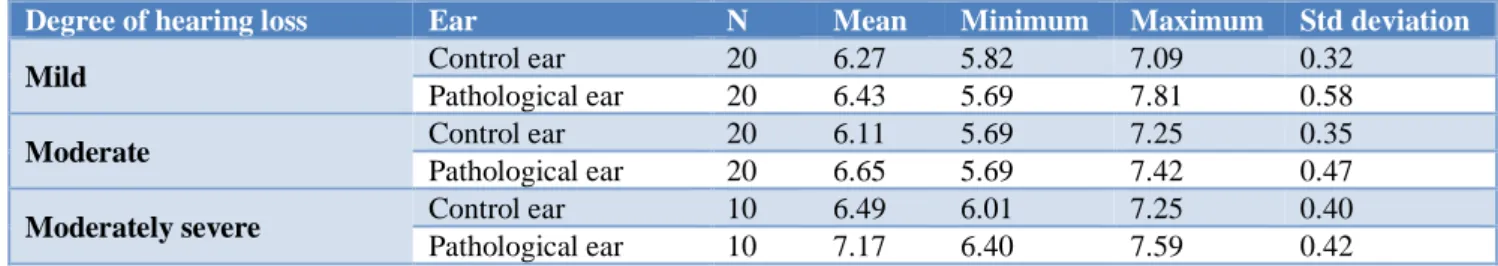
Table 1, the descriptive statistics for mild hearing loss group revealed a mean latency of 6.27 ms in control ear and 6.43 ms in pathological ear. For the moderate hearing loss group, the mean latency in the control group was 6.11 ms and in the pathological group was 6.65 ms. In the moderately severe hearing loss group, the mean latency in the control group was 6.49 ms and in the pathological group was 7.17 ms. Table 2, shows the values of descriptive statistics (Table 2).
Figure 1: a) Right ear and b) left ear, ABR waveform of a case with mild conductive hearing loss in left ear.
Table 2: Descriptive statistics for the three groups.
Degree of hearing loss Ear N Mean Minimum Maximum Std deviation
Mild Control ear 20 6.27 5.82 7.09 0.32
Pathological ear 20 6.43 5.69 7.81 0.58
Moderate Control ear 20 6.11 5.69 7.25 0.35
Pathological ear 20 6.65 5.69 7.42 0.47
Moderately severe Control ear 10 6.49 6.01 7.25 0.40
Pathological ear 10 7.17 6.40 7.59 0.42
On visual inspection, when the between ear is compared, the latency difference is seen between the pathological and control ear. The latencies are delayed in pathological
ear than the control ear. Also, as the degree of hearing loss increases the mean latency of the ABR wave V increases in the pathological group. The latencies are
0 1 2 3 4 5 6 7 8 9 10 11 12 13
1 ms 0.2 μV
V
Va Va V III
50 R 50 R 2
30 R 30 R 2
0 1 2 3 4 5 6 7 8 9 10 11 12 13
1 ms 0.2 μV
Va V V
Va
70 L 70 L 2
50 L 50 L 2
40 L 40 L 2 70 L 3
suggestive of effect of hearing loss on ABR latency due to degree of hearing loss, whereas the control group doesn’t show such a change.
Therefore, on visual inspection, there is effect of CSOM on the ABR wave V latencies in pathological ear and as the degree of hearing loss increases the mean latencies also increases.
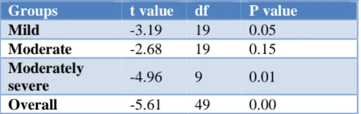
The paired t test for the entire data set revealed a significant difference between the control and the pathological ear. Whereas when the groups were analysed alone, the mild and moderately severe groups showed significance and moderate group did not show significance between the ears. Table 3, shows the paired t test values (Table 3).
Table 3: Paired t test values for different groups.
Groups t value df P value
Mild -3.19 19 0.05
Moderate -2.68 19 0.15
Moderately
severe -4.96 9 0.01 Overall -5.61 49 0.00
DISCUSSION
In the present study, the null hypothesis was rejected. The results of the present study indicated that chronic supportive otitis media reduces the auditory input which in turn delays the latencies. As the degree of hearing loss increases, the latency delay will also increase as seen in the present study. The hearing loss in the pathological ear causes increased conduction time for the stimulus to reach the brainstem than the control ear at the same sensation levels in both the ears.6
CSOM typically produces mild to moderate degree of hearing loss and untreated condition the magnitude of hearing loss increases. Sound vibration entering the ear bypass the perforated Tympanic membrane and strikes the wall of oval and round window. However, 30dB to 60dB reduction of sound stimulation is present which is reflected in ABR test. As we know that ABR latency is inversely proportional to intensity levels, the predictor of latency delay will be the magnitude of conductive hearing loss which in our study is the chronic suppurative otitis media.7
Keeping up with above discussion, our study results are in accordance with a study which shows reduced acoustic attenuation in adults with conductive hearing loss and loss of energy leading to delayed latency.9,10 Correlation
between degree of hearing loss and BAEP latency delay was done and shown no significance difference between them.11 However only moderately severe hearing loss
cases shown altered results between the ears and no difference in interpeak latencies (I-III, III-V and I-V)
which is not in accordance to our study. Due to peripheral involvement in CSOM, reduces the elicitation of responses of external ciliated cells that synopsis with 10% of the afferent nerve fibres that leads to time delay required to stimulate internal ciliated cells that intern enlarges the latency of ABR waveform.11
Also, in case of high frequency conductive hearing loss, wave I was more prolonged and interpeak differences was in lower limits of normal range as seen in study by Gorga 1985 as at lower intensities, the response is dominated by apical regions of cochlea and hence prolonged latency in case of long standing CSOM cases.9
However further investigation is required in case of high frequency conductive hearing loss and correlation between ABR latency shifts and mixed hearing loss cases with definite air borne gap.
CONCLUSION
The present study concludes that there is effect of chronic suppurative otitis media on auditory brainstem response latencies. As the conductive component increases the response latencies get altered due to reduced input to the auditory system. Also, the morphology of ABR peaks on comparison to the normal hearing ear gets affected due to constant conductive pathology in the pathological ear. Hence chronic suppurative otitis media should not be ignored and proper treatment should be provided at the right time.
ACKNOWLEDGEMENTS
We acknowledge the director, MIMS, Mandya for providing the opportunity to carry out research activity in the department of ENT, Mandya. We also like to thank the patients who took part in the data collection and helped to complete the research paper within time.
Funding: Borne by the principle investigator Conflict of interest: None declared
Ethical approval: The study was approved by the Ethical committee of Mandya Institute of Medical science, Mandya
REFERENCES
1. Dhingra P. Diseases of ear, nose and throat. New Delhi: Elsevier; 2010.
2. Verma AK, Vohra A, Maitra A, Banerjee M, Singh R, Mittal SK et al. Epidemiology of chronic suppurative otitis media and deafness in a rural area and developing an intervention strategy. Indian J Pediatrics. 1995;62(6):725-9.
3. Wakode PT, Joshi SV, Gawarle SH. Chronic suppurative otitis media in school going children. Indian J Otolaryngol Head Neck Surg. 2006;58(2):152-5.
South Indian children. Int J Pediatr Otorhinolaryngol. 1999;48(3):217-21.
5. Gorga MP, Reiland JK, Beauchaine KA. Auditory brainstem responses in a case of high-frequency conductive hearing loss. J Speech Hear Disord. 1985;50(4):346-50.
6. Borg E, Lofqvist L. Auditory brainstem response (ABR) to rarefaction and condensation clicks in normal and abnormal ears. Scand Audio. 1982;11(4):227-35.
7. Gee MTJ, Clemis JD. Effects of conductive hearing loss on auditory brainstem response. Ann Otol Rhinol Laryngol. 1982;91(3):304-9.
8. Clarkson C, Antunes FM, Rubio ME. Conductive hearing loss has long-lasting structural and molecular effects on presynaptic and postsynaptic structures of auditory nerve synapses in the cochlear nucleus. J Neurosci Methods. 2016;36(39):10214-27.
9. Gorga MP, Reiland JK, Beauchaine KA. Auditory brainstem responses in a case of high-frequency conductive hearing loss. J of Speech and Hearing Disorders. 1985;50(4):346-50.
10. Steinhoff HJ, Bohnke F, Janssen T. Click ABR intensity-latency characteristics in diagnosing conductive and cochlear hearing losses. Arch Oto-rhino-laryngol. 1988;245(5):259-65.
11. Matas CG, Leite RA, Goncalves IC, Neves IF. Brainstem auditory evoked potential in individuals with conductive and sensorineural hearing losses. Int Arch Otorhinolaryngol. 2005;9(11):337.